UNIVERSITÁ DEGLI STUDI DI NAPOLI
“FEDERICO II”
DIPARTIMENTO DI AGRARIA
DOTTORATO IN
SCIENZE AGRARIE ED AGROALIMENTARI
XXXI° CICLO
Omeprazole, a proton-pump inhibitor on humans, acts as a growth
enhancer and stress protectant in plants
Tutor:
Candidato:
Prof. Albino Maggio
Valerio Cirillo
TABLE OF CONTENTS
Chapter 1 Literature Review………... 1
1.1 Introduction: benzimidazoles………... 1
1.2 Review on benzimidazole effect on plants………... 3
1.3 Proton pump inhibitors………... 6
1.4 Small bioactive molecules……… 6
1.5 Aim of the study………... 9
1.6 References………... 9
Chapter 2 A benzimidazole proton pump inhibitor increases growth and tolerance to salt stress in tomato………... 17
2.1 Abstract………... 17
2.2 Introduction………... 17
2.3 Materials and methods………... 19
2.3.1 Plant Growth Conditions………... 19
2.3.2 RNA Extraction and Quantitative RT-PCR………... 20
2.3.3 Ion Measurements……… 22
2.3.4 Chl a Fluorescence Emission and Gas Exchange……… 22
2.3.5 Statistical Analysis………... 23
2.4 Results………... 24
2.4.1 Plant Growth……… 24
2.4.2 Salt stress tolerance………... 26
2.4.3 Gas Exchange and Chl a Fluorescence Emission…………... 27
2.4.5 Gene Expression……….. 30
2.5 Discussion………... 36
2.5.1 OP Improves Plant Growth and Salt Stress Tolerance……... 36
2.5.2 OP has multiple effects on cellular mechanisms that enhance salt stress tolerance……… 37
2.5.3 OP protects the photosynthetic system……… 39
2.5.4 Possible targets of OP in plants………... 40
2.6 Published paper front-page………... 41
2.7 References………... 42
Chapter 3 Physiological and Metabolic Responses Triggered by Omeprazole Improve Tomato Plant Tolerance to NaCl Stress……….... 51
3.1 Abstract………... 51
3.2 Introduction………...………….... 52
3.3 Materials and methods………... 55
3.3.1 Plant Material, Greenhouse Conditions, and Crop …...Management………... 55
3.3.2 Experimental Design, Omeprazole Application, and Nutrient ---…...Solution Management……….. 56
3.3.3 Yield, Growth Measurements, and Root Characteristics………... 57
3.3.4 Leaf Water Potential, Relative Water Content, and Leaf Gas …….Exchange Measurements………... 57
3.3.5 Ion Analyses……….... 58
3.3.6 Collection of Samples and Metabolomic Analysis……….. 59
3.3.7 Statistical Analysis of Experimental Data………... 60
3.4.1 Morphological Parameters, Yield, and Root Characteristics... 61
3.4.2 Physiological Parameters………... 64
3.4.3 Ion Content and Partitioning………... 65
3.4.4 Metabolic Profiling of Leaves………... 67
3.4.5 Principal Component Analysis………... 72
3.5 Discussion………... 75
3.5.1 Implications of Omeprazole for Morphological and Physiological …….Parameters……….... 75
3.5.2 Implications of Omeprazole for Ion Homeostasis…………... 77
3.5.3 Implications of Omeprazole for the Metabolomic Profile of Tomato …...Leaves………... 78
3.6 Conclusions………... 82
3.7 Published paper front-page……… 84
3.8 References………... 85
Chapter 4 Omeprazole treatment elicits contrasting responses to salt stress in two basil genotypes………... 95
4.1 Abstract………... 95
4.2 Introduction………... 95
4.3 Materials and Methods………...………...……….... 97
4.3.1 Plant material……….... 98
4.3.2 Biometric measurements……….. 98
4.3.3 Relative Water Content and ion leakage assays………... 98
4.3.4 qRT-PCR and gene expression………... 98
4.3.6 Post-harvest experiment………... 99
4.3.7 Statistical analysis………... 99
4.4 Results………... 100
4.4.1 Growth responses induced by OP………... 100
4.4.2 Water relations and ion leakage………...…... 102
4.4.3 Ion content of OP treated plants………...…... 103
4.4.4 OP induced changes in gene expression………..…... 105
4.4.5 OP effects on post-harvest shelf-life………... 107
4.5 Discussion………... 108
4.6 References………... 112
Chapter 5 Omeprazole induces changes in Arabidopsis root system architecture…. 119 5.1 Abstract………. 119
5.2 Introduction………... 119
5.3 Materials and methods………... 120
5.3.1 Plants growth conditions……….. 121
5.3.2 Determination of root traits……….…... 121
5.3.3 Measurement of shoot biomass………...…... 121
5.3.4 Statistical analysis………...……... 121 5.4 Results………... 122 5.5 Discussion………... 124 5.6 References………... 124 General conclusion……….... 127 Appendix………... 129
1
CHAPTER 1. LITERATURE REVIEW
1.1 Introduction: benzimidazoles
Benzimidazole is the benzo derivative of imidazole (Figure 1), synthesized for the first time by Hoebrecker in 1872 (Bansal, 2002). Imidazole is a five member-ring with formula C3N2H4. It is an organic compound contained in many natural products like
alkaloids in plants.
Figure 1. Skeletal formula of benzimidazole, showing the numbering convention for substituent groups
The initial research on benzimidazoles can be traced back to 1944, when Woolley hypothesized that, because of their similarity with purines, benzimidazoles could act as this heterocyclic compound to induce some biological responses (Woolley, 1944). In nature, the benzimidazole moiety is a part of vitamin B12 complex, and some of the first artificial benzimidazoles had a vitamin B12-like activity (Brink and Flokers, 1949). All seven positions on the benzimidazole nucleus can be substituted with a variety of atoms. However, most of the biologically active benzimidazole based compounds have the functional groups at 1, 2 and/or 5, 6 positions (Bansal and Silakari, 2012). Many benzimidazoles derivatives are bioactive molecules widely used for their pharmacological and pharmacokinetic activities. Benzimidazole bioactivity in humans has been well documented (Salahuddin et al., 2017).
Substituents around the benzimidazole nucleus give rise to many drugs with diametrically opposed activities, including antiparasitic, anticonvulsants, analgesic,
2 antiulcer, anticancer, proton pump inhibitors, and many others (Bansal and Silakari, 2012). For this reason, the research on benzimidazoles has gathered broad interested in the last years, due to the need of new therapeutic molecules.
The benzimidazole moiety is also the nucleus of many fungicides (Gupta and Pathak, 2011; Dias, 2012; García et al., 2003). These derivatives control a broad range of fungi at relatively low application rates thanks to their ability to inhibit β-tubulin polymerization, consequently blocking cytoskeleton formation. The most used benzimidazole fungicides are benomyl (a), carbendazim (b), thiabendazole (c) and fuberidazole (d) (Figure 2).
Figure 2 Skeletal formula of the most used benzimidazole fungicides
However, due to benzimidazole resistance by several pathogens their use as fungicide has declined and it is currently prohibited for some of them (Ma and Michailides, 2005; Lucas et al., 2015; https://ec.europa.eu/food/plant_en ).
Interestingly, many authors observed both beneficial and detrimental effects of different benzimidazole based fungicides on plants, which represent their off target activity (García et al., 2003). For this reason, verification of the direct and/or indirect effects of benzimidazoles on plants metabolism and physiology deserves further attention, considering that the target of these molecules is still unknown. While many reviews on the therapeutic potential of benzimidazoles for humans have been published (Bansal and
3 Silakari, 2012; Salahuddin, 2017), a comprehensive review on use, potential use and likely target of benzimidazoles in plants is missing.
1.2 Review on benzimidazole effect on plants
The first paper in which a benzimidazole has been used on plants was published in 1955, with a further 35 papers since then. The majority of the literature involved testing the effects of different benzimidazole derivatives on various plants, reporting a plethora of effects induced by these molecules on various aspects of plant growth and physiology. A comprehensive list of experiments involving benzimidazoles and their effects is enumerated in Table 1. However, considering that the concentrations and the nature of the benzimidazole derivatives used in each experiment was different, it is not easy to find a common trait involved in the bioactivity of these molecules. Indeed, what the authors found in these experiments is contrasting: some papers reported that benzimidazoles had a positive effect on the overall growth of treated plants (IPO-1, Kołodziej et al., 2006; Benzimidazole, Dyar and Shade, 1974; Carbendazim, García et al., 2002; Carbendazim, Debergh, 1993; Benomyl, Skene, 1972; Benomyl/carbendazim, Magnucka et al., 2007; Benzimidazole, Dyar, 1967; Benzimidazole, Hillman, 1955), while others showed reductions in shoot and root biomass and in root length as well as phytotoxicity (Benzimidazole, Hillman, 1955; Benomyl, Boyle, 1973; Benzimidazole, Klingensmith, 1961; Flubendazole, Stuchlíková; 2018; Benomyl, Siddiqui and Zaman, 2004; Mercaptobenzimidazole, Rebstock et al., 1955; Benomyl, Mihuta-Grimm et al., 1990; Carbendazim, Siddiqui and Bano, 2018; Benzimidazole, McCorquodale and Duncan, 1957; Carbendazim, García et al., 2002). In addition to direct effect on plant growth and/or health, benzimidazole have been reported to delay excised leaves senescence (Benzimidazole, Harris-Shultz et al., 2015; Benzimidazole Nowierski et al., 1995; Tripathi et al., 1982; Benzimidazole, Sayeed et al., 1985), the production of specific compounds like phenols (Benomyl, Siddiqui and Zaman, 2004), phytocystatin (Omeprazole, Braga et al., 2018) and ginsenoids (IPO-1, Kołodziej et al., 2006), and in seed germination (Benomyl/carbendazim, Thomas, 1973). It should be noted that the
4 positive effect on plants growth were found only when benzimidazole derivatives were used at low concentrations (less then 1 mM), while with higher doses detrimental effects were observed. A list of the reviewed papers is shown in Table 1.
Year Molecule Dose Specie Reference
1955 Benzimidazole 2.5 mM Lemna minor Hillman, 1955
1955 Mercaptobenzimidazoles 5 mM Cucumis sativa Rebstock et al., 1955
6 mM Phaseolus vulgaris
1957 Benzimidazole 1.9 mM Vicia faba McCorquodale and
Duncan, 1957 1961 Benzimidazole 1 mM Cucumis sativa Klingensmith, 1961 10 mM Solanum lycopersicum 10 mM Bushbean
1967 Benzimidazole 0.5-1.5 mM Nicotiana tabacum Dyar, 1967
1972 Benomyl 80-160 µM
Raphanus raphanistrum
subsp. sativus Skene, 1972 80-160 µM Glicine max
1973 Benomyl 2-4 mM Allium cepa Boyle, 1973
1973
Benomyl 0-3.4 mM Apium graveolens
Thomas, 1973 Thiabendazole 0-5 mM Apium graveolens
Carbendazim 0-5.2 mM Apium graveolens
1973 Benzimidazole 10-100 µM Thelypteris felix-mas Dyar and Shade, 1973
1982 Carbendazim 26-520 µM Triticum vulgare Tripathi et al., 1982
1985 Benzimidazole 0.5 mM Triticum vulgare Sayeed et al., 1985
1990 Benomyl 0-479 µM Solanum lycopersicum Mihuta-Grimm and
Rowe, 1990
1993 Carbendazim 0-800 µM Cordyline terminalis Debergh, 1993
0-800 µM Prunus avium
1995 Benzimidazole 1 mM Hordeum vulgare Nowierski et al., 1995
2002 Carbendazim
1.3 mM Nicotiana tabacum
García et al., 2002 2.5 mM Nicotiana tabacum
5
2004 Benomyl 3.4-6.8 mM Zea mays Siddiqui and Zaman,
2004
2006 IPO-1 unknown MW Panax quinquefolium Kołodziej et al., 2006
2007 Benomyl 34 µM Secale cereale Magnucka et al., 2007
52 µM Secale cereale
2015 Benzimidazole 1 mM Sorghum bicolor Harris-Shultz et al., 2015
1 mM Zea mays
2018 Omeprazole 30-115 µM Allium cepa Braga et al., 2018
2018 Flubendazole 10 µM Plantago lanceolata Stuchlíková; 2018
10 µM Plantago lanceolata
2018 Carbendazim 10-100 µM Allium sativum Siddiqui and Bano, 2018
Table 1 List of the paper reporting the effect of various benzimidazole derivatives on plants from 1955 to 2018
Most of the above experiments were performed to test the phytotoxicity of the most commonly used fungicides on crops. When the literature is examined more closely, it becomes evident that some of these molecules, when used at lower concentrations than optimal as fungicides, can induce an increase in plants biomass production (García et al., 2003). Strikingly, none of these experiments demonstrates the biochemical or molecular targets for growth enhancement benzimidazole activity in plants. This is likely due to the scarce tools available to researchers in the period in which these experiments were performed. For this reason, the aim of this thesis is to understand the mechanisms involved in benzimidazole function on plants, and specifically the family of substituted benzimidazoles known to inhibit proton-pumps activity in humans. Molecules that are able to perturb ion homeostasis in plants can be useful to understand the mechanisms responsible for plant tolerance to osmotic and ionic stresses, with direct agricultural applications and for the study of basic biology.
6 1.3 Proton pump inhibitors
The inhibition of proton pumps is the most effective strategy for the treatment of gastrointestinal diseases, such as gastroesophageal reflux or peptic ulcer. The first proton pump inhibitor (PPI), omeprazole (OP) was approved in humans in 1989, and subsequently followed by other six molecules of the same class, namely pantoprazole, esomeprazole, rabeprazole, lansoprazole, and dexlansoprazole. All of the PPI’s have a benzimidazole nucleus and a pyridine with substituents on both of the rings, which are principally linked with bioavailability and catabolism of the drug. Specifically, omeprazole possesses a methoxy chain attached to its benzimidazole ring. The efficacy of this class of drugs is due to their ability to block the gastric acid secretion through the inactivation of the hydrogen-potassium adenosine triphosphatase enzyme system (H+/K+-ATPase). In the acidic environment of the parietal cells in the stomach lumen,
these molecules undergo to a protonation in both the pyridine and benzimidazole ring that leads to the formation of the active sulfonamide derivative, which then binds covalently to cysteine moieties of the H+/K+-ATPase. This inactivation is irreversible
and long lasting (Strand et al., 2017). After inhibition, the acid production will requires new H+/K+-ATPase protein complexes to replace the inactivated proteins. These
molecules are able to maintain stomach pH higher than 4 from 15 to 21 hours (Wolfe and Sachs, 2000). Moreover, their effectiveness is not reduced with long-term treatment. These two qualities of PPI’s represent the revolutionary aspect of these molecules compared to the ones used previously for the treatments of gastric disease, the histamine H2-receptor antagonists. Indeed, the latter had an efficacy of maximum 8 hours, with a high chance to incur in tachyphylaxis as soon as within 3 to 5 days (Wilder-Smith et al., 1990).
1.4 Small bioactive molecules
While a number of ATPase inhibitor have been demonstrated to inhibit activity of plant ATPases, no studies have tested the effects of benzimidazole based PPIs on plant growth
7 and tolerance to stress (Pedersen and Palmgren, 2017). The use of small bioactive molecules represents a promising approach, not only to study the molecular target divergence between different biological systems, but also to find mechanisms involved in different aspects of plant biology (Kaschani and der Hoorn, 2007). The use of human-to-plant and plant-to-human application of bioactive molecules is well established (Dillworth, 2009). The general trend is to use molecules well-characterized in one system and evaluate their effect on other model systems. This approach has led to a number of molecules now widely used cross-kingdom. In plant biology, many have been applied to study the protein activity, enzymes, aquaporins, and ATPases (Table 2).
Compound MW (Da) Function/target References
Modulators of phosphorylation
PD98059 372.41 MAP kinase kinase
inhibitor Zhang et al., 2006 K-252a 467.47 Kinase inhibitor Dai et al., 2006 Wortmannin 428.43 Kinase inhibitor Robatzek et al., 2006
Cantharidin 168.15 Phosphatase inhibitor Luan, 2003
Modulators of proteolysis
E-64 357.41 Inhibitor of cysteine
proteases Van der Hoorn, 2004 AEBSF 203.23 Inhibitor of serine
proteases Pak and Van Doorn, 2005 MG132 475.3 Proteasome inhibitor Robatzek et al., 2006 Bestatin 308.17 Inhibitor of
8 Modulators of
membrane trafficking
Latrunculin B 395.51 Inhibitor of actin
polymerization Robatzek et al., 2006 N-ethylmaleimide (NEM) 125.13 Inhibitor of exocytosis/actinomyosin complex Davies et al., 2006 Brefeldin A 280.36 Inhibitor of vesicle
trafficking Nomura et al., 2006 Colchicine 399.44 Inhibitor of microtubule
assembly Fiorani and Beemster, 2006 Oryzalin 346.36 Inhibitor of tubulin
polymerization Robatzek et al., 2006
Other modulators
U73122 464.64
Inhibitor of phosphatidyl inositol
phospholipase C
Hetherington and Brownlee, 2004 Quinidine 326.43 Inhibitor of root
exudation Hoat et al., 2006 Paclobutrazol 293.79 Inhibitor of gibberellin
biosynthesis Saito et al., 2006 Naphthylphthalamic
acid 291.3
Inhibitor of auxin
transport Dai et al., 2006 Cerulenin 223.27 Inhibitor of fatty acid
synthase Koo et al., 2005 Cycloheximide 281.35 Inhibitor of protein
synthesis Dreher et al., 2006 W7 340.87 Inhibitor of
calmodulin-dependent proteins Davies et al., 2006 Forskolin 410.5 Activator of adenyl
cyclase Davies et al., 2006 BTH 210.28 Induces systemic
acquired resistance Van Hulten et al., 2006 Mevinolin,
lovastatin 404.54
Inhibitor of the MVA
9
Isoxaben 332.39 Inhibitor of cellulose
biosynthesis Paredez et al., 2006
Table 2 List of small bioactive molecules (<500 Da) active on different aspects of plant biology adapted from Kaschani and der Hoorn (2007)
1.5 Aim of the study
We chose to test OP, the ancestor molecule of benzimidazole based PPI. OP can be considered a small bioactive molecule, being smaller than 500 Da. Using a chemical genetics approach, we hypothesized that substituted benzimidazoles that act as PPIs in humans may have an effect in plants exposed to environmental stresses such as salinization of the root zone. To test this hypothesis, we chose to subject different plant species (tomato and basil) to osmotic and ionic stress. In plants, proton pumps are essential for establishing the proton motive force used to exclude toxic ion and maintain homeostasis (Ji et al 2013; Oh et al., 2010). In addition, we used the model species Arabidopsis thaliana to assess changes in root architecture under the effect of omeprazole. Our results can be valuable as an innovative approach for the study of unknown mechanisms involved with the tolerance to salt stress, that we know is one of the main constrains of plant productivity worldwide. For this reason, considering the high impact that this specific stress has on food safety, this study could give us important insights on this topic from both a biological as well as an applicative point of view, which are pivotal for the comprehensiveness and thoroughness of a scientific work.
1.6 References
Bansal, R. K. (2002). Herocyclic Chemistry (third ed.). Publisher New Delhi, New Age International, 401 pp
Bansal, Y., Silakari, O. (2012). The therapeutic journey of benzimidazoles: A review. Bioorganic Med. Chem. 20, 6208–6236. https://doi.org/10.1016/j.bmc.2012.09.013
10 Boyle, W. S. (1973). Cytogenetic effects of Benlate fungicide on Allium cepa and
Secale cereale, J. Heredity, 64:1, 49–50.
https://doi.org/10.1093/oxfordjournals.jhered.a108340
Braga, A. L., de Meneses, A. -A. P. M., Santos, J. V. de O., dos Reis, A. C., de Lima, R. M. T., da Mata, A. M. O. F., Paz, M. F. C. J., Alves, L. B. dos S., Shaw, S., Uddin, S. J., Rouf, R., Das, A. K., Dev, S., Shil, M. C., Shilpi, J. A., Khan, I. N., Islam, M. T., Ali, E. S., Mubarak, M. S., Mishra, S. K., e Sousa, J. M. de C., Melo-Cavalcante, A. A. de C. (2018). Toxicogenetic study of omeprazole and the modulatory effects of retinol palmitate and ascorbic acid on Allium cepa. Chemosphere 204, 220–226. https://doi.org/10.1016/j.chemosphere.2018.04.021
Brink, N. G., Folkers, K. (1949). Vitamin B12. VI. 5,6 dimethylbenzimidazole, a degradation product of vitamin B12. Journal of the American Chemical Society 71, 2951–2951. https://doi.org/10.1021/ja01176a532
Dai, Y., Wang, H., Li, B., Huang, J., Liu X., Zhou, Y., Mou, Z., Li, J. (2006). Increased expression of MAP kinase kinase7 causes deficiency in polar auxin transport and leads to plant architectural abnormality in Arabidopsis. Plant Cell, 18, 308-320.
Davies, D. R., Bindschedler, L.V., Strickland, T. S., Bolwell, G. P. (2006). Production of reactive oxygen species in Arabidopsis thaliana cell suspension cultures in response to an elicitor from Fusarium oxysporum: implications for basal resistance. J. Exp. Bot., 57, 1817-1827.
Debergh, P. C., Coster, G.D., Steurbaut, W. (1993). Carbendazim as an Alternative Plant Growth Regulator in Tissue Culture Systems. In Vitro Cellular & Developmental Biology. Plant 29P, 89–91.
Dias, M. C. (2012). Phytotoxicity: An Overview of the Physiological Responses of plants Exposed to Fungicides. J. Bot., 1–4. https://doi.org/10.1155/2012/135479
Dilworth, M. F., (2009). Perspective: Plant biology-A quiet pioneer. Plant Biotechnology 26, 183–187. https://doi.org/10.5511/plantbiotechnology.26.183
11 Dilworth, M.F. (2009). Perspective: Plant biology—A quiet pioneer. Plant Biotechnology 26, 183–187. https://doi.org/10.5511/plantbiotechnology.26.183
Dreher, K. A., Brown, J., Saw, R. E., Callis, J. (2006). The Arabidopsis Aux/IAA protein family has diversified in degradation and auxin responsiveness. Plant Cell, 18, 699-714.
Dyar, J. J. (1968). Certain Effects of Benzimidazole on Young Tobacco Plants. Plant Phys., 43, 477–478. https://doi.org/10.1104/pp.43.3.477
Dyar, J. J., Shade, J. (1974). The Influence of Benzimidazole on the Gametophyte of Thelypteris felix-mas. Plant Phys. 53, 666–668. https://doi.org/10.1104/pp.53.4.666
Fiorani, F., Beemster, G. T. (2004). Quantitative analyses of cell division in plants. Plant Mol Biol 2006, 60:963-979. Hetherington AM, Brownlee C: The generation of Ca2+
signals in plants. Annu. Rev. Plant. Biol., 55, 401-427.
García, P. C., Rivero, R. M., Ruiz, J. M., Romero, L. (2003). The Role of Fungicides in the Physiology of Higher Plants: Implications for Defense Responses. Botanical Review, 69(2), 162-172
García, P. C., Ruiz, J. M., Rivero, R. M., López-Lefebre, L. R., Sánchez, E., Romero, L., (2002). Is the Application of Carbendazim Harmful to Healthy Plants? Evidence of Weak Phytotoxicity in Tobacco. Journal of Agricultural and Food Chemistry 50, 279– 283. https://doi.org/10.1021/jf010748g
Gupta, N., Pathak, D. (2011). Synthesis and evaluation of n-substituted imidazole derivatives for antimicrobial activity. Indian Journal of Pharmaceutical Sciences 73, 674. https://doi.org/10.4103/0250-474X.100246
Harris-Shultz, K., Ni, X., Wang, H., Knoll, J. E., Anderson, W. F. (2015). Use of Benzimidazole Agar Plates to Assess Fall Armyworm (Lepidoptera: Noctuidae) Feeding on Excised Maize and Sorghum Leaves. Florida Entomologist 98, 394–397. https://doi.org/10.1653/024.098.0169
12 Hillman, W. S. (1955). The Action of Benzimidazole on Lemna Minor. Plant Phys. 30, 535–542. https://doi.org/10.1104/pp.30.6.535
Hoat, T. X., Nakayashiki, H., Tosa, Y., Mayama, S. (2006). Specific cleavage of ribosomal RNA and mRNA during victorin-induced apoptotic cell death in oat. Plant J., 46, 922-933.
Ji, H., Pardo, J. M., Batelli, G., Van Oosten, M. J., Bressan, R. A., Li, X. (2013). The Salt Overly Sensitive (SOS) Pathway: Established and Emerging Roles. Molecular Plant 6, 275–286. https://doi.org/10.1093/mp/sst017
Kaschani, F., van der Hoorn, R. (2007). Small molecule approaches in plants. Current Opinion in Chemical Biology 11, 88–98. https://doi.org/10.1016/j.cbpa.2006.11.038 Keri, R. S., Hiremathad, A., Budagumpi, S., Nagaraja, B. M. (2015). Comprehensive Review in Current Developments of Benzimidazole-Based Medicinal Chemistry. Chemical Biology & Drug Design 86, 19–65. https://doi.org/10.1111/cbdd.12462
Klingensmith, M. J. (1961). The Effect of Certain Benzazole Compounds on Plant Growth and Development. American Journal of Botany 48, 40. https://doi.org/10.2307/2439593
Kołodziej, B., Kochan, E., Kazimierczak, J., Chmiel, A. (2006). The Effect of Growth Regulators on Quality Parameters and Ginsenosides Accumulation in Panax quinquefolium L. Roots. Plant Growth Regul. 48, 13–19. https://doi.org/10.1007/s10725-005-5088-z
Koo, A. J, Fulda, M., Browse, J., Ohlrogge, J. B. (2005). Identification of a plastid acyl-acyl carrier protein synthetase in Arabidopsis and its role in the activation and elongation of exogenous fatty acids. Plant J., 44, 620-632.
LaPaz, L., Wilson, R. H. (1960). Magnetic Damping of Rotation of the Vanguard I Satellite. Science 131, 355–357. https://doi.org/10.1126/science.131.3397.355
13 Lucas, J. A., Hawkins, N. J., Fraaije, B. A. (2015). The Evolution of Fungicide Resistance, in: Advances in Applied Microbiology. Elsevier, pp. 29–92. https://doi.org/10.1016/bs.aambs.2014.09.001
Ma, Z., Michailides, T. J. (2005). Advances in understanding molecular mechanisms of fungicide resistance and molecular detection of resistant genotypes in phytopathogenic fungi. Crop Protection 24, 853–863. https://doi.org/10.1016/j.cropro.2005.01.011
Magnucka, E. G., Suzuki, Y., Pietr, S. J., Kozubek, A., Zarnowski, R. (2007). Action of benzimidazole fungicides on resorcinolic lipid metabolism in rye seedlings depends on thermal and light growth conditions. Pesticide Biochemistry and Physiology 88, 219– 225. https://doi.org/10.1016/j.pestbp.2006.11.008
McCorquodale, D. J., Duncan, R. E. (1957). Plant Growth Inhibitions by Certain Imidazole Compounds and their Preventions with Metal Ions. American Journal of Botany 44, 715. https://doi.org/10.2307/2438638
Mihuta-Grimm, L., Rowe, R. C. (1990). Fusarium Crown and Root Rot of Tomato in Greenhouse Rock Wool Systems: Sources of Inoculum and Disease Management with Benomyl. Plant Disease 74, 996–1002
Nomura, K., Debroy, S., Lee, Y. H., Pumplin, N., Jones, J., He, S. Y. (2006). A bacterial virulence protein suppresses host innate immunity to cause plant disease. Science, 313, 220-223.
Nowierski, R. M., Zeng, Z., Scharen, A. L. (1995). Age-Specific Life Table Modeling of the Russian Wheat Aphid (Homoptera: Aphididae) on Barley Grown in Benzimidazole Agar. Environmental Entomology 24, 1284–1290. https://doi.org/10.1093/ee/24.5.1284 Oh, D.-H., Lee, S. Y., Bressan, R.A., Yun, D.-J., Bohnert, H. J. (2010). Intracellular consequences of SOS1 deficiency during salt stress. J. Exp. Bot. 61, 1205–1213. https://doi.org/10.1093/jxb/erp391
Pak, C., Van Doorn, W. G. (2005). Delay of Iris flower senescence by protease inhibitors. New Phytol., 165, 473-480.
14 Paredez, A. R., Somerville, C. R., Ehrhardt, D. W. (2006). Visualization of cellulose synthase demonstrates functional association with microtubules. Science, 312, 1491-1495.
Pedersen, J. T., Palmgren, M. (2017). Why do plants lack sodium pumps and would they benefit from having one? Funct. Plant Biol. 44, 473-479.
Rebstock, T. L., Ball, C. D., Hamner, C. L., Sell, H. M. (1955). Inhibition of Plant Growth by 2-Mercaptobenzimidazole Analogs. Plant Phys., 30, 382–384. https://doi.org/10.1104/pp.30.4.382
Robatzek, S., Chinchilla, D., Boller, T. (2006). Ligand-induced endocytosis of the pattern recognition receptor FLS2 in Arabidopsis. Genes Dev., 20537-542.
Rodriguez-Concepcion, M., Fores, O., Martinez-García, J. F., Gonzalez, V., Phillips, M. A., Ferrer, A., Boronat, A. (2004). Distinct light mediated pathways regulate the biosynthesis and exchange of isoprenoid precursors during Arabidopsis seedling development. Plant Cell, 16, 144-156.
Saito, S., Okamoto, M., Shinoda, S., Kushiro, T., Koshiba, T., Kamiya, Y., Hirai, N., Todoroki, Y., Sakata, K., Nambara, E., Mizutani, M. (2006). A plant growth retardant, uniconazole, is a potent inhibitor of ABA catabolism in Arabidopsis. Biosci. Biotechnol. Biochem., 70, 1731-1739.
Salahuddin, Shaharyar, M., Mazumder, A. (2017). Benzimidazoles: A biologically active compounds. Arabian Journal of Chemistry 10, S157–S173. https://doi.org/10.1016/j.arabjc.2012.07.017
Sayeed, S. A., Behera, B. K., Mohanty, P. (1985). Interaction of light quality and benzimidazole on pigment contents and on photochemical activity of chloroplasts isolated from detached senescing wheat (Triticum aestivum) leaves. Physiologia Plantarum 64, 383–388. https://doi.org/10.1111/j.1399-3054.1985.tb03357.x
Siddiqui, M. F., Bano, B. (2018). Exposure of carbendazim induces structural and functional alteration in garlic phytocystatin: An in vitro multi-spectroscopic approach.
15 Pesticide Biochemistry and Physiology 145, 66–75. https://doi.org/10.1016/j.pestbp.2018.01.008
Siddiqui, Z. S., Zaman, A. U. (2004). Effects of benlate systemic fungicide on seed germination, seedling growth, biomass and phenolic contents in two cultivars of Zea mays L. Pak. J. Bot., 36, 577-582.
Skene, K. G. M. (1972). Cytokinin-Like Properties of the Systemic Fungicide Benomyl. Journal of Horticultural Science 47, 179–182. https://doi.org/10.1080/00221589.1972.11514454
Strand, D. S., Kim, D., Peura, D. A. (2017). 25 Years of Proton Pump Inhibitors: A Comprehensive Review. Gut and Liver 11, 27–37. https://doi.org/10.5009/gnl15502 Stuchlíková, L. R., Skálová, L., Szotáková, B., Syslová, E., Vokřál, I., Vaněk, T., Podlipná, R. (2018). Biotransformation of flubendazole and fenbendazole and their effects in the ribwort plantain (Plantago lanceolata). Ecotoxicology and Environmental Safety 147, 681–687. https://doi.org/10.1016/j.ecoenv.2017.09.020
Thomas, T. H. (1973). Growth regulatory effect of three benzimidazole fungicides on the germination of celery (Apium graveolens) seeds. Annals of Applied Biology 74, 233–238. https://doi.org/10.1111/j.1744-7348.1973.tb07743.x
Tripathi, R. K., Tandon, K., Schlösser, E., Hess, W. M. (1982). Effect of fungicides on the physiology of plants. Part IV: Protection of cellular organelles of senescent wheat leaves by carbendazim. Pesticide Science 13, 395–400. https://doi.org/10.1002/ps.2780130409
Van der Hoorn, R. A. L., Leeuwenburgh, M. A., Bogyo, M., Joosten, M. H. A. J., Peck, S. (2004). Activity profiling of papain-like cysteine proteases in plants. Plant Physiol. 135, 1170-1178.
Van Hulten, M., Pelser, M., Van Loon, L. C., Pieterse, C. M., Ton, J. (2006). Costs and benefits of priming for defense in Arabidopsis. Proc. Natl. Acad. Sci. USA, 103, 5602-5607.
16 Wilder-Smith, C. H., Ernst, T., Gennoni, M., Zeyen, B., Halter, F., Merki, H. S. (1990). Tolerance to oral H2-receptor antagonists. Digestive Diseases and Sciences 35, 976– 983. https://doi.org/10.1007/BF01537246
Wolfe, M. M., Sachs, G. (2000). Acid suppression: Optimizing therapy for gastroduodenal ulcer healing, gastroesophageal reflux disease, and stress-related erosive syndrome. Gastroenterology 118, S9–S31. https://doi.org/10.1016/S0016-5085(00)70004-7
Woolley, D. W. (1943). Some biological effects produced by benzimidazole and their reversal by purines. J. Biol. Chem. 152:225-232.
Zhang, L., Tamura, K., Shin-ya, K., Takahashi, H. (2006). The telomerase inhibitor telomestatin induces telomere shortening and cell death in Arabidopsis. Biochim. Biophys. Acta. 1763, 39-44.
Zheng, W., Zhai, Q., Sun, J., Li, C. B., Zhang, L., Li, H, Zhang, X., Li, S., Xu, Y., Jiang, H., Wu, X., Li, C. (2006). Bestatin, an inhibitor of aminopeptidases, provides a chemical genetics approach to dissect jasmonate signaling in Arabidopsis. Plant Physiol. 141, 1400-1413.
17
CHAPTER 2.
A BENZIMIDAZOLE PROTON PUMP INHIBITOR INCREASES GROWTH AND TOLERANCE TO SALT STRESS IN TOMATO
2.1 Abstract
Pre-treatment of tomato plants with micromolar concentrations of omeprazole (OP), a benzimidazole proton pump inhibitor in mammalian systems, improves plant growth in terms of fresh weight of shoot and roots by 49 and 55% and dry weight by 54 and 105% under salt stress conditions (200 mM NaCl), respectively. Assessment of gas exchange, ion distribution, and gene expression profile in different organs strongly indicates that OP interferes with key components of the stress adaptation machinery, including hormonal control of root development (improving length and branching), protection of the photosynthetic system (improving quantum yield of photosystem II) and regulation of ion homeostasis (improving the K+:Na+ ratio in leaves and roots). To our knowledge OP is one of the few known molecules that at micromolar concentrations manifests a dual function as growth enhancer and salt stress protectant. Therefore, OP can be used as new inducer of stress tolerance to better understand molecular and physiological stress adaptation paths in plants and to design new products to improve crop performance under suboptimal growth conditions.
2.2 Introduction
Soil salinization is a major problem for agriculture. It is estimated that by 2050 salinization will lead to up to 30% degradation of cultivated land (Rengasamy, 2006; FAO, 2011; Aragüés et al., 2015; Lal, 2015). The effects of soil and water salinity on plant growth and development have been well documented. Excess of Na+ and Cl− ions in proximity of the roots generate osmotic and ionic stress and activate signals inhibiting cell division and plant growth (Deinlein et al., 2014). Metabolic dysfunction and
18 nutritional disorders associated with Na+ and Cl− loading in plant tissues and organs translate in further growth reduction and eventually irreversible cell damage. Upon exposure to salt stress, the control of growth, ion and water homeostasis becomes an essential part of an adaptation program that helps resuming growth, albeit at a reduced rate (Maggio et al., 2006; Park et al., 2016; Van Oosten et al., 2016; Annunziata et al., 2017). During adaptation, ion movement through cellular compartments is essential to detoxify the cytoplasm and re-establish osmotic balance (Hasegawa, 2013; Ji et al., 2013; Mancarella et al., 2016). Plasma membrane and vacuolar H+-ATPases play a fundamental role in this physiological process since by generating active transport of proton H+ across the membranes they create pH gradients and electrical potentials that drive transport of ions and molecules (including NO3−, PO4−, K+, Na+, sucrose, hexoses,
and amino acids) across membranes (Pardo et al., 2006; Batelli et al., 2007; Deinlein et al., 2014). H+-ATPases can be activated/deactivated in response to many environmental cues such as abiotic stresses (Hasegawa, 2013). It occurs that salinization stimulates vacuolar H+-ATPase and H+-PPase activities which in turn facilitate tonoplast Na+/H+
antiporter function and cytoplasm detoxification (Pardo et al., 2006; Ji et al., 2013). It has been shown that increasing cellular proton pump activity via co-overexpression of the vacuolar H+-pyrophosphatase gene AVP1 and the vacuolar Na+/H+ antiporter gene
AtNHX1, enhanced salt stress tolerance, most likely by potentiating ion compartmentalization functions (Shen et al., 2015). In contrast, cell treatment with vanadate (an inhibitor of the plasma membrane H+-ATPase) increases the Na+/K+ ratio
in plant tissues and enhances sensitivity to salinity (Li et al., 2014).
In animals, homologs of plant proton pumps operate through the H+/K+ ATPase mechanism (Axelsen and Palmgren, 1998). Similar to plants, animal proton pumps working across membranes generate acidification of organismal compartments. For decades proton pump inhibitors (PPIs) have been successfully used to inhibit gastric acid secretion (McTavish et al., 1991). As matter of fact, benzimidazole based PPIs are common treatments used with gastro-esophageal reflux disease (GERD) and peptic ulcers (Baumann and Baxendale, 2013). In animals, omeprazole, the most common
19 benzimidazole PPI, affects P-Type IIC ATPases. These P-Type IIC ATPases represent a large family of ATP driven transporters, which are responsible for moving ions across membranes. These include membrane Ca2+ pumps, Na+/K+ transporters, and H+/K+
transporters. Omeprazole is largely used as PPI that suppresses stomach acid secretion in the gastric mucosa (Wallmark, 1986; Shin et al., 2009). The specific inhibition of the P-Type IIC H+/K+ ATPase located in the parietal cells is irreversible and specific. Plants are not known to possess P-Type IIC ATPases that transport Na+ or K+, instead relying on the family of NHX-type Na+ and K+/H+ antiporters for plasma membrane extrusion and compartmentation into the vacuoles and endosomes. Plants do possess P-Type IIA and III ATPases, primarily SERCA-like, which are not known to transport Na+ or K+. SERCA-like ATPases show very low homology (approximately 25%) to P-Type IIC ATPases that transport Na+ or K+ (Sweadner and Donnet, 2001). The SERCA-like ATPases are primarily endoplasmic reticulum transporters of calcium (Altshuler et al., 2012) and manganese (Mills et al., 2008). Plants also have P-Type III ATPases typically found in the plasma membrane, but none have been functionally characterized as Na+ or
K+ transporters. While plants are not known to possess P-Type IIC ATPases that transport Na+ or K+ which are the target of omeprazole, we wanted to verify whether
plant treatment with omeprazole might actually alter the transmembrane control of ion fluxes and disrupt plant tolerance to saline stress. In contrast to what we may have expected based on our current understanding of plant ATPases and plant responses to salt stress, here we demonstrate that tomato treatment with micromolar concentrations of omeprazole greatly enhanced plant growth and improved its tolerance to saline stress.
2.3 Materials and methods 2.3.1 Plant Growth Conditions
Tomato seeds (cultivar M82, accession LA3475) obtained from the Tomato Genetics Resource Center (TGRC, University of California, Davis, USA) were germinated in plates containing MS media and transplanted to a hydroponic system after 1 week when
20 cotyledons were fully expanded. Air temperature (Ta, °C), humidity (RH, %), and solar radiation (Rs, W m−2) were acquired by a data logger (Spectrum Technologies, Plainfield, IL, United States). The average air humidity and temperature were 58% and 24 °C during the day and 86% and 18 °C during night-time, under short day conditions. Plants were grown in hydroponic solution containing: 1.5 mM Mg(NO3)2 • 6H2O, 3.4
mM Ca(NO3)2 • 4H2O, 1 mM KNO3, 1.8 mM K2SO4, 1.5 mM KH2PO4, and 14 mg/L
Hidromix (Valagro, Atessa (Chieti) Italy). Eight plants per treatment were grown in 4 L tanks with constant aeration. At 26 Days After Sowing (DAS), the first treatment of OP was added to the nutrient solution (1, 10, and 45 µM). Salt stress was initiated 36 DAS by adding 75 mM NaCl to salt treatments. At 42 DAS the NaCl concentration was increased to 150 mM and on 46 DAS the NaCl concentration was raised to 200 mM NaCl. Destructive harvest and biometrics were taken at 50 DAS (14 days of salt stress). Roots were separated from shoots in order to obtain their individual FW. To measure relative water content (RWC), shoots were transferred in deionized water for 24 h to induce maximum turgidity and weighed. Shoots and roots were dried for 3 days at 64°C and weighed individually. Root length was measured with a ruler; root area was measured using ImageJ (National Institutes of Health and the Laboratory for Optical and Computational Instrumentation, University of Wisconsin, USA) as per Yoo et al. (2010).
2.3.2 RNA Extraction and Quantitative RT-PCR
Leaves of 7-week-old hydroponically grown plants (50 DAS, 14 DAST), treated with 1, 10, and 45 µM OP, with 0 and 200 mM NaCl, were harvested and immediately frozen at −80°C. Leaves from the same treatment were mixed and three replicates per bulk were analyzed. 100 mg of fresh leaf tissue per sample was homogenized with liquid nitrogen and extracted with 1 ml of TRIzol (Life Technologies). First-strand synthesis was performed with a QuantiTect Reverse Transcription Kit (QIAGEN) using 1 µg of total RNA. Real-time qPCR reactions, using 10 ng of cDNA per reaction, two experiments, four replicates per experiment, were carried out on an ABI 7900HT qPCR detection system using Platinum SYBR Green qPCR SuperMix-UDG with ROX (Life
21 Technologies). Each qRT-PCR experiment was repeated at least twice to confirm results. Primers were designed based on gene models and EST sequences available in GenBank. All qRT-PCR primers were determined to be within 3% efficiency of each other. Relative expression levels were calculated using EF1α as an internal standard and the ∆∆Ct method for relative quantification. The list of the primers used and the gene name is reported in Table 3.
Primer Name Sequence Accession #
Sl707A1-F CTGAACAGAAAGTTATTTGGCAGTC Solyc04g078900.2
Sl707A1-R ATGATACTAGCCATTCTCAGTGTCTC
Sl707A3-F GCTCCCAAACCCAATACCTAC Solyc04g071150.2
Sl707A3-R CAGTTTGGCGAGTTCATTTCC
SlHKT1.1-F TCTAGCCCAAGAAACTCAAAT Solyc07g014680.2
SlHKT1.1-R CTAATGTTACAACTCCAAGGAATT
SlNCED1-F CATAATCGAAAACCCGGATG Solyc07g056570.1
SlNCED1-R AACTTTTGGCCATGGTTCAG SlNHX1-F CACGATATGGTGGGCTGGTT Solyc06g008820.2 SlNHX1-F GGGTGTGGCCAAATCTCGTA SlNHX2-F ATTGGAGGATCGGCAGGAAC Solyc04g056600.2 SlNHX2-R CCATGGAGCCAGATTGACCA SlNRT1.1-F TAGCGCCGCGATGATATTAGGG Solyc06g074990 SlNRT1.1-R TGTAACGTTGTTGGCTGAACTTGC SlP5CD-F CACAGGTAGCTCAAGGGTGG Solyc02g089620 SlP5CD-R GGCCCAAGGATCTTCCAGTC SlP5CS-F AACTGAGCTTGATGGCAAGG Solyc08g043170 SlP5CS-R ACCAGAGGCTGAGCTGATGT
SlSOS1-F TCGAGTGATGATTCTGGTGG Solyc01g005020
SlSOS1-R ATCACAGTGTGGAAAGGCT
SlLEA-F CGAAGGAGAAGGCTAGTGGA Solyc03g116390.2
SlLEA-R AGCGATGCTCCTCACTTGTT
SlAPX2-F ATGGTAGCTGGAGGAGACCT Solyc06g005150
SlAPX2-R TTGAGGGAGCATGGACCAAC
SlCAT1-F GGACAATAATGGCAGGGCAA Solyc12g094620
SlCAT1-R TTACAGCCAGTTGGTCGCTT
SlCHLBP-F GAGCATTCTAGCAGTATTGG Solyc07g063600
SlCHLBP-R TGTTGCCTTCACCAACTCCA
22
SlPSII-R GTCTCGTGGAGTCCGCATAA
SlFTSH-F AAGGACGTGGATCTGTCAGC Solyc03g112590
SlFTSH-R TGGCATATTTGCATGCTCGC
Table 1 List of primers and gene names used in this study.
2.3.3 Ion Measurements
Ions measurements were performed according to a procedure described by Carillo et al. (2011), with following modifications. 100 mg of powdered dried material was suspended in 10 mL of MilliQ grade water (Milli-Q PLUS, Millipore, United States), and subjected to four freeze-thaw cycles by freezing in liquid nitrogen and thawing at 40°C. Samples were centrifuged at 34000 × g for 10 min and the clear supernatants were analyzed by ionexchange chromatography using a DX500 apparatus (Dionex, Olten, Switzerland) with an IONPACATC1 anion trap column (Dionex), an IONPAC-AG11 guard column (Dionex) and an analytical IONPAC-AS11 4-mm column (Dionex), fitted with an ASRSII 4-mm suppressor for anions (Dionex), and an IONPACCTC cation trap column (Dionex), an IONPAC-CG12A guard column (Dionex) and an analytical IONPAC-CS12A 4-mm column (Dionex), fitted with a CSRS 4-mm suppressor for cations (Dionex), with detection by a CD20 conductivity detector (Dionex), according to the manufacturer’s instructions.
2.3.4 Chl a Fluorescence Emission and Gas Exchange
To determine chlorophyll a (Chl a) fluorescence, a LED light source with emission peaks centered at 465 (blue) and 635 nm (red) generated a PPFD equal to 1500 µmol (photons) m−2 s−1 (90% red, 10% blue). A modulated fluorometer analyzer, Li-6400XT (Li-Cor Biosciences, Lincoln, NE, United States), was used to assess the fluorescence parameters. The measuring beam was set at intensity 5 (according to the instrument manual) with a modulation of 20 kHz. After the measurement of Chl a fluorescence emission at steady-state under light conditions, F’, the maximum fluorescence emission, Fm, was assessed upon induction by a 0.8 s saturating light pulse at 6000 µmol (photons)
23 m−2 s−1 at 20 kHz. After that, actinic light was briefly switched off while a far-red light of 8 µmol (photons) m−2 s−1 for 6s was used to discharge the PSII to allow measurement of the minimum fluorescence emission under light conditions, F0’. Photochemical
quenching (qP) was calculated from the previously mentioned parameters. Net photosynthetic CO2 assimilation rate (A, µmol m−2 s−1) and stomatal conductance of water vapor (gs, mol m−2 s−1) were measured using a portable open-system gas exchange (Li-6400XT).
Measurements of photosynthetic rates (A) and stomatal conductance (gs) were taken at saturating light on well-exposed and fully expanded top leaves of six plants per treatment. Leaf chamber CO2 was set to 400 µmol CO2 mol−1 air. Measurements were
taken for each time point between 10:00 and 13:00 for the duration of the experiment (November 10th– 23rd, 2016). Gas-exchange parameters using the von Caemmerer and
von Farquhar (1981) model were calculated by instrument software (Li-Cor, 2011). The effective quantum yield of PSII photochemistry in light-adapted leaves was calculated using: ФPSII = (Fm’– F’)/Fm’ (Genty et al., 1990). Anatomical analysis on a leaf area of
0.069 mm2 were performed as described in Yoo et al. (2010).
2.3.5 Statistical Analysis
Biometric measurements were statistically analyzed using the Student’s t-test. Photosynthesis and gas exchange results were statistically analyzed using two-way ANOVA procedure with Sidak multiple comparisons test. Ion quantification was analyzed using a two-way ANOVA and Duncan’s multiple range to determine differences between means (P ≤ 0.05). Ions for roots and shoots were statically analyzed in two separate analyses.
24 2.4 Results
2.4.1 Plant Growth
In order to assess the potential for omeprazole to affect growth and salt tolerance, we conducted a hydroponic experiment at increasing OP concentrations. Measurements were taken to separate phenotypes of roots and shoots. Our results indicate that OP induces two specific phenotypes: (1) increased growth of roots and shoots and (2) increased tolerance and growth under high salt stress. Specifically, for growth we observed that OP works in a dose dependent manner (Figure 1). Low doses, 1 µM, stimulated significant increases in growth while a higher dosage either had no stimulatory effect (10 µM) or, in the case of 45 µM, were inhibitory to growth. Application of OP at 1 µM resulted in 49 and 48% increases in shoot FW and DW, respectively (Figure 1). The highest dose (45 µM) inhibited growth of shoots and reduced FW and DW by 37 and 32%, respectively (Figure 1). The stimulatory effect of OP was not limited to shoots; we also observed that 1 µM stimulated root growth and biomass accumulation by increasing FW and DW of roots by 55 and 56%, respectively. Higher concentrations, 10 and 45 µM, did not significantly affect root growth (Figure 1). In control growth conditions, low concentrations of OP did not increase root length significantly. However, at 45 µM root length was severely inhibited, with a reduction of 53% (Figure 2). While OP did not significantly increase the maximal lengths of roots, low doses greatly increased root mass. OP also had effect on later root branching resulting in changes to the overall root area.
Low concentrations of 1 µM increased root area by 19%. Again, 45 µM had an inhibitory effect, decreasing root area by 44% (Figure 2).
25
Figure 1 Omeprazole (OP) enhances shoot and root growth of Solanum lycopersicum var. M82 plants under control and salt stress conditions. Photos (right panel series) show representative plants (E,F) or roots (G) treated with 1 μM OP and controls, with and without 200 mM NaCl. The left panel series indicates Shoot fresh weight (FW) and dry weight (DW) (A,B) and Root FW and DW (C,D). Plants were grown in a hydroponic solution containing 0, 1, 10, and 45 μM OP, with and without 200 mM NaCl. Plants were harvested after 2 weeks of salt treatment (50 DAS, 14 DSS) and average shoot and root FW and DW was calculated. Values indicate average SE (n = 7). Single asterisks denote significant differences according to student (P < 0.1) between untreated controls and OP treated plants, double asterisks denote (P < 0.01) between untreated controls and OP treated plants.
26
Figure 2 Omeprazole enhances root growth of Solanum lycopersicum var. M82 plants under control and salt stress conditions. Average Root Length (top) and Average Root Area (bottom). Plants were grown in a hydroponic solution containing 0, 1, 10, and 45 μM OP, with and without 200 mM NaCl. Plants were harvested after 2 weeks of salt treatment (50 DAS, 14 DSS) and root length and area were measured. Values indicate average SE (n = 7). Single asterisks denote significant differences according to Student (P < 0.1) between untreated controls and OP treated plants, double asterisks denote (P < 0.01) between untreated controls and OP treated plants.
2.4.2 Salt stress tolerance
The role of OP on salt stress tolerance was assessed in hydroponic culture. OP had significant effects on shoot growth and remarkable effects on root growth in the presence of NaCl stress. Plants grown under severe salt stress, 200 mM NaCl, and low concentrations of OP were able to maintain growth. Treatment with 1 µM under 200 mM NaCl increased shoot FW and DW over untreated controls by 56 and 54%, respectively (Figure 1). Similar results in shoots were observed with 10 µM treatments and salt stress. The application of 45 µM did not increase either of these shoot growth parameters under severe salt stress. Growth promotion under severe salt stress was more
27 pronounced in roots. Both root FW and DW of 1 µM OP treated plants was double (103 and 105%, respectively) that of untreated controls under severe salt stress (Figure 1). While 10 µM had no significant effect in control conditions, we observed increased tolerance in salt stress conditions. Salt stressed plants treated with 10 µM demonstrated increases of 52% of FW and DW over untreated plants subjected to 200 mM NaCl (Figure 1). High concentrations of OP (45 µM) did not induce significant gains under salt stress. Average root length for 1 and 10 µM treated plants under salt stress was similar to controls and unstressed plants, showing no reduction in root length, while salt stress reduced average root length in untreated controls by one third (Figure 2). OP treated plants also showed increased root area under severe salt stress, with 1 and 10 µM treated plants having an average of 45% more root area that untreated controls (Figure 2). Treatment with OP did not significantly alter RWC of leaves in either control or salt stressed plants (90 ± 0.04% for controls and 88 ± 0.04% for OP treated plants and 68 ± 0.03% for salt stressed plants and 71 ± 0.01% for salt stressed plants treated with OP).
2.4.3 Gas Exchange and Chl a Fluorescence Emission
Gas exchange and Chl a fluorescence emission were measured to assess direct effects of OP treatment on these physiological parameters. For this purpose, we conducted a second experiment with plants grown in soil. The second soil experiment was conducted using what was deemed to optimal concentration for OP (1 µM) and a NaCl concentration of 150 mM. Before stress imposition, A, Net photosynthetic CO2
assimilation rate (A, µmol m−2 s−1) and gs stomatal conductance of water vapor (gs, mol m−2 s−1) did not show any significant difference with respect to OP treatments, and they averaged 25.8 and 0.397 µmol m−2 s−1, respectively. In non-stress conditions, we did not observe any effect by OP on gas exchange or photosynthesis. Before salt stress imposition there was no statistical difference between untreated controls and OP treated plants where the average, ФPSII and qP were 0.255 and 0.456 respectively.
28 We did observed a protective effect on photosystem integrity in 1 µM OP treated plants subjected to salt stress. Both ФPSII (quantum yield of photosystem II) and qP
(photochemical quenching) were significantly affected by salt stress and OP (Figure 3). After 14 days of salt stress ФPSII was 0.124 and qP 0.279 in OP treated plants, 37 and
43%, respectively, higher than untreated controls (Figure 3). Stomatal Index and Stomatal Density were found to be similar between OP treated plants and controls (data not shown).
Figure 3 Omeprazole enhances growth and tolerance to salt stress of Solanum lycopersicum var. Red Setter plants. Plants were grown in soil, unsalinized or salinized with 150 mM NaCl and irrigated with 0 and 1 μM OP. Photos (A,B) of representative plants were taken after 2 weeks of salt treatment (72 DAS, 14 DSS) and efficiency of Photosystem II (8PSII, C,D) and photochemical quenching (qP, E,F) was measured. Values indicate average ± SE (n = 6). Single asterisks denote significant differences according to Student (P < 0.01) between untreated controls and OP treated plants.
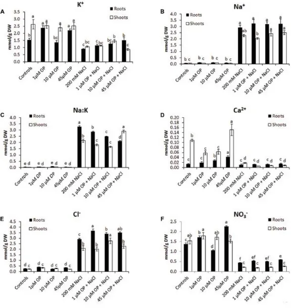
29 2.4.4 Ion Profile of Omeprazole Treated and Salt Stressed Plants
To better understand the mechanisms that OP affects to increase growth and tolerance to salt stress, tissue ion concentrations were profiled in all hydroponic treatments. OP altered the ion accumulation of tomato plants in control conditions and under severe salt stress. In unstressed conditions, OP increased K+ accumulation in roots treated with 1 µM (Figure 4); however, it did not increase Na+ accumulation. OP did affect the Na+/K+ ratio of salt stressed leaves and roots. The root Na+/K+ ratio of roots under salt stress was reduced by 12, 23, and 35% in 1, 10, and 45 µM OP treated plants, respectively (Figure 4). Calcium accumulation was also affected by OP treatment. In shoot, lower OP concentrations, 1 and 10 µM, decreased shoot calcium concentration significantly. Interestingly, 45 µM OP increased calcium concentration in roots and shoots. Root chloride accumulation was observed to be elevated in roots of plants treated with OP when compared to controls only under stress condition. Treatment with OP was observed to increase nitrate content of roots at 1 and 45 µM. OP treatment with salt stress did not result in any significant increase in nitrate accumulation (Figure 4).
30
Figure 4 Ion profiles of Solanum lycopersicum var. M82 plants treated with omeprazole. Plants were grown in a hydroponic solution containing 0, 1, 10, or 45 μM OP, with and without 200 mM NaCl. Plants were harvested after 2 weeks of salt treatment (50 DAS, 14 DSS) and used for ion analysis. Values for K+ (A), Na+ (B), Na:K ratio (C), Ca2+ (D),
Cl- (E), and NO3- (F) are shown. Values indicate average ± SE (n = 6). Different letters indicate significant differences
at P < 0.05 between an OP treated sample and the corresponding untreated control.
2.4.5 Gene Expression
Gene expression analysis was used to characterize the downstream mechanisms affected by OP that resulted in improved growth and salt tolerance in hydroponically grown tomatoes (Figures 5–7). We looked at three general categories of genes: ion transporters, stress signal transduction and osmotic response components, genes involved in
31 antioxidant and photosynthetic systems. Gene expression was evaluated in roots and shoots. We found that OP treatment affected a number of genes in non-stress conditions and augmented responses of numerous key genes involved in salinity stress responses and adaptation.
For ion accumulation, exclusions and transport, we selected a few ion transporters known to play key roles in responses to salinity: the plasma membrane Na+ antiporter SlSOS1 (Salt Overly Sensitive 1), two tonoplast located K+ antiporters, SlNHX1 and SlNHX2 (Sodium/hydrogen exchanger 1 and 2) and the Na+ transporter SlHKT1.1 (High affinity K+ transporter) (Figure 5). SlSOS1 was significantly upregulated in all OP
treatments, with augmented expression over untreated controls under salt stress in roots and shoots (Figure 5). This may have likely contributed to the lower Na+/K+ ratio seen in the ion analysis. For the two tonoplast located potassium antiporters, SlNHX1 and SlNHX2, which also mediate critical functions, including turgor maintenance, stomatal function and ion homeostasis under hyperosmotic stress (Zhang and Blumwald, 2001; Pardo et al., 2006), both genes were significantly upregulated under 1 µM OP treatment with increased expression over controls under salt stress (Figure 5). We also analyzed the expression pattern of HKT transporters, which play an important role in limiting the influx and subsequent accumulation of sodium into the shoot as well as sodium loading into root xylem (Asins et al., 2013; Ali et al., 2016). HKT1.1 expression in shoots was higher in OP treated plants compared to untreated controls in non-stress and salinity stress conditions (Figure5). In roots, we found that OP treatment decreased SlHKT1.1 expression slightly in non-stress conditions and remarkably under salt stress. Decreased expression of HKT1 transporters may have significantly reduced sodium entry into roots, protecting them from ionic stress. Furthermore, HKT1 expression increased in salt stressed shoots. This may have favored sodium recirculation into the xylem, a function that coupled with decreased uptake and loading in roots could be a key role OP plays in salt tolerance. This result is consistent with a reduced Na+/K+ ratio found upon OP treatment. In non-stress conditions, treatment with OP increased the nitrate content of roots and moderately in shoots (Figure 4). In order to link the observed nitrate
32 accumulation profile with gene functions, we examined the gene expression of the bidirectional transporter SlNRT1.1 (High affinity nitrate transporter 2) responsible for uptake and transport of nitrate (Jossier et al., 2010). Expression of SlNRT1.1 was upregulated in OP treated plants, in roots and shoots, under non-stress conditions. We also observed higher SlNRT1.1 expression in salt stressed plants with OP application although no significant increases in nitrate content were detected under stress conditions. With respect to the stress signal transduction components, we analyzed the expression of an ABA biosynthesis gene, SlNCED (9-cis-epoxycarotenoid dioxygenase), and an ABA catabolism gene SlCYP707A3 (ABA 8'-hydroxylase). We found that OP treatment decreased SlNCED expression in roots and shoots in non-stress conditions. Interestingly, SlNCED was induced upon salt stress, yet OP treatment caused an opposite response compared to what was observed in control plants (Figure 6). Specifically, in contrast to control plants, shoot expression of SlNCED was elevated over untreated controls in shoots under salt stress while root expression was significantly reduced. SlCYP707A3 expression was highly dysregulated under 1 µM OP treatment. While shoot expression was less than a third of controls, root expression was nearly three times that of untreated roots. Under salinity stress and OP treatment, expression of SlCYP707A3 was not downregulated. To better explain the expression pattern of ABA related genes, we examined the expression of SlLEA (Late embryogenesis abundant protein) a highly inducible marker in response to abiotic stress (Iovieno et al., 2016). While SlLEA was induced in salt stress conditions, it was less highly upregulated in the roots of OP treated plants under salt stress. This seems to correlate with decreased ABA signal transduction in the roots. In salt stressed shoots treated with OP, SlLEA demonstrated drastic upregulation, 10-fold higher than in untreated salt stress controls.
We also examined the antioxidant machinery and osmotic adaptation expression profiles of genes associated to ascorbate and proline biosynthesis. The cytosolic ascorbate peroxidase, SlAPX2 (Ascorbate peroxidase), was highly upregulated in shoots and roots of salt stressed plants. In OP treated plants under salt stress, root SlAPX2 expression was highly induced, compared to salt stress controls, indicating a more robust ROS
33 scavenging response induced by OP. With respect to proline and osmotic stress response, while expression of pyrroline-5-carboxylate synthetase, SlP5CS was not significantly altered in OP treated controls, it showed increased expression in salt stressed roots and shoots. Expression of the genes encoding for the catabolic pyrroline-5carboxylate dehydrogenase, SlP5CD (Proline dehydrogenase), showed a similar pattern in shoots of OP treated plants under salt stress. This may have contributed to a differential accumulation of proline in the roots and shoots.
The last set of genes we examined were those involved in the protection of the photosynthetic system. Expression levels of the tomato photosystem II reaction center psb28-like protein (PSII) were significantly upregulated under low concentrations of OP. These increases were also observed in OP treated plants under severe salt stress (Figure 7). In the leaves of OP treated plants under salt stress we found significant upregulation of the tomato catalase gene, SlCAT1 (Catalase 1) (Figure 7). High SlCAT1 expression levels have been found to enhance salt stress tolerance by reducing photoinhibition from damage to the photosystem by H2O2 (AlTaweel et al., 2007). We also examined the
tomato homolog of FtsH, an ATP-dependent protease that plays a key role in degradation and repair of photosystem II (Kato et al., 2009; Sun et al., 2010). Expression of SlFTSH was below detectable thresholds in controls and OP treated plants. However, salt stress induced SlFTSH expression with an even greater upregulation under salt stress and OP treatment.
34
Figure 5 Ion transporter gene expression in plants treated with omeprazole. Plants were grown in a hydroponic solution containing 0, 1, and 45 μM OP, with and without 200 mM NaCl. Samples for qRT-PCR were harvested after 2 weeks of salt treatment (50 DAS, 14 DSS) and harvested for ion analysis. Values indicate average ± SD (n = 3).
35
Figure 6 Secondary metabolism and stress signaling gene expression in plants treated with omeprazole. Plants were grown in a hydroponic solution containing 0, 1, and 45 μM OP, with and without 200 mM NaCl. Samples for qRT-PCR were harvested after 2 weeks of salt treatment (50 DAS, 14 DSS) and harvested for ion analysis. Values indicate average ± SD (n = 3).
36
Figure 7 Photosynthetic gene expression in plants treated with omeprazole. Plants were grown in a hydroponic solution containing 0, 1, and 45 μM OP, with and without 200 mM NaCl. Samples from leaves for qRT-PCR were harvested after 2 weeks of salt treatment (50 DAS, 14 DSS) and harvested for ion analysis. Values indicate average ±
SD (n = 3).
2.5 Discussion
2.5.1 OP Improves Plant Growth and Salt Stress Tolerance
In this work we demonstrated that by feeding tomato roots with hormonal concentrations of omeprazole, a benzimidazole PPI in animal systems, we can significantly improve plant growth and ability to tolerate saline stress. OP treatment with 1 µM increased shoot FW by 49% and DW by 48%. FW of roots was increased by 55% and DW by 56% in the absence of stress. Under saline stress, shoot growth was maintained, with a 56% increase in shoot FW and 54% increase in DW. Roots showed the most dramatic phenotype under salt stress, with a doubling of FW and DW over untreated controls (Figures 1, 2). Although this morphological change was not the only component that may have enhanced salt tolerance of OP treated plants, this response may have important implications with respect to growth and adaptation in saline environments (Julkowska et al., 2014; Feng et al., 2016). Longer, more extensive roots may help to escape