UNIVERSITÀ DEGLI STUDI DI SASSARI
SCUOLA DI DOTTORATO IN
SCIENZE VETERINARIE
INDIRIZZO
:
Patologia e Clinica Animale (XXVII CICLO)Use of Two-Dimensional Speckle Tracking Echocardiography to
Assess Left Ventricular Systolic Function in Dogs with Systemic
Inflammatory Response Syndrome
Docente Guida
Prof.ssa Maria Luisa Pinna Parpaglia
Direttore Tesi di Dottorato del
Prof. Sergio Ledda Dott. Andrea Corda
ANNO ACCADEMICO 2014 – 2015
La presente tesi è stata prodotta nell’ambito della Scuola di Dottorato in “Scienze Veterinarie” dell’Università degli Studi di Sassari, a.a. 2014/2015 – XXVII ciclo, con il supporto di una borsa di studio finanziata con le risorse del P.O.R. SARDEGNA F.S.E. 2007-2013 - Obiettivo competitività regionale e occupazione, Asse IV Capitale umano, Linea di Attività l.3.1.
Acknowledgements
A very special thanks goes to Prof. Anne French from the Small Animal Hospital, University of Glasgow, Prof. Pablo Gomez Ochoa from the Department of Animal Pathology, University of Zaragoza and Prof.ssa Maria Luisa Pinna Parpaglia from the Department of Veterinary Medicine, University of Sassari for giving me their valuable teachings and suggestions during the last four years. My research would not have been possible without their helps.
Of course I would like to give particular thanks to Maurizio Chiodi and Massimiliano Porretta, from Esaote company, for giving me the technical support during the study, I would also like to acknowledge Prof. Giovanni Sotgiu, from the Department of Biomedical Science, University of Sassari, for providing the statistical analysis and Dott.ssa Rosanna Zobba, from the Departement of Veterinary Medicine of the University of Sassari, for the laboratory support.
Moreover I want to express my sincere thanks to the University of Sassari and in particular to the Department of Veterinary Medicine for giving me the chance to make the great experience of the European PhD during which I had the opportunity to grow professionally and the fortune to meet many wonderful colleagues and friends from all over Europe.
I want to thanks my parents, my sisters and my aunts Franca and Peppina for being such a huge support through my education and my experiences.
Last, but certainly not least, I’d like to thanks Marzia for her love, support, encouragement and patience during my pursuit of the European Doctorate in Veterinary Science.
Table of contents
LIST OF ABBREVIATIONS ... 3 ABSTRACT ... 4 1. INTRO DUCTIO N ... 5 1.1 SIRS ... 5 1.2 PATHOPHYSIOLOGY OF SIRS ... 6 1.3 PATHOPHYSIOLOGY OF MODS ... 101.4 MYOCARDIAL DEPRESSION IN SIRS ... 12
1.4.1 Cardiac myocyte contraction ... 12
1.4.2 Pathophysiology of myocardial depression in SIRS and sepsis ... 14
1.4.3 Myocardial injury in SIRS ... 18
1.4.4 Myocardial dysfunction in SIRS ... 19
1.5 ADVANCED TECHNIQUES IN VETERINARY ECHOCARDIOGRAPHY ... 24
2. OBJECTIVE O F TH E STUDY ... 34
3. MATERIALS AND METHO DS ... 36
3.1 CASE SELECTION ... 36 3.1.1 SIRS group ... 36 3.1.2 Control group ... 37 3.2 DATA COLLECTION ... 38 3.2.1 Blood collection ... 38 3.2.2 Echocardiographic examination ... 39
3.2.3 Two-dimensional Speckle Tracking Echocardiography ... 41
3.3 VARIABILITY STUDY ... 44
3.4 STATISTICAL ANALYSIS... 45
4. RESULTS ... 46
4.1 SIGNALMENT... 49
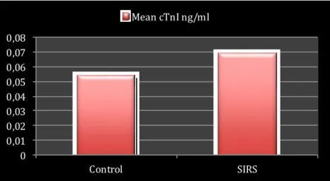
4.2 MEAN CRP, CTNI AND HEART RATE... 49
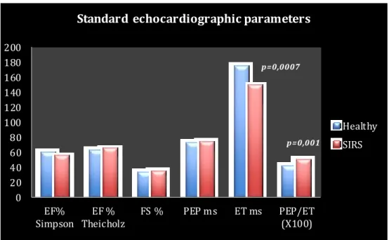
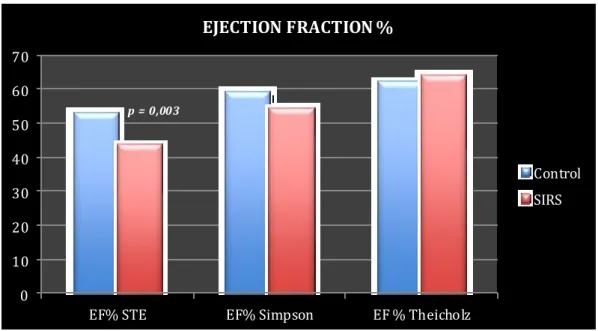
4.3 2D,M-MODE AND DOPPLER DERIVED INDICES OF LV SYSTOLIC FUNCTION ... 50
4.4 MEAN EF% OBTAINED BY SPECKLE TRACKING METHOD... 53
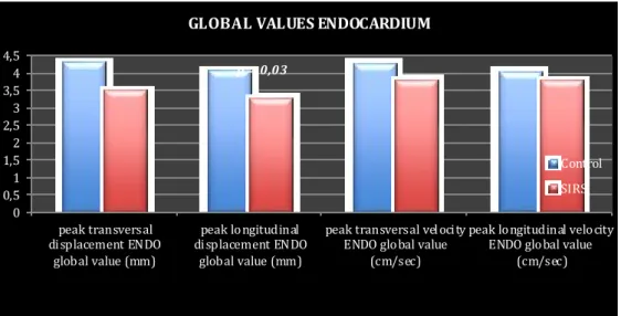
4.5 SPECKLE TRACKING ENDOCARDIAL VARIABLES... 54
4.6 SPECKLE TRACKING EPICARDIAL VARIABLES ... 56
4.7 SPECKLE TRACKING TRANSVERSAL STRAIN AND STRAIN RATE VARIABLES ... 57
4.8 VARIABILITY STUDY ... 58
4.9 CORRELATION BETWEEN VARIABLES ... 58
5. DISCUSSIO N ... 63
6. CONCLUSIO NS... 68
List of abbreviations
2Ch Two Chambers 2D Two-Dimensional
Echocardiography
2D STE Two-Dimensional Speckle Tracking Echocardiography
3Ch Three Chambers 3D Three Dimensional 4Ch Four Chambers
AFI Automated Function Imaging APPs Acute Phase Proteins AT Antithrombin
ATP Adenosine Triphosphate BP Blood P ressure CBC Cell Blood Count CI Cardiac Index CO Cardiac Output COX-2 Cyclooxygenase 2 CPV Canine P arvovirus CRP C-reactive P rotein CV Coefficient of Variation cTn Cardiac Troponin cTnI Cardiac Troponin I
DAMP Damage Associated Molecular Pattern
DCM Dilated Cardiomyopathy DNA Deoxyribonucleic Acid DO Oxygen Delivery ECG Electrocardiogram ECM Extracellular Matrix
ECVIM-CA European College of Veterinary Internal Medicine-
Companion Animals EF Ejection fraction ENDO Endocardial Border or
Endomyocardium EPI Epicardial Border or
Epimyocardium
EPSS E-point to Septal Separation ESVI End Systolic Volume Index ET Ejection Time
ET1 Endothelin-1
ETC Electron Transport Chain FPS Frames per Second FR Frame Rate
FS Fractional Shortening
GLPS Global Longitudinal Peak Strain HMGBP1 High Mobility Group Box Protein 1 HR Heart Rate
IBD Intestinal Bowel Disease IL Interleukin
IL-1 Interleukin One IL-6 Interleukin Six
IMP Index of Myocardial P erformance iNOS Nitric Oxide Synthase
IVS Interventricular Septum LA Left Atrium
LiDCO Lithium Dilution Cardiac Output LPS Lipopolysaccharide
LTCC L-type Calcium Channels LV Left Ventricle
LV DV Left Ventricular Diastolic Volume LV GLS Left Ventricular Global
Longitudinal Strain
LV SV Left Ventricular Systolic Volume LVIDd Left Ventricular Internal Diameter
in Diastole
LVIDs Left Ventricular Internal Diameter in Systole
LVPW Left Ventricular Posterior Wall MDS Myocardial Depressant Factor MODS Multiple Organ Dysfunction
Syndrome
MOF Multiple Organ Failure MRI Magnetic Resonance Imaging NF-ĸB Nuclear Factor Kappa B NK Natural Killer
NO Nitric Oxide
PAF Platelet-Activating Factor PAMP S Pathogen Associated Molecular
Pattern
PCO2 Partial Pressure of Carbon Dioxide
PEP Pre Ejection Period
PMNLs Polymorphonuclear Leukocytes PRR Pattern Recognition Receptor PW Pulsed Wave Doppler RA Right Atrium RBCs Red Blood Cells RV Right Ventricle RyR Ryanodine Receptors SD Standard Deviation
SERCA Sarcoendoplasmic Reticulum Ca2+
-ATPase
SIRS Systemic Inflammatory Response Syndrome
SMOD Simpson’ s Method of Disks SR Sarcoplasmic Reticulum St Strain
STE Speckle Tracking Echocardiography StR Strain Rate
TDI Tissue Doppler Imaging TF Tissue Factor
TLR Tool Like Receptor TM Thrombomodulin TnC Troponin-C
TNF Tumour Necrosis Factor TNF- α Tumour Necrosis Factor Alpha TnI Troponin-I
TnT Troponin-T WBC White blood cell
Abstract
SIRS is a clinical syndrome caused by systemic inflammation of infectious or non-infectious origin. SIRS is characterized by an endogenous cascade of interleukins and others inflammatory mediators such as TNF-α IL-6 and IL-1 which are responsible to the myocardial depression during systemic inflammation. Conventional echocardiographic indices of LV systolic function such as Fractional Shortening (FS%) and Ejection Fraction (EF%) are not sensitive enough to detect mild or early sy stolic dysfunction in dogs suffering from SIRS. Speckle-Tracking Echocardiography (STE) is a new echocardiographic technique that allows an objective and quantitative evaluation of global and regional myocardial function through the analysis of the motion of speckles that are created by the interaction of ultrasonic beams and the myocardium during the 2D exam. In the present study we tested the hypothesis that 2D-STE may detect LV systolic dysfunction, not diagnosed by conventional echocardiography , in dogs with SIRS. 17 dogs with evidence of SIRS and 17 healthy dogs as a control group were included in this prospective study. At the time of Veterinary Teaching Hospital admission each dog was submitted to standard 2D, M -mode, Doppler and 2D Speckle Tracking echocardiography to assess systolic function. Furthermore, blood samples were obtained for the measurements of cTnI and CRP serum levels. To assess the intraobserver within-day and between-day variability of the 2D-STE acquisition and measurements we performed a study of variability on 5 healthy dogs belonging to the control group. The results showed that the 2D-STE had low intraobserver variability and that the LV Global Longitudinal Peak Strain of endomyocardial layer (ENDO GLPS) and the STE-derived Ejection Fraction (EF%) were lower in the SIRS group than in the control group. On the contrary, standard 2D and M -Mode indices of systolic function such as EF%, FS% weren’t significantly different between the two groups. We didn’t find significant correlation between CRP serum levels and 2D-STE variables and between cTnI and ENDO GLPS. Our study demonstrated that 2D-STE was more sensitive than standard echocardiography in detecting early or mild to moderate myocardial dysfunction, not detected by conventional echocardiography, in a population of dogs with SIRS.
1. INTRODUCTION
1.1 SIRS
The Systemic Inflammatory Response Syndrome (SIRS) is a clinical syndrome caused by systemic inflammation of infectious or non-infectious origin. The term SIRS was introduced by the American College of Chest Physician and Society of Critical Care Medicine Consensus Conference held in Chicago in August 1991. The committee defined SIRS as a complex pathophysiologic response to a variety of insults such as infection, pancreatitis, ischemia, multiple trau ma, tissue injury, haemorrhagic shock, immune-mediated organ injury and the exogenous administration of inflammatory mediators such as Tumour Necrosis Factor (TNF) and other cytokines1. Sepsis was defined as a systemic inflammatory response to infection. The pathophysiologic state of the SIRS has been studied extensively. The onset of inflammation results in a complex set of interactions within the immune system to minimize injury and preserve organ function. A contained inflammatory response can become over-exuberant and deregulated by on-going injury, the addition of new insults or a compromised immune system. An uncontrolled generalized activation of the inflammatory reaction in organs remote from the initial insult can progress to secondary multiple organ dysfunction syndrome1 (MODS), multiple organ failure (MOF) and death2. Heart rate, respiratory rate, body temperature and white blood cell count are the clinical criteria used to categorize patients with SIRS in veterinary3 and human medicine1. SIRS criteria have been used to determine the severity of illness an d prognosis in critically ill dogs3 and in puppies with canine parvovirus infection4 in both studies mortality was significantly associated with the number of criteria fulfilled. In the field of canine medicine, SIRS has been described in a lot infectious and non infectious clinical conditions such as bite wound trauma2, soft tissue trauma, gastrointestinal foreign body/obstruction, gastrointestinal ulceration, septic peritonitis, parvovirus enteritis, mast cell tumour, lymphoma, pancreatitis, pancreatic adenocarcinoma and pyelonephritis5 as well as in
tumour, splenic haemangiosarcoma, gastric volvulus, canine distemper virus infection, leptospirosis7, urinary tract infections, pneumonia, polyarthitis8, staphylococcal skin infection9, subcutaneous infections, hepatobiliary bacterial infections10 splenic torsion, bile peritonitis, heat stroke11, babesiosis12, uterine infections, prostatitis, osteomyelitis, abscesses13, discospondylitis, myositis, steroid-responsive meningoencephalitis, prostatic abscess, mastitis and pulmonary carcinoma14. Differentiating between sepsis and non-infectious forms of SIRS can be a diagnostic challenge as the clinical presentation is often similar10.
1.2 Pathophysiology of SIRS
SIRS and sepsis are characterized by the activation of a strong immune response. The innate immune response is the first line of defence against a microbial invader and represents a nonspecific response that can be quickly activated by innate-cell types such as neutrophils and macrophages. These cells have Pattern Recognition Receptors (PRRs) that identify different microbial molecules know as Pathogen-Associated Molecular Pattern (PAMPs). The most widely recognized PAMP is lipopolysaccharide (LPS) of Gram-negative bacteria. When a PRR recognizes a PAMP, cells of the innate immune system generate a strong response that may ultimately result in the death of the invading microorganism. This can be achieved by phagocytosis or by inducing an up regulation of the immune response by interaction with the adaptive immune system, based on a system of T and B-lymphocytes that respond to specific epitopes.
Among the membrane-bound PRRs, the Toll-Like Receptors (TLRs) are the first identified and best characterized. TLRs are highly conserved and expressed ubiquitously throughout species, including mammalian. In mammalian they are expressed in immune cells such as monocytes/macrophages, neutrophils, natural killer cells, dendritic cells, mast cells, specific T and B ly mphocytes and non immune cells such as epithelial cells, skin keratinocytes, fibroblasts, cardiomyocytes and endothelial cells in the heart15. The TLRs are able to recognize and respond to a large numberof microbial organisms including
Gram-negative and Gram-positivebacteria, Mycoplasma spp., fungi, viruses, parasites, bacterialflagellin as well as host cells colonized by viral pathogens. In addition to microbial PAMPs, TLRs can recognize endogenously produced mediators that are released in response to injury or inflammation called Damage-Associated Molecular Patterns (DAMPs). DAMPs are endogenous molecules specifically generated upon tissue injury. So me are intracellular molecules normally inaccessible to the immune system that are released into the extracellular milieu as a result of cell necrosis or activation following injury. Others are extracellular matrix (ECM) mo lecule fragments that are released upon tissue damage or ECM molecules that are specifically up regulated in response to tissue injury. DAMPs are vital danger signals that alert the immune system to tissue damage upon both infectious and sterile insult. DAMPs have implicated in diseases where excessive inflammation plays a key role in pathogenesis, including autoimmune disease and cancer16. PAMPs and/or DAMPs identification by TLRs leads a complex cascade of intracellular signals that culminates in the activation of the transcriptional factor “Nuclear Factor kappa B” (NF-ĸB) leading inflammatory gene expression17. NF-ĸB activates transcription sites for a variety of genes including pro-inflammatory cytokines, chemokines, and other inflammatory mediators such as Tumour Necrosis Factor alpha (TNF-α), Interleukins (ILs) 1, 6, 8, 12 and Interferon (IFN-α and INF-β)18. Cytokines are soluble proteins produced and secreted as part of the immune response to a variety of insults including infection, cancer, and autoimmunity. Chemokines are similar proteins, but they induce chemotaxis and promote the migration of certain cells to areas of infection18. Cells of the immune system secrete most of these pro-inflammatory mediators. The primary function of these molecules is to promote inflammation by signalling pathways that induce expression of inflammatory products, which have direct effects on the host vasculature and facilitate the transmigration of leukocytes to the site of infection. Dogs with SIRS of infectious or non-infectious origin demonstrate high levels of interleukins in their bloodstream14, 19,
TNF-α is predominantly produced by activated macrophages and T-cells but also by mast cells, B-cells, Natural Killer (NK) cells, neutrophils, endothelial cells, myocytes, osteoblasts and fibroblasts. Many of the classical features of inflammation can be attributed to the actions of TNF-α upon the endothelium with increased production of Nitric Oxide Synthase (iNOS) and Prostaglandin Synthase type 2 (cyclooxygenase 2 or COX-2) leading to vasodilatation and local slowing of blood flow. TNF-α also stimulates the expression of endothelial adhesion molecules that lead to the tethering of leukocytes to the endothelial wall and their transmigration into the interstitium accompanied by fluid and plasma macro molecules. In addition to the up-regulation of iNOS, COX-2 and adhesion molecules, TNF-α also induces the expression of pro-coagulant proteins such as Tissue Factor (TF) and down regulates anti-coagulant factors such as Anti-thrombin (AT) and Thrombomodulin (TM), leading to the activation of coagulation cascade21. Monocytes and endothelial cells are the predominant cell type to express Tissue Factor after appropriate stimulation by endotoxin, immune complexes, cytokines (TNF-α and IL-6), and activated platelets22. Increased serum activity of TNF-α correlate strongly with a severe outcome or mortality in canine parvovirus infection23 and acute pancreatitis24. Another important pro-inflammatory cytokine in SIRS is the interleukin-6, it is a powerful pyrogen and it has a strong stimulatory effect upon leukocyte activation and myeloid progenitor cell proliferation25. Many papers report increase of serum concentration of 6 in SIRS and sepsis dogs. Rau et al reported that plasma IL-6 concentration is predictive of outcome in naturally occurring canine SIRS and sepsis14. Do-Hyeon et al found high IL-6 p lasma levels in dogs with infectious and non-infectious SIRS, in the same study IL-6 levels were not related to outcome19. Increased TNF-α and IL-6 serum concentration in dogs has been reported in a canine model of sepsis induced by endotoxin26 and in a group of 45 dogs with natural occurring SIRS (22 with sepsis and 23 with non infectious SIRS). In the last report mentioned above TNF activity and IL-6 concentration were not different in dogs with sepsis compared to dogs with non-infectious
SIRS and authors didn’t find correlation between TNF bioactivity, IL-6 concentration and survival to discharge27. TNF-α and IL-6 are both known for inducing the expression of pro-coagulant proteins such as Tissue Factor (TF). Interleukin-1 (IL-1) acts like TNF-α in the hyper acute period after immune stimulation in sepsis and SIRS. It induces the synthesis of adhesion molecules and cytokines by endothelial cells, stimulates leukocyte activation and endothelial tethering and transmigration into the interstitium. IL-1 also up regulates iNOS and COX2 production, acts as the mayor endogenous pyrogen in fever, and increases corticosteroid release via hypothalamic effects21. Recently another cytokine has been identified as being critical in dogs with SIRS and sepsis, it is the High Mobility Group Box Protein 1 (HMGBP1), an endogenous protein released into the circulation after necrotic or apoptotic cell death, it has direct pro-inflammatory actions. Dogs with SIRS and high plasma levels of HMGBP1 were associated with worse outcome regardless of the aetiology of SIRS19, 28. Under normal circumstances, cytokines exert their actions locally and their effects are restricted to neighbouring cells. Although their local effects benefit the host since they are designed to limit the initial injury or infection, in cases of severe inflammation or infection, the excessive release of pro-inflammatory cytokines overwhelms the host’s compensatory anti-inflammatory response and results in progressive endothelial dysfunction, increased microvascular permeability and activation of the coagulation system7, 29–31.
During systemic inflammation, the pro-inflammatory cytokines stimulate the synthesis and the release of large quantities of acute phase proteins (APPs) from hepatocytes and other cells. For years APPs were thought to be synthetized only by the liver, however, there is growing evidence that they also can be produced in other organs such as kidney, intestine, heart, and different types of white blood cells in humans32, 33. APPs have a variety of functions designed to re-establish homeostasis, assisting in pathogen elimination and overcoming inflammation. The acute phase response is characterized by fever, neutrophilia, activation of the coagulation and complement cascades, serum iron and zinc binding, enhanced
gluconeogenesis, increased muscle catabolism, and altered lipid metabolism34. As a consequence of the up-regulation of APPs production, concentrations of other plasma proteins such as Albumin, Protein C, Antithrombin (AT) and Protein S (collectively know as negative APPs)35, 36 decrease. A number of APPs have been characterized in veterinary species37 including dogs38, and they have been used as biomarkers of systemic inflammation in both research and clinical practice39. C-Reactive Protein (CRP) is a major APP in dogs. The normal levels and the effect of age and sex on the CRP concentration in healthy dogs has been investigated and no correlation has been observed40, 41. CRP increase rapidly after a systemic inflammatory stimulus triggered by various clinical conditions42–45 such as infection46–49, neoplasia50–52, trauma43, immune-mediated diseases53, 54 parasites55–57 and other inflammatory states like acute pancreatitis58 and snake envenomation59. Interestingly, several studies have reported raised serum levels of CRP in pregnant bitches39, 40, 60 from day 21 to day 55 after ovulation. Probably, the endometrial injury caused by the imp lantation itself provokes this response in the maternal circulation. In general, serum CRP concentration provides a sensitive but nonspecific means of measuring inflammation . A recent study of SIRS and sepsis in dogs demonstrated a correlation between decreasing serum CRP concentration and recovery from disease, suggesting its use as a prognostic biomarker in this context61.
1.3 Pathophysiology of MODS
MODS defines the progressive, but potentially reversible, dysfunction of two or more organ systems after acute life-threatening disruption of systemic homeostasis62. Organ systems that can be affected in patients with MODS include the renal, cardiovascular, respiratory, hepatic, hematologic, neurologic, gastrointestinal, endocrine and immune systems63. Mortality rates increase as the number of dysfunctional organ systems increase. A veterinary clinical report in 2010, of 114 dogs with sepsis secondary to gastrointestinal tract leakage, found that the overall mortality rate was 70% with MODS and 25% for those with only
dysfunction of one organ system. In this study the risk of death increased as the number of dysfunctional organs increased, and dysfunction of either the respiratory, cardiovascular, renal or coagulation system was independently associated with a significantly increased odds of death64. The exact pathophysiology of the development of MODS has not been completely defined. It is likely that multiple pathophysiologic mechanisms and mediators are involved in the pathogenesis of this injury process. A number of the potential pathophysiologic mechanisms, which may act individually or collectively in the production of organ system dysfunction, are:
Collateral tissue damage caused by activated immune cells65 and the
direct toxicity (cytopathic changes consequent to cytokine-cell receptor interactions) of the systemically released mediators of the SIRS response.
Inadequate tissue perfusion or the misdistribution of blood flow, which contains oxygen, nutrients, and other important substrates required to support cellular and organ function. In the setting of sepsis and SIRS there is commonly a decrease in the cardiac output, decreased systemic perfusion pressure which may result in hypoperfusion or ischemia of the organ system. The period of ischemia may be followed by reperfusion and oxidative injury, which may produce organ system dysfunction/failure. Tissue ischemia also may result from the aggregation of microthrombi composed of platelets, PMNLs, RBCs, and fibrin, which may obstruct the small capillaries and impair the delivery of blood and substrate to the tissues.
Some authors have speculated that even though adequate blood flow may reach the various tissue beds, there may be an inability of the mitochondria or cells to take up or use the delivered oxygen and substrate. The inflammatory cytokines IL-1β and TNFα promote mitochondrial o xidant production thus increase the state of oxidative stress66. Strong evidence suggests that oxidative stress may be responsible for mitochondrial damage during sepsis and other acute illnesses.
The free radicals can damage mitochondrial proteins, including components of the Electron Transport Chain (ETC) 67 and mitochondrial DNA 68. Mitochondria damage results in impaired oxygen utilization and attendant reduction in the capacity to produce ATP, which is associated with loss of cellular function in vital organs69.
1.4 Myocardial depression in SIRS 1.4.1 Cardiac myocyte contraction
Before describing the pathophysiological mechanisms that are assumed to be responsible for the myocardial depression in sepsis and SIRS, it is appropriate to describe briefly the physiology of myocardial contraction. Cardiac myocytes are a type of striated muscle cells, so called because cross striation are observed microscopically. Myocytes connect to each other by way of specialized cell membranes called intercalated disk forming a branching network of cells that is sometimes referred to a functional syncytium. Within the intercellular regions there are gap junctions, which permit cell-to-cell conduction of electrical currents. Gap junction ensure that the electrical impulse travel to all the interconnected myocytes allowing the myocardium to contract as a unit. The functional unit of the myocyte is the sarcomere, composed of thick and thin filaments. Thick filaments are composed of myosin, whereas thin filaments contain actin and other associated proteins70.
Myocardial contraction and relaxation are regulated by the interaction among contractile proteins (actin and myosin filaments), regulatory proteins (troponin and tropomyosin) and calcium ions. About 300 molecules of myosin packed together compose each thick filament and six thin filaments surround it. Each thin filament is composed of actin, tropomyosin and troponin. Actin is a globular protein arranged as a chain of repeating globular units, forming two helical filaments. Interdigitated between the actin filaments there are regulatory proteins called tropomyosin. Each tropomyosin molecule is associated with seven actin molecules. Attached to the tropomyosin at regular intervals there is the troponin regulatory complex, made up of three subunits: Troponin-T (TnT), which
attaches to the tropomyosin; Troponin-C (TnC), which serves as a binding site for Ca2+ during excitation-contraction coupling and Troponin-I (TnI), which binds to actin. In the resting state, tropomyosin blocks the site of actin –myosin interaction, while the TnI subunit inhibits the actin–myosin ATPase. Following the myocyte depolarization, during phase 2 of the cardiac action potential, extracellular Ca2+ enters the myocyte through sarcolemmal L-type Calciu m Channels (LTCC) also known as the Dihydropyridine Channels. This, in turn, lead to release of intracellular Ca2+, stored in the Sarcoplasmic Reticulu m (SR) via Ryanodine Receptors (RyR). As the intracellular Ca2+ concentration increases, the ion binds the TnC in a concentration dependent manner. This leads to conformational changes in the troponin-tropomyosin regulatory complex, which move away fro m the myosin-binding site on the actin molecule. Actin and myosin are then free to crosslink and contraction proceeds with ATP hydrolysis. Ca2+ channel closure at the end of phase 2 of the action potential prevents extracellular influ x, while the Sarcoendoplasmic Reticulum Ca2+ ATPase (SERCA) pumps cytoplasmic Ca2+ back into the SR. Calciu m is also returned extracellularly via the sodium-calciu m exchanger. As intracellular Ca2+ concentration declines, troponin-tropomyosin complex reassumes its inhibitory configuration, and myocardial relaxation starts. ATP is required both for providing the energy for contraction and for relaxation. In the absence of sufficient ATP as occurs during cellular hypoxia, cardiac muscle contraction and relaxation will be impaired. Sarcoplasmic Ca2+ concentration determines the force of myocardial contraction. Higher intracellular levels lead to greater relief of troponin-tropomyosin complex inhibition, rendering more actin-myosin complexes active. β-adrenergic stimulation augments cardiac contractility by increasing intracellular Ca2+ influx, therefore, noradrenaline (released by sympathetic nerves) and adrenaline (released by the adrenal glands) are positive inotropic agents; β-agonism also promotes myocyte relaxation enhancing SERCA activity and preserving diastolic function. Cholinergic stimulation, in turn, decreases contractility by down regulating intracellular Ca2+ influ x. This
pathway is coupled to Muscarinic Receptors (M2) that bind acetylcholine
released by parasympathetic (vagal) nerves within the heart, therefore, acetylcholine is a negative inotropic agent70.
1.4.2 Pathophysiology of myocardial depression in SIRS and sepsis
Myocardial depression is a major contributor to mortality and morbidity in human patients with sepsis, in fact, in the 40% of patients who experience myocardial dysfunction as a complication of sepsis, the mortality rises from 70% to 90%71. In human medicine contractile dysfunction in the septic patients is defined as biventricular dilatation, reversible decrease in ejection fraction, diminished blood pressure response to intravenous fluids, and reduced ability to augment cardiac output despite increased levels of circulating catecholamines72. Cardiac function is severely impaired in sepsis, severe SIRS73 and MODS74. Although there are numerous causes of SIRS, sepsis is the most investigated, essentially because it is a leading cause of death in critically ill patients. Nevertheless, septic myocardial impairment remains a clinical enigma.
Wiggers in 1947 postulated the presence of a circulating myocardial depressant factor (MDF) in some cases of human shock75. Waisbren in 1951 was the first to report myocardial dysfunction in patients with sepsis, and subsequent experimental studies were able to confirm the suspected myocardial depressant factor in sepsis76. In 1985 Parrillo and colleagues77 demonstrated that serum of septic patients generated concentration-dependent depression of in vitro myocyte contractility. This was the first study that confirmed the long suspected link between sepsis and a circulating myocardial depressant factor. Using hemofiltration techniques, researchers found that the stimulating factor was greater than 10 kDa, heat labile, and water soluble, suggesting a protein or polypeptide78. Lipopolysaccharide (LPS) was postulated to represent the MDS because its infusion in animals and humans produced the hyperdynamic, hypotensive state seen in naturally occurring septic shock78.A second hypothesis suggested that reduced myocardial perfusion, due to peripheral vasodilation and hypotension, was involved in septic myocardial depression79. However, clinical
studies80 evidenced that coronary perfusion during septic shock was preserved or even increased and that hypotension was more likely the product of profound reduction in systemic vascular resistance.
Although endotoxin was sufficient to produce septic shock, it was unlikely to explain the complete mechanism in itself. Experimental studies found that gra m-positive bacteria produced the same pattern of cardiovascular changes as those invoked by endotoxin81. Furthermore, many patients that were culture negative or had no detectable levels of endotoxemia manifested the typical cardiovascular modifications of patients with septic shock. These findings suggested that endotoxin, and several other molecules contributed to the activation of an endogenous cascade of local and systemic inflammatory mediators , which caused myocardial dysfunction in sepsis and SIRS. Nowadays, researchers around the world believe that cytokines such as TNF-α, IL-6 and IL-1 are the main causes of the myocardial depression in SIRS/sepsis. In vitro studies showed significant decreases in measures of contractility in cardiomyocytes exposed to TNF-α, IL-1β 82, and IL-6 83.
The cellular mechanisms underlying cytokine-mediated cardiomyopathy have been extensively studied but remain incompletely elucidated. Alteration of intracellular calciu m currents, overproduction of Nitric Oxide (NO), Endothelin-1 (ETEndothelin-1) and Platelet-Activating Factor (PAF), o xidation–reduction imbalance, altered mitochondrial activities, autonomic deregulation, microcirculatory alterations and changes in load conditions, have all been invoked in explanation.
a) Lew et al in 1996 reported a reduction in cardiac Dihydropyridine Receptors (LTCC) by 25% during induced endotoxemia in rabbits84. Zhong et al reached the same conclusions by performing an experimental study on ventricular myocytes from endotoxemic guinea pigs85. Since Dihydropyridine Receptors are primarily L-type Calciu m Channels (LTCC), their reduction in endotoxemia may contribute to reduce intracellular Ca2+ influ x during the action potential resulting in negative inotropic effect. Rigby et al in 1998, strengthened this hypothesis by
showing that decreased available Ca2+, rather than decreased myofilament Ca2+ responsiveness, led to decreased contractile function of the pig heart during endotoxemia86. Finally, Stengl et al in 2010 demonstrated that septic shock, in pigs, was associated with decreased number of functional LTCC and reduction of myocyte Ca2+ influx. As a consequence of the decreased L-type Calciu m current there was a shortening of cardiac repolarization87.
b) TNF-α and IL-1 induce iNOS up-regulation during inflammatory response. The role of NO is well recognized in SIRS and sepsis. NO has a direct effect on cardiomyocytes, probably inducing contractile dysfunction82. NO inhibits mitochondrial function in various organs, including the heart. The underlying mechanisms of mitochondrial dysfunction induced by NO are still incompletely elucidated. The NO overproduction can cause direct oxidative damage and inhibition of oxidative phosphorylation complexes. As a consequence, mitochondrial ATP generation is compromised, and energy depletion may contribute to cardiac contractile dysfunction. It further induced vasodilation with resulting changes in load conditions (preload and afterload) and myocardial perfusion.
c) Persistently elevated ET-1 plasma levels are associated with myocardial dysfunction in sepsis88 but the exact ro le of ET-1 in the pathogenesis of myocardial depression in sepsis is not completely understood. ET-1 may have a direct myocardial depressive action. It also produces deregulation of systemic and regional vascular tone and itself may trigger increased inflammatory cytokines and NO89.
d) Prostanoids, including thromboxane and prostacyclin, may contribute to impaired cardiac function by altering coronary endothelial function and promoting intracoronary leukocyte activation90.
e) Platelet-Activating Factor has been reported to directly decrease myocardial contractility via a specific, high-affin ity cardiac PAF receptor.
PAF directly decreases velocity of contraction, and of relaxation in cardiomyocytes91.
f) Autonomic impairment, identified experimentally following endotoxin exposure, could be related to alterations within the brain, the autonomic nervous system, or the pacemaker cell itself73. The autonomic deregulation includes resistance to catecholamines, despite elevated circulating levels of these mediators in SIRS/sepsis. The effect is mediated by decreased density or dysfunction of myocardial adrenoreceptors92. Studies have identified that endotoxin-induced alterations of heart rate variability are mediated, at least in part, by direct interaction with the cardiac pacemaker cells93.
g) Changes in loading condition are common in SIRS. Cytokines increase capillary permeability leading to decreased intravascular volume (reduced preload). The release of vasodilating factors like NO, TNF-α and prostaglandins, and the vascular decreased response to vasopressor agents leads to a reduction in afterload and hypotension.
h) Microcirculatory alterations are pathological changes in the capillary bed related to microtrombi formation and activation of circulating cells (leukocytes and platelets). As a consequence of the microcirculatory modifications, organ perfusion and oxygen delivery is impaired and high lactate levels can be produced by anaerobic glycolysis. Progressive hypoxemia94 and lactate acidosis95 limits ventricular oxygen consumption and contractility.
1.4.3 Myocardial injury in SIRS
Myocardial injury, detected by increase of cardiac troponins, is observed in setting of SIRS and sepsis in humans96 and dogs97, 98. Troponins are regulatory proteins of the contractile apparatus of striated muscle cells (skeletal and cardiac myocytes). Each troponin protein has specific functions in contractile mechanism. Card iac Troponin (cTn) is present only in cardiac muscle and Cardiac Troponin I (cTnI) is much more specific for detection of any damage to cardiac myocytes99. During myocardial injury, loss of membrane integrity, causes release of cTnI into the circulation, elevated cTnI levels indicate myocardial damage100. In dogs, cTnI is detectable within 4 to 6 hours and peaked between 10 to 16 h after cardiac injury101. Langhor et al in 2013 showed the clinical importance of cTnI as a marker of myocardial cell in jury occurring in dogs with systemic inflammation and without primary cardiac disease. The study included 42 dogs presented with a diagnosis of trauma, neoplasia, gastrointestinal, respiratory, neurological and ematological diseases. Authors assessed systemic inflammation by measurement of CRP and several cytokines serum activity, they also evaluated myocardial cell damage by measuring cTnI concentration97. cTnI serum levels were higher in disease dogs than in control group and cTnI was found to be predictive of death in critically ill dogs with systemic inflammation97, 98. Myocardial injury associated with systemic inflammation has also been described in dogs with snake envenomation102, ehrlichiosis103, gastric dilation-vulvulus104, babesiosis105, pyometra106, parvovirosis107and leishmaniasis108.
Hagman et al reported that cTnI levels did not differ significantly in a pyometra patient group when compared with healthy dogs the same study showed that presence of SIRS was not associated with increased cTnI values109.
1.4.4 Myocardial dysfunction in SIRS
Several veterinarian studies reveal that myocardial dysfunction accompanies sepsis and SIRS also in dogs. Bulter et al in 2008 demonstrated that cardiac output (CO), assessed by the Lithiu m Dilution Cardiac Output (LiDCO) method, oxygen delivery (DO), and cardiac index (CI) were significantly lower in a group of 18 dogs that meet SIRS criteria co mpared to healthy controls111. DO is the amount of oxygen delivered to the tissue and is calculated as the product of CO and arterial o xygen content, CI is the product of heart rate and stroke volume indexed to body surface area. LiDCO technique allows CO to be measured using only a peripheral or central venous catheter and a peripheral arterial catheter. The LiDCO method has been validated for use in dogs110. Since the heart rate did not differ between SIRS and control group, the authors hypothesized that the main cause of decreased in CO and CI, in the SIRS dogs, must be decreased stroke volume by reduced cardiac contractility secondary to inflammatory cytokines. CI, CO and OD were not significantly different between survivors and non -survivors111. Kenney and colleagues64 reported a large-scale study of the association between outcome and organ system dysfunction in dogs with sepsis secondary to gastrointestinal tract leakage. Cardiovascular dysfunction was defined as hypotension sufficiently severe to require vasopressor t reatment after surgery. Twenty (17.5%) of the 114 dogs met the criteria for cardiovascular dysfunction and only 2 of the 20 (10%) survived to discharge from the hospital. Echocardiography has been used in only a relatively small nu mber of studies to assess cardiac function in dogs with SIRS107, 112. The most commonly used echocardiographic indices to assess global systolic function are the Fractional Shortening (FS%) and the Ejection Fraction (EF%). The FS% is obtained using the M-Mode scan of the left ventricle to measure the internal systolic and diastolic diameters. FS is calculated as follows:
FS% = (LVIDd – LVIDs / LVIDd) x 100
Were LVIDd is the Left Ventricular Internal Diameter in diastole and LVIDs is the Left Ventricular Internal Diameter in systole. FS% has several limitations, first, it measures only the radial contractility without considering the longitudinal and torsional motion of the left ventricle, second, it is highly dependent by preload and afterload conditions. A low FS% may be due to decreased radial contractility as well as increased afterload (vasoconstriction, systemic hypertension) or decreased preload (hypovolemia, dehydration). Conversely high FS% may be secondary to increased preload or decreased afterload. Accord ing to the Frank Starling principle, preload affect FS% because an increase in left ventricular diastolic size stretches the myofibers and increases their ability to shorten during systolic contraction. Moreover, FS% is negatively correlated to the heart rate, it decreases when the ventricle has less time to fill113. Normal FS% value in dogs is 34,4 ± 6,5114. FS < 25% is considered index of systolic dysfunction115, although in some breeds, healthy subjects, can have a lower FS%116.
Ejection Fraction (EF%) is an index of global systolic function derived from ventricular volumes. It is calculated as follows:
EF % = (LV DV – LV SV / LV DV) x 100
Where LV DV is the Left Ventricular Diastolic Volu me and LV SV is the Left Ventricular Systolic Volume.
EF < 40% is considered index of systolic dysfunction115.
LV volu mes can be estimated by M-Mode and by Bi-Dimensional echocardiography. M-Mode derived LV volumes are calculated by the Teicholz equation as follows:
LV DV = (7 x (LVIDd) 3) / (2,4 + LVIDd) and
Were LV DV is the Left Ventricular Diastolic Volu me, LV SV is the Left Ventricular Systolic Volu me, LVIDd is the Left Ventricular Diameter in diastole, LVIDs is the Left Ventricular Diameter in systolie.
The Teicholz formula is a geometric method based upon the assumption that the LV chamber is an ellipse. Teicholz equation overestimates the LV volu me in dogs, especially in the case of volume overload117, this is because the geometric method does not use the LV length but only the transversal diameter.
Two-Dimensional derived LV volumes are achieved by the Simpson method. The ventricular volumes are derived from the left ventricular diastolic and systolic area, obtained by drawing the endocardial contour of the LV on a 2D image (right parasternal long a xis or left apical 4 or 2 chambers views). Simpson’s derived method shows the best correlation with real volumes in the diseased heart117.
Another commonly used index of systolic function is the End Systolic Vo lume Index (ESVI). It represents the LV systolic volume indexed to the body surface area (BSA). The normal values depend on the method used to calculate volume. Spectral Doppler may also be used to obtain left ventricular systolic indices. The LV Pre Ejection Period (PEP) is the time interval between the beginning of the Q wave on the simultaneous ECG and the beginning of the ventricular ejection on the spectral Doppler trace. LV Ejection Time (ET) is the time interval between the beginning and the end of the aortic wave on the spectral Doppler. A systolic dysfunction leads to an increase of the PEP, as a consequence there is an increase in the PEP/ET ratio118.
In 2006, Nelson and Thompson119reported a retrospective study of 16 dogs with left ventricular dysfunction associated to critical illnesses . Critical illness was defined as metabolic derangements that required intensive care to sustain life, and left ventricular systolic dysfunction was defined as a FS < 26% and/or an EF < 46%. Dogs with a LV PEP/ET > 0.4 were also considered to have systolic dysfunction. Mitral valve inflow E/A ratio < 1 were interpreted as ventricular diastolic dysfunction. Dogs that had an obvious endocarditis and breeds of dogs
with an increased incidence of dilated cardiomyopathy were excluded from the study because of their greater risk of cardiac dysfunction. All the dogs were normally hydrated at the time of echocardiographic exam. The two most common diseases identified producing critical illness with left ventricular systolic dysfunction were sepsis (n 5) and cancer (n 5), within the cancer group 3 dogs had lymphosarcoma, 1 mesothelioma and 1 pulmonary adenocarcinoma. Other diseases diagnosed in affected dogs were pancreatitis, intestinal bowel disease (IBD), immune-mediated haemolytic anaemia, immune polyartropathy and coccidiomycosis. Twelve of the 16 dogs (75%) died or were euthanized within 15 days of hospital admission, with an average time until death of 3.6 days. Ten dogs had necropsy examination, which did not reveal gross or histological evidence of cardiac disease in any dog. Treatment regimens for the dogs varied considerably so comparison between survivors and non -survivors was not performed. The 4 dogs that were discharged had follow-up of 20 days, 3.5 months, 4 months, and 2 years respectively. Longitudinal echocardiographic data were available only for a dog with immune-mediated polyarthropathy, anaemia, and hyperglobulinemia that was still alive 2 years after hospitalization. The FS had risen from 21% at the time of hospitalization to 34% 2 years later, suggesting reversible myocardial depression119.
Dickinson et al120 reported a case of a 5-months-old 22 Kg female Rhodesian Ridgeback with reversible myocardial depression associated with septic arthritis. At the time of admission the dog fulfilled the SIRS criteria (tachypnea, fever and band neutrophils). Systolic dysfunction was assessed with an echocardiographic exam, which showed a dilated, hypokinetic left ventricle and a decrease in FS%. Right ventricle was also dilated. The Left Ventricular Systolic Diameter (LVIDs), the End Systolic Volu me Index (ESVI) and the E-Point to Septal Separation (EPSS) were all increased. The dog recovered well, and, three months later an echocardiographic examination showed good left ventricular contractile function and normal cardiac dimensions. All evidence of previously noted myocardial dysfunction had resolved.
Recently Kocaturk et al121 demonstrated myocardial injury and myocardial depression in dogs with SIRS induced by Canine Parvovirus (CPV). EF%, FS% and the Tei index were used to assess left ventricular systolic and diastolic dysfunction. cTnI levels were used to determine myocardial injury. Tei index calculated for non-survival dogs were significantly higher than that of survival and control dogs. Lower FS% and EF% were observed in non -survival dogs, compared with survival and control dogs. There were no significant differences in EF% and FS% between healthy and SIRS survivors dogs. The Tei index, also called Index of Myocardial Performance (IMP) is a Doppler derived index of global myocardial systolic and diastolic function developed by Tei122.
In the 22nd ECVIM-CA congress, Go mmeren112 et al presented an oral research communication about echocardiographic evaluation of cardiac dimensions and left ventricular systolic function in 37 dogs with SIRS (infection n=6, neoplasia n=4, trauma=4, gastric dilation-volvulus n=4, other gastro-intestinal disease n=4 and miscellaneous diseases n=15) and without primary cardiac disease. Left ventricular systolic function was assessed through FS% and EF%. In this study 28 patients (76%) survived. The authors concluded that in this population of SIRS patients, no echocardiographic evidence of cardiac dysfunction was demonstrated112.
The results of these studies suggest that myocardial injury and myocardial dysfunction are common findings in dogs suffering from SIRS of infectious or non-infectious origin. However, the commonly used echocardiographic indices of systolic function, such as FS% and EF%, have not been always sensitive enough to detect systolic dysfunction in dogs suffering from SIRS112. In fact, echocardiographic evidence of systolic impairment has been demonstrated mostly in SIRS dogs that did not survive compared to survivors119, 121. FS% and EF% were reduced only in case of severe cardiovascular impairment as seen in multip le organ dysfunction or failure123. Several studies in humans and in a porcine model of sepsis124 have shown evidence of left ventricular systolic dysfunction with preserved Ejection Fraction in patients with sepsis125, 126 and
other conditions of heart failure127. During SIRS mild to moderate systolic dysfunction may not be revealed by the decrease of FS% or EF% due to the reduction in preload and afterload and the increase in heart rate (HR).
1.5 Advanced techniques in Veterinary Echocardiography
New cardiovascular ultrasound imaging techniques have been developed over the last 15 years. They include Tissue Doppler Imaging (TDI) and Speckle Tracking Echocardiography (STE). These techniques allow analysing myocardial displacement, velocity and deformation that occur during the cardiac cycle. The active systolic movements of the left ventricle are complex and have three major components:
1) Longitudinal motion with the base moving toward the apex;
2) Radial motion, during which the left ventricular posterior wall (LVPW) and the interventricular septum (IVS) converge toward a geometric centre located in the centre of the left ventricular chamber. The radial motion is associated with a circu mferential deformation and radial thickening of the myocardium;
3) Torsional motion of the left ventricle, it is due to the opposite rotational movements of the base and apex in relation to an imaginary long axis line from apex to base. When the heart is viewed from the apex, the base rotate in clockwise direction and the apex rotate in counter clockwise direction128.
The complexity of ventricular kinetic is related to the geometrical layout of the myocardial fibres. They are set in three layers : sub-endocardial, mid-wall and sub-epicardial. Fibre orientation changes gradually from a right handed helix layout in the sub-endocardial layer to a circular layout in the midwall layer, and then to a left-handed helix layout in the sub-epicardial layer113. Radially oriented fibres of the middle layer are predominant at the base compared to the apex wall of the left ventricle.
There are also passive movements of the LV like the translation movements, corresponding to the displacement of the heart as a whole within the thoracic
cavity and the dragging movements by which a myocardial segment is dragged by the contraction of an adjacent segment (e.g. scar tissue which is unable to contract is dragged by adjacent vital myocardium during systole).
Tissue Doppler and Speckle Tracking Imaging have introduced new echocardiographic indices of myocardial function as myocardial displacement, velocity of displacement, deformation (Strain) and velocity of deformation (Strain Rate).
TDI is a Doppler technique that enables to quantify global and regional myocardial function, measuring the radial and longitudinal myocardial velocities throughout the entire cardiac cycle. From the myocardial velocity (expressed in cm/sec), Tissue Tracking Imaging (a technique derived from TDI) allows to calculate the myocardial displacement (expressed in mm) and the deformation parameters as Strain (St) and Strain Rate (StR). TDI has been the first echocardiographic method from which myocardial deformation parameters could be derived. However, TDI, has several limitations, in fact, being a Doppler technique it is angle dependent, and myocardial velocities assessed by TDI do not discriminate between actively contracting myocardium and passive motion due to translational movement and tethering effect129. The derived parameters of deformation, St and StR, partially reduce these limitations.
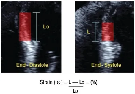
Strain (St) is the percentage of deformation of the myocardium during the cardiac cycle (Figure 3). It is calculated with the following formula:
St = L – L0 / L0
Where L is the length of the myocardial segment after deformation, and L0 is the
basal length of the segment. By convention, depending on the direction, a lengthening or thickening deformation is given a positive value, whereas a shortening or thinning deformation is given a negative one. Strain is expressed in per cent (%).
Strain Rate (StR) is defined as the velocity at which deformation occurs (Figure 4). It is equal to the Strain divided by Δt:
It is expressed in s-1; in other words, if the same Strain value is reached in half the time, the Strain Rate value will be doubled.
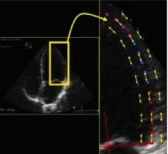
Speckle-Tracking Echocardiography (STE) is a new non-invasive ultrasound imaging technique that allows an objective and quantitative evaluation of global and regional myocardial function using a completely different algorithm compared to TDI-derived methods to calculate deformation. Speckles are groups of myocardial pixels that are created by the interaction (reflexion, scattering and interferences) of ultrasonic beams and the myocardium during the 2-dimensional exam. Single speckles are merged in functional units (kernels) that are in turn univocally identifiable given the peculiar disposition of the speckles. They represent the natural acoustic markers of the myocardial tissue; each kernel constitutes a sort of ultrasound fingerprint that can be tracked by the software from frame to frame during the entire cardiac cycle128, 130.
STE allows distinguishing between active and passive movements of myocardium. STE derived Strain is directly calculated from the frame-to-frame motion of speckle patterns and not from myocardial velocities (Figure 1 and 2). Through analysis of the motion of each kernel that composes a routine 2-dimensional grey scale image, the system, without using the Doppler signal, can measure global and regional displacement, velocity of displacement, Strain and Strain Rate, in longitudinal, radial and circumferential directions130 (Figure 2). It can also quantify rotational movements such as rotation, twist and torsion of the myocardium. The protocols for acquisition are not much different from routine echocardiograms. The endocardial border needs to be well delineated and well visualized for reliable tracking, and for that, images acquired should be of high quality. The optimal frame rate should be 60 – 110 frames per second (FPS). It is preferable to keep the sector width and depth minimal to focus on the structure of interest. As the values are averaged for the final results, using the software during final processing, three consecutive cardiac cycles are obtained. Considering the close dependence of STE and single-cardiac-cycle Strain analysis, it is not possible to conduct a study in patients with non-sinusal rhythm. There are
different algorithms used by different vendors in tracking these kernels. The region of interest (ROI) has to be outlined manually even if vendors have incorporated tools to help users identify tracking reliability. The LV is evaluated by tracing the endocardial border by point and click method. The epicardium is automatically traced by the system, but the wall thickness can be manually adjusted.
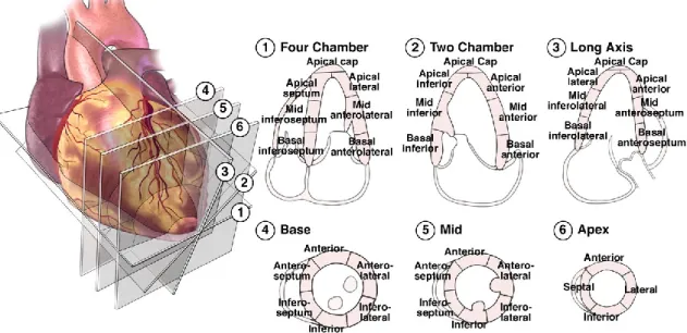
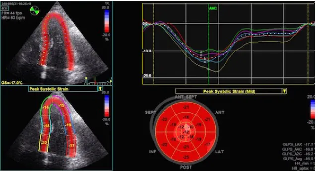
To obtain the complete STE analysis of the global LV longitudinal and radial function from the long axis views the operator needs to analyse three left apical views: left apical two, three and four chambers (Figure 5). The ways myocardial segments are divided widely vary among the vendors, but in general, a 16 to 18-segment LV model is used. Segments for which no adequate image quality can be obtained are rejected by the software and excluded from the analysis. Once the region of interest is optimized, the software generates Strain curves for each selected myocardial segment (Figure 6). From these curves, the operator can obtain regional and global peak and time-to-peak values. Global values of LV deformation parameters are obtained by averaging values observed in all segments. By the Automated Function Imaging (AFI) system, which allows a semiautomatic analysis of STE parameters, the results of each view are merged and represented in the bull’s-eye view of segmental parameters of deformation (Figure 7). Each analysed segment corresponds to a different coronary territory. Parasternal short-axis views one from the basal short-axis view, obtained at the tips of the mitral valve leaflets and the other at the apex, pro ximal to the end of LV cavity, are necessary to analyse radial, circumferential and torsional deformation of the left ventricular myocardium (Figure 5).
Longitudinal Strain (LS) represents the myocardial deformation directed from the base to the apex (Figure 2). During systole, ventricular myocardial fibres shorten with a translational movement from the base to the apex; the consequent reduction of the distance between single kernels is represented by negative St curves (Figure 6). During diastole the distance between kernels increase therefore the St value is positive. The Left Ventricular Global Longitudinal Strain (LV
GLS) has been recently validated as a quantitative index of global LV function in humans131. Culwell et al in 2011 found a significant linear correlation between 2D-STE derived LV GLS and load-independent indices of systolic function obtained through the thermodiluition technique in dogs132. Similarly, A mudsen et al, in 2006, identified a strong positive correlation between 2D-STE derived LV GLS and LV long-axis St obtained from Tagged MRI (r = 0,87; p< 0,001) in humans as well as between 2D-STE derived LV GLS and LV longitudinal St measured by sonomicro metry (r = 0,9; p< 0,001) in dogs133. MRI tagging is currently considered the non-invasive gold standard for evaluation of systolic deformation134. The results of these studies suggest that the LV GLS can be considered as a non-invasive, load-independent index of LV systolic function and therefore of LV contractility.
Radial Strain represents radially directed myocardial deformation, toward the centre of the LV cavity. It indicates the LV thickening (systole) and thinning (diastole) deformation during the cardiac cycle; consequently the radial St values during systole are represented by positive curves. Radial St measurements can be obtained by STE analysis of both LV short-axis and long axis views.
Circumferential Strain represents LV myocardial fibre shortening along the circular perimeter observed on a short-axis view. Consequently, during systole, for circumferential speckle-to-speckle distance reduction, circumferential strain measurements are represented by negative curves.
Parasternal short-axis views, one from the basal short-axis view obtained at the tips of the mitral leaflets, the other at the apex, barely pro ximal to the end of LV cavity, are necessary for rotation, twist, and torsion analysis.
Rotation is the measure of the rotational movement of the myocardium in relation to an imaginary long axis line fro m the apex to the base drawn through the middle of LV cavity. Clockwise rotations are assigned a negative value (e.g.-10°) and counter clockwise rotations a positive value (e.g. +(e.g.-10°).
Twist or torsion measures the algebraic difference in rotation between the apex and the base. For example, if the apex moves 30 ° counter clockwise and the base
moves 15 ° clockwise, then the twist will be 45 ° (30 - (- 15) = 30 + 15 = 45°. The rate and total duration of twist can also be measured.
Untwisting: Growing attention has been also recently given to the role of untwisting in diastolic LV filling mechanics. Untwisting velocity is thought to be a critical initial manifestation of active relaxation, which makes this measurement relevant for investigating diastole and, mainly, isovolumic relaxation because it seems to be less dependent on load compared to other diastolic parameters.
All the 2D-STE measurements can be individualized for each of the myocardial segment or can be expressed as global values when all the segmental values are averaged. St and StR can be measured not only for the LV but also for the Right Ventricle (RV) and Left and Right Atria135 (LA and RA), but these are much less commonly used in the clinical setting and some have not been fully validated. In canine cardiology 2D-STE have been used to measure LV Radial St136, LV Torsion137, Longitudinal St and StR138 for both endomyocardial (ENDO) and epimyocardial (EPI) layers139. In all the studies the 2D-STE has been shown to have a good repeatability and reproducibility. Moreover, 2D-STE has been validated in dogs against sonomicro metry, tagged MRI133, Tissue Doppler Imaging136, 138 and thermodilution technique132. Suzuky et al demonstrated that age140 and heart rate141 did not exert a significant influence on 2D-STE derived longitudinal systolic St and StR in healthy dogs.
Figure 1. Change in distance between kernels during cardiac cycle
Figure 3. Longitudinal Strain of the Left Ventricle
Figure 5. Apical and transversal view commonly used to evaluate the longitudinal, radial an torsional deformations of the LV
2. OBJECTIVE OF THE STUDY
Global systolic function is influenced by several factors including preload, afterload, contractility, distensibility, coordinate contraction and heart rate. Assessment of global left ventricular systolic function is of paramount importance in dogs suffering from SIRS because it could influence the prognosis and the treatment decision. There is evidence that endotoxin77 and several other molecules16 contribute to the activation of an endogenous cascade of local and systemic inflammatory mediators, such as interleukins83, which reduces myocardial contractility in humans and animals suffering from SIRS and sepsis82. It is also known that dogs with SIRS of infectious or non-infectious origin have high levels of interleukins in their bloodstream14, 19, 20. Previous studies conducted in dogs suffering from SIRS suggest that the conventional echocardiographic indices, such as LV Fractional Shortening (FS%) and Ejection Fraction (EF%), are able to detect systolic dysfunction only in severe SIRS or in the late stage of the disease in fact, low FS% and EF%, were found especially in non-survival dogs suffering from SIRS and sepsis107, 119. EF% and FS% as well as systolic time intervals are not sensitive enough to detect mild to moderate or early systolic dysfunction in dogs because they are highly dependent on loading conditions and heart rate132. Tagged MRI is considered to be the absolute non-invasive gold standard for measurement of LV contractile function134, but it is expensive and requires anaesthesia, which depresses contractility and may be a risk for critically ill patients. Several studies, including people and other animal models with sepsis, have revealed the ability of the 2D-STE deformation parameters in detecting LV systolic dysfunction before the reduction in the LV EF% could be seen124, 126. The aim of the present study was to evaluate the systolic function in dogs with SIRS using 2D-Speckle Tracking Echocardiography (2D-STE). We tested the hypothesis that 2D-STE may detect LV systolic dysfunction not diagnosed by conventional echocardiograp hy in dogs with Systemic Inflammatory Response Syndrome of both infectious and non-infectious origin. A second objective of the study was to compare serum
Cardiac Troponin I (cTnI) levels among dogs with SIRS and healthy dogs as well as to evaluate if there were correlation between systolic function, serum cTnI and C-reactive Protein (CRP) in dogs suffering from SIRS.
3. MATERIALS AND METHODS
Data were collected during 2014 at the Small Animal Hospital, University of Sassari. Permission for the participation of each dog was obtained from the owner.
3.1 Case selection 3.1.1 SIRS group
Dogs presented to the Critical Care Serv ice of the Small Animal Hospital, with SIRS, secondary to various pathologies, were prospectively included in this observational clinical study. SIRS was diagnosed when a dog fulfilled at least two of the established SIRS criteria as described in a previous publication3:
1. Hypo-or-hyperthermia (≤ 37,8° C or ≥ 39,7° C) 2. Tachycardia (≥160 beats per minute)
3. Tachypnea (≥ 40 breaths/min) 4. PCO2 (≤ 32 mm/Hg)
5. A WBC count ≥ 12000 WBCs/μl (leucocytosis) or ≤ 4000 WBCs/μl (leukopenia) or ≥ 10% immature (band) neutrophils .
To increase specificity of SIRS criteria we excluded from the study group dogs with CRP values lower than 1,07 mg/dl142.
The exclusion criteria for the study group were:
Giant breeds or breed predisposed to dilated cardiomyopathy (DCM), in particular Doberman Pinscher, Newfoundland, Portuguese Water dog, Boxer, Great Dane, Cocker Spaniel, Saint Bernard and Irish Wolfhound; Dogs less than 1 year of age;
Previous diagnosis of myocardial disease;
Echocardiographic evidence of congenital cardiac disease;
Moderate or severe mitral regurgitation (Regurgitant Jet Area/ Left Atrium Area ≥ 30%)143, 144 with or without symptoms;
Echocardiographic evidence of moderate to severe pulmonary hypertension 146, 147;
Have received anaesthetic, sedative or opioid treatment during the previous 12 hours;
Have received corticosteroids or non-steroidal anti-inflammatory drugs during the previous 12 hours ;
Have been treated with a know cardio toxic drug (e.g. Doxorubicin); Presence of persistent arrhythmias identified during the echocardiographic
exam;
Clinical evidence or previous diagnosis of pregnancy; 3.1.2 Control group
The control group consisted of healthy dogs recruited from the veterinary community as a control population of this study. Dogs of the control group were considered healthy based on physical examination, cell blood count (CBC), biochemical profile, serum protein electrophoresis, indirect blood pressure measurement, echocardiographic exam (with simultaneous ECG) and serum levels of CRP and cTn-I.
The exclusion criteria for the control group were:
Giant breeds or breed predisposed to dilated cardiomyopathy (DCM), in particular Doberman Pinscher, Newfoundland, Portuguese Water dog, Boxer, Great Dane, Cocker Spaniel, Saint Bernard and Irish Wolfhound; Previous diagnosis of myocardial disease;
Echocardiographic evidence of congenital cardiac disease;
Moderate or severe mitral regurgitation (Regurgitant Jet Area/ Left Atrium Area ≥ 30%)143, 144 with or without symptoms;
Echocardiographic evidence of cardiac remodelling (stage B2 or more)145;
Echocardiographic evidence of moderate to severe pulmonary hypertension 146, 147;