SCUOLA DI DOTTORATO IN SCIENZE BIOMEDICHE
CURRICULUM FISIOPATOLOGIA MEDICA
(CICLO XXXII)
EXPLORING STEM CELL FATE FROM
ADIPOSE TISSUE:
NOVEL APPROACHES TO MODULATE
STEM CELL SIGNATURES
Direttore:
Prof. Andrea Piana
Tutor:
Prof.ssa Margherita Maioli
Prof. Roberto Manetti
Tesi di Dottorato di:
Dott.ssa Sara Cruciani
La presente tesi è stata prodotta durante la frequenza del corso di dottorato in Scienze Biomediche – Curriculum Fisiopatologia Medica dell’Università degli Studi di Sassari, A.A. 2018/2019 – XXXII ciclo, con il sostegno di una borsa di studio finanziata con le risorse del P.O.R. SARDEGNA F.S.E. 2014-2020 Asse III - Istruzione e Formazione - Obiettivo Tematico 10 “Investire nell’istruzione, nella formazione e nella formazione professionale per le competenze e l’apprendimento permanente”.
ABSTRACT
Stem cells (SCs), undifferentiated elements able to acquire specific phenotype upon stimulation, represent an important source for regenerative medicine, restoring function of compromised organs. The purpose of regenerative biology is to identify the cellular and molecular differences that distinguish normal tissue turnover from scar repair, in order to create an ideal microenvironment suitable for regeneration in damaged adult tissues. Stem cell differentiation is a complex process controlled by signaling pathways and molecular mechanisms, acting to maintain tissue homeostasis. A wide range of natural molecules and compounds, known as nutraceuticals or functional foods, are widely used for their therapeutic or preventive effects. These natural and synthetic molecules exert their action via epigenetic modulations of a specific molecular differentiation program and gene expression of lineage-specific markers. Within this context, unraveling the cellular mechanisms involved in the activation and differentiation of the adipose resident stem cells, could help in identifying innovative and preventive tools to counteract obesity and its related diseases. The aim of this project was to evaluate cell behavior in the presence of conditioned media, drugs or natural molecules, in the attempt to counteract the molecular mechanisms involved in inflammatory-associated adipogenesis. Understanding the molecular mechanisms involved in the decision of this fate could lead to the development of drugs capable of influencing stem cell behavior, for future in vivo clinical applications.
TABLE OF CONTENTS
INTRODUCTION………7
1. Stem cells……….8
1.1 Embryonic stem cells………..10
1.2 Adult stem cells………...………11
1.2.1 Mesenchymal stem cells……….12
1.2.2 Adipose-derived stem cells……….14
2. Adipose tissue……….15
2.1 Types of adipose tissue……….17
2.1.1 Beige adipose tissue………18
2.1.2 White adipose tissue………...19
2.1.3 Brown adipose tissue………..19
2.2 Features of cells in adipose tissue……….20
2.3 Understanding adipocytes features a lesson from the clinic: obesity and obesity related disorders………...22
3. Stem cell plasticity………...23
4. Epigenetic of self-renewal and differentiation………24
5. Orchestrating stem cell identity………...26
5.1 Focus on: Melatonin……….27
5.2 Vitamin D……….28
5.3 Natural extracts from plants………...30
6. Translation to clinical applications………...31
AIM OF RESEARCH………....34
MATERIAL AND METHODS………35
7.1 Adipose-derived stem cell isolation………..35
7.2 Cells magnetic separation……….36
7.3 ADSC characterization by flow cytometry………..36
7.4 ADSC culturing and treatment……….37
7.4.1 Culture in the presence of melatonin and Vitamin D………...………..37
7.4.2 ADSC exposure to natural extracts……….38
7.5 Cell viability assay………38
7.6 Nitric Oxide Production………39
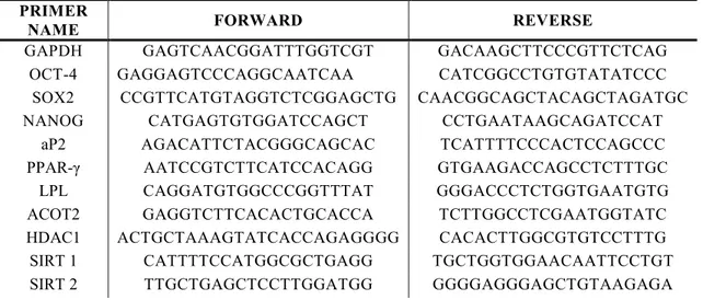
7.7 RNA extraction and gene expression analysis………..39
7.8 Immunofluorescence Microscopy……….41
7.9 Red Oil O and Alizarin Red staining………41
7.10 Senescence Associated β-galactosidase Staining………...42
7.11 Statistical Analysis………..43
RESULTS……….44
8.1 Features of ADSCs after treatment with conditioned media………44
8.2 Biomolecules maintains cell viability reducing nitric oxide production after H2O2 exposure………..44
8.3 Modulation of stem cells regenerative potential in inflammatory response………..44
8.4 Cellular response to stress and cell differentiation involve Sirtuin-Dependent Epigenetic Changes……….45
8.5 Melatonin and vitamin D counteract adipogenesis inducing the molecular pattern of osteogenesis………45
8.6 Melatonin and vitamin D inhibit intracellular lipid accumulation inducing ADSCs mineralization……….46
8.7 ADSCs survive to premature senescence induced by oxidative stress…………46
CONCLUSIONS……….47
FIGURES………..51
FIGURES AND TABLES
Figure 1. Regenerative medicine in biological reestablishment of tissue functions……8
Figure 2. Characteristics of stem cells………10
Figure 3. Structure of adipose tissue………..17
Figure 4. Epigenetic modulators and stem cell fate………26
Figure 5. Vitamin D metabolism………30
Figure 6. Bioactive molecules modulate stem cell fate………..49
Figure 7. The balance between osteogenesis and apipogenesis……….50
Figure 8. Isolation from adipose tissue………...51
Figure 9. Analysis of cell morphology after culturing in conditioned medium……….52
Figure 10. Cell viability assay by MTT………..53
Figure 11. NO concentration after induction of oxidative stress………..….54
Figure 12. Gene expression levels of proinflammatory cytokines………..…...55
Figure 13. Gene expression levels of stemness related genes………....56
Figure 14. Gene expression levels of SIRT and HSP………...57
Figure 15. Gene expression levels of SIRT and HDAC in adipogenic conditioned medium………58
Figure 16. Gene expression levels of adipogenic related genes……….59
Figure 17. Gene expression levels of osteogenic related genes………..60
Figure 18. Immunohistochemical analysis of adipogenic related proteins (TMEM26).61 Figure 19. Immunohistochemical analysis of adipogenic related proteins (ASC-1)…..62
Figure 20. Immunohistochemical analysis of adipogenic related proteins (PAT2)…...63
Figure 21. Evaluation of lipid accumulation………..64
Figure 22. Evaluation of calcium accumulation……….65
Figure 23. β-galactosidase activity……….66
Table 1. Cell characterization by CytoFlex Beckman Coulter………...37
INTRODUCTION
Regenerative medicine identifies the area of research and therapies based on the use of stem cells for tissue regeneration [1]. Several diseases lead to loss of functionality or aging of tissues and organs [2]. In the last years, the interest of researchers has been oriented toward regenerative medicine, with the aim of restoring function of compromised organs [3]. The aim of regenerative biology is to identify the cellular and molecular differences that distinguish normal tissue turnover from scar repair, in order to create an ideal microenvironment suitable for regeneration in damaged adult tissue [4][5]. Regenerative medicine encompasses a set of interdisciplinary activities, including the possibility of inserting growth factors in the damaged site, or alternatively the development of biomaterials for tissue engineering [5]. Most of these, as biomimetic polymers and bioactive three-dimensional scaffolds, are capable of inducing specific cellular responses and the formation of new tissues to be implanted in vivo [6][7]. Stem cells (SCs) represent an important source for regenerative medicine, to ameliorate genetic and degenerative diseases, as cardiovascular, musculoskeletal and neurological [8][9]. To be considered ideal for tissue regeneration, SCs must be easily expandable in
vitro, multipotent and able to permanently restore tissue function avoiding neoplastic
transformation [10][11]. The extra cellular matrix (ECM) plays a crucial role in stem cell differentiation [12]. External stimuli coming from the microenvironment in which they reside, known as niche, finely regulate stem cell fate in order to maintain homeostasis and remodel tissues in physiological and pathological conditions [13][14]. SCs could then be harnessed for patients with serious disease, with a high yield and minimizing the risk of rejection [15][16]. Transplantation of bone marrow-derived SCs has been applied for the treatment of celiac disease and chronic inflammatory bowel diseases such as Crohn's disease (CD) and ulcerative colitis (UC)[17], but also for diabetes mellitus (DM) [18]. Novel therapeutic strategies have been proposed for the treatment of DM, such as the administration of growth factors, transplantation of pancreatic islands and infusion of SCs to replace dysfunctional beta-cells [19]. Nevertheless, a possible side effect of SCs based therapies consists in their possible neoplastic transformation [20].
Unravelling the cellular mechanisms governing stem cell behavior could lead to the development of new therapeutic approaches for tissue repair and other clinical applications.
Figure 1. Regenerative medicine in biological reestablishment of tissue functions. Tissue engineering, gene and cell therapies, and stem cell research are some of the methodological approaches commonly used.
1. Stem cells
The origin of the term "stem cells" can be traced back to the late nineteenth century, in 1868 precisely, when Ernst Haeckel, a major supporter of Darwin's theory of evolution, used the term "stem cells” to describe the unicellular ancestor organism from which all multi-cellular organisms presumably evolved [21][22]. Stem cells are in fact undifferentiated primitive cells, with the unique ability to differentiate in the approximately 200 different cell types that form the body: neurons, skin cells, muscle cells, bone cells, liver cells, starting from the embryo and throughout the life span of each individual [23][24]. Therefore, they can be described as cells whose fate has not yet been decided. Four criteria are used to define a stem cell. First of all, the cell must be able to undergo multiple and sequential cell divisions of self-maintenance, a prerequisite for supporting the cell population [25][26]. Subsequently, from each cycle of replication by asymmetrical division, two daughter cells are generated, one of which may differentiate toward a particular cell line, while the other retains the features of stem cells. This is involved in replacing damaged elements in origin tissue and thus
maintaining tissue homeostasis [27][28]. The third criterion for defining stem cells deals with the ability to repopulate tissues when transplanted into a damaged recipient site, a property widely demonstrated for hematopoietic stem cells and more recently for progenitors of liver cells and nervous stem cells [29][8]. A last criterion is that stem cells must contribute to a differentiated progeny in vivo even in the absence of tissue damages. They can undergo differentiation, giving rise to specialized cells through a process that depends on signals inside and outside the cell [30]. Internal signals are controlled by specific genes, while external signals include physical contact with adjacent cells and specific molecules of their microenvironment, called “niche” [13][31]. The process of cellular specialization is genetically controlled by the cell nucleus and it is fundamental to allow the development of tissues [32]. According to the level of specialization, stem cells can be classified as totipotent pluripotent, multipotent, oligopotent and unipotent [33]–[35]. In mammals, only the zygote, produced by the fusion of an egg with a spermatozoon, and the cells originating from the first divisions of the fertilized egg, the blastomeres, are totipotent and represent the founder cells of all the cells in the body [36]. By successive divisions, embryonic stem cells originating from the zygote, are capable of generating all types of cells in the body, both in vivo and in vitro, but are uncapable of generating extra-embryonic tissues [37]. Then, following the process of gastrulation, the three germinative leaves and differentiated cells are generated, including tissue-specific or somatic stem cells, which are responsible for the formation of the residing tissue [38]. These cells have the function of maintaining the structural and functional integrity of the tissues through the replacement of damaged mature cells [39].
Figure 2. Characteristics of stem cells [40].
1.1 Embryonic stem cells
Embryonic Stem Cells (ESCs) derive from the internal mass (Inner Cell Mass - ICM) of the blastocyst and can be easily isolated from the degradation of an embryo of 5-7 days [41]. The first lines of embryonic stem cells were isolated from mouse blastocyst at the beginning of the ‘80s, by Martin Evans and Gail Martin, followed by studies carried out in the late 1990s by James Thomson [42][43]. They are cells that can not only be expanded indefinitely in culture while maintaining an undifferentiated state, but are also able to give rise to all adult cell types if injected into the blastocyst [44]. Human embryonic stem lines were obtained from cells of the internal cellular mass that were surgically isolated, dissociated and grown on a layer of murine fibroblasts, which act as "feeder" [45][46]. The term indicates a cell type capable to supply the nutrients or trophic factors necessary for the maintenance of cells in an undifferentiated state [47]. ESCs can generate disorganized masses of mature cells of ectodermal, endodermal and mesodermal origin, called embryoid bodies (EB), and give rise to teratomas if injected into immune depressed mice [48][49].
This ability, defined as pluripotency, make them extremely interesting as a possible therapeutic tool for any type of disease, but, at the same time, highlights their tumorigenic potential for uncontrolled differentiation [50]. ESCs grown in vitro maintain self-renewal, telomerase activity, also retaining a stable diploid karyotype. The key genes implicated in the mechanisms of proliferation and self-maintenance are known as “Oct4-like” [51]. Oct4 is the main transcription factor of ESCs, expressed by the zygote during segmentation, being downregulated in the blastocyst and silenced in adult tissues [52]. FoxD3 is another transcription factor that presents the same pattern of expression as Oct4 [53]. Other genes involved in self-renewal are Sox2 and Nanog. Nanog is a transcription factor belonging to the family of homeobox proteins and its expression seems instead limited to the cells of the internal mass and the primordial germ cells [54]. In addition, there are also enzymes used as markers for human ESCs, such as alkaline phosphatase and telomerase [55].
1.2 Adult stem cells
The use of human embryonic stem cells arises ethical implications, as isolation of stem cells from blastocyst occurs only after destroying embryos. For these ethical concerns, somatic stem cells attracted the attention of scientists [56]. Somatic or adult stem cells (Adult Stem Cells-ASCs) are capable to acquire a specific phenotype, according to the features of mature cells in their residing tissue. They show reduced levels of telomerase activity and a limited capability to differentiate, if compared to embryonic cells [57][58]. Indeed, adult stem cells undergo an irreversible senescence process that involves a progressive differentiation toward the mature stage. For these reasons they are defined multipotent stem cells [34][59].
It has been demonstrated that each tissue in the organism, can be a source of stem cells that could be used in autologous transplants, for the ability to differentiate into mature phenotypes [60]. Bone marrow- derived stem cells (BM-MSCs), for example, can differentiate into hematopoietic line, glia, skeletal muscle or bone cells [61]. Stem cells from dermis can differentiate into skeletal muscle, adipose tissue or cartilage. This ability to make different lineages has been defined as “plasticity” of adult stem cells [62]. The degree of plasticity suggests their possible use in the treatment of many diseases, even if their pluripotency does not reach that of embryonic stem cells [8].
The first adult cells extensively characterized are hematopoietic stem cells (HSC). They support hematopoiesis, giving rise to all the blood elements [63].
They were isolated for the first time in mice, based on the antigenic characteristics of the surface: they express CD45, Thy1, c-kit and Sca-1 while not express lineage (Lin) and CD34 antigens [64]. Even human HSCs do not express lineage and CD38 antigens, while they express CD45, c-kit and Thy1 and, unlike mouse antigens, are present in the CD34+ fraction of bone marrow, peripheral and cord blood [63]. Recently, these criteria have been applied to identify adult stem cells from other sources, for example neural stem cells, discriminated by a combination of CD133 and CD24 [65].
1.2.1 Mesenchymal stem cells
Human adult mesenchymal stem cells (hMSCs) can contribute to the maintenance of normal homeostasis in all tissues through the replacement of the degenerated elements [66]. Their definitive fate is regulated by specific signals, as biological molecules and biophysical factors [24]. The presence of stem cells has been identified also at the level of the bone marrow stroma. The interest of researchers has thus focused no longer on the role of stroma as a support for hematopoiesis but on the potential of stem cells located there [67]. Alexander Friedenstein and Maureen Owen were the first to use in
vitro cultures and transplants in animal to characterize the cells that compose the bone
marrow stroma [68]. The cells isolated from the stroma are also called marrow stromal cells, mesenchymal stem cells (mesenchymal stem cell-MSC) or mesenchymal progenitor cells (MPC) [10]. They have a “fibroblast-like” morphology with very little cytoplasm, few mitochondria and a poorly developed Golgi apparatus, can be easily expanded in vitro and show differentiating potential towards various cell types: osteocytes, chondrocytes, adipocytes, astrocytes [69][70]. The classical model of mesenchymal cell is derived from the stromal fraction of bone marrow. The isolation procedure involves the surgical removal of the portion of the matrix that is subsequently mechanical disintegrated to obtain from 0.001 to 0.01% of mononuclear cells to put in culture [71].
In vitro studies have shown that only some colonies derive from mesenchymal stem
differentiation potential. Hence characterizing and identifying the real mesenchymal stem cells actually represent a crucial issue [72].
Since 1992 the existence of a precursor common to mesenchymal and hemopoietic progeny on fetal liver cells, characterized as CD34+, CD38-, HLA DR-, was hypothesized [73]. In the spinal cord microenvironment, stromal cells produce the growth factors needed to promote the activation, proliferation and differentiation of responsive stem cells. Most of these cells are quiescent (G0 of cell cycle) in their niche and only a small fraction respond to paracrine messages, differentiating into CD34+ or mesenchymal stem cells [74][75]. The CD34 marker, identified as a surface glycoprotein, belonging to the sialomucin family, and involved in the cell-cell and cell-matrix adhesion mechanisms, is expressed in hematopoietic progenitors and vascular endothelial cells but cannot be found on ex vivo expanded MSCs [76]. In general, MSCs express numerous molecules important for cell adhesion and interaction with the microenvironment of the niche [13]. They also express receptors for chemokine growth factors, integrins and markers shared with other cytotypes while they exhibit negative staining for typically hematopoietic markers such as CD45, expressed on myeloid cells, and CD14, receptor of bacterial lipopolysaccharide, found on monocytes [77][78]. Three antigens have been identified whose simultaneous presence on MSCs, is commonly used for their characterization. First of all the antigen SH2, epitope of the endoglin CD105 or TGF beta-IIIR [79][80]. It plays a key role in endothelial cell interactions and vessel development, mediates interactions between MSCs and hematopoietic cells in the marrow and could play a key role in chondrogenic differentiation by signal transduction [81].
On the other hand, the monoclonal antibodies SH3 and SH4 recognize two different epitopes of CD73, an antigen present in many cell types and in particular in lymphoid tissue but not in hematopoietic precursors [82]. In the spinal cord microenvironment, CD73 could mediate cell-cell interactions, representing a common element between stromal and lymphocyte development [83]. Moreover, its expression can be related to the immunomodulatory activity of mesenchymal cells. Another interesting marker is the CD166 glycoprotein, also involved in osteogenic differentiation, although the precise mechanism of this process has to be clarified yet [84][85].
It is well known that MSCs bind monoclonal antibodies directed against the low affinity receptor for the nerve growth factor, also known as CD271 [86]. This property has recently been successfully exploited to develop a method for cell selection by immunomagnetic sorting.
Due to the low rate of availability and the invasive method to isolate BM-MSCs, new sources of mesenchymal stem cells have been recently evaluated: the human body hosts multipotent stem cells within several niches, including dental pulp, dermis, Wharton jelly of umbilical cord and adipose tissue [87][88].
1.2.2 Adipose-derived stem cells
Since the discovery in 2001 of a population of mesenchymal stem cells within its stromal compartment, human adipose tissue has been evaluated as an important suitable source for regenerative medicine [89]. These cells, called adipose derived stem cells (ADSCs), have been shown to be available for adipose tissue generation, the creation of tissue banks, being also potentially cryopreserved for a long time [90]. ADSCs are multipotent stem cells obtained from the vascular stromal fraction (SVF) of adipose tissue, consisting of CD34+/CD31- adipocyte precursors, CD34+/CD31+ endothelial cells and CD14+/CD31+ macrophages [91][92]. According to several authors, ADSCs isolated from SVF can probably be considered “pericytes” or “perivascular progenitor cells” [93]. Like BM-MSCs, ADSCs generally show a fibroblast-like morphology and have properties and features equivalent to multipotent cells isolated from other tissues. The characterization of the ADSCs surface markers shows positivity towards the epitopes CD13, CD49a, CD49b, CD49d, CD90, CD105, STRO1, CD166, CD117 and CD29, CD44, CD49 and CD54, CD34, CD133, CD144 [25][94].
In vitro and in vivo studies have also shown a high degree of plasticity of these cells.
ADSCs can be committed towards mesodermal derived tissues, as adipocytes, chondrocytes, osteoblasts, muscle cells, and towards non-mesodermal tissues, as angiogenic, hepatic, pancreatic, epithelial and neurogenic phenotypes [95][96].
The adipose tissue can be collected from different sources through various methods of isolation. The first application of adipose tissue for self-/homo-grafts belongs to the end of the 19th century [97].
The introduction of the modern liposuction technique at the end of the 1970’s, allowed the removal of subcutaneous adipose tissue through the use of cannula of variable diameter, through small incisions in the area of interest [98].
This technique has the advantage of obtaining large amounts of adipose tissue with minimally invasive procedure, simplifying isolation and making it less traumatic [99]. In the following years, several improvements were made in the instrumentation, techniques and devices used. The isolation of the cellular component residing in the adipose tissue involves mechanical trituration followed by enzymatic digestion [100]. The recovery of ADSCs is higher than that of other classical adult sources such as bone marrow: the ADSCs constitute 2% of the lipoaspirate cells, while the MSCs in the marrow represent less than 0.1% [101]. An innovative technique to minimize the manipulation of any cellular product, in order to maintain the native characteristics of the tissue, was developed by Tremolada and collaborators. It is based on non-enzymatic processing of lipoaspirates, called Lipogems®, to obtain a minimally manipulated preparation containing vital ADSCs, with elimination of the oil and blood residues [102][103]. In vitro tests revealed the differentiating and immunomodulatory capability of these cells, representing an excellent candidate in regenerative medicine approaches. Moreover, unlike other lipoaspirates, this product can be frozen without losing the vital and functionally active stem cell population [104][102].
2. Adipose Tissue
Adipose tissue (AT) is a connective tissue responsible for different physiological functions [105]. It is composed of adipocytes, surrounded by a stroma of connective fibers, intercellular matrix, fibroblasts, immune system cells and blood vessels [106]. For many decades, the adipose tissue has been viewed as a passive organ dedicated to lipids storage during excessive caloric intake, capable of releasing energy when needed by other organs [107][108].
It represents an important energy reserve for the body, prevents the dispersion of internal heat, protects and supports the internal organs [109]. Adipose tissue exerts also important endocrine function, secreting hormones as leptin, adiponectin and resistin [110].
Adiponectin is involved in regulation of glucose and lipid metabolism and energy homeostasis, while resistin is responsible for insulin resistance and inhibits the activity of AMPK (AMP-activated protein kinase) in liver and skeletal muscle [111][112]. On the other hand, leptin is related to the inhibition of food intake and stimulation of energy consumption [113].
The distribution of adipose tissue in human body depends on age and gender differences. In men, fat is mainly stored around the waist, while fat accumulation in woman is around the hips [114][115]. Moreover, with aging, changes in body composition are associated not only with modifications in total adiposity and body weight, but also with increase in bone marrow fat content, leading to lower bone mineral density and the risk of elderly fractures [116]. Adipose tissue can be classified in subcutaneous adipose tissue (SCAT), with mechanical and protective functions [117], and visceral adipose tissue (VAT), a hormonally active component of total body fat presents in the abdominal cavity [118]. Accumulation of adipose tissue is also formed between muscles and in the connective sepia that separate the various bundles. The subcutaneous adipose tissue affects the entire body surface and above all the deep layers of skin [119]. The 50% of the fat is located in the subcutaneous panniculus, where it performs both an insulating and a mechanical function; 45% in the abdominal cavity, forming the visceral adipose tissue, and, finally, for 5% in the muscle tissue, where supports muscular activity [120]. SCAT and VAT show difference in their structures and in endocrine system regulation. In mammals, VAT contains greater numbers of large APCs than SCAT, which generally contains small adipocytes but a higher lipolytic activity [110][121]. In addition, non-adipocyte resident cell population secrete higher levels of proinflammatory cytokines in VAT then in SCAT, responsible for increased risk of developing insulin resistance and metabolic syndromes [122]. Adipocytes are responsible of storing triglycerides during calories intake and to mobilize these reserves in the form of fatty acids, according to the tissue needs [123]. Mature adipocytes retain a large number of enzymes and regulatory proteins crucial for lipolysis and lipogenesis [124].
Some of these molecules, such as adipokines, are secreted in the blood, and are able to regulate the biological activity of other cells, including central nervous system,
pancreas, liver, skeletal muscle tissue, kidneys, endothelium and immune system [125][126].
Therefore, adipose tissue plays a key role in homeostatic regulation of the energy balance, insulin sensitivity and vascular-endothelial function [109][110]. Alterations in distribution of adipose tissue and its endocrine function are related to insulin resistance, increased risk of developing type 2 diabetes mellitus, atherosclerosis and cardiovascular disease [127].
The molecular mechanisms involved in this kind of mutations are not fully known yet. It is believed that these alterations are mainly related to a dysregulation in VAT activity and adipokines secretion [128][129].
Figure 3. Structure of adipose tissue. Mature adipocytes are the main population composing adipose tissue. Images are acquired by optical microscope. Scale bar=100µm.
2.1 Types of adipose Tissue
In mammals, the fat is organized as white (WAT) and brown (BAT) adipose tissue, having typical structures and functions [130]. WAT stores energy as triacylglycerol (TAG), whereas BAT dissipates energy as heat by acid fatty metabolism [131][132]. In addition, an inducible thermogenic fatty tissue type has been identified, known as beige [133].
2.1.1 Beige adipose tissue
Beige adipose tissue, also called “brite”, derives from the differentiation of precursor cells in mature adipocytes or from the transformation or trans-differentiation of pre-existing white adipocytes [134]. This kind of cells, classified as beige/brite or brown-induced, have intermediate features between WAT and BAT but are able to respond to thermogenic stimuli [135][136].
WAT, following certain stimuli, can transdifferentiate from a WAT phenotype toward a BAT-like one, modifying its morphology, gene expression and mitochondrial activity. At the beginning, they exhibit large lipid drops and the absence of UCP1[137][138]. When they receive thermogenic stimuli, the adipocytes changes their morphology to brown like features: multilocular lipid drops, with a low expression of UCP1 [139][140]. The process of WAT transforming into beige is called “browning”[141]. This transformation may depend on various stimuli, including hormonal signals as catecholamines. These are hormones released in stressogenic situations and may have relevant therapeutic applications for obesity and obesity-related syndromes in humans [142]. The factors able to induce the darkening of the WAT are classified in the class of activators of the SNS [143]. This mechanism leads to the activation of lipolytic phenomena and fatty acid oxidation, and is also associated with over-expression of genes involved in mitochondrial function and thermogenesis typical of brown adipocytes [144]. Among the activators of the sympathetic nervous system, we distinguish the β-adrenergic agonists and the endogenous signal neuropeptides. Transcription factors as peroxisome proliferator-activated receptor-γ (PPARγ), CCAAT/enhancer-binding protein beta (C/EBPβ), and FOXC2, growth factors, as Bone morphogenetic protein 7 (BMP7), FGF21, BDNF, and BMP8B, enzymes and proteins associated with lipid drops, as perilipin, are also included [145]–[147]. Understanding the cellular mechanisms controlling the plasticity of WAT-BAT could lead to the design of future treatments and drugs for type 2 diabetes and obesity, also improving the outcome of tissue transplantation and gene therapy [148].
2.1.2 White adipose tissue
The WAT, also called unilocular tissue, consists of cells in close contact with each other, with a poor extracellular matrix [149].
The adipocytes are very huge and can reach 150-200 µm in diameter. The adipocytes are arranged to form aggregates, between which numerous blood vessels are placed [150].
The lipid drop is one, unilocular, which lodges almost the entire intracellular space, while the cytoplasm is reduced and located on the periphery of the cell. This tissue is constantly exposed to mechanical stresses [151][152]. The principal functions of WAT are the storage of lipids during fasting and the release of fatty acids through β-oxidation process, providing adenosine triphosphate (ATP) for cellular biological processes [153]. The white adipose tissue is also known as an important secretory organ, producing cholesterol, retinol, steroid hormones, prostaglandins and adipokines [154][155]. Alterations or inadequate release of these molecules, can lead to the onset of diseases, as obesity, insulin resistance, increased risk of metabolic syndrome, cardiovascular diseases and other more [156][157]. WAT can be distinguished in subcutaneous, under the skin, or omental, that can be localized inside the abdomen (gonadal, mesenteric, omental, and perirenal), with a less innervation than BAT [158].
2.1.3 Brown adipose tissue
The brown adipose tissue, also called multilocular, consists of adipocytes widely distributed and significantly smaller than the WAT. However, adipocytes form aggregates with a glandular-like appearance, with a real endocrine function [159][136]. BAT is a highly vascularized and innervated tissue, and brown adipocytes possess multilocular lipid droplets and a high number of mitochondria, whose cytochromes are responsible not only for vascularization, but also for the characteristic brown color of the tissue [160][161].
In fact, a special transmembrane channel protein called thermogenin is placed in the internal membrane of the mitochondria, which allows the passage of protons to the matrix without involving the complex of ATP synthase [162][163].
This tissue is responsible for the metabolism of fatty acid, thanks to the presence of mitochondria uncoupling protein 1 (UCP1). Activation of UCP1 stimulates the uptake of lipids and glucose from circulation to support thermogenesis [131][164].
The thermogenesis of BAT is physiologically stimulated by noradrenaline released by the nerve fibers of the sympathetic system (SNS) [165].
The transduction of the thermogenesis signal occurs mainly through the β-adrenergic receptors present on the membrane of brown adipocytes [166]. The activation of adenylate cyclase (AC, adenylyl cyclase) leads to increased levels of cytosolic cyclic adenosine monophosphate (cAMP). Increased cAMP induces hydrolysis of triglyceride reserves through the activation of protein kinase A (PKA) [167][168]. Through this process, the fatty acids are released and act as a substrate for mitochondrial oxidation and, at the same time, activate the UCP1 [169]. BAT is typical of rodents and hibernating animals. In humans it provides thermoregulation in the first years of life. The amount of BAT tends to decrease with age and persists in adulthood in a variable way in the perirenal, cervical, mediastinal and mesenteric areas [170][171].
2.2 Features of cells in adipose tissue
Adipose tissue is a heterogeneous cell population, including mature adipocytes, endothelial cells, macrophages, pericytes, and pre-adipocytes or adipose-derived stromal/stem cell [172]. Mature adipocytes originate from precursor cells differentiation placed in the connective tissue and capable to differentiate into adipocytes, chondrocytes, osteoblasts, and myocytes [10]. These precursor cells are called lipoblast or preadipocyte or adipoblasts [173]. During development, these cells progressively accumulate lipid drops in their cytoplasm, becoming globular, with a very reduced cytoplasm that forms a ring around a single large vacuole rich in lipids [174][175].
WAT formation is the results of increasing adipocyte size and number. The potential to produce new fat cells from lipoblast continues throughout the lifespan [176]. In the course of differentiation, a series of chronological changes in the expression of numerous genes occur. This event is represented by the appearance of mRNAs and protein markers and in the accumulation of triglycerides in the cytoplasm of the cells [177].
Among the various transcription factors involved in adipocyte differentiation, CCAAT/reinforcing binding protein (C/EBP-) and peroxisome proliferator-activated receptor (PPAR- γ) are involved in the activation of adipo-specific genes and in the cell-cycle arrest, required for adipocyte differentiation [178][179].
C/EBP-is expressed slightly before the first phases of gene transcription as compared to PPAR- γ. C/EBP transcription factor interact also with Co-activator-3 steroid receptor (SRC-3) in regulating PPAR2 expression and white adipocytes formation [180][181]. Although the expression of C/EBP-α and PPAR-γ increases dramatically during adipocyte differentiation, the low level of these factors expressed as preadipocytes may be enough to mediate the growth arrest preceding differentiation [182]. Lipoprotein lipase (LPL) is secreted by mature adipocytes, but also by other cell types such as heart muscle cells and macrophages, and is important in controlling lipid accumulation and in catalyzing the hydrolysis of triglycerides [183]. Preadipocyte factor-1 (Pref-1) also participates in the maintenance of the pre-adipogenic phenotype. During the last phase of differentiation, cultured adipocytes significantly increase lipogenesis and become insulin-sensitive [182][184]. Adipocytes also synthesize other specific adipose tissue products not directly related to lipid metabolism. These include aP2, an adipocyte-specific fatty acid binding protein found in adipose tissue and plays an important role in intracellular metabolism and fatty acid transport [185]. Acyl-coenzyme A (CoA)-binding protein (ACBP) is also significantly induced during differentiation, regulating the number of acyl-CoA esters available for various metabolic processes [186].
2.3 Understanding adipocytes features a lesson from the clinic: obesity and obesity-related disorders
Obesity is a multifactorial worldwide disease in adults, as well as among children and adolescents, associated to a great risk of morbidity and mortality due to an abnormal and uncontrolled fat accumulation in adipose tissue [187][188]. Obesity has become a global epidemic in the past decades, associated with high medical costs for its
diagnosis and treatment. Premature mortality, disability, loss of productivity and absenteeism are only a few of the consequences related to it [189]–[191].
Moreover, a large part of the health expenditure for treatment of obesity comes from public insurance. The excess of fat depots is measured according to Body Mass Index (BMI) determination, a parameter derived from the weight in kilograms divided by the square of the height in meters (kg/m2) of the individual [192]. Based on the data from the Global BMI Mortality Collaboration, a BMI of 20.0-25.0 kg/m2 is associated with the lowest mortality rate. According to World Health Organization (WHO) a BMI greater or equal to 30 Kg/m2 is consistent with obesity and a BMI between 25 and 30 Kg/m2 identifies overweight [193][194]. Obesity is classified according to BMI in:
Normal weight 18-25 Overweight 30-35 1st degree obesity 30-35 2nd degree obesity 35-40 Severe obesity 40-50 Super obesity >50
The distribution of adipose tissue in the body has important implications for health [195]. Obesity is the result of the intake of excessive calories needed for body activity. In mammals, a complex set of hormones and nerve signals acts to keep energy and nutrient consumption balanced [196].
In maintaining the body's homeostasis, the signal produced in the adipose tissue is able to influence the brain centers that control eating behavior and metabolic activity [197]. Adipokines and in particular leptin secreted by adipose tissue, provide the information on the availability of energy tissue reserves, stimulating the production of the anorexic hormone that suppresses appetite [198]. Obesity is often associated with metabolic syndrome and onset of type II diabetes, followed by cardiovascular disease, kidney failure, blindness, difficulty in healing wounds and, first of all, development of insulin resistance [199][200]. There is also a low-degree of chronic inflammation. With the
increase in adipose tissue, in particular the omental tissue, the number of macrophages, responsible for the elimination of damaged adipocytes [201] also grows. Macrophages in obese subjects control the production of most cytokines, followed by a drastic increase in insulin production [202]. Cytokine production, together with the reduced sensitivity to insulin is the main cause of the complications associated with obesity: cardiovascular diseases (CVD), hypertension and other more, leading in the most serious cases to premature death [127][203]. Not all obese patients show the symptoms of obesity-related disorders. This fraction of patients has a regular insulin sensitivity and reduced amount of visceral fat compared to obese patients with metabolic syndrome [204]. The metabolic syndrome is associated with dyslipidemia, hyperglycemia, hypertension, doubling the risk of coronary heart and cerebrovascular disease [205]. Therefore, obesity depends on body composition rather than on body weight and in particular on the number of adipocytes. The treatment of obesity requires a prolonged and determined effort [206]. Actually, interest of research is aimed at finding methods leading to a decrease in weight by eliminating adipose tissue without affecting the non-lipidic mass [207][208]. Understanding the mechanisms of adipocytes differentiation could pave the way for future therapeutic application and development of new drugs and methodologies to counteract severe obesity in patients [209].
3. Stem cell plasticity
MSCs are able to acquire different phenotypes replacing injured elements under external signals from their microenvironment, acting to maintain tissue homeostasis [210].
In the regeneration processes, stem cells residing in tissues not affected by the damage may also contribute to the function recovery after injury [211]. The plasticity of stem cells is the ability of adult tissue stem cells to acquire a new identity. The term plasticity also indicates the potential of stem cells to differentiate into adult cells phenotypically different from those of their original tissues [212][62][213]. In response to different types of signals, stem cells are able to rearrange their membrane and cytoskeleton structure. Cell polarity is a fundamental feature of migrating cells, and
represent the ability to redistribute cellular organelles and proteins for cell division and migration [214][215]. Cell viability, cell proliferation and tissues homeostasis are regulated by a perfect and controlled balance between symmetrical and asymmetrical division of MSCs [27]. The asymmetric division leads to the formation of two different daughter cells. One with self-renewal properties, like the mother cell, and the other one that differentiate toward a specific cell type and has a reduced regenerative potential [28][216]. Alterations in this system are related to the loss of tissue function, as occurs in aging [217], when stem cells lose their regenerative potential by dividing mainly symmetrically, or to uncontrolled cell proliferation, as occurs in cancer progression [218].
4. Epigenetic of self-renewal and differentiation
Stem cell differentiation is a complex process controlled by signaling pathways and molecular mechanisms that influence the expression of the main stem markers Transcription Factor 4 (Oct-4), sex determination region Y-box 2 (Sox-2) and Homeobox Nanog protein (Nanog) [219][220]. These transcription factors are essential in maintaining MSC pluripotency and are also involved in adult somatic cell reprogramming. If these factors are downregulated, cells immediately start the differentiation process [221][221]. These transcription factors are also able to suppress a group of genes needed by the embryo for development. If repressed, these set of genes leads to production of additional transcription factors responsible for the activation of genes needed for differentiation [222]. When the embryo begins to grow, Oct4, Sox2 and Nanog are inactivated and the ESCs lose their pluripotency. These transcription factors could thus be considered positive stem switchers: they regulate the expression of other genes by direct interaction with chromatin or through the presence of other factors, changing the conformation and making certain regions accessible or not for transcription [223][224]. The expression of pluripotency genes influence cell cycle and prevent the spontaneous differentiation of cells even when they are expanding in culture[225].
In vitro studies on MSCs show that an over-expression of Oct4 and Sox2 results in the
promotion of proliferation, with an increase in the duration of the S phase of the cell cycle, and in a greater expression of the markers of the adipogenic and osteogenic
differentiation [226][227]. On the other hand, the over-expression of Nanog has been shown to slow down adipogenesis in MSCs [228]. Epigenetics refers to the range of hereditary changes in the structure of chromatin that can influence gene expression and represents the molecular reaction to all environmental changes [229].
These chromatin modifications, involving miRNA, DNA methylation and chromatin remodeling, are orchestrated by different types of enzymes, such as DNA methyltransferases (DNMTs), histone deacetylases (HDACs) and histone acetyltransferases (HATs) in a still unclear fashion [230][231]. Epigenetic mechanisms are able to guide the MSCs behavior from the undifferentiated to the differentiated state. Environmental and developmental stimuli influence chromatin structure and the activation of a specific transcription program, playing a central role in maintaining MSC regenerative potential [232][233]. DNA methylation plays a key role in reprogramming somatic cells, through repression of differentiating genes and regulation of the MSC undifferentiated state [234]. Additionally, microRNAs (miRNAs), small non-coding RNAs, have been discovered as regulators of cell pluripotency and reprogramming, suggesting their possible therapeutic application of various disorders, including myocardial infarction, neurodegenerative and muscle diseases [235]–[237]. Nevertheless, further studies and continuous innovations are needed in the attempt to develop novel strategies in the epigenetic control of stem cell fate and tissue homeostasis.
Figure 4. Epigenetic modulators exert a role in modulating stem cell fate, finely tuning stemness related genes and tissue-specific markers [238].
5. Orchestrating stem cell identity
Since the discovery of stromal cells from adipose tissue, several researchers have been interested in the use of molecules in the attempt to modulate stem cell commitment [233][238]. These natural and synthetic molecules exert their action via epigenetic modulations of a specific molecular differentiation program and gene expression of lineage-specific markers [239][240]. ADSCs cultured in the presence of calcium, or ascorbic acid, β-glycerol phosphate and dexamethasone in the growth medium, are capable to differentiate into osteoblasts, inducing an increase in the levels of expression of involved transcription factors [241][242]. Colorimetric tests (e.g. Alizarin Red) and gene expression analysis of the main osteogenic genes, highlight the accumulation of intracellular calcium deposits and reduced expression of stemness genes [243]. Retinoic acid (RA) promotes neural differentiation in mouse embryonic cells [244], or cardiac differentiation where used in combination with butyric acid and hyaluronic acid esters (HBR), increasing the transcription of cardiogenic genes and various growth factors such as VEGF (Vascular Endothelial Growth Factor) and HGF (Hepatocyte Growth Factor) [245]–[247]. More recently, a mixture of HBR and melatonin
has been successfully used to induce an osteogenic phenotype in dental pulp stem cells (DPSCs) [248]. Some authors found that the activin A, bone morphogenetic protein 4 (BMP4), VEGF or Dickkopf-related protein 1 (DKK1), can optimize cardiac development in human stem cell lines and mice [249][250]. Different combination of activin A, nicotinamide and resveratrol are able to induce mouse ADSCs differentiation into Insulin-Producing Cells [251][252]. The addition of dexamethasone, HGF, fibroblast growth factor 4 (FGF4) and other growth factors in the culturing medium induce ADSCs differentiation towards functional hepatocyte-like cells [253][254]. These findings highlight the chance to apply in vitro modulation of stem cells potential for future in vivo applications with a great impact in regenerative medicine [255], [256].
5.1 Focus on: Melatonin
A wide range of natural molecules and compounds, known as nutraceuticals or functional foods, are widely used for their therapeutic or preventive effects [257].
Melatonin (N-Acetyl-5-methoxytryptamine), synthetized from serotonin by pineal gland during the night, is a neurohormone involved in regulating circadian rhythm and reproduction, and in a number of other pleiotropic effects [258][259]. Melatonin can exert many other functions on mitochondrial activity and on the immune cell production, as well as anti-apoptotic properties, improving the outcome of stem cell transplantation [260][261]. In addition, melatonin also exhibits various effects on stem cells, controlling cell viability and differentiation. It is a molecule greatly preserved with physiological and pathophysiological functions, and whose synthesis decays with aging [262][263]. Melatonin is able to induce neural differentiation of stem cells in transplanted mice reducing the production of reactive oxygen species [264]. Moreover, melatonin can influence the differentiation of MSCs towards osteogenic, chondrogenic, adipogenic and myogenic lineages [265]. These processes involve the activation of Wnt/β-catenin pathway, MAPKs and TGF-β signaling, and the recruitment of AMPK-activated protein kinase (AMPK), and the protein Forkhead box O3 (FOXO3) [266][267]. AMPK activation is also involved in the regulation of the expression of the peroxisome proliferator-activated receptor gamma γ (PPARγ), the main adipogenic orchestrator gene and molecular target of the natural compounds used in the management of obesity [268].
These physiological effects exerted by melatonin, are mediated by the interaction with specific G protein-associated receptors, including MT1 and 2 [269][270] and with epigenetic modulators as HDACs or Sirtuins, a class of proteins involved in a wide range of cellular processes [271][272]. In particular, SIRT 1, 3 5, are mostly involved in metabolic controls, while SIRT2 and SIRT6, are mainly related to aging processes. The ability of melatonin to decrease body weight and inflammation, promoting adipocyte pyroptosis [273] and protecting tissue against mitochondrial dysfunction, suggest an application of this molecule during inflammatory obesity [274][275].
5.2 Vitamin D
Vitamin D is a fat-soluble vitamin with numerous biological properties, capable of influencing the physiology of the body system through the epigenetic regulation of over 200 genes [276]. It is involved in the maintenance of calcium and phosphate homeostasis and in the processes of bone mineralization, and its deficiency determines the onset of various diseases [277]. It is synthetized from a liposoluble prohormone that is exposed to a series of hydroxylation reactions in the skin, liver and kidney [278], starting from a cholesterol precursor, 7-dehydrocortisol, becoming vitamin D2 (25-hydroxyvitamin D3) or ergocalciferol, and vitamin D3 (1,25-dihydroxy vitamin D3) or cholecalciferol, its biologically active form [279][280]. Once synthetized, it is released into the blood where, thought vitamin D-binding protein (DBP), it can be absorbed by adipose tissue or transported to the liver for subsequent activation [281][278][282]. The enzymes responsible for both vitamin D activation and inactivation are part of the cytochrome P450 monooxygenase (CYP) family [283][284]. In humans, the main CYPs associated with vitamin D are CYP24A1, CYP27A1, CYP27B1 and CYP2R1 [285]. CYP27A1, CYP2R1, CYP3A4 and CYP2J3 are mainly located in the liver and are involved in the first phase of pre-vitamin conversion at the c-25 site into 25-hydroxy-vitamin D3 or calcitriol [286]. CYP27B1 and CYP24A1 are mainly located in the renal tubules and complete the activation pathway with hydroxylation at the c-1, producing 1,25-dihydroxyvitamin D3 or biologically active cholecalciferol [287]. In order to perform its function, the 1,25-(OH)2D3 bind to a nuclear receptor, the vitamin D receptor (VDR) [277]. The VDR is a member of the superfamily of the nuclear receptor
and acts as a transcription factor induced by ligand [288][289]. The binding between vitamin D and the VDR induces conformational changes in the activation domain, allowing the formation of the transcriptional complex associated with the retinoid receptor X (RXR) [290]. The heterodimer formed binds to the specific DNA sites called VDRE (Vitamin D response elements), modulating the enzymes responsible for the chromatin modification [280]. 1,25-(OH)2D3 is able to influence chromatin remodeling by modulating HDACs and DNA methylation. VDR is expressed in adipocytes and regulates adipogenic gene expression and apoptosis, and is involved in the control of calcium homeostasis and osteogenic-associated genes (osteocalcin, osteopontin, calbindin, calcium channels) [291][292]. Many genes activated by vitamin D are involved in various other systems, such as the regulation of proliferation and cell differentiation, and in oxidative stress, inflammation and immune response [293][294]. Recent studies highlighted the role of 1,25(OH)2D3 in influencing the gene expression of key molecules involved in adipogenesis, fat oxidation and lipolysis through the activation of the nicotinamide-dependent adenine dinucleotide (NAD)-signaling pathway [182][295][296]. Vitamin D deficiency leads to altered bone development, such as rickets in children and, in adults, to a variety of health problems as osteoporosis, muscle weakness, cardiovascular disease, metabolic syndrome, multiple sclerosis, mental defects, a variety of cancer diseases and many other disorders [297]–[299]. Vitamin D and VDR deficiency has also recently been proposed as a cause of obesity [300][301]. Deficiencies are commonly caused by an insufficient intake or imbalance of enzymes involved in the activation and/or metabolism of vitamin D [302].
Figure 5. Vitamin D metabolism involving P450 enzymes [303].
5.3 Natural extracts from plants
Nutraceuticals are applied in regenerative medicine for their potential to modulate physiological processes and cure a variety of disorders [304][305]. Medicinal plants are known for their antiseptic, antibacterial and anti-inflammatory properties [306]. They also have antioxidant activities and stimulate tissue regeneration counteracting acute inflammation [307]–[309]. This protective mechanism exerts a key role in restoring tissue function.
Myrtus communis L. belongs to this class of plants, known for the amount of bioactive
polyphenols, mainly anthocyanins, in its berries and seeds [310]. Commonly used for the production of liqueur in the food industry [311], it represents an important resource that can be used as a food supplement or extracts with antioxidant and anti-inflammatory activity [312]. Chronic inflammation leads to excessive secretion of proinflammatory cytokines (TNF-α and IL-6) involved in the onset of several diseases [313][314]. The production of reactive oxygen species (ROS) contribute to worsening the fall of the inflammatory state, increasing the release of cytokines, aging, cancer, diabetes, and atherosclerosis [315][316]. Polyphenols, as anthocyanins, gallic acid derivatives and flavonoids, counteract the release of toxic molecules and stimulate physiological defenses [317]. The class III NAD-dependent histone deacetylase sirtuin-1 (SIRT1) regulates various physiological processes and is involved in metabolism, stress response and aging [318]. Several studies have shown that downregulation of SIRT1 promotes inflammatory cytokine secretion, whereas when upregulates protects from premature senescence and oxidative stress [319][320]. In addition, phytochemicals are involved in the heat shock-induced response as a mechanism of self-defense [321]. Heat Shock Proteins (HSPs) are responsible for protein homeostasis and play an important role in aging, also protecting cells from oxidative stress damages [322]. Limited ROS production, together with modulation of TNF-α release, play a critical role in the enrolment of stem cells at injured sites, indorsing cell migration and tissue regeneration [323][324]. For these reasons, natural extracts can be applied in the attempt to drive cell proliferation and activate natural defenses through epigenetic regulations and post-transcriptional modifications [325][326].
6. Translation to clinical applications
Adult stem cells have attracted the attention in the field of regenerative medicine for their easily in vitro expansion and the ability to differentiate towards different phenotypes, overcoming ethical issues related to the use of ESCs [327]. MSCs are currently used in gene therapy and in the treatment of serious diseases, sometimes representing the only alternative to conventional treatments to improve the quality of life of the patients [328][329]. Transplant of MSCs is currently the most frequently used cell therapy in hematological diseases [330], and the main actor in several clinical
studies for neurological amyotrophic lateral sclerosis (ALS) [331][332]. ALS is characterized by progressive spinal cord motor neurons degeneration, involving toxicity and inflammatory processes associated with resident cell populations [333]. Currently drugs that suppress oxidative stress, can be used in the attempt to maintain motor neuron function [334]. Autologous MSC transplantation represent an alternative to conventional therapy. MSCs are used as therapeutic tools in stem cell transplants for their immunomodulatory properties, secretion of growth factors and regeneration of damaged tissues, especially in patients refractory to conventional therapies [335][336]. Autologous transplants are applied in leukemias, lymphomas, multiple myeloma and other hematological neoplasms [337]. The discovery of a population of ADSCs residents in stromal fraction of adipose tissue, has paved the way for several possible applications of lipoaspirate in the field of regenerative medicine [338]. Stem cells derived from adipose tissue have been shown to improve wound healing, ulcers and skin defects [339]; they can be used for the regenerative treatment of large head and face defects due to trauma or simply age, or used for joint reconstruction in arthritis [340]. The use of adipose tissue is very promising in breast reconstruction in patients who have undergone mastectomy for breast cancer and subsequent radiotherapy [341]. After the infiltration of adipose tissue, a marked improvement was observed due to the deposition of new collagen, skin hypervascularization and dermis hyperplasia in the case of face burns of 2 and 3 degrees [342][343]. Co-transplantation could also play an important role in preventing or contrasting the Graft-versus-Host disease (GvHD), one of the most frequent clinical complications of rejecting allogeneic bone marrow transplants [344][345]. For these reasons, adipose tissue is considered an ideal source of stem cells that can be used for tissue generation and reconstruction and, above all, for autologous transplants [346]. For acute diseases such as heart attack or immunologically based diseases as diabetes, allogenic therapy may be necessary unless cells can be obtained in large quantities from the patients [347]. Nowadays, several clinical trials with MSCs are ongoing, being successful in the treatment of imperfect osteogenesis, in improving the rooting of transplanted HSCs and in the control of autoimmune diseases (multiple sclerosis) and the rejection of transplants, in the repair of metabolic diseases (juvenile diabetes), cardiovascular system (heart attack, stroke) and the nervous system, as Alzheimer and Parkinson [347]–[349]. Unravelling the factors that
govern these processes, should lead to an improvement in the methods for stem cell differentiation, making this phenomenon clinically relevant.
AIM OF RESEARCH
The stromal fraction of adipose tissue is an important source of stem cells exploitable in regenerative medicine. MSCs residing in adipose tissue play an important role in the maintenance of cellular and tissue homeostasis, replacing dead or non-functioning cells. Fat tissue seems to play a central role in inducing inflammation, as excessive nutrition leads to changes in its cellular composition and to the production of pro-inflammatory cytokines. Hence, inflammation occurring in the injured tissue acts by recruiting resident MSCs. Abdominal obesity is associated with a state of chronic inflammation, which leads to insulin resistance and metabolic disorders. Understanding the cellular mechanisms involved in the activation and differentiation of the adipose resident stem cells, could help in identifying innovative and preventive tools to counteract obesity and the related diseases. The aim of this project was to evaluate cell behavior in the presence of conditioned media, drugs or natural molecules, in the attempt to counteract the molecular mechanisms involved in inflammatory-associated adipogenesis. Within this context, we aimed at modulating the epigenetic mechanisms involved in stem cell differentiation and cellular behavior, reducing both the production of pro-inflammatory cytokines and the inappropriate activation of adult stem cells in stressful conditions. The findings obtained here could open up novel strategies for future therapeutic approaches in epigenetic controlling stem cell fate and tissue homeostasis.
MATERIAL AND METHODS
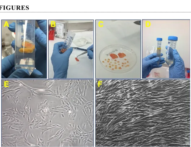
7.1 Adipose-derived stem cell isolation
Adipose-derived Stem cells (ADSCs) were isolated from subcutaneous and omental adipose tissue harvested during surgery processes for different reasons, from human adult female and male patients (n = 12, age = 45 ± 15 years, BMI: 22 ± 3 kg/m2). All the patients participating at the study, approved by the Ethics Committee Review Boards for Human Studies in Sassari (n◦ ETIC 240I/CE 26 July 2016, Ethical committee, ASL Sassari), signed a written informed consent. Cell isolation procedures were performed by mechanical and enzymatic digestion of fat tissue, which was washed repeatedly with sterile Dulbecco’s phosphate buffered saline (DPBS) (Euroclone, Milano, Italy) containing 200 U/mL penicillin and 0.1 mg/mL streptomycin (Euroclone, Milano, Italy), to remove the blood cells. Subsequently, fat sample was mechanically reduced to small fragments by sterile scalpels and then enzymatically digested for 1 hour at 37°C in a solution of 0,1% Collagenase Type I (Gibco Life Technologies, Grand Island, NY, USA). The enzyme activity was neutralized with 10% of fetal bovine serum (FBS) (Life Technologies, Grand Island, NY, USA) and the fat digestion solution was filtered with 70 μm cell strainer (Euroclone, Milano, Italy) and centrifuged at 600× g for 10 min, to separate the two distinct cell fractions of mature adipocytes, that were removed, and the stromal vascular fraction (SVF) that includes adipose-derived stem cells. The SVF in fact, is composed of preadipocytes, endothelial cells, pericytes, fibroblasts, adipose-derived stem cells (ADSCs) and hematopoietic stem cells. After collagenase digestion, mature adipocytes with a high fat content are separated as a floating layer. The adipocytes were transferred into a 12 cm2 culturing flasks filled with basic Dulbecco's modified Eagle's Medium (DMEM) (Life Technologies Grand Island, NY, USA) supplemented with 20% fetal bovine serum (FBS) (Life Technologies, Grand Island, NY, USA), 200mM L-glutamine (Euroclone, Italy), and 200 U/mL penicillin—0.1 mg/mL streptomycin (Euroclone, Milano, Italy). The flasks were placed upside-down in incubator at 37°C with 5% CO2. Once attached, the adipocytes were washed and put in incubator with fresh medium.
All cells remaining after the removal of mature adipocytes constitute the SVF. The pellet of SVF was resuspended into a basic medium, plated in 12 cm2 culturing flasks and transferred in incubator at 37°C and 5% CO2. After 48 h of incubation, the cultures were washed with DPBS and kept in the fresh medium. The culture medium was changed every 3 days. When the cells reached 80–90% confluence, they were harvested using 0.25% Trypsin EDTA (Euroclone, Milano, Italy), counted and passed into new flasks.
7.2 Cells magnetic separation
For isolation of adipose progenitor cells from the stromal vascular fraction (SVF), CD117 MicroBeads (Miltenyi Biotec, Minneapolis, MN, USA) were used. CD117, also known as c‐ kit, is a stem cell receptor, encoding a 145kD cell surface glycoprotein belonging to the class III receptor tyrosine kinase family [350]. According to the manufacturer's instructions, cells were trypsinized and resuspended in a specific buffer containing phosphate‐ buffered saline (PBS) pH 7.2, 0.5% bovine serum albumin (BSA), and 2 mM EDTA. Cells were then incubated with a primary monoclonal anti-c/kit (CD117) antibody for 1h at 37°C and, after washing, with a secondary antibody directly conjugated to MicroBeads for 15 min at 4°C. Then, the cell suspension was loaded onto a MACS® Column, which was placed in the magnetic field of a MACS Separator. The magnetically labeled CD117+ cells were retained within the column, while the unlabeled cells eluted through. After removing the column from the magnetic field, the magnetically positively selected CD117+ cells can be eluted in another tube, recovered and put in culture for subsequent experiments.
7.3 ADSC characterization by flow cytometry
To evaluate the percentage of mesenchymal markers on isolated population, flow cytometry analysis was performed. A total of 1×10⁶ ADSCs were trypsinized, centrifuged and fixed in 1% formaldehyde for 10 min at room temperature. After fixation, cells were permeabilized using a permeabilization buffer (eBioscienceMilano,
Italy) for 30 min at 4◦C, and then washed and incubated with fluorescein isothiocyanate (FITC)- or phycoerythrin (PE)-conjugated primary antibodies for 1h at 4°C.
ADSCs were tested for CD73-PE, CD90-PE (BD Biosciences, San Jose, CA, USA), CD105-PE (Santa Cruz Biotechnology, Heidelberg, Germany), CD45-FITC and CD31-FTC (Sigma-Aldrich, Munich, Germany) (all at 1 μg/106 cells). After washing, cells were analyzed on a flow cytometer (CytoFlex, Beckman Coulter, Milan, Italy) by collecting 10,000 events. Cells were positive for CD73, CD90 and CD105, and negative for CD31 and CD45 (Table 1). The data analysis was performed using CytExpert Software (Beckman Coulter, California, USA).
Table 1. Cell characterization by CytoFlex Beckman Coulter.
Antigen Expression CD31 - CD45 - CD73 + CD90 + CD105 +
7.4 ADSC culturing and treatment
7.4.1 Culture in the presence of melatonin and Vitamin D
After characterization, ADSCs were grown in a basic medium (BM) and propagated until the quantity required to carry out the subsequent in vitro tests has been reached. Cells at passage 5 were thus exposed to various treatments. One group of cells, used as undifferentiated control, was maintained in a growing (BM). Another group of ADSCs was induce to adipogenic differentiation by culturing in a conditioned adipogenic differentiation medium (DM or ADM), composed of basic medium supplemented with 1 μM dexamethasone, 0.5 μM hydrocortisone, 60 μM indomethacine, and 0.5 mM 3-isobutyl-1-methylxanthine (IBMX Sigma Aldrich Chemie GmbH, Munich, Germany). Positive control of adipogenic differentiation was represented by mature adipocytes.

![Figure 2. Characteristics of stem cells [40] .](https://thumb-eu.123doks.com/thumbv2/123dokorg/8364733.134878/11.892.233.669.75.428/figure-characteristics-stem-cells.webp)

![Figure 4. Epigenetic modulators exert a role in modulating stem cell fate, finely tuning stemness related genes and tissue-specific markers [238]](https://thumb-eu.123doks.com/thumbv2/123dokorg/8364733.134878/27.892.185.762.59.505/figure-epigenetic-modulators-modulating-stemness-related-specific-markers.webp)
![Figure 5. Vitamin D metabolism involving P450 enzymes [303] .](https://thumb-eu.123doks.com/thumbv2/123dokorg/8364733.134878/31.892.198.727.56.709/figure-vitamin-d-metabolism-involving-p-enzymes.webp)

![Figure 6. Bioactive molecules modulate stem cell fate inducing epigenetic modifications and influencing the expression of specific differentiation markers [238] .](https://thumb-eu.123doks.com/thumbv2/123dokorg/8364733.134878/50.892.187.774.56.440/bioactive-molecules-modulate-epigenetic-modifications-influencing-expression-differentiation.webp)
![Figure 7. The balance between osteogenesis and apipogenesis involves epigenetic modification influencing the expression of tissue-related genes [256]](https://thumb-eu.123doks.com/thumbv2/123dokorg/8364733.134878/51.892.187.766.56.343/figure-osteogenesis-apipogenesis-involves-epigenetic-modification-influencing-expression.webp)