Faculty of Pharmacy and Medicine
PhD Programme in
Morphogenesis and Tissue Engineering 31st Cycle
(A.A. 2015/2016 - 2017/2018)
Development Of a Biological Scaffold From Adult Human Skin For Cardiovascular Repair and Regeneration
PhD student Clotilde Castaldo
Tutor Coordinators
Prof. Stefania Montagnani Prof. Sergio Adamo
Prof. Antonio Musarò
CONFIDENTIALITY NOTICE
Reviewers and PhD committee members are obliged to keep the files confidential and to delete all records after completing the review process.
Il ricevimento degli elaborati scientifici, per l’ottenimento del titolo di Dottore di Ricerca, in qualità di Membro del Collegio dei Docenti del Dottorato in Morfogenesi ed Ingegneria Tissutale richiede di osservare le seguenti normative:
i. considerare le Informazioni confidenziali e riservate come strettamente private e ad adottare tutte le ragionevoli misure finalizzate a mantenerle tali;
ii. utilizzare le Informazioni confidenziali e riservate unicamente allo scopo per le quali sono state fornite o rese note, impegnandosi a non divulgarle a soggetti terzi le informazioni contenute negli elaborati ricevuti;
iii. a garantire la massima riservatezza, anche in osservanza alla vigente normativa in materia di marchi, di copyright e di brevetti per invenzioni industriali e in base alla normativa sulla privacy, ai sensi del D.Lgs. 196/2003, riguardo il know-how e tutte le informazioni acquisite, che non potranno in alcun modo, in alcun caso e per alcuna ragione essere utilizzate a proprio o altrui profitto e/o essere divulgate e/o riprodotte o comunque rese note a soggetti terzi.
Index
The thesis explained………...7
List of Abbreviations……….9
1 Introduction ... 13
1.1 Cardiovascular diseases ... 13
1.1.1 Definition ... 13
1.1.2 Epidemiology and risk factors ... 13
1.2 The Ischemic Heart Disease (IHD) ... 15
1.3 Current Treatment of IHD ... 20
1.4 Future Treatment of IHD ... 21
1.4.1 Cardiac Cell Therapy ... 21
1.4.2 Cardiac Tissue Engineering ... 22
2 Aims ... 25
3 Results and discussion ... 27
3.1 The decellularization of human skin from living subjects can be swiftly and efficiently accomplished ... 27
3.2 Decellularized Human Skin (d-HuSk) consists of structural and functional ECM proteins that are key components of the cardiac matrix ... 29
3.3 d-HuSk has the potential to deliver mechanical stimuli . 33 3.4 The profile of growth factors of d-HuSk is comparable to that of the cardiac native matrix ... 39
3.5 d-HuSk is a suitable environment for the engraftment and survival of human Cardiac Progenitor Cells (hCPCs) ... 42
3.6 hCPCs retain on d-HuSk the potential to differentiate towards cardiac myocytes in vitro ... 47
4 Conclusions ... 53
6 Materials and methods ... 57
6.1 Tissue specimens ... 57
6.2 Decellularization of human skin ... 57
6.3 Decellularization of human myocardium ... 58
6.4 Quantitative measurement of DNA content ... 58
6.5 Histochemistry ... 59
6.6 Immunohistochemistry ... 59
6.7 Quantitative measurement of elastin ... 60
6.8 Quantitative measurement of GAG ... 61
6.9 Growth Factor Array ... 61
6.10 hCPC isolation and culture ... 62
6.11 Preparation of d-ECM scaffolds for 3D cultures of hCPCs 63 6.12 In vitro assessment of d-HuSk cytocompatibility ... 63
6.13 Assessment of ability of d-HuSk to support hCPC engraftment in vitro ... 64
6.13.1 Live cell imaging analysis of repopulation of d-HuSk by hCPC residing on d-HuM ... 65
6.13.2 Scanning Electron microscopy analysis ... 65
6.14 Analysis of the effects of d-HuSk on hCPC myogenic differentiation potential in vitro ... 65
6.14.1 Gene expression analysis by Real-time PCR ... 66
6.14.2 Immunofluorescence ... 66
6.15 Statistical analysis ... 67
7 References ... 69
List of publications………...85
The thesis explained
Cardiovascular diseases (CVDs) are still the leading cause of death and disabilities globally. Among CVDs, ischemic heart disease (IHD) has remained the leading cause of death worldwide in the last 16 years. IHD is caused by a sudden blockage of blood flow through coronary arteries that prevents the supply of oxygen and nutrients to the region of myocardium fed by the affected vessels. This condition causes the necrosis of the myocardium that is followed by a reparative process that starts from the infarcted area, but then involves, at later stages, also the uninjured myocardium, causing progressive fibrosis that may lead eventually to heart failure. Unfortunately, there is no cure for IHD and therapy can at best control symptoms and prevent a second ischemic event. The induction of post-infarction cardiac regeneration by the means of three factors, cells, scaffold and signals, is currently the target of cardiac tissue engineering. However, the field is still at its infancy and all three factors are yet to be defined. Since the ECM is the naturally occurring scaffold loaded with uncountable biological and mechanical signals, we aimed at obtaining and characterizing a biological three-dimensional scaffold for cardiac repair and regeneration from the adult human skin.
Our results provided evidence that the scaffold of decellularized human skin (d-HuSk) was acellular and had a preserved architecture, retained components of the ECM that are also typical of cardiac matrix and are critical for cardiac functions and mechanical properties of the ECM, like collagen, fibronectin, laminin, tenascin, elastin and GAGs. Additionally, growth factors stored in d-HuSk matrix were similar to those found in cardiac matrix and, as similar were the signals, similar were the effects of d-HuSk and cardiac matrix on human cardiac progenitor cells (hCPCs). Indeed, as emerged from cytocompatibility study, the environment offered by d-HuSk did not differ from the cardiac native one in supporting engraftment and survival of hCPCs. Furthermore, d-HuSk attracted hCPCs from the cardiac native
matrix and sustained their differentiation and differentiation towards cardiac myocytes.
Therefore, d-HuSk is a biological scaffold that is easily obtained and might be used as an autograft. It shares to a large extent the composition of the cardiac native matrix, exerts on hCPCs similar effects in vitro and is also capable of stimulating their mobilization and engraftment. Overall, d-HuSk fulfills the key requirements needed for a scaffold to warrant its use in tissue engineering and, then, holds great promise as substitute for cardiac environment. Additionally, consisting of ECM proteins and being a storage of growth factors, d-HuSk might alone provide two of the three pillars of tissue engineering, namely the scaffold and the signals, and might be exploited as stand-alone scaffold to boost cardiac regeneration by recruiting resident cardiac progenitor cells, or as a cellularized scaffold by preparing a cardiac engineered tissue in vitro with the cell population of choice.
List of Abbreviations
ACE Angiotensin-Converting Enzyme
ACTC Cardiac muscle Actin
ANOVA Analysis of Variance
bFGF basic Fibroblast Growth Factor
BMMNCs Bone Marrow-derived Stem Cells
CA California
CABG Coronary Artery Bypass Graft (CABG)
CAD Coronary Artery Disease
CDCs Cardiosphere-Derived Cells
CPCs Cardiac Progenitor Cells
CVDs Cardiovascular Diseases
CX Connexin
DAB Diaminobenzidine
DALY Disability-Adjusted Life Year
DAPI 4’,6-diamidino-2-phenylindole
d-HuM decellularized Human Myocardium
d-HuSk Decellularized Human Skin
DNA Deoxyribonucleic acid
dsDNA Double-stranded Deoxyribonucleic acid ECM Extracellular Matrix
EDTA Ethylenediaminetetraacetic Acid
EGF Epidermal Growth Factor
ESCs Embryonic Stem Cells
F12K Nutrient Mixture F-12 Ham
FBS Fetal Bovine Serum
FESEM Field-Emission SEM
GA Georgia
GAPDH Glyceraldehyde 3-phosphate dehydrogenase
GMCSF Granulocyte-Macrophage Colony-Stimulating Factor
H&E Hematoxylin and Eosin
HBSS Hank’s Balanced Salt Solution
hCPCs Human Cardiac Progenitor Cella
HGF Hepatocyte Growth Factor HRP Horseradish Peroxidase
HuM Human Myocardium
IGF Insulin-like Growth Factor IHD Ischemic Heart Disease iPSCs induced Pluripotent Stem Cells
MA Massachusetts
MACS Magnetic-Activated Cell Sorting
MD Montana
MEF Myocyte-specific Enhancer Factor MI Myocardial Infarction
MO Missouri
MSCs Mesenchymal Stem Cells
MYH MutY homolog
NCDs Non-Communicable Diseases
NJ New Jersey
PBS Phosphate Buffer Saline
PCI Percutaneous Coronary Intervention PCR Polymerase Chain Reaction
SEM Standard Error of Mean
SMs Skeletal Myoblasts
TBX T-box
TGF-beta Transforming Growth Factor (bFGF)
UK United Kingdom
USA United States of America
VEGF Vascular Endothelial Growth Factor
VT Vermont
WA Washington
WHO World Health Organization
YLDS Years of Life Lived with Disability
1 Introduction
1.1 Cardiovascular diseases 1.1.1 Definition
Cardiovascular diseases (CVDs) are disorders of the heart and blood vessels that include coronary heart disease, cerebrovascular disease, peripheral arterial disease, rheumatic heart disease, congenital heart disease and deep vein thrombosis and pulmonary embolism (1). CVDs and other chronic diseases like chronic respiratory diseases and diabetes are classified by World Health Organization (WHO) as non-communicable diseases (NCDs).
1.1.2 Epidemiology and risk factors
Based upon WHO computations, NCDs are responsible every year for 41 million deaths that account for 71% of all deaths globally (2). Remarkably though, cardiovascular diseases (CVDs), cancers, chronic respiratory diseases, and diabetes collectively account for 70% of all NCD deaths, while CVDs alone are responsible for 31% of all deaths worldwide (1). Furthermore, CVDs are major contributors to overall disease burden, expressed as the disability-adjusted life year (DALY), which is a measure of both the quality of life and the life expectancy of affected patients expressing the number of years of life lived with disability (YLDs) and years of life lost (YLLs) due to ill-health.
Among CVDs ischemic heart disease (IHD) was responsible for more than 15% of all deaths in all countries from the low-income to the high-income economies in 2016. Although most IHD deaths can be prevented or delayed by addressing behavioral risk factors, they continue to be an important public health challenge in all countries and IHD has remained the leading cause of death in countries ranging from lower-middle to high income in the last 16 years (Fig. 1) (3).
However, this difference is likely due to the great variation in life expectancy, since people from poorer countries die from other conditions before reaching an age where they would develop IHD (4).
Behavioral risk factors, including tobacco use (5), unhealthy diet (6), lack of physical activity (7) and the harmful use of alcohol (8), lead in turn to obesity, diabetes, hypertension and high blood cholesterol (9). All these conditions dramatically increase the risk of developing CVDs (10-13). Hence, the burden of IHD can be effectively reduced by tackling modifiable behavioral risk factors (14). Governments have made strong political commitment to implement measures to reduce the harmful use of alcohol and to promote healthy diet and physical activity and to strengthen health systems through primary health care and universal health coverage interventions aimed at promoting and supporting healthier lifestyle. However, although the progress was tangible in western countries (15-17), it has been highly uneven. In fact, the burden of IHD is rising disproportionately among low-income and lower-middle-low-income countries, due to other important risk factors like poverty, psychosocial status (18), low educational status, globalization of food industry, rapid urbanization and population growth (19).
1.2 The Ischemic Heart Disease (IHD)
IHD is due to coronary artery disease (CAD), a condition characterized by the formation of the atherosclerotic plaque within the wall of the vessels that feed the myocardium, namely the coronary arteries (Fig. 2, A). This process, termed atherosclerosis, occurs through the gradual accumulation of lipids in the vessel wall and involves inflammatory cells, endothelial cells and smooth muscle cells (20). Eventually, the plaque in a carotid artery hardens or ruptures and causes reduction or interruption of blood flow whose clinical manifestation is IHD.
Myocardial infarction (MI) occurs when the blood flow is suddenly blocked more often due to the formation of a clot on a
ruptured atherosclerotic plaque. This blockage prevents the supply of oxygen and nutrients to the region of myocardium supplied by the affected arterial branch. If this condition persists it leads inexorably to the death of cardiac myocytes (Fig. 2, B).
Figure 2. Coronary Artery Disease and Ischemic Heart Disease. Coronary artery disease is characterized by the formation of the atherosclerotic plaque
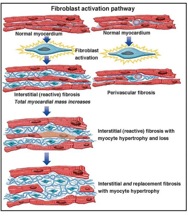
Due to the sudden ischemic death of cardiac myocytes as to the very limited regenerative ability of human myocardium, cardiac tissue damage triggers a series of complex events that leads to the reorganization of cardiac extracellular matrix (ECM) at site of injury and to the replacement of dead myocardium with permanent scar tissue. Such "local" early remodeling that involves the infarcted area occurs as a beneficial response to prevent ventricular wall rupture and is referred to as reparative or replacement fibrosis (Fig. 3). Specifically, tissue injury causes the releases of endogenous molecules known as alarmins (21) that are involved in cardiac repair by eliciting an inflammatory response that clears the necrotic area of degraded ECM and of dead cells. Furthermore, the release of soluble factors, like IL-1, IL-6 and TGF-beta 1, causes the activation of cardiac fibroblasts and their transdifferentiation to myofibroblasts (22) that synthesize and secrete large quantities of ECM proteins, including type I collagen, fibronectin, and tenascin C (23, 24). The formation of scar stabilizes the infarcted zone and the region immediately adjacent to it, but it also causes a dilation (25). However, cardiac remodeling following MI causes geometric changes that involve both the infarcted and non-infarcted myocardium (26, 27). Indeed, replacement fibrosis is followed by the remodeling of border zone and remote uninjured myocardium, where myocytes undergo hypertrophy in response to an unbalanced distribution of wall stress and ECM undergoes extensive rearrangement with excessive deposition of type I collagen, at the expense of the much less rigid type III collagen, to stabilize heart chambers and prevent further deformation. Such fibrotic response involves the left ventricle globally and is referred to as reactive or interstitial fibrosis (28) (Fig. 4).
Figure 4. Compensatory cardiac remodeling of the remote myocardium leads to the reactive fibrosis. The reparative fibrosis causes ventricular geometric changes that are responsible for an uneven distribution of mechanical forces through the myocardium. Healthy myocytes in regions remote to the infarcted area respond to such modified mechanical stimulation by undergoing hypertrophy, while fibroblasts are activated and synthesize excessive collagen. This fibrotic response, termed reactive or interstitial fibrosis, involves the left ventricle globally (from JACC 2014;63:2188-2198).
This progressive fibrosis occurring in the myocardium remote to the infarcted region disrupts myocardial architecture and have adverse effects on ventricular function (29). Therefore, the inflammatory response that is essential for cardiac repair is also implicated in the pathogenesis of post-infarction remodeling and may eventually lead to heart failure (30), a complex clinical syndrome caused by a structural or functional cardiac disorder that results in systolic or diastolic dysfunction responsible, in turn, for heart inability to perform sufficient pumping to meet body’s needs (31, 32).
1.3 Current Treatment of IHD
Lifestyle changes, drugs, and medical procedures can help prevent or treat coronary heart disease.
The initial therapy of MI is directed toward restoration of perfusion as soon as possible to limit myocyte loss. This may be accomplished through a pharmacological fibrinolysis in selected patients or by mechanical means, such as percutaneous coronary intervention (PCI), or coronary artery bypass graft (CABG) surgery.
Successively, pharmacological therapy aimed at reducing mortality, relieve symptoms and signs, improve quality of life, prevent the occurrence of further myocardial damage and the remodeling of the myocardium is established.
The medical therapy relies on several drugs, including anti-thrombotic agents, vasodilator agents, beta-adrenoceptor antagonists (beta blockers), statins, diuretics, aldosterone receptor antagonists, angiotensin-converting enzyme (ACE) inhibitors, angiotensin II receptor blockers, whose combinations are dictated
symptoms, reduce myocardial remodeling and the risk of having another ischemic event.
As a result, the induction of post-infarction cardiac regeneration in adult mammals is currently the target of intensive research and drug discovery attempts.
1.4 Future Treatment of IHD
Regenerative medicine is aiming at restoring structure and function of damaged tissues and organs. The goal of this approach rises from the needing to develop a strategy to treat previously incurable injuries and highly deadly pathologies, as cardiovascular diseases (35). A combination of approaches and tools, such as tissue engineering, cellular therapies, and medical devices or artificial organs can enhance the healing process naturally occurring in human body as result of an injury (36). The close cooperation of diverse fields, like biology, chemistry, tissue engineering and even robotics is required to deal with such a challenging task (37).
1.4.1 Cardiac Cell Therapy
Cell therapy has been experimented for more than a decade, with a number of cell types used in clinical trials in seeking to replace damaged cells or to repair injured tissue (38).
A pioneer study with the first cells used for cardiac regeneration, was performed in 1998 by Taylor et al. and demonstrated that skeletal myoblasts (SMs) have the ability to enhance and partially restore cardiac function (39). This study was an inspiration for the further use of many other cell types, such as bone marrow-derived stem cells (BMMNCs) (40-43), Mesenchymal stem cells (MSCs) (43), cardiac progenitor cells (CPCs) (44), cardiosphere-derived cells (CDCs) (45, 46), embryonic stem cells (ESCs) (47) and more recently induced pluripotent stem cells (iPSCs) (48, 49). There are still more controversies about the use of cells in therapy: on one hand they
produced encouraging results in improving the global cardiac function, and this is probably due to a paracrine action more than an effective integration and proliferation or survival in vivo, on the other hand the results of pre-clinical studies conducted in animal models and clinical trials conducted in humans were variable, not reproducible, and a number of obstacles and side effects were frequent (38).
One of the major obstacles about cell therapy was to find the appropriate way to deliver a sufficient number of cells to the damaged area in the less invasive way (34). Intracoronary and intramyocardial injection after myocardial infarction were the preferred strategies, even if the integration, the engraftment and the long-term survival of cells administrated was very weak (50).
An alternative to the direct injection of cells is the implantation of engineered tissue to offer a both mechanical and biological support to enhance cell retention and function in vivo (51).
1.4.2 Cardiac Tissue Engineering
Tissue engineering technologies aim at the generation of three-dimensional tissue-like scaffolds for therapeutic use. Three factors are pivotal for a successful approach: cells, extracellular matrix, and biomimetic signals (52) (Fig. 5).
Scaffolds, in fact, require specific features to produce the desirable therapeutic outcome, as they have to guide cell organization, growth and differentiation, and have to ensure structural stability and a suitable environment where cells can produce new biological tissue (53).
Biocompatibility, biodegradability and appropriate mechanical properties are the major criteria used for the production
Figure 5. The tissue engineering triad. Tissue engineering strategies are based on three pillars: cells to rebuild the tissue, scaffolds to recreate the tissue environment and physical and chemical signals to support and stimulate cellular functions.
A scaffold must be non-permanent and for this reason, biodegradability is a notable characteristic. In fact, once cells begin to produce their own new and healthy extracellular matrix, the scaffold is no more needed (52).
It is crucial to customize the scaffold depending on the anatomical site of destination, in particular, mechanical properties and architecture must be balanced to guarantee the surgical handling during the implantation procedure and the adequate porosity to allow cell infiltration and vascularization (52).
Several biomaterials, either natural or synthetic, have been tested thus far as substitute for cardiac environment to support cell-based regeneration (55).
However, on the one hand the spatial organization of structural ECM components and the biochemical complexity of the ECM can be fully recapitulated only by the naturally occurring cardiac ECM, but, on the other hand, biomaterials are tunable in their physical properties that have been demonstrated to drive stem cell differentiation and behavior (56). Therefore, finding a substitute able to recapitulate, at least partially, mechanical and biological features of the myocardium is a primary goal in cardiac regenerative medicine.
2 Aims
Cardiac cell therapy demonstrated that regeneration cannot occur without restoring the extracellular compartment along with the cellular compartment, as cell death causes a dramatic rearrangement of myocardial environment that triggers the replacement fibrosis and sustains the reactive fibrosis. The replacement of noncompliant scar tissue with newly formed and fully functional myocardium in the infarcted region would compensate the limited regenerative ability of human adult myocardium and oppose the pathological remodeling that inexorably takes place in the remote region of the ischemic heart.
Cardiac tissue engineering promise to heal the infarcted heart relies on the combination of a scaffold with growth factors and cells. Scaffold and growth factors are employed to restore the microenvironment in its composition, architecture and signaling activity, while cells are needed to form new vessels and replace cardiac supporting and parenchymal cells.
The aim of this study is to explore in vitro the possibility of constructing a three-dimensional scaffold for cardiac repair and regeneration from the adult human skin. The skin is the largest organ in the body that can be easily and opportunely harvested to provide a viable biological and autologous substitute for cardiac environment. Furthermore, cardiac and dermal ECMs are rich in collagen, laminin and elastin. Therefore, the decellularized human skin may also provide biological signals and mechanical properties, like elasticity, that are, at least partially, shared with the microenvironment of the myocardium. Based on the mounting evidence that the adult human heart hosts a population of resident cardiac progenitor cells, a scaffold carrying biological and mechanical signals might yet be used as stand-alone bioengineering product.
To test our hypotheses, we prepared biological scaffold of decellularized human skin (d-HuSk) and decellularized human myocardium (d-HuM). Then, we evaluated whether d-HuSk
showed promise to serve as substitute for cardiac environment by analyzing its composition, biocompatibility and capability of delivering biological signals like the signals delivered by the native cardiac environment. Moreover, we analyzed in vitro the effects of d-HuSk on the engraftment and differentiation of resident cardiac progenitor cells to assess the potential of d-HuSk to serve as both cell-delivering and stand-alone scaffold.
3 Results and discussion
3.1 The decellularization of human skin from living subjects can be swiftly and efficiently accomplished
To be considered suitable for regenerative medicine applications, scaffolds of decellularized matrix should not elicit an immune response. This implies that decellularization process should ensure a complete removal of cellular antigens (57, 58). The currently accepted criteria to satisfy the intent of decellularization include a residual double-stranded DNA (dsDNA) content of less than 50 ng dsDNA per mg of dry decellularized tissue and lack of visible nuclear material in tissue sections stained with Hematoxylin and Eosin (H&E) (59). Snap-frozen skin samples were decellularized following a recently published protocol proven successful for a fast and effective decellularization of human myocardium (60). The first obvious change that occurred with the decellularization was in the color of samples that turned completely white with the respect to the brownish color of native skin (Fig 6, A and B). Then quantitative measurements of DNA content and H&E staining were used to evaluate the effectiveness of decellularization and suitability of d-HuSk as biological scaffold for tissue engineering, in accordance to aforementioned criteria. Analysis of DNA content clearly showed the virtual absence of dsDNA in d-HuSk (Fig 6, C), whose residual amounts were well below the currently accepted standard resulting as low as 7.50 ± 2.162 ng per mg of dry tissue (Fig 6, B). Such dramatic result was confirmed by the histological analysis as H&E staining demonstrated the absence of nuclei in d-HuSk (Fig. 6, F) when compared with native skin (Fig. 6 E). Notably though, decellularization might be a very harsh treatment for soft tissues. The combination of chemical and mechanical methods of decellularization can cause both a biological impoverishment of the ECM and a disruption of tissue integrity and architecture (61).
Figure 6. Evaluation of the effectiveness of the decellularization procedure of skin. A and B: Representative images of macroscopic examination of native human skin (HuSk) (A) and decellularized human skin (d-HuSk) (B) showing an obvious change in color, inasmuch as d-HuSk samples were completely white. C and D: Representative gel electrophoresis revealing an extremely low content of residual dsDNA in d-HuSk (n = 6) with bands barely visible (C) and concentration of DNA well below that of the native skin (n = 4) and the
Therefore, it is crucial to find the right balance between strength, to ensure the complete removal of cells and debris, and subtlety, to avoid marked alteration of native structure and composition of matrix. Indeed, the histological analysis showed an overall well-preserved architecture of the dermal matrix in d-HuSk (Fig 6, F). Therefore, d-HuSk not only fulfilled the requirements proposed for the evaluation of the effectiveness of decellularization, but also preserved the histological organization of native tissue, providing compelling evidence of a highly efficient decellularization method.
3.2 Decellularized Human Skin (d-HuSk) consists of structural and functional ECM proteins that are key components of the cardiac matrix
The ECM is a complex network of fibrous proteins, polysaccharides and soluble factors. On the one hand, the ECM is continuously produced and remodeled by resident cells, but, on the other hand, its unique composition and three-dimensional organization provide biological and mechanical cues that are responsible for directing stem cell fate (62) and cell behavior both in physiological and pathological states. Unquestionably, the ideal scaffold for cardiac regeneration would be the naturally occurring cardiac ECM and the most ambitious - and still unmet - aim of cardiac tissue engineering is to replicate its architecture and composition. Aiming at investigating on the suitability of d-HuSk as substitute for cardiac ECM, we evaluated by immunohistochemistry the presence and localization in d-HuSk of structural and non-structural proteins of the ECM (63) that are described as main components of both cardiac (64) and dermal matrix (65, 66) and that we previously reported as retained by decellularized cardiac matrix (60).
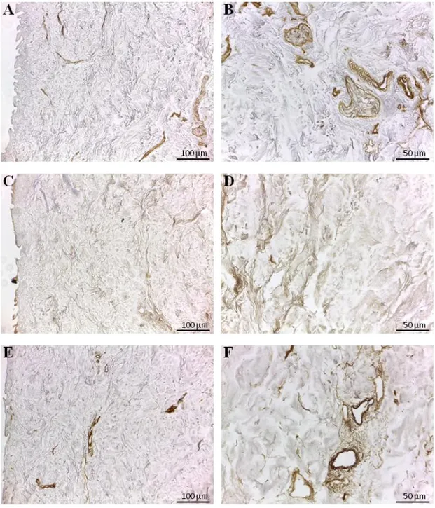
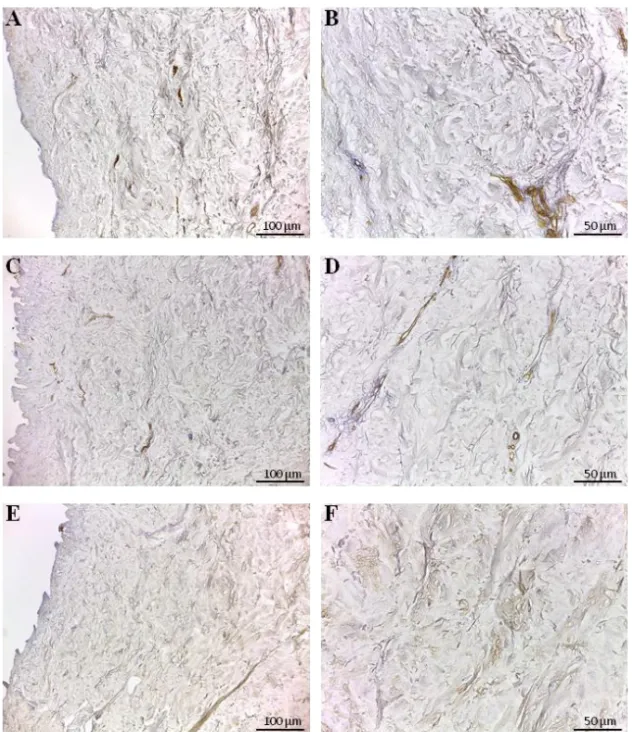
Immunohistochemical analysis revealed that type I, III and IV collagens (Fig. 7) and the non-collagenous proteins, like fibronectin, laminin and tenascin (Fig. 8), were preserved in d-HuSk as well. Type I and III collagens resulted scattered throughout the entire dermis (Fig. 7, A-D), along with fibronectin
(Fig. 8, A-B) and tenascin (Fig. 8, E-F), while type IV collagen (Fig. 7, F) and laminin (Fig. 8, D) were localized mostly in the basement membrane of vessels. Moreover, type III collagen (Fig. 7, D) and tenascin formed a delicate texture (Fig. 8, F), while type I collagen (Fig. 7, B) and fibronectin (Fig. 8, B) were visible as thicker bundles.
The investigated ECM components are the main structural and functional proteins of cardiac matrix and their retention in d-HuSk provide evidence to substantiate the ability of d-d-HuSk to deliver a combination of structural support and biological signals that act in cardiac microenvironment. Specifically, fibril-forming type I and III collagens are the main collagenous component of the cardiac matrix (67) that provide tensile strength and are responsible for the integrity of cardiac interstitial connective tissue. Additionally, type I collagen promotes cardiomyogenesis of MSCs (68, 56), while fibronectin, laminin and tenascin, other than playing major role in cell adhesion and migration or in tissue repair, are involved in embryonic development of the heart (69-71). Particularly, fibronectin is required during cardiac morphogenesis for the formation of the cardiac outflow tract and nodes and it is involved in the differentiation of neural crest cells into vascular smooth muscle cells by regulating Notch signaling (72), tenascin C is transiently expressed during the development of the heart at restricted sites to promote differentiation of cardiomyocytes (73), while laminin in the developing heart orchestrates the assembly of the cardiac ECM and in the adult heart lines the myocardial cells (74). Such functions are collectively of strategic importance when considering the application of d-HuSk in a stem cell-based cardiac regeneration approach, conferring to d-HuSk the potential to support and boost cardiac differentiation.
Figure 7. Immunodetection of collagen type I, III and IV in d-HuSk. Representative images of the immunohistochemical analysis showing the presence and localization of the main cardiac collagenous components in d-HuSk: the fibrillar type I (A, B) and III (C, D) collagen and the collagen type IV (E, F) with its typical localization in the basement membrane. (Scale bar: 100 µm for A, C and E, and 50 µm for B, D and F).
Figure 8. Immunodetection of fibronectin, laminin and tenascin in d-HuSk. Representative images of the immunohistochemical analysis showing the presence and distribution of fibronectin (A, B), laminin (C, D) and tenascin (E,
3.3 d-HuSk has the potential to deliver mechanical stimuli
When evaluating biological scaffold for regenerative medicine applications it is crucial to focus on the main requirements that the bioconstruct must fulfil to be eventually translated to the clinics. As for myocardium, along with the biochemical cues regulating cellular processes, like proliferation, differentiation and apoptosis, muscle cells need a compliant matrix that allows contraction and relaxation cycles while preserving its integrity. Hence, mechanical properties of scaffolds should be considered as important as biological signals.
Other than collagens, also elastin and glycosaminoglycans (GAGs) are known to be responsible for the biomechanical properties of ECM (75, 76). Particularly, elastin is the dominant mammalian elastic protein and, as the main component of elastic fibers, is responsible for tissue flexibility (77), while glycosaminoglycans (GAG) are highly hydrophilic chains of proteoglycans whose significant water-binding capacity confers mechanical stability to tissues (78).
Therefore, we analyzed the retention and distribution of elastin and GAGs in d-HuSk by Paraldehyde fuchsin Gomori’s, Weigert Van Gieson and Alcian Blue stainings. Then we performed comparative analysis between d-HuSk and adult human myocardium by quantitative dye-binding assay for elastin and GAG content to gain an insight into what might be the mechanical properties of d-HuSk with the respect to human myocardium (79).
Paraldehyde fuchsin Gomori’s (Fig. 9, A and B) and Weigert Van Gieson (Fig. 9, C and D) stainings clearly showed the presence of elastic fibers in native skin (Fig. 9, A and C), and in d-HuSk (Fig. 9, B and D), where the retention of elastic fibers surrounding well-preserved blood vessels resulted glaringly obvious (Fig. 9, B and D, red and yellow arrowheads, respectively). Quantitative Fastin Elastin assay not only confirmed the presence of elastin in d-HuSk but showed also that elastin
content of d-HuSk did not differ significantly from the elastin content of native skin 34,2054 ± 2,529 µg/mg of dry tissue in d-HuSk vs. 37,5772 ± 2,561 µg/mg of dry tissue in d-HuSk) (Fig. 8, E).
Similarly, Alcian Blue staining revealed the presence of GAGs in native skin (Fig. 10, A), and in d-HuSk (Fig. 10, B), while quantitative Blyscan assay confirmed the retention by d-HuSk of amounts of GAGs that did not differ significantly from the content of native skin (76,89 ± 14,22 µg/mg of dry tissue vs. 100,7 ± 17,36 µg/mg of dry tissue in HuSk) (Fig. 10, C).
Conversely, the content of elastin and GAGs differed between human myocardium and d-HuSk. Indeed, native human myocardium contained significantly (p < 0.05) higher amount of elastin when compared to d-HuSk (116,50 ± 6,499 µg/mg of dry tissue vs. 34,2054 ± 2,529 µg/mg of dry tissue). Interestingly though, the elastin content of d-HuSk did not differ significantly from that of d-HuM (34,2054 ± 2,529 µg/mg of dry tissue vs 48,18 ± 9,629 µg/mg of dry tissue) (Fig. 11, A). Although the difference observed between d-HuSk and native myocardium must be taken into account when evaluating the overall composition of d-HuSk, it is of note that, as far as we are concerned, the best substitute for cardiac ECM in terms of composition is the decellularized cardiac ECM. Therefore, the comparison between d-HuSk and d-HuM is more appropriate to assess the suitability of d-HuSk as a scaffold for cardiac regenerative medicine. As regards GAGs, instead, quantitative analysis revealed a significantly (p < 0.001) lower content of GAGs in both native (18,69 ± 2,344 µg/mg of dry tissue) and decellularized (12,60 ± 2,300 µg/mg of dry tissue) human myocardium, when compared with the content of d-HuSk (76,89 ± 14,220 µg/mg of dry tissue) (Fig. 11 B).
Figure 9. Histochemical and quantitative analysis of elastin content in native human skin (HuSK) and in d-HuSk. A-D: Representative images of the histochemical analysis performed by Paraldehyde fuchsin Gomori’s (A, B) and Weigert Van Gieson (C, D) stainings showing the presence of elastic fibers in the dermis of native skin (A and C) and in d-HuSk (B and D). Patent blood vessels surrounded by abundant elastic fibers are apparent in d-HuSk (red and yellow arrowheads). (HuSk: native human skin). (Scale bar: 50 µm). E: Quantification of elastin showing comparable content of elastin in d-HuSk and native skin (HuSk). Data are expressed as mean ± SEM (n = 8).
Figure 10. Histochemical and quantitative analysis of GAG content in native human skin (HuSK) and in d-HuSk. A and B: Representative images of the histochemical analysis performed by Alcian Blue staining and showing by the light blue color the presence of GAGs in the dermal connective tissue of native skin (A) and in d-HuSk (B). (Scale bar: 50 µm) C: Quantification of GAGs showing no statistically significant difference in content of GAGs in d-HuSk and native skin (d-HuSk). Data are expressed as mean ± SEM (n = 5).
Interestingly, due to the presence of elastin and GAGs d-HuSk holds the potential to deliver mechanical stimuli that support and promote cardiac differentiation (56, 80, 81).
Indeed, a close relationship between the differentiation of cardiac myocytes and the accumulation of elastin in embryoid bodies has been recently reported (82), along with the evidence that elastin content in the left ventricle of developing mammalian heart increases throughout the developmental stages but decreases soon after birth probably as a stimulus for complete maturation of cardiac myocytes (83).
Furthermore, the early tubular heart is formed by the endocardium and the myocardium with the interposed earliest form of cardiac ECM, the cardiac jelly. The cardiac jelly consists primarily of collagen, glycoproteins and GAGs (84). At this very early stage of cardiac development, by controlling cardiac jelly hydration, GAGs play a major role in exerting compressive strength on the myocardium that drives cardiac chamber expansion (85). Hence the higher content of GAGs in d-HuSk might be crucial for both conferring elasticity and driving cardiac differentiation. Such hypothesis is supported by both the evidence emerging from preliminary studies on mechanical characterization that the stiffness of d-HuSk, measured as the elastic modulus at increasing levels of strain by uniaxial tensile tests, closely matches the range of end diastolic values of human myocardium (86, 87) and the evidence that the lack of GAGs in mouse embryo leads to severe defects in cardiac chamber formation and loss of trabeculation (88).
Figure 11. Comparative analysis of the content of elastin and GAGs in human myocardium and d-HuSk by quantitative dye-binding assays. A: Graphical representation of quantitative analysis of elastin content showing the presence of significantly higher content of elastin in native human myocardium (HuM) than in decellularized human myocardium (d-HuM) or in d-HuSK (n = 8). There is no statistically significant difference in the elastin content between d-HuM and d-HuSk, instead. B: graphical representation of quantitative analysis of GAG content showing significantly higher content of GAGs in d-HuSK. than
3.4 The profile of growth factors of d-HuSk is comparable to that of the cardiac native matrix
Growth factors are signaling molecules known to regulate a plethora of tissue and cellular functions and their importance has been widely recognized also in tissue engineering (89). The ECM is known to function as a storage for growth factors (90) that are bound to ECM components like GAGs.
Therefore, we performed a comparative analysis of the growth factor profile of d-HuSk and d-HuM by protein array. Interestingly, the analysis revealed that d-HuSk contained, to a large extent, the same growth factors of the cardiac native matrix (Fig. 12, A-C). Indeed, both d-ECMs contained growth factors, like hepatocyte growth factor (HGF), insulin-like growth factor (IGF), stem cell factor (SCF), platelet-derived growth factor (PDGF) and vascular endothelial growth factor (VEGF), but d-HuSk resulted enriched with growth factors that were virtually absent in d-HuM, like granulocyte-macrophage colony-stimulating factor (GMCSF) and transforming growth factor (TGF-beta) and also contained significantly higher amount of growth factors like basic fibroblast growth factor (bFGF) and epidermal growth factor (EGF) (Fig. 12, D).
These factors have been reported to be involved in a variety of cardiac processes. Particularly, HGF in the developing heart, influences cardiomyocyte proliferation and differentiation, while in the adult heart controls heart homeostasis, prevents oxidative stress in normal cardiomyocytes (91) and is thought to act as a modulator of cardiac repair (92). Additionally, the ligand-receptor systems HGF/Met has been extensively investigated for its role in cardiac regeneration and evidence supports its involvement in boosting migration, engraftment and commitment of cardiac stem cells (93) and in regulating MSC proliferation and differentiation into cardiac myocytes (94).
Similarly, PDGF and VEGF, other than playing a pivotal role in promoting and controlling angiogenesis and
neovascularization, have been reported to enhance the proliferation of MSCs and their differentiation into cardiac myocytes (94-96).
SCF is a growth factor abundantly expressed in the normal heart and for its ability to mobilize stem cells from the bone marrow is an interesting factor for stem cell-mediated cardiac repair. A role in controlling and attenuating cardiac remodeling has also been proposed for this growth factor (97).
An intriguing relationship between IGF-1 and cardiac myocytes has emerged from several studies, as IGF-1 has been shown to control differentiation, transcription, protein synthesis, and cell death of cardiac myocytes (98, 99). Further, IGF have cardioprotective functions mediated by the inhibition of cardiomyocytes apoptosis (100) and stimulates cardiac muscle regeneration mediated by the myocardial adult stem cells (101).
As mentioned above, d-HuSk contained significantly higher amount of bFGF, EGF, GM-CSF and TGF-beta. These factors are also involved in processes like cardiac stem cell migration and proliferation (102), cardioblast specification (103), cardiac development (104) mobilization and proliferation of endothelial progenitor cells (105), cardioprotection (106).
The abundance of growth factors in d-HuSk strengthens its suitability as a promising tool for cardiac tissue engineering. Noteworthily, considering the already described similarities between d-HuSk and the native cardiac matrix, d-HuSk might alone provide two of the three pillars of tissue engineering (Fig. 5), namely the scaffold and the signals, and then be exploited as stand-alone scaffold to boost cardiac regeneration by recruiting resident cardiac progenitor cells, or as a cellularized scaffold by preparing a cardiac engineered tissue in vitro with the cell population of choice.
Figura 12. Analysis and quantification of growth factors evaluated by specific protein array. The representative images of membrane array (on the left) show that most of the 41 targets were present both in d-HuSk and d-HuM. However, it is immediately apparent a higher content of bFGF, EGF, GM-CSF and TGF-beta in d-HuSk, as shown by the reference array map in the upper left corner. The quantification of the aforementioned factors (on the right) shows their significantly higher content in d-HuSk. Each value expresses the mean ± SEM (n = 4). Asterisks were used to report significance in each comparison as follows: significant (* p < 0.05), very significant (*** p< 0.001). O.D.: optical density.
3.5 d-HuSk is a suitable environment for the engraftment and survival of human Cardiac Progenitor Cells (hCPCs)
In order to be considered suitable for regenerative medicine purposes, any scaffold must meet some key requirements with biocompatibility being one of them (52). This implies that the scaffold must have the ability to attract cells and sustain or promote their adhesion and survival exactly like the naturally occurring tissue microenvironment.
Thus, to evaluate the ability of d-HuSk to serve as a viable microenvironment capable of supporting hCPC homing, engraftment and survival, we seeded and cultured hCPCs on 600-μm-thick sections of d-HuSk and cultured them for up to four weeks. Additionally, to evaluate the effects of d-HuSk on hCPCs survival as compared to those of the cardiac native matrix, we seeded and cultured in the same conditions hCPCs on 600-μm-thick sections of HuM as a reference. Last, to assess whether d-HuSk had the potential to attract resident hCPCs and, then, to be used as a cell-free scaffold for cardiac tissue engineering, we subjected hCPCs seeded and cultured on d-HuM for four weeks to d-HuSk, by placing d-HuSk scaffolds in culture in the close proximity of recellularized d-HuM scaffolds.
Then, we analyzed by time-lapse microscopy the migration of hCPC from d-HuM to d-HuSk, while by the fluorescent staining with DAPI and SEM analysis we evaluated the engraftment of hCPCs on d-HuSk. Finally, by trypan blue exclusion assay we assessed the cell death rate and cell viability of hCPCs cultured on d-HuSK and on d-HuM, calculating the mean percentages of dead and alive cells over total cells at each timepoint considered.
Figure 13. Migration of hCPCs from d-HuM to d-HuSk. Representative images at phase contrast microscope showing hCPCs engrated onto d-HuM migrating towards d-HuSk at different timepoints. A: time 0, B: after 24 hours, C: after 48 hours, D: after 72 hours, and E: after 96 hours. Cells reached d-HuSk within 90-116 hours. (Scale bar: 100 µm).
By binding the nuclear DNA the DAPI staining showed the presence of hCPCs that engrafted onto d-HuSk either after direct seeding or after having migrated in vitro to d-HuSk from d-HuM (Fig. 14).
SEM analysis not only confirmed the engraftment of hCPC on d-HuSk but allowed also to examine the morphology of hCPCs, that appeared either as mesenchymal-like cells characterized by elongated irregular shape and multiple filopodia or as rectangular/polygonal-shaped cells. Further, cell-to-cell contacts were apparent in some microscopic fields (Fig. 15).
48 hours after seeding the death rate of hCPCs seeded on the d-HuM or on d-HuSk did not differ significantly (9.814 ± 1.792% and 9.112 ± 1.532%, respectively) and on both matrices the death rate of hCPC decreased remarkably and progressively with time (3.402 ± 0.681% on HuM and 3.035 ± 0.529% on d-HuSK after 72 hours, 2.348 ± 0.779% on d-HuM and 1.857 ± 0.771% on d-HuSK after 96 hours, 1.2723 ± 0.678% on d-HuM and 1.190 ± 0.659% on d-HuSK after 120 hours, 0.750 ± 0.303% on d-HuM and 0.688 ± 0.239% on d-HuSK after 144 hours) till it reached values well below 1% of total cells, without any statistically significant differences between the two matrices (0.257 ± 0.107 % on d-HuM and 0.253 ± 0.104% on d-HuSk). Obviously, cell viability showed an inverted trend and increased with time on both d-HuM and d-HuSk. Specifically, the mean percentage of alive cells resulted 90.186 ± 1.792% and 90.888 ± 1.792% after 48 hours on d-HuM and d-HuSk, respectively, but then increased up to 99.743 ± 0.107% and 99.747 ± 0.107% after 7 days on d-HuM and d-HuSk, respectively (Fig. 16).
Based on the evidence emerging from the in vitro assessment of biocompatibility, d-HuSk environment was as safe
HuSk holds great promise as a stand-alone scaffold for cardiac tissue engineering applications.
Figure 14. Fluorescent staining with DAPI of hCPCs engrafted on d-HuSk. Representative images showing by the nuclear staining with DAPI and the phase contrast microscope the presence of hCPCs engrated onto d-HuSk either after direct seeding (A, C, E) or after having migrated to d-HuSk from d-HuM (B, D, F). The merged pictures clearly show the distribution of hCPCs in d-HuSk. (Scale bar: 50 µm).
Fig. 15: SEM analysis of hCPCs engrafted onto d-HuSk. Representative images of SEM analysis of hCPC cultured on d-HuSk. White asterisks indicate the cells. In (A) two cells clearly spread onto d-HuSk that contact each other are shown. In (B) one cell with elongated irregular shape characterized by multiple filopodia is shown. This morphology is compatible with mesenchymal-like or mesenchymal derived cell. In C and D cells with an elongated rectangular/poligonal shape compatible with differentiating cardiomyocytes are shown. (Scale bar: 10 µm for A and 2 µm for B, C and D).
Figure 16. Quantification of cell death rate and viability of hCPCs on HuSk and HuM. Mean death rate and mean viability of hCPC cultured on d-HuSk or d-HuM, as measured by trypan blue exclusion assay. Cells were detached and assayed every day started at day 2 to allow them to attach. Then based on the ability of healthy cells to exclude the dye, dead cells were recognized by the blue staining with trypan blue and, hence, at any given timepoint the number of alive and death cells was obtained, averaged and calculated as the mean percentage ± SEM (n = 4). With time cell death rate dramatically decreased from around 9% to less than 1%, without any statistically significant differences between cells cultured on d-HuM or d-HuSk.
3.6 hCPCs retain on d-HuSk the potential to differentiate towards cardiac myocytes in vitro
Evidence collected thus far supported the hypothesis that d-HuSk might constitute a myocardial-like matrix. Loaded with signals that direct and control cardiac development and the homeostasis of the adult heart, d-HuSk could also be capable of supporting and promoting the differentiation of hCPCs into cardiac myocytes.
Hence, to assess the suitability of d-HuSk to serve as a cardiogenic environment for hCPCs, we analyzed the expression of genes specific for cardiac program (GATA4, TBX5) and for cardiac myocytes (CX43, CX37, TBX3, TBX5, MEF2C, ACTC1, MYH7) in hCPCs cultured on d-HuSk for four weeks by real-time PCR. To gain better insight into the possible effects of d-HuSk on hCPC expression of cardiac myocyte differentiation markers, we used as a reference the expression of the same markers in hCPC cultured in the same conditions on d-HuM.
Interestingly, from gene expression analysis emerged that the transcription of all cardiac specific genes investigated, either typical of early or late stages of differentiation, was significantly upregulated (p ≤ 0.05) up to two-fold in hCPCs cultured on d-HuSk for 4 weeks (Fig. 17).
Additionally, to confirm the results of gene expression analysis, we evaluated at the protein level the expression of the cardiac myocyte markers Nkx 2.5, alpha-sarcomeric actin, connexin-43, desmin and dystrophin in hCPC cultured on d-HuSk for 4 weeks, by immunofluorescence.
The immunocytochemical analysis showed the immunopositivity in hCPC engrafted onto d-HuSk for the markers investigated. Clearly, the main cell population of CPC consisted of cells expressing Nkx 2.5 and alpha-sarcomeric actin (Fig. 18), as well as desmin, dystrophin and connexin-43 (Fig. 19) that are all markers specific for cardiac myocytes. Moreover, the alignment of actin filaments is suggestive of striated muscle. even though further investigation is needed to demonstrate the electrical coupling of the connexin-43 expressing cells in order to demonstrate their effective ability to form functional syncytia.
Figure 17. Analysis of gene expression of cardiac myocyte markers. Real-time PCR analysis of the expression of genes characteristic of cardiac myocytes showing an upregulation of the transcription for all markers in hCPCs cultured on d-HuSk when compared with hCPCs cultured on d-HuM. Data are expressed as mean ± SEM (n = 3). Asterisks are indicators of the p value obtained in each comparison as follows: significant (* p ≤ 0.05), very significant (** p < 0.001 and extremely significant (*** p ≤ 0.001).
Fig. 19: Confocal microscopy analysis of hCPC cultured on d-HuSk for four weeks. Representative images of immunofluorescence experiments, showing the distribution pattern of desmin connexin-43 and dystrophin in hCPC cultured on d-HuSk.Most of CPC expresses such markers. Moreover, the actin filaments appear distributed in approximately parallel rows. This pattern is compatible with cardiomyocyte differentiation. In the lower panel negative control, obtained omitting primary antibodies, is shown.
markers, which, along with the morphology of cells observed at the SEM, supports the hypothesis that the decellularized human skin makes a myocardial-like environment that attracts resident CPCs, ensures their survival and sustains their differentiation.
4 Conclusions
In the scenario of an urgent need for more effective therapy of ischemic heart disease, tissue engineering has emerged as the only therapeutic approach that holds the potential to reestablish the structural, biomechanical and functional integrity of ischemic myocardium. However, the search for the best-performing and cost-effective scaffold capable of recapitulating biological and mechanical cues of cardiac microenvironment is still a primary goal of cardiac regenerative medicine.
With the present work we developed and evaluated in vitro a biological tool for cardiac regeneration that holds the potential to become a powerful alternative for both native matrix and synthetic scaffold.
d-HuSk fulfills the key requirements needed for a scaffold to warrant its use in tissue engineering. The biological nature of d-HuSk ensures a full biodegradability and, indeed, d-d-HuSk should be considered a temporary implant providing support to resident cells until its complete replacement by native tissue, as resident cells are expected to degrade the ECM and produce their own. Furthermore, the dermal ECM provides both sites of attachment for cell adhesion and a vascular network critical to ensure diffusion of nutrients, and, then, addresses two serious problems related to synthetic biomaterials.
However, the groundbreaking features of d-HuSk are its autologous origin and ability to support, in vitro, the engraftment, proliferation and differentiation potential of resident human cardiac progenitor cells (hCPCs). In fact, using d-HuSk as an autograft would imply transplanting a fully compatible scaffold capable of averting the immunological response and the risk of rejection that affect the heterologous and xenogeneic transplant or implant. Additionally, despite the different anatomical site of origin d-HuSk proved to be a suitable environment for hCPC, at least as cozy as the native one. If proven in vivo, such evidence has a tremendous potential, as d-HuSK might be used as an autologous and
stand-alone scaffold that promotes and sustains cardiac regeneration by resident hCPCs. Noticeably yet, as adult fibroblasts have been recently successfully reprogrammed to cardiac mature cells (107), the preparation of scaffolds of decellularized dermal ECM holds great promise to eventually evolve into the development of a complex therapeutic approach that offers, with one single intervention of skin harvesting, the possibility of constructing an engineered – fully autologous – myocardium using cardiac direct reprogramming of fibroblast to restore the cellular compartment and the decellularized dermal ECM to replace the extracellular compartment.
5 Limitations of the study
In this study we mainly focused on the effects of d-HuSk on CPCs, however further investigation is needed to provide more extensive insights on cell-matrix cross-talk and, specifically, to analyze whether and at to what extent CPCs can modify the composition of d-HuSk and adapt it to their requirements. This should include elucidating whether CPCs are capable to remodel and to convert d-HuSk from a fetal-like cardiac matrix to a mature cardiac matrix.
The native ECM influences cell behavior by its biological, chemical, physical and mechanical properties. Thus, a mechanical characterization at the macro and the micro scale should be performed to gain better knowledge of the signaling that are possibly delivered by d-HuSk and that might influence cell viability, proliferation, migration and differentiation.
Although the evidence that CPCs retain their ability to differentiate on biological scaffold obtained from different and more easily accessible ECM represents an important advance in cardiovascular regenerative medicine, as it overcomes problems related to the preparation of myocardial biological scaffolds, such evidence emerges from an in vitro study. Undoubtedly, in vivo studies designed to understand also mechanical properties, should advance its clinical utility.
6 Materials and methods 6.1 Tissue specimens
Skin specimens were collected from patients undergoing abdominoplasty (n = 8, mean age 42.25 ± 7.94 years). Upon receipt, specimens were washed in physiological saline, then subcutaneous fat tissue was removed, and multiple samples were cut. Resulting samples were then promptly processed for histological analysis, decellularization, or snap frozen until use. Cardiac specimens were harvested from macroscopically uninjured areas of the free wall of the left ventricle of hearts of cardiac transplant recipients (n = 10, mean age 49.5 ± 4,7 years). Specimens were washed in physiological saline and then cut into multiple samples that were processed for decellularization, or enzymatically digested to isolate hCPCs, or snap-frozen until use. Patients provided written informed consent and specimens were collected without patient identifiers, following protocols approved by the hospital and in conformity with the principles outlined in the Declaration of Helsinki.
6.2 Decellularization of human skin
Human Skin (HuSk) specimens were cut into smaller samples (2 x 1 cm, length by width) and incubated in decellularizing solution containing 1% SDS (w/v) and 1% Triton (v/v), for 24 hours under constant stirring. The solution was replaced every 8 hours. During the procedure the epidermis detached from the dermis and was removed. Samples were then rinsed for 24 hours in antibiotic solution containing 0.25 μg/ml Amphotericin B, 100 U/ml Penicillin, 50 U/ml Streptomycin in PBS, and then for an additional 30 minutes in sterile bidistilled water. After decellularization, samples of d-HuSk were either fixed in formalin for paraffin embedding and histological analysis or stored at -80°C until use for decellularization and molecular analysis.
6.3 Decellularization of human myocardium
Following a recently published protocol (60), frozen samples of human myocardium were mounted on a cryostat chuck using Tissue Freezing Medium (Leica Microsystems, Wetzlar, Germany) and cut into 600-µm-thick sections by a Leica CM1950 cryostat (Leica Microsystems). Cryosections were collected into sterile 50-ml sterile plastic tubes and then incubated for 24 hours in decellularizing solution containing 1% SDS (w/v) and 1% Triton (v/v). Cryosections were decellularized under constant agitation on an orbital shaker. The decellularizing solution was replaced every 8 hours. Sections were then rinsed for 24 hours in antibiotic solution containing 0.25 μg/ml Amphotericin B, 100 U/ml Penicillin, 50 U/ml Streptomycin in PBS, and then for an additional 30 minutes in sterile bidistilled water. After decellularization, sections of d-HuM were either stored at 4°C in sterile bidistilled water or at -80°C until use for comparative analysis of d-HuSk and d-HuM.
6.4 Quantitative measurement of DNA content
Genomic DNA was extracted from frozen native (n = 4) and decellularized human skin (n = 6), using the AllPrep DNA/RNA Mini Kit (Qiagen, Hilden, Germany), according to the manufacturer's instructions. Briefly, tissue samples were lysed and homogenized in a highly denaturing buffer containing guanidine-isothiocyanate. The buffer ensured isolation of intact DNA and RNA by inactivating DNases and RNases. Then the passage of lysates through an AllPrep DNA spin column allowed selective and efficient binding of genomic DNA. The columns were washed
6.5 Histochemistry
Specimens of native skin (n = 8) and d-HuSk (n = 8) were fixed in 10% neutral-buffered formalin and then processed for paraffin embedding, following a routine procedure. Briefly, formalin-fixed samples were dehydrated in an ascending series of alcohols, infiltrated with liquid paraffin and then embedded in paraffin in molds. Paraffin blocks were then sliced into serial 5-µm-thick sections that were mounted onto microscope slides. Following standard protocols, after deparaffinization and rehydration in a descending alcohol series, sections were stained with Fast H&E (Hematoxylin and Eosin) staining kit (Bio-Optica, Milan, Italy) to evaluate tissue architecture and effectiveness of decellularization, with Paraldehyde Fuchsin Gomori’s Method and Weigert - Van Gieson Quick Method staining kits (Bio-Optica) to detect elastic fibers and with Alcian Blue pH 2,5 staining kit (Bio-Optica) to detect GAGs and Hyaluronic Acid. Stained sections were observed and evaluated by at least three independent observers using a light microscope DM2000 Led (Leica Microsystems) equipped with the ICC50HD camera (Leica Microsystems) for microphotography.
6.6 Immunohistochemistry
Paraffin sections of d-HuSk were prepared as described for histochemistry and immunostained using indirect immunoperoxidase technique to detect the presence and analyze the distribution of collagens, fibronectin, tenascin and laminin using specific antibodies. The presence and localization of antigen‐antibody complexes were revealed by the UltraVision LP Detection System HRP Polymer & DAB Plus Chromogen (Thermo Scientific, Waltham, MA, USA), following the manufacturer’s protocol. First, sections were incubated with UltraVision Hydrogen Peroxide Block to reduce non-specific background due to endogenous peroxidase. Next, sections were washed in PBS and incubated with Ultra Vision Protein Block, to reduce non-specific
bindings of antibodies. Then, slides were incubated with primary antibodies against human type I collagen, fibronectin, tenascin and laminin (all from Sigma-Aldrich, St. Louis, MO, USA). After further washes in PBS, sections were incubated with Primary Antibody Enhancer, and then with HRP Polymer. Finally, sections were incubated with DAB chromogen for the visualization of the polymer complex. Nuclei counterstaining with Mayer’s hematoxylin (Bio‐Optica, Milan, Italy), dehydration and coverglass mounting with Bio Mount mounting medium (Bio‐Optica) were the last steps before analysis. All steps were performed at room temperature, except for the incubation with primary antibody that was performed at 37°C. Stained sections were evaluated and documented by at least three independent observers using a light microscope DM2000 Led (Leica Microsystems) equipped with an ICC50HD camera (Leica Microsystems).
6.7 Quantitative measurement of elastin
Elastin was extracted from samples of native skin (n = 8) weighting 20 mg each and from samples of HuSk (n = 8) and d-HuM (n = 8) weighting 20 mg each before decellularization. The extraction was performed by heating specimens at 100°C for three one-hour periods in 0.25M oxalic acid, in accordance with manufacturer's directions. Tissue extracts were then assayed in the Fastin Elastin Assay quantitative dye-binding method (Biocolor, Ltd, Carrickfergus, UK), following instructions provided by manufacturer. Briefly, samples were incubated with Elastin Precipitating Reagent to allow the precipitation of α-elastin. Dye Reagent was then added and the Elastin-Dye complexes that formed were exposed to the Dye Dissociation Reagent before