SAPIENZA Università di Roma
Facoltà di Scienze Matematiche Fisiche e Naturali
DOTTORATO DI RICERCA
IN GENETICA E BIOLOGIA MOLECOLARE
XXXII Ciclo (A.A. 2018/2019)
Studying the role of chromatin remodelers
during cell division
Dottoranda
Francesca Delle Monache
Docente guida Coordinatore
Prof. Patrizio Dimitri Prof. Fulvio Cruciani
1 Index
1. Summary………2
2. Introduction ... 5
2.1 Chromatin organization in eukaryotes... 5
2.2 Human SRCAP and Tip60/p400 chromatin remodelling complexes... 10
2.3 The Drosophila Tip60 complex ... 14
2.4 Cell division: an overview ... 16
2.5 Subunits of Tip60/p400 and SRCAP chromatin remodelling complexes localize to the mitotic apparatus in human cells ... 22
3. Aims ... 27
4. Results ... 28
4.1 RNAi knock-down of SRCAP and Tip60/p400 subunits impair successful cell division – part one ... 28
4.2 SRCAP and Tip60/p400 subunits interact with some important cell cycle regulators ... 33
4.3 Aurora B kinase promotes recruitment of some subunits at the midbody ... 37
4.4 Subunits of dTip60 chromatin remodelling complex localize to the mitotic apparatus in Drosophila S2 cells ... 39
4.5 RNAi knock-down of the subunits impair successful cell division – part two ... 41
5. Discussion and conclusion ... 45
6. Future perspectives ... 50
7. Materials and Methods ... 51
7.1 Cell colture, RNA interference and ZM447439 treatment ... 51
7.2 Immunofluorescence ... 52
7.3 Cell cycle synchronization ... 53
7.4 Chromatin fractionation and immunoprecipitation ... 55
7.5 Western Blotting ... 55
7.6 RNA interference in Drosophila S2 cells ... 56
7.7 Statistical analysis ... 60
8. Reference ... 61
2 1. Summary
The genome of eukaryotic cells is organized in a nucleoproteic structure defined chromatin, consisting of DNA, RNA and associated proteins. A dynamic balance is needed between chromatin packaging and genome access to promote essential functions, such as DNA replication and repair, transcription. In these processes, multiple molecules are involved, including histone chaperones, chromatin remodelers and enzymes that modify histones and DNA. The structural characteristic of the remodelling enzymes belonging to a family of remodelling complex, INO80, is the ATPase domain interspersed with a long insertion.
The SRCAP (SNF2-Related CBP Activator Protein) and Tip60 (HIV1 Tat Interacting Protein, 60 kDa) remodelling complexes belong to the INO80 family (subfamily Swr1) and they are conserved from yeast to human. These complexes share some subunits. Evolutionary speaking, these two complexes derive from duplication of Drosophila dTip60 complex. The main function is to promote the exchange of the canonical histone H2A with the histone variant.
Previous studies suggested that chromatin and remodelling proteins might also play roles in cell division, yet evidence remains sporadic.
In past years, in our lab my colleagues found that subunits of the human SRCAP and Tip60/p400 complexes, besides the canonical localization in the interphase chromatin, at different times during cell division target different sites of the mitotic apparatus (centrosomes, spindle and midbody, the bridge connecting the daughter cells at the finale step of mitosis). So, my PhD project aimed at studying the involvement of chromatin remodelling complex in ensuring proper cell division in both human and Drosophila cell lines, two species separated by 700 million years of evolution.
3 First, I found that RNAi-mediated depletion of SRCAP, Tip60, Gas41, YL1 and MRG15 subunits in human cells and DOM-A, Tip60, MRG15 and Yeti in Drosophila cells affect cell division. In particular, cells have shown aberrant spindle morphology, chromosome misalignments, long intercellular bridges (LIBs) connecting the daughter cells and multinucleation. Defects found are consistent with their localization in both human and Drosophila cells.
Then, to study how these remodelers are recruited, I performed interaction assays with already known cell division players. Co-immunolocalization and co-immunoprecipitation assays performed thus far on HeLa telophase extracts suggested that the aforementioned players contribute to the relocation of the Tip60 subunit from chromatin to the mitotic apparatus. Moreover, The inhibition of kinase activity of Aurora B, a well-known cell division regulator, caused mislocalization of TIP60, SRCAP and BAF53a remodelers.
Finally, RNAi-mediated depletion of SRCAP, Tip60, Gas41, YL1 and MRG15 caused mislocalization of key components of the midbody, such as Aurora B, MKLP2, CIT-K and PLK1.
In conclusion, we found that subunits of ATP-dependent remodelling complexes:
i) are recruited to the mitotic apparatus;
ii) interact in telophase with main cell division regulators (Aurora B, Cit-K, etc.);
iii) prevent the failure of cell division and genome instability.
Together, our findings suggest a cross-talk between SRCAP and TIP60 complexes and the main regulators of cytokinesis and highlight the existence of a previously unexplored and evolutionarily conserved phenomenon where chromatin remodelling factors translocate from the interphase nucleus to the
4 mitotic apparatus in order to ensure faithful cell division in both human and Drosophila cells.
5
2. Introduction
2.1 Chromatin organization in eukaryotes
The genome of eukaryotic cells is organized in a nucleoproteic structure defined chromatin, consisting of DNA, RNA and associated proteins. The complex was initially identified by the German cytologist Walter Flemming, who defined the "chromatin" as the cellular structure detectable under a microscope after staining with basic dyes such as safranin and hematoxylin (Flemming, 1882).
Chromatin has hierarchical levels ranging from the repetition of the unit of basic, the nucleosome, at higher order levels (Ou et al., 2017). This organization, finely regulated, represents a dynamic balance between packaging of the genome and its accessibility. The linear length of DNA chromosome in a human cell presents a significant topological challenge since about 2 meters of DNA must be packed in the core characterized by a diameter of only 6 μm (Andrews and Luger, 2011; Khorasanizadeh, 2004). Nucleosomes are positioned along DNA in a “beads on a string” configuration to create a 10-nm fiber.
The nucleosome is an octamer, consisting of two copies of each of the four canonical histone proteins (H3, H4, H2A, and H2B), around which there are wrapped 147 bp of DNA. The histone residues, positively charged, interact with the phosphate skeleton of DNA every 410.4 bp, determining thus a high conformational stability (Clapier and Cairns, 2009).
A dynamic balance is needed between chromatin packaging and genome access, as essential functions, such as replication and transcription, require a certain degree of DNA accessibility.
There are two main mechanisms by which chromatin becomes more accessible: the enzymatic modification of histones by adding
6 acetyl, methyl or phosphate groups (Fischle et al., 2003) or displacement, expulsion and histone substitution, mediated by chromatin remodelling complexes (Smith and Peterson, 2005) (Fig.2.1).
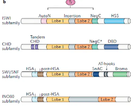
Figure 2.1. Functional classification of remodelers. The ATPase–translocase
subunit of all remodelers is depicted in pink; additional subunits are depicted in green (imitation switch (ISWI) and chromodomain helicase DNA-binding (CHD)), brown (switch/sucrose non-fermentable (SWI/SNF)) and blue (INO80) (Clapier et al., 2017).
In fact, in addition to the four canonical histones, there are histone variants, which are incorporated within the nucleosome, (locally determining a change in its composition) for example in the case of DNA damage or the request for transcriptional activity. In particular, the currently known histone variants are those of histone H2A and histone H3. Four variants of H2A are currently known, but the function is not yet known:
• H2A.Z, associated with chromatin in active transcription • H2A.X, associated with DNA damage (when phosphorylated) • MacroH2A, mainly located in regions that are transcriptionally inactive
7 • H2ABBD variant, not yet well characterized.
In addition, there are two H3 histone variants: • H3.3, associated with transcriptional activation • CENP-A, mainly associated with centromeres
Furthermore, it must be considered that the peripheral portion of the octamer consists of aminoterminal histone tails, which protuded from the nucleosome and represent the first target for post-translational modifications. In particular, the two histones that are more subjected to chemical modifications at the level of the N-terminal tails are H3 and H4. These modifications consist of the addition of chemical groups to amino acid residues, this influences the organization of the chromatin and represents a way of regulation for events such as transcription. (Talbert and Henikoff, 2017).
Chromatin is therefore subjected to continuous structural and functional alterations.
In these processes, multiple molecules are involved, including histone chaperones, chromatin remodelers and enzymes that modify histones and DNA. They, together with readers of post-translational modifications (PTM), transcription factors and RNA, generate specialized genomic domains that contribute to the formation of a versatile chromatin scenario (Yadav et al., 2018). Four different families of chromatin remodelling complexes have been identified conserved in eukaryotes (Clapier and Cairns, 2017), that share a similar ATPase domain but that are distinguished by the uniqueness of the flanking domains (Flaus et al., 2006) and for the different specializations (Fig. 2.2).
8 - ISWI family
Members of the ISWI family (Imitation SWItch) are characterized by two domains adjacent to the C-terminal, SANT and SLIDE domains, which together define a form of recognition of nucleosomes that binds the non-histone tails modified and DNA (Boyer et al., 2004). Many complexes belonging to this family promote chromatin assembly and are generally involved in transcriptional repression (Langst and Manelyte, 2015).
- CHD family
The members of the CHD family (Chromodomain-Helicase-DNA binding) are characterized by two chrome-domains arranged in tandem at the N-terminal. One of the most studied complexes is NURD (nucleosome remodelling and deacetylase) containing the proteins HDAC1 / 2 (histone deacetylases) and MBD (methyl CpG-binding domain). Numerous data have shown an involvement of this complex in the transcriptional repression of specific sets of genes during development in mammals, in D. melanogaster and in C. elegans (Murawska and Brem, 2011).
- SWI/SNF family
The members of the SWI / SNF family (switching defective / sucrose nonfermenting) are characterized by the presence of an HSA domain (helicase-SANT) at the end N-terminal, necessary to recruit actin and ARP (actin-related protein), from a bromodominio to the C-terminal, necessary for the link with the lysines acetylated histones (Filippakopoulos and Knapp, 2012). This family of remodelers plays numerous roles, such as slipping and mediates expulsion of nucleosomes, as well as participates in the assembly of chromatin (Langst and Manelyte, 2015).
9 The structural characteristic of the remodelling enzymes belonging to the INO80 family (INOsitol requiring 80) is the ATPase domain interspersed within a long insertion. The members of this family are classified into two subfamilies, INO80 and Swr1. Unlike the remodelers of other families, the INO80 complex exhibits helicase activity and binds in vitro particular structures of DNA similar to Holliday junctions, consistent with the role of the complex in homologous recombination and replication of DNA. Furthermore, INO80 and Swr1, in yeast, promote the exchange of histone H2A with the histone H2A.Z, by replacing the H2A-H2B dimers with the H2A.Z-H2B dimers (Bao and Shen, 2011).
Figure 2.2. Domain organization of remodeler subfamilies. The ATPase–
translocasedomain (Tr) of all the remodelers is sufficient to carry out DNA translocation. Remodelers can be classified into four subfamilies based on the length and function of the insertion and on their domain organization (Clapier et al., 2017).
10
2.2 Human SRCAP and Tip60/p400 chromatin remodelling complexes
The SRCAP (SNF2-Related CBP Activator Protein) and Tip60 (HIV1 Tat Interacting Protein, 60 kDa) remodelling complexes belong to the INO80 family and specific to the subfamily Swr1 and it’s conserved from yeast to human (Fig. 2.3). These complexes share some subunits.
Evolutionary speaking, these two complexes derive from dTip60 complex duplication.
Figure 2.3. Subunits composition of SWR1 chromatin remodelling complexes
11 SRCAP complex
SRCAP complex is a complex of ~ 1MDa consisting of 10 subunits between including SRCAP, DMAP1, VPS72 (YL1), RUVBL1, RUVBL2, ACTL6A (BAF53a), ACTR6 (ARP6), ACTIN, YEATS4 (GAS41) and ZNHIT1. The homologous complex Swr1 of yeast consists of 10 conserved subunits and four specific subunits, Swc3, Swc5, Swc7 and Bdf1. Mass spectrometry and co-immunoprecipitation data have shown that the human ortholog protein of Swc5, called CFDP1 (Cranio Facial Development Protein 1), interacts with the members of the SRCAP complex (Havugimana et al., 2012; Messina et al., 2017). This suggests that CFDP1 also could be a member of the complex.
The main function of the SRCAP remodelling complex is to promote the exchange of the canonical histone H2A with the variant H2A.Z (Monroy et al., 2001).
Recently, a possible mechanism for histone exchange has been proposed according to which the ATPase domain of the SRCAP protein binds DNA and directs it towards the ARP6 subunit, resulting in a partial detachment of the nucleosomal DNA e the loss of H2A-H2B dimer contacts within the histone octamer (Feng et al. 2018) (Fig. 2.4).
Moreover, Dong et al. identified the human SRCAP chromatin remodelling complex as a factor that promotes DNA-end resection. Indeed, SRCAP confers resistance to DNA damage- inducing agents and is recruited to DSBs (Dong et al., 2014).
Truncated SRCAP protein variants have been implicated in the mechanism of Floating-Harbor Syndrome, a rare human disease characterised by delayed bone mineralisation and growth deficiency, associated with mental retardation and skeletal and craniofacial abnormalities, but the molecular bases underlying the
12 disease must be elucidated (Messina et al., 2016; Greenberg et al., 2019).
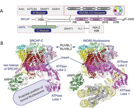
Figure 2.4. A: structural domain organization of SRCAP complex. SRCAP
serves as a scaffold. B: cryo-EM structure of SRCAP complex, in comparison with INO80 complex (Feng et al. 2018).
Tip60/p400 complex
The Tip60 complex in humans consists of 16 subunits (Fig. 2.5) and is involved in multiple cellular processes including transcriptional regulation, DNA repair, the chromatin remodelling, cell migration and genomic instability. The KAT5 protein (Tip60),
A
13 the catalytic subunit of the complex, is an histone acetyltransferase (HAT) belonging to the MYST family. This enzyme acetylates histones, implicated in the organization of chromatin, and some non-histone proteins, implicated in transcriptional regulation and response to DNA damage (Squatrito et al., 2006). Recent studies have also allowed us to identify a new role for this complex in cell cycle progression. Indeed, Tip60-dependent p53 acetylation is regulated to modulate cellular outcome. p53 is acetylated at Lysine 120 by Tip60 to trigger apoptosis when severe DNA damage is revealed (Wang et al., 2019).
Moreover, it has been shown that the KAT5 (Tip60) acetylates Aurora B kinase, a key component of the checkpoint mitotic, in order to guarantee a correct chromosomal segregation (Mo et al., 2016).
As concerns the other subunits, literature reports that Pontin and Reptin belong to the AAA+ family of helicases (ATPases associated with diverse cellular activities) (Kanemaki et al.,1999), while MRG15 is adaptor module which can bind to a modified histone H3 and down-regulates intragenic histone H3 lysine 4 methylation, so it’s involved with active transcription (Zhang P. et al. 2006; Hayakawa et al., 2007).
So, proteins that are co-present in different multi-protein complexes, can also function independently from each other.
14 Figure 2.5. Tip60 complex composition (Judes et al., 2015).
2.3 The Drosophila Tip60 complex
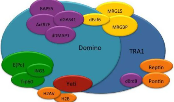
The Tip60 chromatin remodelling complex in Drosophila melanogaster (dTip60) consists of 16 subunits, that have been identified by immunoprecipitation followed by immunoblotting experiments in S2 cells (Kusch et al., 2004) and share a high level of identity with those of human SRCAP and tip60/p400 complex. It catalyzes the replacement of canonical H2A.V by phospho-H2A.V histone variant and it is involved in many cellular processes. dTip60 complex is composed of 16 subunits that share a high level of identity with those of human orthologous SRCAP and
15 Tip60/p400 complexes: dMRG15, dTRA1, dGAS41, dIng3, E (Pc), dPontin, dReptin (Kusch et al., 2004) (Fig. 2.6). The dTIP60 complex catalyzes the dislocation of the canonical histone H2A, and the YETI subunit plays an essential role in subsequent deposition of the H2A.V (Messina et al., 2014; Prozzillo et al., 2019). Nucleosomes containing the H2A.V-H2B dimer seem to facilitate gene activation by transcription factors.
Figure 2.6. Cartoon of dTip60 composition.
Recently, a genetic interaction map of cell cycle regulators showed a link between DOMINO and Yeti and Anaphase Promoting Complex/ Cyclosome (APC/C) (Billman et al., 2016).
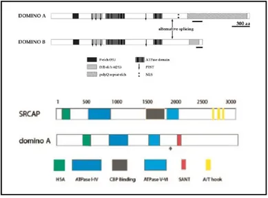
Ruhf et al characterized domino as a gene encoding novel members of the SWI2/SNF2family of DNA-dependent ATPase. domino encodes two protein isoforms of 3202 (DOM-A) and 2498amino acids (DOM-B), which contain a common N-terminal region but divergent C termini (Ruhf et al., 2001).
A study from Eissenberg and colleagues highlighted a significant overlap between the binding sites of DOMINO and RNA
16 polymerase II on Drosophila polytene chromosomes, suggesting for DOMINO a role as a transcriptional co-activator. A similar role was demonstrated for the human homologue of DOMINO, SRCAP, when expressed in Drosophila (Eissenberg et al., 2005) (Fig. 2.7).
Fig. 2.7. Domain organization of DOM-A and DOM-B and comparison with the
human homologous SRCAP (Modified from Ruhf et al., 2001 and Eissenberg et al., 2005).
2.4 Cell division: an overview
Eukaryotic cells duplicate by carrying out an ordered sequence of events by which they duplicate their nuclear content and then divide. These processes together constitute the cellular cycle, which it can be divided into four main phases: G1, S, G2 and M, where the two gap phases divide the two major cell cycle events which are the duplication of genetic material (S phase) and the division into two daughter cells (M phase) (Fig.2.8).
17 Figure 2.8. Phases of cell cycle.
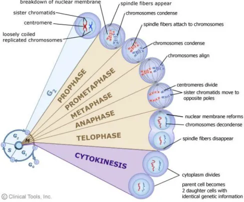
M phase, or mitosis, can be divided into several phases (Fig. 2.9), which lead first to condensation of chromatin and to the segregation of the two centrosomes (prophase), then to the disintegration of the nuclear membrane and the formation of the metaphasic plate, in which the duplicated chromosomes are aligned at the center of the cell and contacted by microtubules of the mitotic spindle (metaphase). During the anaphase the sister chromatids begin to segregate at the two opposite poles of the cell due of microtubule dynamic activity. The telophase is the last stage of the division, which allows the separation of the two daughter cells. These are still connected by a cytoplasmic bridge called midbody. For the final event of cytokinesis it is necessary to train one throttling of the contractile ring, so as to resolve the midbody (MB) and lead to separation definitive of the two daughter cells. The MB has become the focus of intense investigation in cancer cell biology. It forms from the bipolar microtubule array of central
18 spindle and plays essential functions in localizing the site of abscission, and hence of physical separation of daughter cells during cytokinesis (Glotzer et al., 2005; Barr et al., 2007). It acts as a platform for the recruitment and organization of proteins that regulate final withdrawal. MB is a dynamic structure composed of a central region, the MB ring, and two flanking regions.
Figure 2.9. Phases of mitosis.
The cell cycle is a finely regulated process. Transitions between the different phases of the cell cycle are induced by oscillating levels of cyclins and cyclin-dependent kinases (CDK).
Successful cell division requires the balanced distribution of chromosomes and cytoplasmic material to daughter cells. In eukaryotes, this is achieved via a series of coordinated cytoskeletal
19 processes which include spindle assembly, chromosome segregation, spindle positioning and cytokinesis.
The mitotic spindle is assembled from microtubule arrays and their associated proteins that orchestrate chromosome segregation during mitosis (Cleveland et al., 2003; Pollard 2017). It is a highly dynamic phenomenon and evolutionarily conserved, with a significant number of components shared by humans and simpler organisms. In addition to tubulins, proteins involved in spindle function include motor proteins and other microtubule-associated proteins (MAPs), as well as microtubule organizing centers, regulatory kinases and phosphatases, kinetochore protein complexes and chromatin-associated proteins.
The correct sulcus localization is guaranteed by the microtubules of the mitotic spindle: they reorganize forming a structure called central spindle. The formation of this structure derives from the cooperation of multiple cytoskeletal proteins and signalling.
Cytokinesis requires a complex interplay among effector components related to cytoskeleton, chromosomes, cell cycle, lipid raft, vesicle and membrane trafficking factors which map to MB. Alterations of these factors cause cytokinesis failure (Fujiwara et al. 2005; Ganem et al., 2007; Sagona and Stenmark 2010; Hu et al., 2012; Bassi et al., 2011; Bassi et al., 2013) and result in: 1) inhibition or regression of the cleavage furrow, with the ensuing formation of tetraploid cells, or 2) persistence of connections between daughter cells, with the formation of long intercellular bridges (LIBs) originating syncytial cells (Lacroix et al., 2012; Normand et al., 2010).
In particular, the known essential MB/cytokinesis players are Aurora B, Citron kinase (CIT-K), PLK1 and at least molecular motors kinesin-like: KIF23 (MKLP1), KIF20A (MKLP2) and
20 KIF4A (D'Avino et al., 2015). Aurora B, the catalytic subunit of the Chromosomal Passenger Complex (CPC), which includes INCENP, Survivin and Borealin., promotes the interaction between PRC1, involved in the collating of microtubules, and KIF4A, deputed to transport Aurora B intermediate spindle (Nunes Bastos et al., 2013). Instead, dimers of KIF23 and RacGAP1 form a complex called centralspindlin, essential for the formation of central spindle. Aurora B functions include regulation of chromosome interactions with microtubules, chromatid cohesion, spindle stability and cytokinesis. Mo et al. identified a signaling axis in which Aurora B activity is modulated by CDK1-cyclin B via acetyltransferase TIP60 in human cell division. CDK1-cyclin B phosphorylates Ser90 of TIP60, which elicites TIP60-dependent acetylation of Aurora B Lys215 and promotes accurate chromosome segregation in metaphase-anaphase transition (Mo et al., 2016).
The CPC associates with the inner centromere until metaphase and then transfers to the spindle midzone, equatorial cell cortex and MB in late mitosis and cytokinesis (Bassi et al., 2013). It regulates the function of many microtubule-associated proteins at the MB, including KIF4 and MKLP1, and control abscission timing through the ESCRT-III complex (Douglas et al., 2010). Furthermore, PRC1 is responsible for the recruitment of the mitotic kinase Polo-like kinase 1 (Plk1) to the spindle midzone. Plk1 controls furrow ingression and abscission timing during cytokinesis and co-localizes with Tip60 and Cyclin B1 at spindle poles and at the MB (Subramanian et al., 2010; Bastos et al.,2010; D’Avino et al., 2007; Zhang et al., 2012)
CIT-K acts at the top of the MB formation hierarchy by controlling a network of contractile ring and central spindle components (Grunerberg et al., 2004). MKLP1 is the motor component of the centralspindlin complex, which controls multiple events during
21 cytokinesis, including MB formation (Guse et al., 2005). MKLP2 (also known as KIF20A) is essential for the recruitment of the CPC to the spindle midzone (Fig.2.10).
Thus, the midbody components show specific localization patterns. Some proteins such as anillin, centralspindlin, CIT-K and KIF14 localize at the level of the midbody ring up to the final stages of abscission and persist even in remnants.
On the contrary the members of the CPC (Aurora B, INCENP, Borealin and Survivin) accumulate at the level of the midbody arms and delocalize after the formation of the two lateral constraints.
Figure 2.10 CPC and MB players crosstalk (McKenzie et al., 2016).
Moreover, abscission is mediated by ESCRT complexes (Endosomal Sorting Complex Required for Transport), a multi-protein family already known for its role in vesicle detachment in late endosomes (Morita, 2012). Proteins of the ESCRT-III complex (CHMP), recruited to the midbody thanks to the interaction between Cep55 and the TSG101 and ALIX proteins (Carlton and
22 Martin-Serrano, 2007; Morita et al., 2007), form spirals of filaments that remodel the membrane causing its separation (Elia et al., 2011; Guizetti et al., 2011). It is important to note that the chronology of events during the discharge it is regulated by the CPC and by Plk1.
However, the roles that the different components of the midbody play in training and in stability of this organelle is not completely understood.
Mass spectrometry assays on midbody-purified extracts have identified proteins not only related to the cytoskeleton, but also involved in other pathways, such as lipid rafts and vesicle trafficking (Barr et al., 2007).
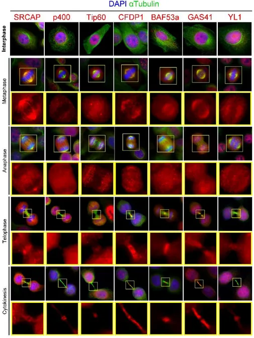
2.5 Subunits of Tip60/p400 and SRCAP chromatin remodelling complexes localize to the mitotic apparatus in human cells In past years, experiments carried out in our lab have shown that subunits of the SRCAP and Tip60/p400 complexes, besides the canonical localization in the interphase chromatin, target to different sites of the mitotic apparatus (centrosomes, spindle and midbody) at differing times during cell division (Messina et al., in preparation).
Using specific antibodies for the endogenous proteins, they performed high-resolution immunofluorescence (IF) experiments in fixed HeLa cells, to study the subcellular distribution of 13 endogenous subunits of SRCAP and Tip60/p400 ATP-dependent chromatin remodelling complexes during cell division: SRCAP, CFDP1, p18-Hamlet (SRCAP complex), Tip60, P400 and MRG15 (Tip60/p400 complex), BAF53a, Myc-Arp6, Pontin, Reptin, GAS 41, YL1 and (both complexes).
23 As expected, for each chromatin protein tested, the antibody staining revealed IF signals in interphase nuclei. In addition, signals were found to different sites of the mitotic apparatus, i. e. centrosomes, spindle and midbody (Fig. 2.11).
24
Figure 2.11. Dynamic localization of human SRCAP and tip60/p400 proteins
25 In particular, SRCAP, CFDP1, BAF53a, GAS41 and YL1 were found at the spindle in both metaphase and/or anaphase; SRCAP and GAS41, also localize at the centrosomes together with Tip60. Moreover, all the subunits tested were found at the midbody (MB): P400, Tip60 and BAF53a in the flanking zone, SRCAP, YL-1 and CFDP1 along the bridge, except the central area (Fig. 2.11).
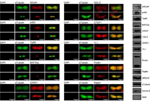
Moreover, the MB associations found by IF on fixed HeLa cell preparations were validated on isolated MBs, by both IF and Western blotting experiments (Fig. 2.12).
Fig. 2.11. IF and WB of isolated midbodies from HeLa cells. IF images show
26 The results of these experiments confirmed that all the SRCAP and tip60/p400 proteins tested by IF are recruited at the MB. By contrast, the human ISWI/hSNF2H remodeler was not found at the MB.
These findings suggest that the subcellular localization of all the proteins tested during cell division is dynamic and, besides their canonical localization in interphase nuclei, all the subunits are recruited to the mitotic apparatus. Remarkably, SRCAP and Tip60/p400 complexes belong to the INO80 family of ATP-dependent chromatin remodelers, whose main function is to govern the deposition of the H2A variant histone in animal and plant species, thus such a bulky association with the mitotic apparatus was unexpected.
27
3. Aims
My PhD project is aimed at studying the involvement of ATP-dependent chromatin remodelling complexes in ensuring proper cell division in both human and Drosophila cell lines, two species separated by 700 million years of evolution.
Experimental in vitro approaches involved the use of human HeLa cells and Drosophila S2 cells of embryonic origin.
In particular, experiments have been carried out to:
i) Characterize the cellular distribution of subunits of Drosophila Tip60 complex;
ii) Analyze the cell cycle progression following removal of specific subunits of both human and Drosophila chromatin remodelling complexes by RNAi knock-down;
28
4. Results
4.1 RNAi knock-down of SRCAP and Tip60/p400 subunits impairs successful cell division – part one
First, to investigate the impact of chromatin remodelers during cell division, I performed experiments of RNAi-mediated depletion of SRCAP, TIP60, GAS41, YL1 and MRG15 subunits (Fig. 4.1) to characterize the MB localization of six known regulators of cytokinesis, i.e. Aurora B, INCENP, MKLP1, MKLP2, PLK1 and Cit-K.
29 Figure 4.1. On the top: WB of HeLa WCE (whole cell extracts) after
RNAi-depletion of remodelers. ISWI is used as control. At the bottom: decrease of fluorescence intensity after RNAi-depletion of each remodelers compared with the mock. Fluorescence intensity was assessed using the ImageJ software.
As shown in Fig. 4.2, the localization of the all tested MB players was affected. In particular, SRCAP depleted cells showed the strongest effects on mislocalization of all MB players, with the exception of Cit-K. Notably, MKLP1 and PLK1 were strongly delocalized by the independent depletion of SRCAP and Tip60. IF images show examples of typical MB players mislocalization found after RNAi depletion of different remodelers. Thus, RNAi-mediated depletion of remodelers affect cell division in HeLa cells.
31 Figure 4.2. Examples of MB players mislocalizations after RNAi-depletion of
SRCAP, Tip60 and GAS41 remodelers (immunofluorescence) and quantitative analysis.
Moreover, RNAi knock-down of the considered subunits produced mitotic defects.
Defects found include multinucleation, aberrant multipolar spindles, LIBs (Long Intercellular Bridges) connecting the daughter cells, chromosome misalignments and chromatin bridges. Examples of these alterations are shown in Fig. 4.3. Importantly, mitotic defects are consistent with the localizations of the studied remodelers. In particular, Tip60 normally localizes at the spindle poles and kinetochores (Fig. 2.11) and RNAi-mediated depletion of this protein causes a statistically significant alteration of the spindle, generating chromosome misalignments in metaphase and multipolar spindles. SRCAP depletion causes a strong impact in cytokinesis, as percentages of multinucleated cells and LIBs increase after RNAi treatment. These results suggest that subunits
32 of ATP-dependent remodelling complexes prevent the failure of cell division and genome instability.
Figure 4.3. Examples of defects found after RNAi-depletion of remodelers
(Immunofluorescence). IF images: green: alfa-tubulin; blue: DAPI. Quantitative analysis of mitotic defects and intercellular distance.
33
4.2 SRCAP and Tip60/p400 subunits interact with some important cell cycle regulators
The aforementioned results strongly suggest that SRCAP, TIP60, GAS41, YL1 and Mrg15 regulate cytokinesis in HeLa cells. An intriguing possibility is that these subunits play a role in the localization/action of crucial cell division players to the MB. To test this hypothesis, first we performed a series of simultaneous IF experiments aimed at studying the co-localization at MB of some remodelers with known MB players, such as Aurora B, INCENP, MKLP1, MKLP2 and CIT-K(Fig.4.4).
34 Figure 4.4 Co-immunolocalization of Tip60 and some MB players in fixed HeLa
35 Tip60 showed a sharp signal at the central zone of MB and it is an acetyl-transferase that interacts and acetylates Aurora B and PLK1 (and Cyclin B1) during interphase and metaphase (Zhang et al., 2012; Mo et al., 2016).
Remarkably, the results of these experiments showed that Tip60 co-localizes with Aurora B, MKLP1, MKLP2, CIT-K at the central zone of MB, while it doesn’t co-localize with INCENP. Moreover, Aurora B co-localizes with Baf53a and SRCAP also.
The results of these experiments were suggestive for possible interactions that can be validated by biochemical approaches and are also mandatory to the study of localization dependencies between MB players and chromatin remodelers, and viceversa. Our IF colocalization experiments suggested that chromatin remodelers interact physically with MB/cytokinesis players during the final stages of cell division.
To test this hypothesis, I focused my attention on Tip60 by performing immunoprecipitation (IP) experiments on cytosolic protein extracts from telophase-synchronized HeLa cells, in order to eliminate the background represented by the chromatin component (detailed protocol is in the Materials and methods section) (Fig.4.5).
36 Figure 4.5. On the left: Chromatin fractionation of HeLa cells synchronized in
telophase. WCE: whole cell extract. P3: nuclei fraction. S2: cytoplasmatic fraction. MKLP1 is expressed in late stages of mitosis. On the right: Co-immunoprecipitation of telophase protein extracts (S2 fraction) demonstrating the interaction of Tip60 with main MB players.
The IP results indicate that Tip60 physically interacts with CITK, Aurora B and MKLP2, in accordance with the previously described co-localization results. These IP experiments also show that Tip60 interacts with α-Tubulin, the structural component of the spindle and midbody, and with Baf53a and MRG15 protein, subunits of the SRCAP and Tip60/p400 complex.
37
4.3 Aurora B kinase promotes recruitment of some subunits at the midbody
Aurora B kinase is a key regulator of cytokinesis and phosphorylates the CHMP4C subunit of the ESCRT-III complex necessary for abscission (D'Avino et al., 2015). Furthermore, the acetylation of Aurora B by Tip60 is necessary for a correct regulation of the mitotic checkpoint (Mo et al., 2016).
The results of our IP assays indicate physical interactions between Aurora B and other midbody players with the subunits, suggesting the existence of a cross regulation feedback between Aurora B and the remodelling complex subunits in cytokinesis.
A crucial question in this study is how SRCAP and tip60/p400 chromatin remodelers are recruited to MB. In order to deepen this aspect, we asked whether the inhibition of Aurora B kinase activity affects the recruitment of Tip60, SRCAP at MB.
The ZM447439, a specific selective ATP-competitive inhibitor of Aurora B kinase (Girdler et al., 2006), was employed to inhibit the activity of Aurora B in these experiments. To evaluate the efficacy of inhibition, phosphorylation of the histone H3 during mitosis was considered as control, as it is target of Aurora B. As it can be seen in Fig. 4.6, cells treated with ZM447439 show a significant mislocalization of TIP60, SRCAP and BAF53a remodelers, as well as of the already known midbody players targets (positive control). These results support the hypothesis of a cross-regulation occuring during cytokinesis between kinases, remodelers and midbody players.
38 Figure 4.6. ZM447439 treatment. On the top: WB testing efficiency of
ZM447439 treatment (Histone H3 is a target of Aurora B kinase). Immunostaining of HeLa cells for Aurora B, before and after ZM447439 treatment, as control. Immunostaining of HeLa cells for PLK1, a well-known Aurora B interactor, before and after ZM447439 treatment, as positive control. At the bottom: Immunostaining of HeLa cells for Tip60, SRCAP and Baf53a before and after ZM447439 treatment. Quantitative analysis of remodelers and MB mislocalization after ZM447439 treatment.
39
4.4 Subunits of dTip60 chromatin remodelling complex localize to the mitotic apparatus in Drosophila S2 cells
In a past work mutations in the Drosophila melanogaster heterochromatic l(2)41Aa gene, now called Yeti, were found to affect chromosome organization and cytokinesis (Cenci et al., 2003; Messina et al., 2014).
We then asked whether the recruitment of chromatin remodelers to mitotic apparatus is limited to human cells or, on the contrary, is an evolutionary conserved phenomenon.
To answer this question, I studied the distribution of four subunits of the Drosophila melanogaster Tip60 complex during cell division in embryonic S2 cells. In particular, I focused my attention on DOM-A, Tip60, YETI and MRG15 proteins, which are orthologous to human SRCAP, Tip60, CFDP1 and MRG15 human, respectively.
40 Figure 4.7. Dynamic localization of dTip60 subunits in Drosophila S2 fixed
41 First, I performed immunofluorescence experiments on Drosophila S2 cells, using specific antibodies against endogenous proteins. The results of these analysis show that DOM-A, TIP60, YETI and MRG15, in addition to their canonical localization in interphase nuclei, are all found at the mitotic apparatus (Fig. 4.7) similarly to their human orthologs. In particular, DOM-A and TIP60 are also found at the spindle, while MRG15 is strongly localized to the centrosomes. In addition, they all localize at the midbody.
Thus, the recruitment of remodelling factors to the mitotic apparatus appears to be an evolutionary conserved phenomenon.
4.5 RNAi knock-down of the subunits impair successful cell division – part two
To investigate the impact of chromatin remodelers during Drosophila cell division also, I performed experiments of RNAi-mediated depletion of the dTip60 complex subunits.
I generated dsRNA to induce RNAi depletion of DOM-A, Tip60, Mrg15 and Yeti in Drosophila S2 cells (details in Materials and Methods).
After 96 hours-treatments, cells were harvested to perform blotting or sqRT-PCR and immunofluorescence analysis.
The efficiency of RNAi-mediated depletion was tested by IF experiments and Western blotting. In the case of DOM-A, I performed the sqRT-PCR analysis because the antibody was not suitable for WB.
IF experiments and Western blotting clearly show the amount of the tested subunits after RNAi induction. Similarly, the DOM-A expression was significantly affected after RNAi induction (Fig. 4.9).
42 Notably, the RNAi-mediated depletion of the dTip60 complex subunits under investigation caused mitotic defects, similar to those found after depletion of remodelers in HeLa cells.
Figure 4.9. On the top: WB and sq-RT PCR of dTip60 subunits after RNAi
43
Tubulin was used as loading control. At the bottom: decrease of fluorescence intensity after RNAi-depletion of each remodelers compared with the mock. Fluorescence intensity was assessed using the ImageJ software.
Again, defects found include multinucleation, aberrant multipolar spindles, LIBs (Long Intercellular Bridges) connecting the daughter cells and chromosome misalignments. Importantly, defects found are consistent with the remodelers localizations. For example, Mrg15 show a strong signal to the centrosomes at the spindle poles and its depletion causes around 50% of multipolar cells.
In conclusion, the recruitment of remodelling factors to the mitotic apparatus and, supposedly their function, appears to be an evolutionary conserved phenomenon.
44 Figure 4.10. Examples of mitotic defects and quantitative analysis of them
found in remodelers-depleted Drosophila cells. IF images: green: alfa-tubulin; blue: DAPI.
45
5. Discussion and conclusion
Previous studies suggested that chromatin and remodelling proteins might also play roles in cell division, yet evidence remains sporadic (Gartner et al., 2003; Ducat et al. 2008; Sigala et al., 2005).
In my PhD work, I combined cell biology, functional genetics and biochemical approaches to investigate in depth the role of five subunits of SRCAP and P400 chromatin remodelling complexes in cell division in HeLa cells. In parallel, the same approach has been carried out to study four subunits of the Tip60 chromatin remodelling complex of Drosophila melanogaster, which is structurally and functionally related to the human Tip60/p400 complex.
We found that, besides the canonical localization in interphase, all the subunits of human SRCAP and Tip60/p400 complexes and Drosophila Tip60 complex under investigation are recruited at different sites of the mitotic apparatus (centrosomes, spindle and midbody) during cell cycle progression. Such a bulky association with the mitotic apparatus is surprising, since the main function of these complexes is to govern the deposition of the H2A variant in animal and plant species which impact gene expression and DNA repair.
RNAi knock-down experiments showed that the depletion of each protein tested yields an array of aberrant mitotic outcomes, including multipolar spindle formation, chromosome mis-segregation and cytokinesis defects. Overall, these categories of defects are known to occur as a consequence of the loss of crucial regulator of spindle and/or midbody organization/function.
Similarly, I found that DOM-A, TIP60, YETI and MRG15 are recruited to the MB in Drosophila cells and their depletion affect different aspects of cell division.
Moreover DOM-A and MRG15 localize at the centrosomes and their RNAi-mediated depletion causes centrosomes amplification.
46 This defect can arise by centriole overduplication or by cell doubling events, including the failure of cell division and cell-cell fusion. Karki et al proposed a model in which leaky APC/C activity during mitotic delay triggers precocious centriole disengagement (Karki et al., 2017).
Interestingly, a century ago, Theodor Boveri proposed that increased centrosome numbers can cause cancer (Boveri).
Two alternative hypotheses could be considered to explain how the knock-down chromatin remodelling proteins affects cell divisions: i) the recruitment of chromatin remodelling proteins to the mitotic apparatus only reflects a passive accumulation of protein to be disposed of, and cell division defects after their depletion/loss is merely the consequence of general chromatin perturbations, that in turn result in deregulation of genes involved in cell division; ii) chromatin remodelling proteins are functional components of centrosome, spindle and MB and their depletion/loss results in disfunctions of mitotic apparatus, thus giving rise to mitosis and cytokinesis failure. This hypothesis implies specific roles of remodelers in the organization/function of mitotic apparatus and regulation of cell division;
Several lines of evidence favour the second hypothesis, suggesting that the bulky association of chromatin remodelling with mitotic apparatus has a functional relevance.
First, the recruitment of remodelling factors to mitotic apparatus and the consequent disruption of cell division following their depletion has been observed in both human and Drosophila melanogaster, two species separated by over 700 million years of evolution.
Second, the effects observed after depletion of chromatin remodelling factors in both human and D. melanogaster cells do not appear to be simply a chaotic disruption of cell division, as one would expect from simultaneous malfunctioning of genes encoding
47 cell division regulators. By contrast, those effects fail into specific classes which are generally consistent with the mitotic apparatus localization of each remodeler tested and are similar to those occurring after the loss of already known spindle and/or midbody (MB) regulators.
Third, immunofluorescence experiments have shown that subunits of SRCAP and Tip60 complexes colocalize at MB with known components/regulators of spindle and/or MB, such as Aurora B, INCENP, CIT-K, MKLP2, PLK1 and alfa-tubulin, suggesting physical interactions.
Fourth, Co-IP assays carried out in telophase synchronized HeLa cells have shown that Tip60 indeed physically interacts with Aurora B, INCENP, CIT-K, MKLP2, PLK1 and alfa-tubulin, factors specifically required for proper cytokinesis. This result strongly suggests a specific role of Tip60 in telophase also.
Moreover, reciprocally, the inhibition of Aurora B kinase activity affects the recruitment at the MB of SRCAP, BAF53a, Tip60. In addition, Tip60 co-localizes with PLK1 and Cyclin B1 at spindle poles and at the MB (Subramanian et al., 2010; Bastos et al.,2010; D’Avino et al., 2007; Zhang et al., 2012) and acetylates Aurora B during metaphase (Mo et al., 2016).
Overall, these results suggest the occurrence of a cross-talk between the subunits of the SRCAP and Tip60 complexes and some of the main regulators of mitosis and cytokinesis, with a positive feedback loop where regulators of mitosis and cytokinesis promotes the recruitment of remodelers to the mitotic apparatus and in turn the latter partecipates to stabilize the former.
In particular, the Aurora B kinase activity promotes the recruitment of SRCAP and Tip60 to the MB and in turn, SRCAP and Tip60 contribute to control Aurora B localization at the midbody. Thus,
48 the crosstalk between chromatin and the mitotic apparatus is much more intertwined than previously supposed.
Frequent mutations in genes encoding chromatin regulatory factors and histone proteins have been identified in human cancer (Morgan M. et al., 2015). Substantiating such a role might be important to investigate as yet poorly understood routes to tumorigenic transformation in cancer types in which these factors can be aberrantly expressed and/or dysfunctional and will help expand our understanding of the growing link between cell division, in particular cytokinesis, and cancer.
Thus, elucidating the molecular mechanisms underlying the roles of chromatin remodelling proteins in cell division is challenging to clarify as yet poorly understood routes to tumorigenic transformation in cancer types in which these factors can be aberrantly expressed and/or dysfunctional and will help expand our understanding of the growing link between cell division and cancer. Notably, most subunits of the complexes studied have been implicated in cancer biology, which are summarized in Table 5.1.
Table 5.1. Examples of subunits of SRCAP and P400 complexes implicated in cancer biology
Subunit Implication in cancer biology
BAF53a Overexpressed in bladder, cervix, myeloma, colon, and ovarian cancers; aberrant expression correlated with the progression of rhabdomyosarcoma, osteosarcoma,
hepatocellular carcinoma and head and neck squamous cell carcinoma (Mayes et al., 2014); BAF53a is considered to promote cancer progression via EMT
epithelial-mesenchymal transition (Meng et al., 2017)
Tip60 Acts as a haplo-insufficient tumour suppressor (Gorrini et al., 2007)
SRCAP Interacts with the CREB binding protein (CBP), an acetyltransferase encoded by a haplo-insufficient tumor suppressor gene in B-cell lymphoma (Zhang et al., 2017). It
49
has been identified as a physiologically relevant mediator of PSA expression in prostate cancer cells (Slupianck et al., 2010).
GAS41 Overexpressed in glioblastoma cell lines (Munnia et al., 2001; Park et al., 2006)
Reptin & Pontin Overexpressed in bladder, colon, liver cancers and melanoma (Huber et al., 2008)
YL-1 Overexpressed in prostate cancer (Barboro et al., 2005) MRGBP/MRGX/MRG15 Overexpressed in colorectal cancer tissues (Yamaguchi et
al., 2010)
P400 Its siRNA-mediated decrease favours the response to 5-fluorouracil of colon cancer cells (Mattera et al., 2009)
In conclusion, our results reveal the existence of a previously undetected, massive and evolutionarily conserved phenomenon, whereby subunits of ATP-dependent chromatin remodelling complexes, in addition to their role of epigenetic regulators, have a functional relevance in cell division. Moreover these results emphasize a surprising scenario in which chromatin remodelling, cell cycle and tumorigenesis are closely interlinked (Fig. 5.1).
Fig.5.1 Model of remodelers translocating from interphase chromatin to the
50
6. Future perspectives
Despite RNAi is a widely used system to achieve protein down-regulation at mRNA level, when RNAi is induced, the protein products already translated remain unaffected. Thus, a long time treatment is required before down-regulation can be induced, making RNAi application for depleting target proteins in specific phases of the cell cycle quite tricky.
To bypass this problem, the Auxin-Inducible Degron (AID) experimental system will be employed in our laboratory to specifically target the protein rather than its mRNA and to induce a total and sudden depletion of the protein of interest. This system is adapted from a gene expression regulation mechanism found in plants, where Auxin induces the degradation of transcriptional repressors by promoting their association with the TIR1 domain leading to polyubiquitination and degradation via the proteasome (Teale W. et al., 2006). By fusing a degron sequence (Aid-tag) to the targeted protein, the mechanism can be exploited for degradation experiments. The AID system has been established successfully in S. cerevisiae, C. elegans, Drosophila, mouse, monkey, humans (Nishimura et al., 2009; Morawska et al., 2013; Trost et al., 2016). Since the AID system relies on a plant hormone that is generally expected to be biologically silent in eukaryotic organisms other than plants, secondary effects should not arise. This might be even more promising using a shorter version of the tag, the mini-Auxin-induced Degron (mAiD), which has been shown to work in HTC116 cell line (Natsume et al., 2016).
51
7. Materials and Methods
7.1 Cell colture, RNA interference and ZM447439 treatment HeLa human cervical cancer cells were grown in Dulbecco’s modified Eagle’s medium (DMEM) supplemented with 10 % foetal bovine serum (Gibco-BRL) and 10% Penicillin/Streptomycin solution (SIGMA). Cell cultures were maintained at 37°C in a humidified atmosphere of 5 % CO2.
Transient transfections were performed using Lipofectamine RNAi-MAX (Thermo Fisher Scientific), according to the manufacturer’s instructions.
The siRNA-mediated interference experiments for SRCAP, Tip60, Gas41 and Mrg15 expression were performed by transfecting a pool of specific double-stranded RNA oligonucleotides:
SRCAP: pool of three sc-93293 (Santa Cruz) Tip60 (Kat5): EHU139921-20UG (Sigma) Gas41: pool of three sc-77331 (Santa Cruz) Mrg15 (Morf4L1): EHU127521-20UG (Sigma)
48, 72 or 96h after transfection cells were harvested for cytological and immunoblotting analysis.
Asynchronous HeLa cells were treated for 45 min with the Aurora B inhibitor ZM447439 at a final concentration of 5 µM at 37°C in a humidified atmosphere of 5 % CO2. An equal volume of DMSO was used as negative control. Then they were fixed and stained. ZM 447439 is a selective and ATP-competitive inhibitor for Aurora A and Aurora B with IC50 of 110 nM and 130 nM, respectively
52
7.2 Immunofluorescence
HeLa cells were seeded on glass coverslips and 24 h later they were fixed for 15′ at room temperature (RT) in 2% formaldehyde in PBS or 5’ methanol in ice. After permeabilization in 0.2% Triton X-100 solution (5’ in ice) and washing in PBS the cells were incubated in 3% bovine serum albumin (BSA) for 1 h and, subsequently, over-night (O/N) with primary antibodies (table 7.1) at 4°C. After several washes in PBS, the cells were incubated with secondary antibodies (table 7.1) for 1h at RT and 1’ with DAPI. The coverslips were then mounted onto slides with ProLong Antifade Mountant for Fixed Cells (Thermo Scientific). Preparations were analyzed using a computer-controlled Nikon Eclipse 50i epifluorescence microscope equipped with a CCD camera. Images optimization
was assessed using the ImageJ software
(http://rsbweb.nih.gov/ij/). About 2000-3000 cells were scored in at least three independent experiments.
Table 7.1. Antibodies used for immunofluorescence experiments
Primary Antibody Dilution Supplier
αTubulin, rabbit polyclonal 1:500 Abcam, ab6046
αTubulin, mouse monoclonal 1:1000 Sigma-Aldrich T9026 Tip60 (KAT5), rabbit polyclonal 1:1000 Abcam, ab137518 H3(phosphoS10), rabbit polyclonal 1:500 Abcam, ab5176
MRG15, rabbit polyclonal 1:1000 Genetex, GTX128495
GAS41, rabbit polyclonal 1.1000 Abcam, ab167495
GAS41, mouse monoclonal 1:50 Abcam, ab205018
YL1, mouse monoclonal 1:50 Epigen, EXM10001
Baf53a, mouse monoclonal 1:100 Santa Cruz, sc-137062
MKLP2, rabbit polyclonal 1:10000 Thomas Mayer
MKLP1, mouse polyclonal 1:1000 Abcam, ab168964
SRCAP, rabbit polyclonal 1:500 SantaCruzBio,sc133312
53
INCENP, rabbit polyclonal 1:1000 Cell signalling, 2807
AuroraB (AIM-1),mouse
monoclonal
1:1000 BD Trans. Lab, 611083
AuroraB, rabbit polyclonal 1:100 Abcam, ab3254
Secondary Antibody Dilution Supplier
anti-Mouse IgG, AlexaFluor 488 1:200 Invitrogen, A21200 anti-Rabbit IgG, AlexaFluor 488 1:200 Invitrogen, A21441 anti-Mouse IgG, AlexaFluor 555 1:200 Invitrogen, A21422 anti-Rabbit IgG, AlexaFluor 555 1:200 Invitrogen, A21430
7.3 Cell cycle synchronization
For immunoprecipitation experiments, HeLa cells were synchronized in telophase using thymidine/nocodazole blocks (Fig. 7.1). Briefly, cells were treated with 2 mM thymidine (Sigma, T9250) for 19h, released from G1/S block in fresh media for 5h, incubated with 40 nM nocodazole (Sigma, M1403) for 13h and, harvested by mitotic shake-off. Mitotic cells were washed three times with PBS and released in fresh medium for 70’ before harvesting and freezing in liquid nitrogen. Telophase cell samples were prepared by resuspending cells in Buffer A (10 mM HEPES at pH 7.9, 10 mM KCl, 1.5 mM MgCl2, 0.34 M sucrose, 10% glycerol, 1 mM dithiothreitol
[DTT], 1 mM NaVO4, 5 mM β-glycero-phosphate, 0.1 mM
phenyl methane sulphonyl fluoride [PMSF], 0.5 mM NaF, protease inhibitor cocktail [Roche]) for subsequent subcellular fractionation assay.
54 Figure 7.1. Scheme of cell cycle synchronization and subsequent subcellular
55
7.4 Chromatin fractionation and immunoprecipitation
HeLa cells (2 x 10⁷) were resuspended in 1 mL of Buffer A. Triton X-100 was added at 0.1% and cells were incubated for 5’ on ice. After centrifugation (3500 rpm for 5’ at 4°C), the cytosolic fraction (supernatant) was stored for the next immunoprecipitation experiments and the nuclear fraction (pellet, P3 fraction) was resuspended in 1 mL of Buffer B (3 mM EDTA, 0.2 mM EGTA, 1 mM DTT, 0.1 mM PMSF, protease inhibitors) and incubated for 5’ on ice. All the fractioned samples were load in a polyacrylamide gel and transferred onto PVDF membrane for western blot analysis. Cytosolic S2 fraction (or soluble proteins) from subcellular fractionation assay was used as input (IN) to immunoprecipitated Tip60 protein using 4 µg of rabbit anti-Tip60 (Abcam, ab137518) antibody O/N at 4°C, followed by 2 h of incubation with 40 µl of agarose-conjugated protein A/G (Santa Cruz Biotechnology). Beads were then washed three times in buffer A and co-immunoprecipitants were analyzed by immunoblotting.
7.5 Western Blotting
HeLa cells were lysated in sample buffer at 10000 cells/μl. All the samples were load in a polyacrylamide gel, transferred onto PVDF membrane and probed with specific antibodies (table 7.2) with appropriate HRP-conjugated secondary antibodies (table 7.2). The bands were immunodetected using the Enhanced chemiluminescence (ECL) kit from Thermo Scientific.
Table 7.2. Antibodies used for western blotting
Primary Antibody Dilution Supplier
Tip60 (KAT5), rabbit polyclonal 1:1000 Abcam, ab137518
56
ISWI, rabbit 1:5000 Tatsuya Hirano
αTubulin, mouse monoclonal 1:5000 Sigma-Aldrich T9026
β-actin, mouse monoclonal 1:10000 Abgent, AM102b
H3 (phospho S10), rabbit polyclonal 1:500 Abcam, ab5176
H3, rabbit Polyclonal 1:10000 Abcam, ab1791
MRG15, rabbit polyclonal 1:1000 Genetex, GTX128495
GAS41, rabbit polyclonal 1.1000 Abcam, ab167495
YL1, rabbit polyclonal 1:250 Genetex, GTX102152
MKLP2, rabbit polyclonal 1:10000 Thomas Mayer
MKLP1, mouse polyclonal 1:1000 Abcam, ab168964
SRCAP, rabbit polyclonal 1:500 Santa Cruz Bio, sc133312
CITK, mouse monoclonal 1:1000 BD Trans. Lab, 611376
Baf53a, mouse monoclonal 1:100 Santa Cruz Bio, sc137062 Aurora B (AIM-1), mouse monoclonal 1:1000 BD Trans. Lab, 611083
Secondary Antibody Dilution Supplier
anti-Mouse IgG conjugate HRP 1:2500 Santa Cruz Bio, sc-2005 anti-Rabbit IgG conjugate HRP 1:2500 ImmunoReagent,
GTXRB-003-DHRPX
7.6 RNA interference in Drosophila S2 cells Cell Culture and RNAi Treatments
S2 cells derive from a primary culture of late stage (20–24 hours old) Drosophila melanogaster embryos (Schneider, 1972). They were cultured at 25°C in Schneider’s Drosophila Medium (Biowest) supplemented with 10% heat-inactivated fetal bovine serum (Corning), 50 units/ml penicillin, and 50 μg/ml streptomycin (Carlo Erba) in 75-cm2 T-flasks (Corning).
RNAi treatments were carried out according to Clemens et al. (2000). Cells were suspended in serum-free Schneider’s medium at a concentration of 1 × 10⁶ cells/ml, and plated, 1 ml/well, in a six-well culture dish (Nalge Nunc). To perform RNAi, each culture
57 was inoculated with 15 μg of double-stranded dsRNA. After 1-h incubation at 25°C, 2 ml of medium supplemented with 10% foetal bovine serum was added to each culture. Control cultures were prepared in the same way but without addition of dsRNA. Both RNA-treated and control cells were grown for 96 h at 25°C and then processed for either immunofluorescence, or blotting analysis. dsRNA preparation
Individual gene sequences were amplified by polymerase chain reaction (PCR) from genomic DNA obtained from S2 cells.
The primers used in the PCR reactions were 48 nt base long and all contained a 5’ T7 RNA polymerase binding site
(5’-GAATTAATACGACTCACTATAGGGAGAC-3’) joined to a gene-specific sequence.
The sense and antisense gene-specific primers were as follows:
DomA for - TCTGGTGCTCAGATCGTGTC
rev - GTTGTCTGCAGCACCTTCAA TIP60 for - CCCTTCAGAAACGCATCA
rev - ACCTTGTTTTTGCGTCCATC
YETI for - ATCGCGGTCGGATGCTTTAT
rev - CCAAATACCCGTCCTTGCCT MRG15 for - TACGCAGCTAAGGTGGAGGT
rev - CTGTGCATTTCGCACGTACT
Individual DNA fragments were approximately 500 bp in length. Amplified sequences were used to produce dsRNA through MEGAscript RNAi Kit (Invitrogen), according to the manufacturer’s instructions.
58 Figure 6.2. Agarose gels to control dsRNAs produced.
In all RNAi experiments, protein depletion was systematically verified by Western blotting and/or semi-quantitative RT-PCR. Western Blot analysis
Cells were harvested by centrifugation at 800g for 5 min. Pellets were lysed appropriate volume of Laemmli buffer to obtain 5000 cells/ul. 20 ul of lysate was used for SDS-PAGE and transferred to a PVDF Immobilion membrane (Millipore) by using a transfer apparatus (Bio-Rad). The membrane was blocked for 1 h in a 5% no-fat dry milk in PBS-T (Phosphate Buffered Saline 1X + 0.05% Tween 20, pH 7.4) and then incubated overnight at 4°C with any of the primary antibodies, diluted in PBS-T + 3% BSA (Bovine Serum Albumin, Sigma), and 1 h at room temperature with any of the secondary antibodies conjugated to HRP (table 7.3). Bands were detected using the Enhanced Chemiluminescence (ECL) kit (Thermo Scientific).
59
Table 7.3. Antibody used for WB
Primary Antibody Diluti
on
Supplier
αTubulin, mouse monoclonal 1:1000 Sigma-Aldrich, T9026 anti-dTip60, guinea pig 1:100 SE. Ehrenhofer Murray
anti-Yeti, rabbit 1:100 P. Dimitri/R. Caizzi
anti-Mrg15, rabbit 1:200 T. Kusch
anti-DomA, rabbit 1:100 ML. Ruhf
Secondary Antibody Dilution Supplier
anti-Mouse IgG conjugate HRP 1:2500 Santa Cruz Bio, sc-2005 anti-Rabbit IgG conjugate HRP 1:2500 ImmunoReagent,
GTXRB-003-DHRPX anti-Guinea Pig IgG conjugate HRP 1:2500 SIGMA, A7289
Immunofluorescence
S2 cells were seeded on glass coverslips and 24 h later they were fixed for 20′ at room temperature (RT) in 4% formaldehyde in PBS or 10’ methanol on ice. Then they were permeabilized 20’ at RT in 0.1% Triton X-100 solution. The cells were incubated in 3% BSA for 1 h in humid chamber and then over-night with primary
antibodies (table 7.4) at 4°C. After several washes in PBS, the cells were incubated with secondary antibodies for 1h at RT (table 7.4). The coverslips were mounted onto slides with ProLong Antifade Mountant for Fixed Cells (Thermo Scientific). Preparations were analyzed using a computer-controlled Nikon Eclipse 50i
epifluorescence microscope equipped with a CCD camera. About 2000-3000 cells were scored in at least three independent
60
Table 7.4. Antibody used for immunofluorescence
Primary Antibody Dilution Supplier
αTubulin, mouse monoclonal 1:1000 Sigma-Aldrich, T9026
anti-dTip60, guinea pig 1:100 SE. Ehrenhofer Murray
anti-Yeti, rabbit 1:100 P. Dimitri/R. Caizzi
anti-Mrg15, rabbit 1:200 T. Kusch
anti-DomA, rabbit 1:100 ML. Ruhf
Secondary Antibody Dilution Supplier
anti-Mouse IgG, AlexaFluor 488 1:200 Invitrogen, A21200
anti-Guinea Pig IgG, Cy3 1:200 Jackson IR, 106165003
anti-Rabbit IgG, AlexaFluor 555 1:200 Invitrogen, A21430
7.7 Statistical analysis
Data analyses were performed using the GraphPad Prism software (GraphPad Software, Inc., La Jolla, CA, USA). All results are expressed as mean ±SD values from three independent replicate experiments. P values of less than 0.05 (*P < 0.05, compared with the control group) are considered statistically significant by using two-tailed Fisher’s exact test.


![Accés als recursos informàtics [text]. Curs 2020-21](data:image/gif;base64,R0lGODlhAQABAIAAAP///wAAACH5BAEAAAAALAAAAAABAAEAAAICRAEAOw==)