U
NIVERSITÀ DIP
ISAD
OTTORATO DIR
ICERCA INM
ORFOLOGIA EF
UNZIONEN
ORMALE EP
ATOLOGICA DIC
ELLULE ET
ESSUTIPRESIDENTE
Chiar.mo Prof. Antonio Paparelli
Study of a Model of Focal Limbic Epilepsy in Rats
Candidato Relatori
FENG XIA Dott. FILIPPO SEA N GIORGI
Prof. FRA NCESCO FORNA I
Prof. RICCA RDO RUFFOLI
SSD-BIO/16
Table of Contents
Abstract ... 1
Chapter 1 General Introduction... 4
1. Epilepsy ... 4
a) Idiopathic epilepsy (Table 1)... 5
b) Symptomatic epilepsy (Table 1) ... 6
2. Some molecular mechanisms underlying seizure onset ... 7
3. Electrophysiological abnormalities of epilepsy... 11
4. Anatomical basis for seizure spreading and synchronization of
different cortical structures ... 14
5. General aspects on experimental models of seizures and epilepsy
... 16
6. Mesial temporal lobe epilepsy: anatomical specificity, clinical
features and experimental models ... 17
a) The limbic system ... 18
b) Human mesial tempral lobe epilepsy syndrome ... 22
c) Animal models of limbic seizures and epilepsy ... 25
c1) Focal induction of acute limbic seizures ... 25
c2) Systemic injection of chemocovulsants ... 26
c3) A classical model of limbic seizures in the rat: the
“kindling” ... 29
c4) Focal microinjections in the APC of rat ... 31
7. Neuromodulators of seizures and epilepsy: the role of Locus
Coeruleus ... 32
a) The nucleus Locus Coeruleus (Figure 4) ... 32
b) The role of NE in epilepsy and experimental seizures ... 34
c) The role of Locus Coeruleus on seizures evoked from limbic
sites and APC ... 37
Chapter 2 Experimental Section... 39
1. Introduction to the experimental section... 39
2. Specific aims ... 43
3. Methods ... 44
3.1 Animals ... 44
3.2 Experimental design (Figure 5) ... 44
3.3 Stereotactic surgery (Figure 6) ... 45
3.4 Acute seizures monitoring ... 46
3.5 Electroencephalography ... 47
3.6 Lesion of the Locus Coeruleus ... 48
3.7 Seizure induction ... 49
3.8 Histology ... 49
3.9 Semiquantitative damage evaluation ... 52
3.10 Statistical analysis ... 53
1. Acute seizures (Figure 7) ... 54
2. Chronic monitoring (Figure 8)... 54
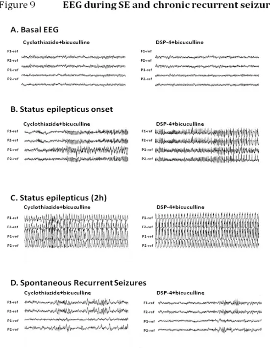
3. Electroencephalography (Figure 9) ... 55
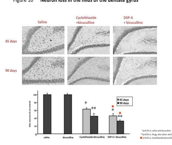
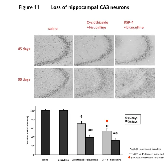
4. Morphological data (Figure 10, 11)... 55
Chapter 4 Conclusions ... 57
Tables and Figures ... 61
Table 1-3... 61
Figure 1-13 ... 64
References ... 76
Abstract
Epilepsy is a neurological disorder characterized by the recurrence of spontaneous, unprovoked epileptic seizures. Mesial temporal lobe epilepsy (more briefly, MTLE) is a very common form of epilepsy which is featured by the occurrence of focal limbic seizures, and associated to a specific neuropathological alteration, the so-called Ammon’s horn sclerosis (AHS, from now on abbreviated as AHS), whose main features are a selective loss of the CA1 and CA3/4 section of the Ammon’s horn (CA, from Latin Cornu
Ammonis, abbreviated as CA), a selective cell loss of inhibitory interneurons
in the hilus of the dentate gyrus (DG), and the abnormal sprouting of granule cells mossy fibers (the so called mossy fiber sprouting, MFS). The onset of spontaneous recurrent seizures (SRS) is the hallmark of a good model of epilepsy. For temporal lobe epilepsy (TLE), the most used models consist in administering systemically chemoconvulsants inducing limbic status epilepticus (i.e. seizures lasting for more than 30’, SE) and evaluating the occurrence of SRS. However, in these models, the widespread involvement of different structures which complicates the interpretation of experimental
findings obtained with this experimental approach, since any
morphological/functional effect of these models might be due either to the direct action of t he chemoconvulsant or to the SE. The morphological features of many structures of the limbic system are highly phylogenically conserved
through the evolution from rodents to primates and humans; it has been recently shown that it is possible to evoke limbic seizures and SE from a small structure, the deep extent of the anterior piriform cortex (from now on abbreviated as APC) by focally infusing picomolar concentration of chemoconvulsants; this structure roughly corresponds to the periamygdaloid cortex in humans. It is the brain region most densely innervated by the noradrenergic fibers originating from the nucleus locus coeruleus (LC), and we recently showed that microinfusing bicuculline (a GABA A receptor antagonist) into the APC of rats with a lesion of LC (induced by a selective neurotoxin, DSP-4, i.p.), induces SE, similarly to the SE obtained by microinfusing into the APC of rats with an intact noradrenergic system, cyclothiazide+ bicuculline. LC plays a critical role in modulating several models of seizures, and it plays a critical role in plastic mechanisms and neuroprotection in the brain. Thus, we compared the group DSP -4+bicuculline and cyclothiazide+bicuculline, to evaluate whether the focal SE evoked from the APC is capable of inducing SRS and AH S, and whether LC plays a significant role in this phenomena.
We found that: a) despite a similar duration and severity of SE in the two models of SE, in the group DSP-4+bicuculline there was a higher incidence of SRS; b) the cell loss in the hippocampal DG hilus and CA3 was higher in the group DSP-4+bic, while MFS was more intense in the group cyclothiazide+bicuculline; also the loss of parvalbumin-positive neurons was
more represented in the DSP-4+bicuculline group, while GFAP expression (an index of reactive gliosis), was similar in the two groups.
In conclusion, our study confirms that focal induction of SE from the AP C represents a good model of TLE, and that NE released from the fibers originating from the LC plays a significant role both in the hippoc ampal damage occurring after SE, and in the incidence of SRS. Differently from what observed in other models, our findings challenge a prominent role of MFS in the occurrence of SRS, since this phenomenon was less intense in the group with more frequent SRS (DSP-4+bicuculline) than in the one with an intact LC.
Chapter 1 General Introduction
1. EpilepsyThe term Epilepsy describes a group of multifaceted diseases afflicting about
0.5-2% (van den Broek and Beghi, 2004) of world population. Epilepsy is
defined by the recurrence of spontaneous, unprovoked epileptic seizures. A seizure is the effect of hypersyncronous and excessive electrical discharge of a group of cortical neurons (Wyllie, 2001). There are several different types of epilepsies and seizures, depending on the etiology of the disease, and on the site of onset of the epileptic seizure. Seizures and epilepsies can be subdivided in to “generalized” or “focal” ones, depending on the localization of the epileptic discharge in a circumscribed region of the cortex, or the involvement of the whole cortex. Generalized seizures can be further subdivided in primarily generalized (in which the seizure involves, from its onset, the whole cortex) or secondarily generalized ones, in which after a focal onset of the epileptic discharge there is a fast recruitment of the whole cortex.
Depending on the aetiology, epileptic syndromes can be further subdivided in Idiopathic epilepsy or in symptomatic epilepsy, depending on the lack (in the former) or presence (in the latter) of any organic, structural brain defect. In many patients a diagnosis of “cryptogenic epilepsy” has been often made, when, even in the absence of a clear brain lesion at the neuroimaging, a symptomatic cause was still hypothesized ( Commission on Classification and
Terminology of the International League Against Epilepsy 1989). More recently (Engel, 2001), the latter form has been defined as “probably symptomatic”.
a) Idiopathic epilepsy (Table 1)
As said, Idiopathic epilepsies are characterized by the lack of any organic alteration of the brain, and are considered as likely to be linked to a genetic alteration. However, usually these disorders are not monogenic and transmitted through the generations as Mendelianan inheritance. Furthermore, as to now we have not identified yet any specific mutation present in all of the patients with a similar clinical phenotypes (e.g. childhood absences or myoclonic juvenile epilepsy), but just the mutations in single families, not confirmed in patients with similar disorders from other families (e.g., concerning Juvenile myoclonic epilepsy, see Delgado-Escueta et al., 1990; Durner et al., 1991; and for childhood absence epilepsy, see Delgado-Escueta et al., 1990). Again, the single gene mutation responsible for the disease has been recently discovered only for some very rare idiopathic epilepsies, such as the sodium channel gene subunit genes (SCN1B, SCN1A, and SCN2A) and the GABA(A) receptor gamma2 subunit gene (GBRG2) mutations in “generalized epilepsy with febrile seizures plus” (Spampanato et al., 2004;
Liao et al., 2010; Mashimo et al., 2010); voltage-gated K+channels of the
al., 2008); and the mutations of the nicotinic acetylcholine receptor (nAChR) genes CHRNA4, CHRNB2, and CHRNA2 (Liu H et al., 2011) in “Autosomal dominant nocturnal frontal lobe epilepsy”, among others.
b) Symptomatic epilepsy (Table 1)
This group of syndromes comprises cases in which a well defined organic cause of epilepsy has been identified. This group represents by far the largest one, among epilepsies, especially after the introduction in the clinical setting, more than two decades ago, of magnetic resonance imaging (MRI). Among the different causes of symptomatic epilepsy it is worth being mentioned the role of brain inflammation, tumors, trauma, stroke or infectious diseases
(Singhi, 2011;Krakow et al., 2010;Yemadje et al., 2011; ). A different role is
played by disorder affecting metabolism/electrolytes or the effect of poisoning by drugs or alcohol: in this case patients often experience a seizure only during such a systemic alteration, even though they are not “epileptic” sensu
strictu: these are defined mainly as “occasional”, “provoked” seizures, and are
not considered as part of symptomatic epilepsy.
Among focal symptomatic epilepsy there is one syndrome which is worth being described more in detail: the so called “MTLE associated to hippocampal sclerosis”. The reasons for its specificity are the frequent occurrence of it among epileptic patients, and the specific brain morphological alteration accompanying such syndrome. Furthermore, the most important
experimental models of focal epilepsy tried to model such a syndrome indeed, including the model we used in the present thesis. Thus, a specific chapter later will be devoted to such a syndrome and to its experimental models.
2. Some molecular mechanisms underlying seizure onset
One of the main functions of glial cells is regulating the ion environment in the brain. Glial dysfunction increases the extracellular K+ concentration, and this is considered as a potentially relevant factor for the onset of epileptic
seizure (Frohlich, 2008; Traynelis and Dingledine, 1988; Feng and Durand,
2006; Rutecki et al., 1985; Yaari et al., 1986). For the onset of an epileptic
discharge, Ca++ influx into the neuron seems to represent the trigger step in
most of the cases. There are two kinds of calcium channel which are the Ligand-gated ion channels (LGICs) and the voltage-dependent calcium channels (VDCC). In the physiological classical scenario, LGICs cause the depolarization current to reach the threshold of the activation of sodium
channel causing the Na+ influx into the neuron. Afterwards, the VDCC is
activated by the voltage change, and cause a further, massive and fast Ca++
influx. After that, K+ and C1- channels open, and Ka+ and Cl- outflow trigger the steps inducing neuronal repolarization. Such a neuronal cycle can be affected by several modulatory (or sometimes pathologic ones) mechanisms. When an excitatory neurotransmitter binds to its specific excitatory receptor, it
can activate the Ca + + channels to cause the Ca + + over-influx, resulting in a ion abnormal distribution inside and outside of the neuronal membrane, to cause an explosive release.
By the same token, an excessive activation of neurons c an be caused by an abnormal reduction of the inhibitory tone on neurons. The permeability of the membrane to Cl- is increased when gamma-aminobutyric acid (GABA) binds to its receptors on neurons, thus maintaining the membrane potential in a stable level of resting potential and weakening its responses to afferent excitatory stimuli (see below).
GABA concentration in the brain and spinal fluid has been found to be lower than in controls, both in epileptic patients and experimental epilepsy models
(Wood et al., 1979; Petroff et al., 1998; Podell et al., 1997). A similar finding
has been obtained in experimental models of seizures, such as the genetically epilepsy-prone rat (Lasley, 1991). As a further indirect proof for the role of GABA in experimental seizures, it has been shown that inhibition of GABAa receptor and of GABA/Cl- channel and the GABA synthetic enzyme-glutamic acid decarboxylase (GAD) dysfunction, (Ushijima et al., 1998; Schwartz et al., 1989; Treiman, 2001; Walls et al., 2010) all could cause seizure. On the other hand, enhancing the activity or increasing GABA concentration can prevent seizure as witnessed by the potent antiepileptic effect of many antiepileptic drugs, which indeed act by potentiating the GABAergic inhibitory function (Jones-Davis and Macdonald 2003; Rogawski and Loscher 2004; White et al. ,
2007).
The GABA(A) receptor, the most represented subtype of GABA receptor, is a ligand-gated ion channel. It selectively conducts Cl-, and when it is open, Cl- enters the neurons causing hyperpolarization, i.e. an inhibitory effect. Interestingly, in some epileptic patients it has been found a mutation of the gene encoded the subunit of this receptor (Kumari et al., 2011, Kang et al., 2010). Recently, a relationship between GABA(A)/central benzodiazepine receptor (GABA(A)/cBZR) density and the neuron loss and the mossy fiber
sprounting (MFS) has been shown(Vivash et al., 2011).
Glutamic acid and aspartic acid are the main excitatory neurotransmitters in the central nervous system (CNS). There are 2 kinds of glutamic receptors, the ionotropic glutamate receptors and metabotropic glutamate receptors(mGLuR). The N-methyl-D-aspartate receptor (NMDAr) is an ionotropic glutamate
receptor and voltage-dependent channel, and allows Ca++ to enter into cells,
thus depolarizing the membranes themselves. In patients with temporal lobe epilepsy it has been found that the concentration, synthesis and release of glutamate and aspartic acid increase, in parallel with an increase of NMDAr activity (Sherwin, 1999). In many experiments, it has been found that the inhibition of the NMDAr by competitive or non-competitive NMDA
antagonists could stop or reduce seizures (Bausch et al., 2010; Obara. 1995;
Ushijima, 1998; Cakil, 2011). Additionally, the NMDAr is related with the activation of the extracellular signal-regulated kinases (ERK) and the
Brain-derived neurotrophic factor (BNDF) signaling pathway. The cross -talk between BDNF and NMDAr in modulation of synaptic plasticity might have a relevant role in the chronic effects of epilepsy (Yamada and Nabeshima, 2004). On contrary, the agonist NMDA enhances the severity of seizures (Toscano et al., 2008). Recently, it has been found the subunit NR1of NMDAr is increased in patients with TLE (de Moura et al., 2010).
The glutamic receptor a-amino-3-hydroxy-5-methyl-4-isoxazolepropionic acid receptor (AMPAR), also defined as non-NMDA receptor, is another cation channel which mediates fast excitatory neurotransmission. Its permeability to
Ca++ is governed by the subunit GLUR2 which prevents the Ca++ to pass. By
blocking this subunit it can prevent the excitotoxicity caused by several
models of epileptic seizures (Kim et al., 2001).
The kainate receptor (KAR) is the third type of ionotropic glutamate receptor; it is less known than the previous ones due to its low expression in the CNS. For several classes of neurons, KAR seems to be located presynaptically, and thus regulates glutamate and GABA release (Matute, 2010). It is believed to play a significant role in epilepsy in hum ans, even though we still lack direct evidences. However, incidentally it is worth being mentioned that the most popular model of limbic seizures and status epilepticus (from now on abbreviated as SE) in rodents, is the “Kainic acid (KA) model”, in which this agonist is administered either systemically of intracerebroventricularly (i.c.v.). Recently, some authors started studying also the role of mGluRs in the
mechanism underlying of epileptogenesis. For instance, it has been shown in vitro that the application of an antagonist of group I mGluR (S)-3,5-dihydroxyphenylglycine(DHPG), induces long-lasting epileptiform discharges (Bianchi et al., 2009) and that after this drug application, spontaneous inhibitory postsynaptic currents (IPSCs) are inhibited in the hippocampal CA3 region, due to the decrease of GABA release from the presynaptic nerve terminals (Inada H et al., 2010).
3. Electrophysiological abnormalities of epilepsy
More than three decades ago, it has been hypothesized that the onset of an epileptic discharge is associated, at the neuronal level, to the so -called “Paroxysmal depolarizing shift” (PDS) of the neurons within the epileptic foci, which is suggested to be the hallmark for epileptic activity in partial-onset
seizures, followed by giant hyperpolarization (Ayala et al., 1983). It has been
observed that the PDS can be initiated by release of glutamate from
extrasynaptic sources (Tian et al., 2005). The characteristic of the PDS is the
100-1800ms long depolarization caused by the activated AMPA channel to
allow Na + influx to be depolarization, by which to activate the NMDA
channel to caused the Na+ and Ca++ influx to cause action potential (Hwa et al.,
1991). This process is followed by the hyperpolarization which is caused by
influx, increased excitability, and decreased inhibition (Alger and Nicoll, 1980;
Neckelmann et al., 2000; Traynelis and Dingledine,1988; McNamara, 1994;
Timofeev et al., 2002). The epileptiform activity demonstrated at the EEG (Electroencephalography) level is the result of the synchronization of a large group of neurons undergoing PDS. (See figure 1)
According the involved ion influx into cells in PDS, the PDS could be divided
as Ca++ dependent and Na+ dependent (Pathak et al., 2009; Ure and Altrup
2006). The synchronization due to the neurons respond to the PDS from other neurons by non-synaptic communication (Altrup, 2004 ; Altrup and Wiemann , 2003) This PDS could be inhibited by the antagonist of NMDA and
non-NMDA (Hwa, 1991; Segal, 1991;Gean and Chang, 1991), but is greater
by antagonist of non-NMDA (Lee and Hablitz, 1991); the GABAAr and
GABABr play different role in the PDS duration and frequency and the
afterdischarge (Siniscalchi et al., 1998;Bijak and Misgeld, 1996).
As said, another feature of a seizure is the hypersynchronization of a group of neurons. While for primarily generalized seizures it has been postulated that subcortical “broadcasting structures” (such as for instance some thalamic nuclei) can simultaneously involve large part of the cortex through effective non-specific projection pathways, groups of cortical neurons can be hypersynchronised with each other by several ways, which are often non-synaptic in nature. For instance, by electrophysiological techniques on brain slices, by ion-selective electrodes (Heinemann et al., 1977; Pumain et al.,
1985) it has been shown during repeated discharges, such as those occurring in epileptic tissue, extracellular calcium levels decrease in such a way to even abolish the chemical synaptic transmission. At the level of the hippocampus, it has been shown that epileptiform activity is accompanied by negative shifts (field bursts) in the extracellular field potential slowly propagating across the CA1 pyramidal cell layer, and by a transient increase in extracellular potassium and diffusion of an extracellular potassium wave, which could facilitate the non-synaptic burst propagation (Yaari et al. 1986). Again, a synchronization of a number of neighbo ur neurons with each other can be due to trans-synaptic positive feedback mechanism: this has been speculated to be the case for hippocampal sclerosis at the le vel of mossy fibers (see below). Other mechanisms proposed for hypersynchronisation is abnormal/excessive electrical coupling between neurons, or diffuse massive release of neurotransmitter in a relatively wide area. When both the abovementioned hyperexcitability (e.g. PDS, see above) and hypersyncronisation of a certain number of neurons reach a “threshold” it can overwhelm self-limiting mechanisms, giving rise to a seizure occurrence.
In this scenario, it can be understood why a GABA receptor blockade can trigger seizure occurrence (blocking the inhibitory effect of a tonic GABA receptor activation), or why a hyperactivation of glutamatergic receptors can, vice versa, trigger a seizure by the opposite mechanism.
routinely used to shorten, stop or even prevent seizures, and (but mainly in the experimental setting, due to the relevant adverse effects of such class of drugs) agents blocking AMPA or NMDA glyutamatergic receptors can have a similar effect.
4. Anatomical basis for seizure spreading and synchronization of different corti cal structures
Apart from the above mentioned cellular/molecular mechanisms, several authors have studied in details, in the last decades, the pathways involved in the onset and spreading of epileptic seizures. Among them, Dr Karen Gale, from the Department of Neuroscience of the Georgetown University proposed an elegant model explaining several experimental findings in different animal
models of epilepsy(Gale, 1992).
In particular, she defined a scenario in which, the main characters of a certain type of seizure, from an anatomical point of view are:
1) TRIGGER AREA: This is the specific site in the brain from which seizure onset can be triggered by focal electrical or chemical stimulation (Piredda and Gale, 1985). Gale pointed out, that this area is not necessarily the first area exhibiting ictal activity during the development of seizure, and to show the same ictal activity. In other words, this implies that even non-cortical
features even very different from the “epileptic cortex” can be the starting point of a seizure. Some nuclei of the amygdala, and the deep pre-piriform cortex (the site of seizure induction we chose for this thesis, see below) are great examples of this concept, since they possess an extremely low threshold to triggering limbic and generalized seizures after microinfusions of nanomolar or even picomolar amounts of chemoconvulsants, and to very low electrical stimulation (see below).
2) TARGET AREA: this is an area which is particularly prone to the development of ictal activity, either due to its anatomical connection with the trigger area, or by its intrinsic circuits. It is the first cortical site recruited by the seizure, and, even more important, the one determining the early phenotypic features of a seizure.
3) PATHWAY OF SEIZURE PROPAGATION: This is the pathway
connecting the trigger and the target areas with each other. It can create in some circumstances positive feedback circuits, and even allow the seizure to spread to other brain regions (Gale, 1988). They also include the commissural pathways involved in the bilateral spreading of the seizures.
4) GATING AREAS: This is perhaps the most innovative concept of the Gale’s model. The Gating Areas are areas whose activity modulates the
excitability of the trigger areas or target areas, but which do not trigger nor cause any seizure per se when stimulated (Gale, 1985). Experimental models of seizures have disclosed the existence of several gating areas, among which there are, for instance, the substantia nigra pars reticulat a, the superior colliculus or some thalamic nuclei. The importance of this concept is the fact that, at least theoretically, pharmacological/surgical manipulation of these structures could strongly affect seizures.
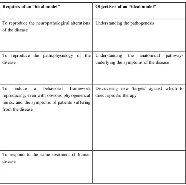
5. General aspects on experimental models of seizures and epilepsy
In table 2, we report the main features of a “good” animal model of neurological disease. Despite the first attempts to study epilepsy in experimental models date back to several decades ago, thus far there aren’t many models of human epileptic syndromes. In fact, the phylogenetic distance between humans and rodents is often the cause of the difficulty to reproduce in the animal, despite having the same pat hological lesion or genetic mutation, a pattern of seizure equivalent in the human being. Moreover, an implicit assumption of this kind of models is the possibility to reproduce in the animal a real epileptic condition, considered as low epileptogenic threshold and the occurrence of SRS.
On the contrary, there are many models of epileptic seizure s. In this case the main objective of the experimenters is to induce acute critical episodes
resembling human seizure, on the behavioral and/or EEG and/or
pathophysiologic perspective (see 1981 ILAE classification of seizure). In this
setting, however, unlike the previous group of models, seizures are not spontaneous, but acutely induced after various experimental manipulations done by the researchers. It is easy to imagine how the two types of approaches are used for two different purposes: the first, i.e. the reproduction of an epileptic condition, which is the most recent, has the aim of understanding the pathogenesis of epilepsy. In the second case, i.e. the induction of acute seizures, experiments are performed mainly with the aim of developing fast and repeatable tools to test the effectiveness of anti-epileptic drugs (AEDs) on the different kind of human seizure.
It is obvious, however, that the latter approach is of limited usefulness in studying epilepsy proper.
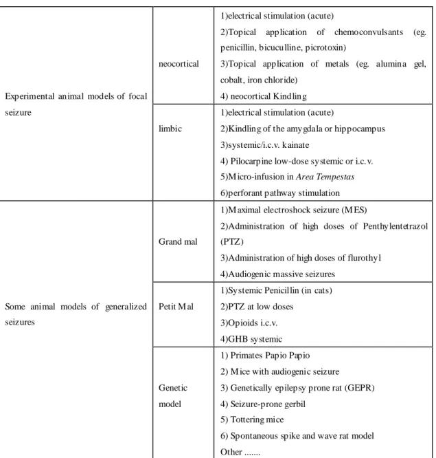
In the following paragraph, we will focus on models of limbic seizures and epilepsy, since one of these models form the basis of the present thesis, while a complete description of the remaining experimental approaches for different types of epilepsy is beyond our aims. However, a concise list of the available experimental models of epilepsy is presented in (Table 3)
6. Mesial temporal lobe epilepsy: anatomical specificity, clinical features
“limbic seizures” and “limbic epilepsy”. It has been developed for addressing
specific physiopathological issues related to a very common form of epilepsy,
the so-called MTLE. In this paragraph, we will address at first the anatomical structures forming part of the limbic system, and then we will briefly discuss the experimental models of MTLE.
a) The limbic system
The term limbic system dates back to the 19th century, to indicate cortical
structures which, without interruption, sourround (thus form a “ring”, i.e.
limbus in latin) the brainstem and the corpus callosum. This term has been
used, along the last decades, to indicate different brain structures slightly varying from author to author. Nowadays, there is a general agreement by many authors in the field, in defining the limbic system as that part of the brain which is directly involved in emotional modulation, memory storing, and in the regulation of visceral functions. Thus, being such a definition exquisitely functional, from an anatomical point of view it includes both telencephalic and diencephalic structures. The concept of limbic system as a “ring” surrounding the brain stem and hippocampus has been more recently substituted by the term “limbic lobe”, while actually the term limbic system includes: a) the limbic lobe, b) the hippocampus, c) the hypothalamic mammillary bodies, d) the mediodorsal and anterior thalamic nuclei, e) the
amygdala. A slightly wider definition includes also: a) the claustrum; b) the anterior perforated substance, c) the piriform nuleus; d) the olfactory tubercle; e) the septal nuclei.
The limbic lobe is formed by the orbital part of the frontal cortex, the parahippocampal cortex and the pole of the temporal lobe, which includes the so-called uncus of the hippocampus formed by the cortex laying right above the amygdale, which can be further defined, according to some authors, as “piriform lobe”, due to its shape in rodents, and which can be further divided in: a) prepiriform cortex; b) periamygdaloid cortex (also called perirhinal cortex), and c) enthorinal cortex. The limbic lobe is considered as an associative limbic area, involved in memory (especially the memory for old events), and in generating the emotional drive for different behaviors, involving both ancient instinct and evaluation of risk/benefits of events (due to its connections with several neocortical associative areas).
Conversely, the part of limbic system formed by the amygdala and hippocampus is involved in recent memory and fast, instinctive visceral and emotional behaviors.
The hippocampal formation is placed within the mediodorsal part of the temporal lobe. It is involved, among other functions, in working memory formation. It is formed by: a) the CA, b) the dentate gyrus (from now on abbreviated as DG, and subiculum, which are cortical formations forming a continuum with each other, c) the fimbria, which is formed by white matter
and is placed above the DG. The CA is formed by a pyramidal cell layer which is the continuation of the subiculum and ends at the level of the hilus of the DG: the latter part of this pyramidal layer is called the CA4, or endfolium, while the part closer to the subiculum is called CA1 region. The DG is a c-shaped neuronal layer formed by the so-called granule cells, which are glutamatergic neurons, which give rise to fibers called “mossy fibers”, which end at the level of the hilus of the DG itself, impinging on interneurons, and at the level of the CA4 and CA3 pyramidal cells dendrites. The interconnections existing between the different parts of the limbic system and the hippocampus are complex in nature, as well as the wide numer of intrahippocampal connections (often involving different kinds of interneurons), and we will not describe them in detail.
It is worth being mentioned in this paragraph that:
a) the main source of afferent signals to the hippocampus is through the excitatory projection of neurons from the enthorhinal cortex to the granule cells of the DG, via the so called “perforant path”;
b) the DG and the CA are strongly connected by the glutamatergic mossy fibers (see above);
c) the efferent fibers originating from the CA pyramidal cells converge in the alveus, but CA3/CA4 neurons send also axon collaterals, the so-called “Schaffer collaterals”, contacting the dendritic trees of pyramidal cells of CA1at the level of stratum radiatum and stratum oriens.
Efferent fibers from the CA get, through the Fimbria, to the Fornix and, l ater on, to the mammillary bodies; the latter send connections to the “limbic thalamic nuclei”, i.e. the mediodorsal and anterior thalamic nucleus.
Other efferent fibers from the hippocampus originate from the CA1 cells, and reach the enthorhinal cortex; t he latter one sends in turn projections to other associative isocortices. Interestingly, while most authors in the last century considered the Fimbrio-Fornix efferent pathway (being part of the so-called Papez memory circuit), as the most important hippocampal efferent pathway, more recently it has been shown that the projection back to the enthorhinal cortex is the most important one, and plays a significant role in memory processes.
Finally, it is important to note that the amygdala is in strict interact ion with the periamygdaloid and pre-piriform cortex, which in turn are widely interconnected with the enthorhinal cortex (the main source of afferents to the hippocampus-see above). Incidentally, the prepiriform cortex is the primary olfactory cortex, and in rodents it plays a critical role in integrating olfaction (the main source of sensitive impulses in these species) with the visceral and instinctive functions of the limbic system: in this experimental study we microinfused chemoconvulsants in a portion of this brain region in rat (see the remaining parts of the thesis).
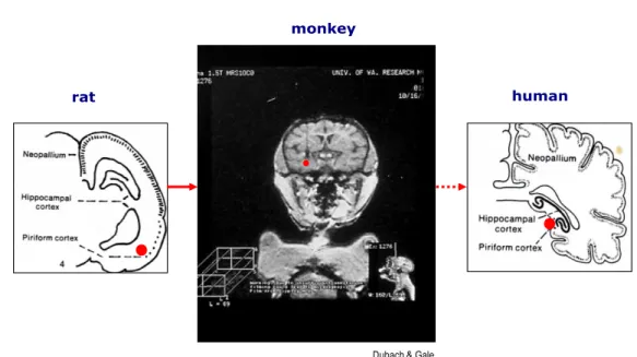
Finally, since the limbic structures are all phylogenically very old, they show a significant persistence of the general cytoarchitecture through evolution: thus,
the same areas, even though, of course, placed differently in the context of the brain, can be identified, and share similar connectivity with each other, in rodents and in primates and humans (See figure 2) exemplifies as the prepiriform cortex (which is the site chosen to be microinfused in our model of limbic seizures), and other limbic structures can be identified through phylogenesis.
b) Human mesial tempral lobe epilepsy syndrome:
In humans epilepsy frequently affects limbic structures, determining the so-called “MTLE”. This form of epilepsy accounts, by itself, for almost 30% of all patients affected by epilepsy. As it can be hypothesized intuitively, the limbic system can be affected by several different kinds of lesions. Nevertheless, since more than 70% of all patients affected by MTLE bear the so-called “hippocampal sclerosis” (see below for a detailed description), and several clinical aspects are specific for most of the patients affected, the latter has been definitely defined as a specific epileptic syndrome (Engel, 2001; Panayiotopoulos, 2002). From now on we will simply refer to MTLE with hippocampal sclerosis just as MTLE.
Patients affected by this syndrome experience mainly focal seizures, starting at the age of 10-15 years. These seizures are featured mainly by an impairment of consciousness and by these reason they have been defined for long time as “psychomotor seizures”, or “complex partial seizures”, as opposite to “simple
partial seizures” in which there are either only motor or only sensitive ictal manifestations without any consciousness involvement. Typically, the patients have a sudden arrest of their attention/speech. During this time they can continue speaking in a stereotyped manner, mainly by repeating passe-partout words. After the seizure, which us ually lasts not longer than 1-2 min, they start again with their normal behavior: sometimes they continue the conversation they were having before the seizure. These seizures are very often associated with motor manifestations usually in the forms of oroalimentary automatisms (in 70% of cases), or stereotyped movements of the arms, and sometimes associated also with an increase in muscular tone on one side’s limbs. In some cases these focal seizures can generalize giving rise to generalized tonic-clonic seizures, but the latter are rare when the patients are under appropriate antiepileptic drugs. Finally, a very typical feature of the seizures occurring in MTLE is that these seizures are preceded by symptoms which, up to very recently were called “aura”, featured by either an epigastric discomfort/nausea, or cacosmia, or a déjà-vu/déjà-veçu sensation. This “aura” is actually a focal seizure per se, which manifests with one of these phenotypes depending on the precise site of origin.
Most of patients affected by MTLE have experienced during infancy febrile seizures, and expecially the most “atypical ones”, which are featured by long duration and post-ictal paralisis. Etiologically it has been believed for long time that these seizures per se, either caused by a lesion nearby, or occurring
without specific reasons, caused the occurrence of hippocampal sclerosis, and, subsequently, MTLE.
As said the hallmark of MTLE is the “hippocampal sclerosis”. This is called also AHS. It was first described in 1966 by Margerison and Corsellis in a cohort of epileptic patients submitted to surgery for TLE (Margerison and Corsellis 1966). The main morphological features of AHS are: a) atrophy of one hippocampus at gross inspection; b) selective piramidal neuron loss at the level of CA1, CA3 and CA4 sub-field of the Cornu Ammonis; c) loss of interneurons within the hilus of the DG: these neurons have been claimed to be mainly GABAergic interneurons; d) Mossy fiber sprouting: by selective coloration (by an histochemical method, TIMM staining, which selectively stains Zn-containing fibers) mossy originating from the glutamatergic granule cells of the DG, it has been shown that in AHS there is a dense re-innervation of the dendrites of granule neurons by their axons themselves. This, functionally, configures the anatomical basis for an autoexcitatory circuit; e) ractive gliosis of the hippocampus, which is usually revealed by testing the expression of glial fibrillary acid protein (GFAP) expression (a hallmark of astrocytes); f) more recently, also granule cell dispersion within the DG has been observed in most of AHS cases. (Blumcke et al., 2002)(See figure 3) Often, hippocampal sclerosis in unilateral; however in many cases as well it has been observed, even though less evident on one side versus another, the occurrence of AHS bilaterally: this has been considered by several authors as
the effects of the spreading of seizures on the contralateral side of a first hippocampal lesion, and the cause of the so-called “mirror focus”, i.e. the onset of new seizures from the contralateral hippocampus (Morrell and deToledo-Morrell, 1999).
Hippocampal sclerosis can be, nowadays, clearly defined also on MRI images, and this facilitates significantly the diagnosis of MTLE in mild forms. Furthermore, this epileptic syndrome is particularly resistant to antiepileptic drugs, since up to 50% of patients, in the different casistics, need more than one drug and is often not seizure-free. The etiological link between the presence of AHS and seizure occurrence has been demonstrated by the good remission in many pharmacoresistant MTLE patients undergoing surgical removal of the affected temporal lobe.
c) Animal models of limbic seizures and epilepsy
c1) Focal induction of acute limbic seizures
Limbic seizures can be induced acutely by stimulating electrically different brain sites, but also by applying electrical stimuli through the corneas . In this setting, just by increasing the intensity of electrical stimulation it is possible to recruit gradually various cerebral structures and, accordingly, induce different seizure types. Thus, for low current facial clonus, forelimb clonus till to lift the hind limbs then fall down can be observed, and this can be considered a
quite “limbic onset” stereotyped seizure pattern in rats, since a similar behavior is observed after low stimulation of the amygdale or hippocampus . Higher currents, however, even induce “running-bouncing”, and even higher currents cause hypertonia with flexion or extension of hind limbs: the latter behavioral phenotypes show a progressive involvement of the whole cortex as well as of brainstem structures.
It is of interest to note that when an electrical stimulation is delivered through the electrodes earphone the first manifestation are those “running-bouncing” which is a sign of early recruitment of the structure of hindbrain (brainstem) .
c2) Systemic injection of chemocovulsants
Systemic administration of KA (a glutamatergic agonist) or pilocarpine (a muscarinic cholinergic agonist) can induce limbic seizure, in a fair percentage of the animals treated. Actually, in those animals these substances induce often a true “SE”, i.e. seizures lasting longer than 30’ without interruption. In particular, the systemic (or i.c.v) injection of KA causes tonic-clonic seizure, through the same sequence of behavioral patterns described above, thus testifying an initial involvement of limbic structures (which bear the
lowest threshold to KA induced seizures). In line with this, Lothman and
Collins (1981), classified the seizure evoked by KA in rats in four distinct stages, which rose to 6 in the latest classification of Zhang et al. (1997).
In the latter classification Stage 6 defines the occurrence of SE, independently from the behavior. Typically, stage 1, 2 and 3 describe staring, clonus of on forelimb and of both ones, respectively. Afterwards rearing and falling can be observed. All these motor manifestation continue up to the occurrence of SE, which occurs between 1.5 and 3 hours after injection and the most severe situations is accompanied by jumps, rotational movements, staggering, intense agitation and wild ride, which continues for hours. Often, animals experience SE till to die.
Only <1% of the injected KA can penetrate into the brain because of the existence of BBB (Blood Brain Barrier). After KA administration the neurons of olfactory cortex, amygdala complex, the APC get lesioned within 24-36h. The major selective lesions to hippocampus are the CA3 area pyramidal cells, interneurons of the hilus of the DG, and to the CA1 region, but much less to area CA2 and the granule cells. the fact that hippocampus is always involved, with the main damage at the level of neurons of areas CA1, CA3 and some interneurons of the hilus and of the DG, has provided the basis for considering this approach, by some authors, as a model of MTLE.
Another model of acute “limbic” seizures is the pilocarpine model. High doses of pilocarpine (up to 400 mg/kg, Turski et al 1984) induce, even in this case, limbic seizures at first, followed soon by secondarily generalized seizures together with the appearance of SE. The lethality in the next 24 hours is very variable and strain-dependent. Even in the case of pilocarpine, brain
damage widely involves the forebrain (Turski et al., 1984); hippocampus, thalamus, amygdala, olfactory cortex, neocortex and substantia nigra are interested with a pattern similar to that induced by of glutamate excitotoxic
drugs (Olney et al., 1986). EEG recording performed during the seizure show
actually the sequential development of the abnormal activity in the forebrain, with the earliest changes at the level of the hippocampus, amygdala, and subsequently the involvement of the whole neocortex (Turski et al., 1983a; Turski et al., 1983b; Turski et al., 1984).
Even in this model of limbic severe seizures one of the earliest lesions regards the interneurons of the hilus of the DG and the pyramidal neurons of the areas CA1 and CA3 of the hippocampus.
In both KA- and pilocarpine-induced SE, chronic plastic changes, such as MFS can be observed (Sloviter, 1994).
As said, both models cannot be claimed as models of temporal lobe epilepsy, since the systemic injections of chemocovulsants induced cellular losses in sites distant from the limbic areas. They cannot be even considered as good models of “the deleterious effects of limbic SE”, since the degenerating phenomena are due not only to the limbic SE, but also to the direct effect of the chemoconvulsant on several brain sites (i.e. those bearing the specific receptors). Consequently, the pattern of neurodegeneration, resulting by these treatments, doesn’t correspond necessarily to the result of the pure epileptic activity, but could be a result of the direct stimulation of large brain areas.
c3) A classical model of limbic seizures in the rat: the “kindling”
Kindling of limbic structures has been considered by several authors a model of limbic epileptogenesis resembling what occurring in TLE. It consists in repeated sub-threshold electrical stimuli of the same brain site (either amygdale, the most frequently tested, or hippocampus or perforant pathway) , resulting in gradual increase in the intensity of seizure activity, culminating in generalized seizures, (i.e. a “Kindling” of the brain, in terms of excitability). Such effects, furthermore, are persistent (or in some cases permanent), since a low electrical stimulus of the same brain site weeks/moths after the kindling induction, still induce severe seizures. Furthermore, sometimes rats even experience spontaneous seizures and chronically abnormal EEG, even though no specific lesions can be observed at the level of the stimulation site. Furthermore, epileptogenicity often is transferred, after several stimulations, also to the contralateral same site, to form the so -called “mirror” focus, a phenomenon often observed also in humans affected by TLE this is similar to the clinical characteristics of human epilepsy (Van de Bovenkamp -Janssen et al., 2004). For these reasons limbic “kindling” is considered by several authors as an interesting animal model of epilepsy. However, one of the main weaknesses for considering it as a good tool to study TLE, is the fact that, independently of the site of kindling induction in the limbic system, in no case
it has been observed a pattern of hippocampal degeneration similar to AHS. Thre “kindling” method has been developed by Goddard starting in the 70’s (Goddard, 1982). This author initially used trains of 60 Hz frequence, 1ms of wavelength, 1s of string length, to stimulate the amygdal a once a day. Subsequently this model had been improved by Lothman with different current and the different position (Lothman et al., 1985).
The limbic structures more often stimulated in order to induce kindling are the amygdala, the piriform cortex, the hippocampus, the enthorhinal cortex and some other limbic regions. In any case, among them the most sensitive to kindling are the amygdala and the hippocampus.
Some authors have also induced “chemical” kindling by administering rats with low, repeated, doses of chemoconvulsants, such as PTZ, penicillin, picrotoxin, kanic acid and pilocarphine. In some cases these drugs induce kindling in a few hours (the so-called “fast kindling”), and often induce spontaneous chronic seizures (Giorgi et al., 2003). The chemical kindling is different with intracerebral kindling, as its no harm to the brain tissue, but the drugs has its own toxicity at high dose, and to give lesion to the brain. Interestingly, in the case of kindling, differently from what observed when using the same chemoconvulsants at high doses, no specific brain damage are usually observed.
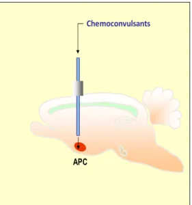
c4) Focal microinjections in the APC of rat
In recent years a new experimental model has been developed in order to minimize the possibility of the nonspecific effect which accompany the traditional models of TLE induction in vivo. This model is based on the microinfusion of the chemoconvulsants into specific “trigger” areas (according to Dr Gale’s definition, see above). As already said, once triggered these neurons, the seizure propagates through the normal interneuronal connections to target areas. It is worth being stressed again the concept that, i n the case of neuropathological, or even just “functional” and “molecular”, changes in sites distant from the trigger area, these effects can be claimed to be the sole effect of seizure propagation per se rather than of the direct effect of the chemoconvulsants.
In a study performed to test the threshold to trigger seizures of different cortical sites in the limbic system of the rat, Dr gale discovered some decades ago, that the anterior extent of the deep prepiriform cortex, also called more briefly as APC is by far the most sensitive trigger site: the microinfusion of picomolar dose of bicuculline (a GABA-A antagonist) in this site is sufficient to trigger seizures (Piredda and Gale, 1985; Gale, 1992; 1995). Furthermore, it has been recently shown that even SE can be elicited from the APC, by the combined infusion of substance which acts on the different receptor systems. It has been observed with bicuculline+carbachol (a cholinergic agonist), KA+ carbachol, bucuculline + cyclothiazide (an inhibitor of the desensitization of
the receptor of the AMPA subtype of glutamate receptor), or AMPA (2-amino-3-(5-methyl-3-oxo-1,2- oxazol-4-yl)propanoic acid) propanoic acid, an antagonist for the AMPA receptor) + carbachol in APC (Fornai et al., 2005). Surprisingly, the SE obtained by these approaches persists beyond the half-life of the microinfused substances, thus being defined as “self sustaining”. Such a phenomena is somehow generated by the interaction between the cholinergic, glutamatergic and GABAergic neurons localized in the micro-infusion areas, since to prevent SE it is sufficient to remove one of these components.
However, we do not know in detail, yet, the specific mechanisms by which such a phenomenon appears, nor it has been investigated in detail the long lasting effect of SE evoked from the APC.
7. Neuromodulators of seizures and epilepsy: the role of Locus Coeruleus
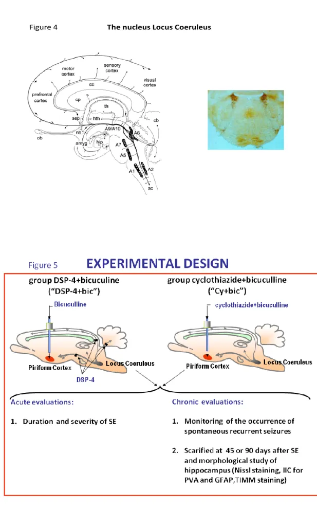
a) The nucleus Locus Coeruleus (Figure 4)
The LC is the main noradrenergic nucleus in the brain. It is located in the pons at the level of the upper part of the floor of the fourth ventricle. A specificity of its neurons is their extremely branched projections which allow each one of them to innervate many subcortical structures and almost the whole cortex. LC receives afferents from the hypothalamus, cerebellum and raphe nuclei, and the amygdala. The two main ascending fiber systems originating from the
LC are the dorsal bundle and the much smaller rostral limb of the dorsal periventricular pathway are innervations. Norepinphrine (Herein abbreviated as NE) released by the LC neurons is considered to play mainly neuromodulatory effects. In fact, the efferent fibers originating from the LC possess a lot of varicosities, and NE is released from those extra-synaptic structures rather than at the level of classical synaptic formations; thus, NE released along LC fiber terminals affect the surrounding structures in a paracrine fashion. As the NE terminals are in close contact with astrocytes, microglia and microvessels, LC affect significantly the function of the BBB and glial function (Harik and McGunigal, 1984). By the releasing of NE into many brain structures, LC can regulate a variety of important CNS functions: it modulates electroencephalographic (EEG) activity (Foote et al., 1983); regulates the sleep-waking cycle (Jouvet, 1969; Aston-Jones and Bloom, 1981) and significantly affect arousal and vigilance (Aston-Jones et al., 1991); furthermore, it seems to play a specific role in alerting and orienting to novelty (Aston-Jones et al., 1994).
From a molecular point of view, most of the above mentioned effects appear to be related more or less directly, to transcription in the target neurons of the so-called immediate early genes (IEG) such as c-fos and nerve growth factor-induced A (NGFI A), nur 77, tis-7, zif-268 and tis-21 in LC target
neurons (Gubits et al., 1989; Bing et al., 1991). This effect has been
stimuli (Stone et al., 1993) and during seizures (Simler et al., 1999). In line with this, LC-dependent circadian rhythm of early gene expression can regulate the state of phosphorylation of cyclic adenosine monophosphate
response element-binding (CREB) proteins (Cirelli et al., 1996). The IEGs are
suppressed during the REM (Rapid eye movement) sleeping and express strongly during the wakefulness (Cirelli et al., 1996). Furthermore, it has been shown that a previous lesion of LC terminals, by DSP-4, significantly attenuated the expression of Fos protein associated with SE (see below-), and this might have relevant effects in the plastic mechanisms related with epileptogenesis attenuated the expression of Fos protein associated with SE (Giorgi et al., 2008).
Finally, it is worth being mentioned in this setting that LC neurons spontaneously degenerate in physiological ageing, as it has been estimated that while in young adults LC contains approximately 60000 neurons, in healthy aged people this number decreases up to approx. 40000 neurons (Baker et al., 1989; Iversen et al., 1983)
b) The role of NE in epilepsy and experimental seizures
It is worth to be mentioned that while in the last dec ades there have been a huge amount of experimental data on the role of NE in a variety of epilepsy models, there are, on the contrary, only a few indirect evidences for its role in
human epilepsy, mainly because of the lack, nowadays, of neuroimaging techniques allowing to investigate in vivo the structure and function of LC in humans. Among the indirect clues, it is worth being mentioned that there are robust epidemiological evidences for a much higher incidence of endogenous depression among epileptic patients (independently from any therapeutic drug) than in the control population, and it has been demonstrated by several authors a significant role of NE in depression pathogenesis (Kanner, 2011; El Mansari et al., 2010). Thus some authors even suggested t hat, in selected cases, NE deficit might contribute to the onset of both epilepsy and depression in the same patient (Jobe et al., 1999).
As said there are several experimental evidences for an anticonvulsant effect of NE. A proconvulsant effect of damage to the NE system has been proven, by profiting of monoamine-depleting agent reserpine (Chen et al., 1954) , or by selectively lesioning the LC in models of audiogenic seizures (Jerlicz et al., 1978), metrazol-induced seizures and seizures induced by electroshock (Mason and Corcoran, 1979). A reduced threshold to different epileptogenic insults has been observed by using the selective LC neurotoxin DSP-4, (Mishra et al., 1994). Conversely, LC stimulation suppresses seizures induced by P TZ (Pentilentetrazolo), amygdala kindling and focal hippocampal
penicillin application (Libet et al., 1977; Weiss et al., 1990; Ferraro et al.,
1994).
observation that both in genetically epilepsy prone rats (GEPRs) and tottering mice, which are susceptible to seizures, there is a congenital alteration of LC . In particular, GEPRs have been discovered due to their proneless to develop seizures after audiogenic stimuli; they also show a low threshold to several epileptic stimuli, such as electroshock and PTZ (Browning et al., 1990), fluorothyl (Franck et al., 1989), limbic kindling (Savage et al., 1986). In this rat strain several NE parameters alterations have been observed, such as reduction of NE levels (Jobe et al.,1973; 1984; Dailey and Jobe, 1986; Dailey et al., 1991), dopamine-beta-hydroxylase (DBH) actvity (Browning et al.,1989) and immunoistochemistry (Lauterborn and Ribak, 1989) reduction, as well as NE uptake sites reduction (Browning et al.,1989). In tottering mice, it has been shown, conversely, an excessive autoinnervation of LC cell bodies, which is believed to induce auto-inhibition of the nucleus itself (Levitt et al., 1987).
Other strains of mice bearing specific, known mutations affecting noradrenergic parameters have an abnormal threshold to epileptic seizures. For instance, the D79N mice bearing a mutation of alpha-2 adrenergic receptor which induces a significant loss of receptorial function (Ceresa and Limbird, 1994): these mice are much more susceptible to amygdale kindling than wild type (WT) mice (Janumpalli et al., 1998). It has been observed a significant incidence of spontaneous seizures when mice overexpressing alpha 1B-adrenergic receptors; furthermore, the degree of activity of the
overexpressed alpha1B-adrenergic receptors (alpha1B ARs) is related with the severity of spontaneous seizures; in these mice, seizures can be partially reversed by the alpha1 ARs antagonist, confirming that the alpha1 ARs signaling sustains seizure activity (Kunieda et al., 2002).
c) The role of Locus Coeruleus on seizures evoked from limbic sites and AP C
Stepping back to the role of NE in acquired experimental seizures, there are a lot of evidences of the involvement of LC in limbic seizures and epilepsy. Concerning amygdale kindling, it has been clearly shown that there is a significant increase in the kindling rate in the amygdala after a selective lesions of NE fibers belonging to the dorsal forebrain bundle , which indeed produces a parcellar selective loss of the NE innervation to limbic and
neocortical areas (Corcoran and Mason, 1980). An opposite effect is provided
by increasing NE activity, either by NE uptake blockade (McIntyre et al., 1982) or by direct stimulation of LC (Jimenez-Rivera et al., 1987): both approaches delay amygdala kindling. The antiepileptogenic effect of NE on kindling is
confirmed by in vitro studies on slices collected from the
piriform/periamygdaloid cortices (McIntyre and Wong, 1986).
The AP C is a part of the olfactory cortex, which possesses the highest NE content in the brain (Giorgi et al., 2003; 2006) and is particularly prone to
seizures evoked by bicuculline infusion into the APC are isolated, sporadic seizures, each one lasting not longer than 30”-1 min’, the microinfusion of bicuculline into APC of rats previously treated with DSP -4 converts the seizure into a long-lasting, self sustaining status epileptic which shows NE deficit provoked a persistent modification in the responsiveness of neural epileptic circuitries (Giorgi et al., 2003). Furthermore, the NMDA antagonist and non-NMDA antagonists prevent the SE in the rats with intact LC but the NMDA antagonist is ineffective in the rats with the LC lesioned. AHS is observed in many patients with MTLE, this neuronal damage could be caused with less seizure duration caused by bicuculline infusion into the APC in NE lesion rats, suggesting a specific neuroprotective effect of endogenous NE (Giorgi et al., 2003).
Chapter 2 Experimental Section
1. Introduction to the experimental sectionAs said repeatedly in the General Introduction, MTLE is by far the most diffuse form of focal epilepsy. The main feature of epilepsy is the presence of “SRS”. There are several models of limbic seizures, as extensively described above. However, only recently authors involved in the epilepsy field have started to describe models of SRS, rather than of just acute limbic seizures. It has been shown repeatedly that after a strong insult, such as the induction of limbic SE by systemic administration of chemoconvulsants (e.g.KA or pilocarpine), both in rats and in mice there is the development of SRS
(Paradiso et al., 2011; Okamoto et al., 2003; Pallud et al 2008; Hellier and
Dudek, 2005). This has given rise to the “double hit” hypothesis for the pathogenesis of MTLE. According to this hypothesis, there is an initial event, either occurring early on in life (e.g. prolonged febrile seizures or SE occurring very early post-natally), or during adolescence/early adulthood (subtle ischemic/traumatic insult? Subtle cortical malformations giving rise to
focal unnoticed epileptic discharges?) (Loscher and Brandt, 2010;Baram et al.,
2011), which might be responsible for the onset of plastic phenomena which, at least in some patients, give rise to the onset of SRS.
AHS has been considered for more than 50 years as a potential pathological substrate for the onset of SRS in MTLE. In fact, from a pathological and
functional point of view, there are several aspects of AHS which might justify such an assumption: a) there is a selective loss of GABAergic interneuro ns at the level of the hilus of the DG; b) there is a marked sprouting of the mossy fibers (the MFS above described), which causes auto-excitation of the glutamatergic granule cells by their axon terminals. c) several studies in vitro have shown a hyperexcitability of the hippocampus bearing AHS signs (Blumcke et al., 2000).
Other authors, however, think that this anatomical alteration is not sufficient by itself to cause SRS and to justify seizure susceptibility in MTLE. They claim, for instance, that: a) kindling of different limbic regions, such as the amygdala, the piriform cortex or the hippocampus itself, are not usually associated with AHS, even though they are often associated to the
development of SRS (Brandt et al., 2004; Muller et al., 2009). b) in rats in
which MFS has been prevented by protein synthesis blockage, SRS after SE have been shown by some authors (Longo and Mello, 1997).
AHS has been observed to occur with the classical models of limbic SE by
KA or pilocarpine (Curia et al., 2008;Bouillere et al., 2000); in both models it
has been shown the occurrence of SRS (Brandt et al., 2003). However, both of these models bear the huge disadvantage of inducing a widespread damage throughout the whole brain, which is likely to be largely independent of SE, but rather a concomitant phenomenon (see for instance Sloviter, 2009). Thus, both the occurrence of AHS, and the presence of SRS might be due to
one/some different lesions occurring in the rodents experiencing this SE. Conversely, by profiting of the model of APC microinfusions, we can test the sole effect of the spreading of seizures via the anatomical physiological pathways of the limbic system. The picomolar amount of chemoconvulsants microinfused in this model do not spread for more than 1 mm apart from the site of infusion and thus do not affect the diffusion of seizures to other sites of the limbic system and brain. Furthermore, by this experimental approach it is possible to discriminate the effect of sporadic seizures versus prolonged seizures (SE) in determining the onset of late SRS, while KA or pilocarpine give rise only to an all-or-nothing phenomenon, i.e. their lightest seizure effect is SE!
As already mentioned, there are at present two different models of SE evoked from the APC, which have been developed in the last few years (Fornai et al., 2005; Giorgi et al., 2003). In one case SE is evoked by co-administering 2 substances (cyclothiazide and bicuculline) into APC in intact rats, and in the other one the simple administration of low dosages of bicuc ulline into the AP C, in rats which have been submitted to LC lesion, induces prolonged SE. As outlined in the introduction, LC seems to play a critical role in the expression of IEGs, which are genes involved in the generation of plastic mechanisms, such as learning and memory phenomena (for a review, see Giorgi et al., 2006). We have recently shown that the lesion of LC significantly affects the pattern of expression of c-Fos protein after SE evoked
from the APC.
At present, nobody has ever investigated whether SE evoked from the APC is associated with SRS, the presence and role of AHS in this model, and, eventually, the role of LC in these phenomena. The latter aspect is particularly important due to the lack of enough information from human studies, and the fact that, however, LC neurons decrease throughout adult life in humans.
2. Specific aims
In this study we wanted to test:
A) Whether there are EEG and behavioral differences between the SE
induced by APC in the presence of intact LC and that induced by the same site in rats bearing a lesion of LC.
B) Whether sporadic seizures evoked from the APC are sufficient to
induce SRS.
C) Whether the model of SE induce d by APC, either in the presence or in
the absence of LC, is associated to the development of SRS, i.e. whether one of the two models can be considered as a good model of epilepsy, rather than acute seizures only.
D) Whether the model of SE induced by APC is associated with the
classical hallmarks of human MTLE, such as AHS and MFS.
E) Whether the lack of NE affects the presence of AHS and MFS, and in
parallel the onset of SRS, or rather these phenomena are, at least in part, independent from one another.
3. Methods
3.1 Animals
We used male Sprague Dawley rats aged between 90 and 120 days at the time of seizure induction (weight 200-250 g). They were kept under controlled environmental conditions and handled in accordance with the Guidelines for Animal Care and Use of the National Institutes of Health
3.2 Experimental design (Figure 5)
Rats were microinfused with chemoconvulsants or saline into the APC at t=0; they were monitored be haviorally and by EEG starting at t=0 and for up to document the last seizure. Animals were submitted either to sporadic limbic seizures (by bicuculline 118 pmol infused into APC) or to SE. SE was induced by a) microinfusing cyclothiazide and bicuculline, 3 min. apart, into the AP C in rats bearing an intact LC (“Cy+Bic” group); b) by microinfusing bicuculline into the APC of animals in which it had been induced a selective LC lesion 2 weeks before (“DSP-4+Bic” group). In the following weeks they were monitored both behaviorally and, in some cases, by EEG. They were sacrificed either 45 days (T1), or 90 days (T2) after the induction of seizures, and their brains were collected and processed for morphological analysis. (See