UNIVERSITA' DEGLI STUDI DI PISA
FACOLTA' DI MEDICINA E CHIRURGIA
DOTTORATO DI RICERCA
Fisiopatologia e Clinica dell’Apparato Cardiovascolare e
Respiratorio
Determinats of exercise tolerance in COPD patients
Relatore
Chiar.mo Prof. Pierluigi Paggiaro
Candidato Dr. Francesco Costa
* * *
Tablet of contents
Summery
3
Abbreviation
9CAPITOLO I Introduction 10
Methods for the assessment of exercise capacity in COPD 14
Other methods for assessing exercise limitation in COPD 21
Determinants of the exercise capacity in COPD patients 26
CAPITOLO II
Aim of the study 29
CAPITOLO III
Patients and methods 30
Statistical analysis 37
CAPITOLO IV
Results 39CAPITOLO V
Discussion 55References
62SUMMERY
Chronic Obstructive Pulmonary Disease (COPD) is a preventable and treatable disease characterised by airflow limitation that is not fully reversible with some significant extrapulmonary effects that may contribute to the severity in individual patients. Progressive decline in airflow is associated with an abnormal inflammatory response of the lung to noxious particles or gases (1).
Limitation in the exercise capacity is a major characteristic of COPD, particularly in moderate-severe disease. This may be expressed as dyspnoea occurring during an effort of different intensity, and as reduction in the possibility to perform a normal or almost normal active life. In more severe patients, exercise limitation is so relevant to avoid the patient to leave home and also limiting them in the normal home daily activity. Limitation in exercise capacity in COPD patients has been demonstrated to be related to different mechanisms, which may be differently represented in the same patient. These mechanisms are: a) abnormal mechanical properties of the respiratory system, leading to ventilatory limitation; b) abnormal cardiovascular response to the exercise (cardiac limitation); c) reduction in the peripheral muscle strength and endurance, leading to muscular limitation.
Cardiopulmonary exercise testing (CPET) is considered the gold standard for exercise intolerance evaluation. During the test, patients are subjected to symptom limited incremental exercise, breath-by-breath monitoring of cardiopulmonary variables (e.g. pulmonary O2 uptake, pulmonary CO2 output, minute ventilation (Ve ), heart rate (HR), assessment of perceptual responses (e.g. dyspnea, leg discomfort) and measurements such as exercise-related arterial oxygen desaturation, dynamic hyperinflation and limb-muscle strength. Other methods for assessing exercise limitation in COPD are: endurance time , incremental shuttle walking test (ISWT), endurance
life through motion sensors like Armband SenseWear becomes important in the valuation of COPD patients.
Several studies have attempted to correlate the tolerance effort of COPD patients with various physiological parameters, clinical, metabolic and inflammatory. However, few studies have attempted to simultaneously consider many different factors that can contribute to determine the limitation exercise in these patients. This is important to determine which of several pathophysiological, biological and metabolic mechanisms are primarily responsible for limiting the effort in individual patients with COPD.
The aim of the present study was to assess in a large group of patients with COPD of different severity, the relative role of the different components of the disease which may have influence on the response to the exercise.
In this attempt, we chose the response to the CPET as the more accurate method for evaluating the pulmonary, cardiovascular and metabolic responses to the exercise, and we tried to correlated the response to CPET with several baseline findings. We included a large, although not exhaustive, number of measurements, including: a) clinical findings (rate of dyspnoea, exacerbation rate, comorbidities); b) pulmonary function measurements; c) respiratory muscle strength measurements; d) metabolic findings (BMI, FFM, blood haemoglobin); e) markers of airway and systemic inflammation (derived from induced sputum, exhaled breath or peripheral blood) or cardiac involvement (pro-BNP). We tried also, in a multivariate analysis performed of all examined patients and in subgroups of patients according to the main factor limiting exercise, to assess the main determinants, among those already mentioned, which may have influence on the exercise limitation.
The study is a cross-sectional study and took place in three visits, along a study period of 2 weeks.
We studied fifty-one outpatients with stable COPD between July 2008 and May 2010, recruited among those attending to the clinics of our Respiratory Pathophysiology Unit of the Cardio-Thoracic and Vascular Department of the University of Pisa.
Three patients didn‟t perform the cardiopulmonary exercise test (CPET), two patients dropped out before performing the CPET, and one patient was defined at the examination as affected by a relevant asthma-like component of the disease. Forty-five patients were therefore included in the analysis.
The patients performed: pulmonary function test, arterial blood analysis, maximal inspiratory (MIP) and expiratory (MEP) pressures, incremental shuttle walking test, cardiopulmonary exercise test (CPET). Physical activity was measured over at least 7 consecutive days, using a multisensory Armband. Metabolic status was evaluated: body mass index (BMI), Fat Free Mass (FFM) and Fat Free Mass Index (FFMI). The Saint George‟s Respiratory Questionnaire (SGRQ) was used for the assessment of the health-related quality of life. Charlson index and CIRS index (Cumulative illness rating scale ) were used as valid and reliable methods to measure comorbidity. In a sample of venous peripheral blood, the following biomarkers were measured: C-reactive protein (CRP), total and differential inflammatory cells, haemoglobin and N-terminal pro–B-type natriuretic peptide (NT-pro-BNP) concentration, as a systemic biomarker of heart failure. Sputum was induced and measured: macrophage, lymphocyte, neutrophil, eosinophil percentages were expressed as percent of total inflammatory cells, excluding squamous cells. In the sputum were measured supernatant, TNF-alfa, IL-8 and neutrophilic elastase (NE). Exhaled nitric oxide (NO) were measured and exhaled breath condensate (EBC) was collected.
Statistical analysis was performed on the data obtained in the 45 patients who performed all measurements. All statistical analyses were performed using a SPSS 16.0 statistical package.
Results
We considered as dependent variable VO2peak/kg, which may be considered the best way for expressing exercise limitation during CPET, and as independent variable all these findings which may potentially have some influence in determining the amount of the exercise limitation. In an univariate single regression analysis, V02peak/Kg was significantly correlated with MRC score, FEV1% FVC %, SVC % FEV1/SVC % IC % RV/TLC %, DLCO %.
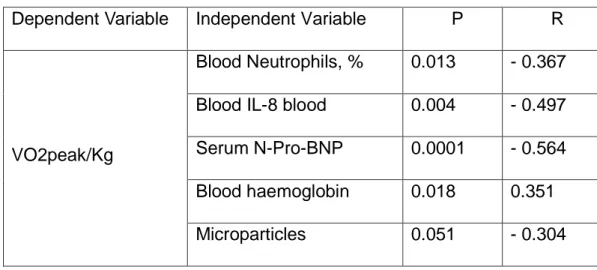
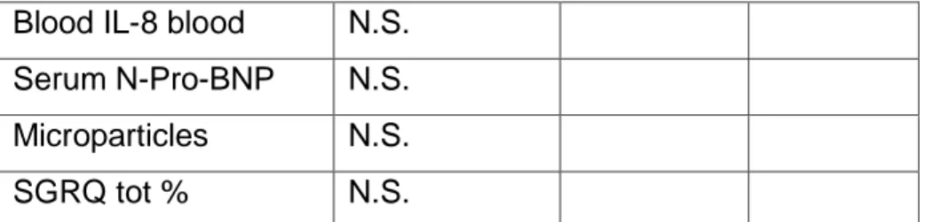
As regards the correlation with biomarkers measured in the blood, sputum and exhaled condensate we can see that there was no significant correlations of VO2peak/kg for many biomarkers, except for some indices of systemic inflammation blood neutrophil %, serum IL-8, blood microparticles, blood haemoglobin and serum N-Pro-BNP.
In a stepwise regression analysis we observed that the only variable that explains the model was blood haemoglobin and total variance explained by this model was 31.5%.
Excluding haemoglobin aspecific parameters, the variable that explains the model is MRC score, with total variance explain of 20.5 %.
If we consider one independent variable for each clinical , functional and biological field, DLCO % is the variable that explains the model and total the variance explain was 19.6%.
Other correlations for VO2peak/kg with: ISWT, as expected, steps/day and SGRQ.
Divided the patients in flow limitation (FL) and no flow limitation(NFL), according Hyatt technique, only in the FL group the VO2peak/Kg correlated in univariate analysis with a several clinical functional and biological indices (MRC, FEV1%, IC%, DLCO%;
steps/day, SGRQ tot %). In stepwise regression analysis MRC and IC% were the parameters that enter in the analysis, with cumulative R2= 0.454.
Considering the patients that had interrupted the CPET for ventilator limitation (30 patients out of 45) in univariate analysis VO2peak/Kg correlated with MRC, FEV1%, IC%, DLCO%, MIP, serum pro-BNP, TEE, ISWT, SGRQ tot %. In stepwise regression analysis were excluded ISWT and parameters of Armband; only MRC entered in the model (p= 0.021, R= 0.589) and explained the 34.7% of variance of exercise tolerance.
In the patients that interrupted the CPET for muscular limitation (15 patients), the exercise tolerance not correlated with functional parameters but several indices: age, blood neutrophils %, blood eosinophils %, sputum neutrophils %, ISWT. This patients had less compromise in FEV1% and higher MRC score than the 30 patients that stopped CPET for ventilator limitation.
Conclusion
In the present cross sectional study, we tried to examine the determinants of the exercise limitation which is a characteristic of the patients with moderate-severe COPD. We included a large, although not exhaustive, series of clinical, functional and biological measurements, in order to find which variable, among those considered, might have the predominant role in determining the limitation in exercise capacity. We chose the maximum specific oxygen consumption (VO2peak/kg), obtained during a CPET, as a good marker of the exercise limitation in our COPD patients. We found that several clinical, functional and biological variables were significantly correlated with VO2peak/kg. Many of these correlations were expected according to the pathophysiology of the disease (like MRC, IC, DLCO), but some of them were fairly unexpected (like some
correlations with other variable which may be considered as possible determinants of the exercise capacity (like respiratory muscle strength, BMI or FFM). When me included the variables which resulted significantly correlated with VO2max/kg in a multivariate step-wise regression (according to different models), we found that the man determinants were haemoglobin and, in a second position, MRC dyspnoea score and diffusing capacity. This was particularly true when only subjects with pulmonary mechanical factors which may limit exercise (like the presence of flow limitation), or with a respiratory limitation to the CPET, were considered. This suggest that the main factors limiting the peak oxygen consumption during exercise are represented by non-pulmonary mechanisms in all unselected COPD patients, and by pulmonary mechanisms in COPD patient with clear limitation to the exercise related to the pulmonary disease.
Limitation of the study
Our study has some limitation. Firstly, we have not directly measured the strength of lower limb muscle, which are main determinants of the maximum work load sustained during the CPET.
Secondly, we have no independent measure of the heart function (like ejection fraction or other indices derived from echocardiography). We know that exercise limitation is strongly related to the performance of the cardiovascular system.
Abbreviation
VO2peak Oxygen uptake at peak exercise
VCO2 carbon dioxide output
WR Wark rate
RER respiratory exchange ratio
Vt tidal volume
Vd Dead space
Ve Minute ventilation
HR Heart rate
HRR Heart rate reserve
FEV1 forced expiratory volume in 1 second
VC vital capacity
FVC Forced vital capacity
IC Inspiratory capacity
TLC Total lung capacity
FRC Functional residual capacity
RV Residual volume
DH Dynamic Hyperinflation
CHAPTER I
INTRODUCTION
Chronic Obstructive Pulmonary Disease (COPD) is a preventable and treatable disease characterised by airflow limitation that is not fully reversible with some significant extrapulmonary effects that may contribute to the severity in individual patients. Progressive decline in airflow is associated with an abnormal inflammatory response of the lung to noxious particles or gases (1). COPD is a leading cause of disability and death worldwide, with variable data of population prevalence rates 5% to 13% (2). Prevalence rates are related directly to tobacco smoking and indoor air pollution, and are expected to rise as smoking rates continue to increase, notably among women and in developing countries, as a consequence by 2030, COPD is expected to represent the third leading cause of death in middle-income countries. In addition, COPD accounts for a significant proportion of health care budgets, mainly due to hospitalisations for exacerbations (2).
COPD is characterised by progressive decline in respiratory function and health related quality of life (HRQoL) with substantial risk for premature death (3). The pathologic effects of COPD on the respiratory system affect the proximal and peripheral airways, lung parenchyma, and vasculature (4). Moreover, comorbidities such as cardiovascular disease, diabetes mellitus, and depression, as well as associated systemic consequences, including weight loss and muscle dysfunction, increase the overall burden of disease (5). The clinical course of COPD has been viewed as a progressive decline in lung function over time (measured in terms of forced expiratory volume in one second (FEV1) (6). However, recent work suggests that disease
progression is very heterogeneous, the clinical course and presentation depend on contributing phenotypes (7). A majority of patients show no statistical significant decline of FEV1 or increase in the Body mass index, degree of airflow Obstruction, functional Dyspnoea, and Exercise capacity (BODE) index. The multidimensional evaluation of COPD should offer insight into response to COPD management (8).
Limitation in the exercise capacity is a major characteristic of COPD, particularly in moderate-severe disease. This may be expressed as dyspnoea occurring during an effort of different intensity, and as reduction in the possibility to perform a normal or almost normal active life. In more severe patients, exercise limitation is so relevant to avoid the patient to leave home and also limiting them in the normal home daily activity. Limitation in exercise capacity in COPD patients has been demonstrated to be related to different mechanisms, which may be differently represented in the same patient. These mechanisms are: a) abnormal mechanical properties of the respiratory system, leading to ventilatory limitation; b) abnormal cardiovascular response to the exercise (cardiac limitation); c) reduction in the peripheral muscle strength and endurance, leading to muscular limitation.
Expiratory flow limitation (EFL) is the pathophysiologic hallmark of COPD and arises because of the dual effects of permanent parenchymal destruction (emphysema) and airway dysfunction, which in turn reflects the effects of small airway inflammation (mucosal edema, airway remodeling/fibrosis, and mucous impaction). In COPD, the increased compliance of the lung, as a result of emphysema, leads to a resetting of the relaxation volume of the respiratory system to a higher level than in health. This has been termed “static” lung hyperinflation. In patients with EFL during spontaneous resting breathing, end-expiratory lung volume (EELV) is also dynamically
determined relaxation volume of the respiratory system. In flow-limited patients, the time-constant for lung emptying (i.e., the product of compliance and resistance) is increased in many alveolar units, but the expiratory time available (as dictated by the respiratory control centers) is often insufficient to allow EELV to decline to its normal relaxation volume, thereby resulting in air retention (or trapping) with further lung hyperinflation. The rate and magnitude of dynamic lung hyperinflation (DH) during exercise is generally measured in the laboratory setting by serial inspiratory capacity (IC) measurements.
The negative effects of acute DH during exercise are now well established:
1) DH leads to increase in the elastic and threshold loads on the inspiratory muscles, thus increasing the work and oxygen cost of breathing;
2) DH results in functional inspiratory muscle weakness by maximally shortening the muscle fibers in the diaphragm and other inspiratory muscles;
3) DH reduces the ability of tidal volume to expand appropriately during exercise, and this leads to early mechanical limitation of ventilation;
4) In some patients, this mechanical constraint on tidal volume expansion in the setting of severe pulmonary V/Q abnormalities (i.e., high fixed physiological dead space) leads to CO2 retention and arterial oxygen desaturation during exercise; 5) DH adversely affects dynamic cardiac function.
The role of static and dynamic hyperinflation in determining the symptoms of COPD patients is demonstrated also by the improvement in dyspnoea after bronchodilators and lung volume reduction surgery, which correlates with increased IC, a measure of reduced lung hyperinflation. (9).
Tzani et al. (10) have found that dynamic hyperinflation is strictly associated to a poor cardiovascular response to exercise in COPD
patients with different degree of severity. In 48 COPD patients there was a significant and inverse relationship between the peak exercise EELV and the change in oxygen pulse achieved on exercise. The oxygen pulse does not directly measure the stroke volume, but it may be considered as a surrogate marker, when arterial oxygen content can be assumed to be normal (11)
Dynamic hyperinflation may reduce left ventricle stroke volume since increases intrathoracic pressures and, consequently, decreases the preload by reducing both venous return and the volume of the left ventricle. As a consequence, there is a reduced filling of the left ventricle and consequently a reduced cardiac output. Moreover, DP (double product) reserve was inversely related to the dynamic hyperinflation during exercise. In addition, heavy hyperinflators had a high likelihood to have a DP reserve value ≤ 10,000 mmHg · bpm. Lastly, in all patients, DP reserve was inversely related to breathlessness perception at peak exercise, suggesting that in COPD patients the exertional dyspnoea may be due not only to ventilator constrains, but also to a functional cardiovascular impairment. DP reserve is used as an estimate of the maximal performance of the left ventricle. DP reserve reflects myocardial oxygen uptake during exercise, since the three major determinants of myocardial oxygen uptake are the tension in the wall of the ventricle, the contractile state of the heart and the heart rate.(12).
A reduced capacity for muscle aerobic metabolism may influence exercise tolerance in several ways. Increased lactic acidosis for a given exercise work rate, a common finding in COPD increases ventilatory needs. This imposes an additional burden on the respiratory muscles already facing increased impedance to breathing. Premature muscle acidosis, a contributory factor to muscle task failure and early exercise termination in healthy subjects, may be an important mechanism contributing to exercise intolerance in COPD.
This is exacerbated by a tendency to retain CO2 during exercise, further increasing acidosis. (13)
Unexplained weight loss occurs in about 50% of patients with severe COPD, but it can also be seen in about 10 to 15% of patients with mild to moderate disease. It is mostly due to loss of skeletal muscle mass and, whenever it occurs, it highlights poor prognosis. Weight loss in COPD is mostly due to skeletal muscle atrophy. Furthermore, the remaining muscle mass is often dysfunctional. This combination contributes significantly to reduce the exercise capacity (and thus, the health status) of patients with COPD.
The mechanisms underlying skeletal muscle abnormalities in COPD are not precisely defined, but they are probably multiple and interdependent. Systemic inflammation is a likely contributor. For instance, cytokines, particularly TNF-alpha, activate the transcription factor nuclear factor (NF)-KB, up-regulate the inducible form of the nitric oxide synthase, and facilitate the degradation of myosin heavy chains through the ubiquitin–proteasome complex .They can also promote apoptosis in skeletal muscle cells, as recently shown to occur in patients with COPD and low body weight. (14)
Methods for the assessment of exercise capacity in COPD
Cardiopulmonary exercise testing
Cardiopulmonary exercise testing (CPET) provides a global assessment of the integrative response to exercise involving respiratory, cardiovascular, haematopoietic, neuropsychological and skeletal muscle systems, which can‟t be adequately studied through measurements of individual organ (15). This test is more suitable to study the functioning of the several systems involved in exercise, with better results than classical cardiologic protocols with ECG only (16).
CPET allows evaluation of several parameters involved in exercise, both during (real-time) and after the test. They are:
Metabolic parameters: oxygen consumption (VO2), CO2 output
(VCO2), respiratory exchange ratio (RER = VCO2/VO2), anaerobic
threshold (AT);
Cardiovascular parameters: maximum workload (Wmax,
measured in Watt), heart rate (HR) and heart rate reserve (HRR = [220-age] – HRpeak), blood pressure (BP), oxigen pulse (O2pulse =
VO2/HR, which provides information about heart and peripheral
circulation efficiency during exercise), percutaneous blood saturation (SpO2);
Respiratory parameters: external ventilaton (VE), tidal volume (Vt), breath frequency (Bf) and ventilatory reserve (Br = MCV – VEpeak, with MCV as Maximum Ventilatory Capacity, calculated as FEV1 x 40, that in COPD patient is an acceptable assessment of
Maximum Voluntary Ventilation (I)), Vd/Vt ratio (a
ventilation/perfusion mismatch index), inspiratory capacity (IC, to evaluate dynamic hyperinflation), end-tidal PaO2 and PaCO2 (PETO2
e PETCO2).
In order to collect these measurements, an adequate equipment is needed: a cycle ergometer, a 12-lead ECG, a sphygmomanometer, a pulseoxymeter and a pneumotachograph to measure volumes and pressure of inspired and expired gases, and a sowftare able to show results on a monitor. Actually, more reliable measurements could be possible using more invasive methods, such as arterial catheterism for invasive blood pressure assessment, blood gases sampling, oxygen
saturation and anaerobic threshold: usually non-invasive
measurements are sufficiently reliable to avoid use of invasive tools. The previously mentioned parameters are integrated with the real-time evaluation (during CPET), of dyspnea and muscular fatigue according to Borg scale. This last evaluation is useful to correlate measured parameters with subjective feelings of patients, in order to better assess exercise tolerance.
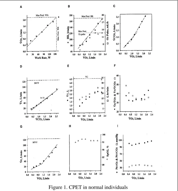
The correlation between the several variables recorded during the test, according to the method proposed by Wassermann and coworkers (16) allows a comprehensive evaluation of individual response to exercise. Figure 1 shows a maximal effort of a normal individual.
The mechanisms of the adaptation to the exercise in a normal subjects involves different steps and physiologic effects. Once contraction of locomotor muscles begins, there deliver both central and peripheral mechanisms governing the appropriate regulation of cardiopulmonary responses. VO2 rises fairly linearly with work rate through-out progressive exercise because of an increase in cardiac output (HR x SV) and increased oxygen extraction at the tissues. Early exercise is associated with a combined increase in stroke volume (not shown) and heart rate, whereas late in exercise the rise in cardiac output is mostly dependent on increases in heart rate. Oxygen pulse also rises linearly early in exercise, but tends to plateau later in exercise as both SV and the C(a–v)O2 begin to plateau. VO2 is relatively insensitive to small changes in VE (e.g., hyperventilation) as long as the VE is adequate to maintain PaO2. Ventilation rises early in exercise as a result of an increase in tidal volume (Vt) and breath frequency (Bf) of breathing. As exercise progresses, Vt tends to level off at about 50% of the VC (likely in part to limit the elastic load associated with high and low lung volumes) and subsequent increases in VE are due to increases in Bf (increasing three- to fourfold from resting levels).
Ventilation increases linearly withVO2 and VCO2 until the approximate time when lactate begins to increase in the arterial blood. At this point, VE and VCO2 begin to increase out of proportion toVO2. The rise in VE is initially sufficient to compensate for the metabolic acidosis. Buffering of the lactic acidosis yields the increase in VCO2 with respect to VO2. Thus the ventilatory equivalents for VCO2 fall
with light exercise and stay constant with moderate exercise while the ventilatory equivalents for VO2 begin to rise.
The dead space ventilation (in absolute terms) increases with exercise; however, the dead space–tidal volume ratio (Vd/Vt) falls. There is a tendency for Vd/Vt to increase slightly near peak exercise as increases and in some cases Vt falls. (15)
With respect to the subject show in figure 1 (A), a COPD patient shows a different pattern in many variables:
VO2peak: it is usually reduced, greater than predicted maximum
value, as the disease progresses (even if the slope of VO2/WR ratio is
usually normal), because of reduced exercise tolerance (ventilatory limitation, skeletal muscle or heart impairment);
Anaerobic threshold (AT): it can be normal or, more usual, reduced (that is, AT reached at a VO2 value lower than predicted); in
more advanced stages of disease, the subject may be not able to reach AT;
Maximum heart rate (HRpeak) and heart rate reserve (HRR): HRpeak is usually reduced (and therefore HRR is higher), indicating a sub-maximal cardiovascular effort, because of ventilatory limitation to
exercise; with the disease progression, HRpeak becomes
progressively lower, and HRR higher; this pattern may be different in case of coexisting heart disease (for example heart failure);
Oxigen pulse (O2pulse): it is usually reduced in proportion to
VO2peak reduction; this may be due to several causes: ventilatory
limitation, deconditioning, hypoxemia, arterial disease;
Ventilation (VE), breath frequency (Bf) and breathing reserve (Br): ventilation is increased to the same load compared with healthy individuals (because of higher Bf), with a reduced Br, that tends to zero, showing the ventilatory limitation to exercise.
Tidal volume (Vt), end-expiratory lung volume (EELV) and inspiratory capacity (IC): because of the dynamic hyperinflation, Vt is
reduced to same load compared to healthy individuals, while EELV is higher and IC is lower, because patient‟s respiratory system is working on higher volumes, to ensure adequate flows;
Vd/Vt ratio: usually is higher, and it‟s an index of ventilation/perfusion mismatch (also showed by high VE/VCO2 ratio),
that contributes to the increase ventilatory request and, therefore, to impaired exercise capacity;
Oxygen arterial partial pressure (PaO2) and alveolar-arterial
difference in PaO2 (P[A-a]O2): while in healthy individuals PaO2 is
almost constant during the test, and P(A-a)O2 slightly increases
because of increased alveolar ventilation, in COPD patients usually a reduction in PaO2 is observed, with out-of-proportion increase in
P(A-a)O2, both for ventilation/perfusion mismatch and for diffusion
impairment (especially in advanced stage of disease), both these conditions being aggravated during the exercise which reduces the transit time of the blood in the alveolar capillaries. Figure 2 shows a CPET performed by a patient affected by COPD
Figure 1. Progressive changes in several indices of ventilary, cardiovascular and metabolic response to the exercise, measured as work rate (in watts) or in VO2max (in L/min), in a normal subject (16)
Figure 2. Progressive changes in several indices of ventilatory, cardiovascular and metabolic response to the exercise, measured as work rate (in watts) or in VO2max (in L/min), in a patient with COPD (16)
Other methods for assessing exercise limitation in COPD
Endurance time
Endurance time during high-intensity exercise is increasingly used to assess exercise tolerance before and after a therapeutic intervention. Work-rates used in recent clinical studies were 75–80% of VO2peak or peak work-rate measured during symptom-limited incremental CPET. In addition to the limit of tolerance, comparisons of symptom intensity (e.g. dyspnoea, leg effort) or physiological variables of interest (e.g. IC, VE, VO2, V9E/VCO2, fC) at a standardised time (isotime) have proven very useful in identifying the underlying physiological mechanisms responsible for increases in exercise tolerance induced by a particular intervention. Recent data from the literature support the notion that in COPD patients, endurance tests are more sensitive than other exercise protocols in detecting exercise-related physiological changes induced by interventions, and are thus very useful in clinical practice (17).
High-intensity constant-load protocols (e.g. 75–80% peak work-rate) have been used to demonstrate the positive effects of interventions such as bronchodilator therapy; oxygen and heliox administration during exercise; bronchoscopic lung volume reduction and rehabilitation. By using this approach, it is possible to demonstrate a significant improvement in endurance time, mostly due to a reduction in lung dynamic hyperinflation and dyspnoea at isotime. The „„highintensity‟‟exercise endurance protocol should therefore now be considered the test of choice in evaluating the effects of therapeutic interventions in COPD. (18)
Walking tests
1) Self-paced test, in which the patients self-selects the speed of walking to meet the aims of the test, as six minutes walking test (6MWT);
2) Externally paced test, in which the speed of walking is imposed by pre-recorded signals as incremental shuttle walking test (ISWT) and endurance shuttle walking test (ESWT)
The 6-MWT has been widely used in many large trials exploring the benefits of rehabilitation, pharmaceutical intervention, oxygen supplementation and surgery in cardiorespiratory disease. The test is used to investigate the effects of interventions on patients‟ walking endurance capacity. Measurements of Sp,O2 and Cf may be included to detect physiological improvements associated with improvements in exercise tolerance. Toosters et al. proposed that a meaningful difference in performance for patients with COPD was 52 m in a study of the short- and longer-term benefits of pulmonary rehabilitation delivered over a 6-month period. (19). Results from similar rehabilitation studies confirm the magnitude of change.
The 6MWT is a practical simple test that requires a 100-ft hallway but no exercise equipment or advanced training for technicians. It evaluates the global and integrated responses of all the systems involved during exercise, including the pulmonary and cardiovascular systems, systemic circulation, peripheral circulation, blood, neuromuscular units, and muscle metabolism. It does not provide specific information on the function of each of the different organs and systems involved in exercise or the mechanism of exercise limitation, as is possible with maximal cardiopulmonary exercise testing. (20). The 6-MWT has been shown to be a sub maximal high-intensity constant-load test (21) and most activities of daily living are performed at submaximal levels of exertion ,so the 6MWD may better reflect the functional exercise level for daily physical activities.
The incremental SWT (ISWT) has not been extensively incorporated into pharmacological studies, but has been cited in a
number of significant rehabilitation studies, although it relationships with spirometric data and health-related quality of life not been widely reported. There is a correlation between ISWT performance and grades of MRC dyspnoea scale. Bestall et al. (22) reported significant difference in performance of the ISWT for each MRC grade. The ISWT has also been usefully employed in elderly patients with COPD, the mean distance completed in this group was 177 m (23). The ISWT evoked a lower VCO2 peak and blood lactate concentration than incremental test, suggesting a reduced contribution from nonaerobic to energy production, influenced by the different muscle groups employed for the two activities. (24). The ISWT appears to provoke a miximal response that may be more appropriate for the evaluation of functional capacity. A large rehabilitation study reported mean changes of 75.9 m (25).
The Endurance shuttle walking test (ESWT) is derived from ISWT, but it is designed to assess endurance capacity. The speed of walking is calculated from the distance completed on an initial ISWT. A speed approximating to a percentage of peak performance is identified, this is commonly 60-85%. The ESWT is more sensitive to change after a course of rehabilitation than the ISWT. Revill et al. reported a mean increase in ESWT of 160% compared to the ISWT change of 32%. (26)
Monitoring of physical activity
There are the two kinds of instruments more commonly used to quantify the amount of physical activity performed by COPD patients in daily life: subjective methods (questionnaires, diaries) and motion sensors (electronic or mechanical methods).
Quantifying physical activity in daily life through questionnaires and diaries has the advantage of being inexpensive and easy to apply. These techniques are known to depend on the following factors, which
1) Accurate perception and recall of information by the subject. 2) The questionnaire‟s design. Questionnaires with an interval
response option (e.g. how many days in a week walking is performed) showed higher self-reported amounts of physical activity when compared to „„open‟‟ questions.
3) Individual characteristics such as age, cultural factors, work status and cognitive capacity.
4) The table of energy costs used in case the outcomes of questionnaires and diaries are converted into an estimate of energy expenditure. Energy cost of different activities varies substantially among subjects as it depends on factors such as body mass and movement efficiency and not all activities carried out in daily life have a known energy cost.
Motion sensors are instruments used to detect body movement, which can be used to objectively quantify physical activity in daily life over a period of time. These instruments basically include pedometers (measurement of steps) and accelerometers (detection of body acceleration). Pedometers are small, simple and inexpensive instruments. They are usually worn on the waist and contain a horizontal, spring-suspended lever arm that deflects with vertical acceleration of the hips during walking (the up-and-down motion during ambulation). Since pedometers were designed to detect vertical movement, they most logically assess number of steps. Any movement in the vertical plane, like getting up from a chair, can be eventually detected and will also result in a motion count. The use of pedometers has been promoted to stimulate and monitor walking in the general population, since it is suggested that 10,000 steps per day could be effective for prevention of disease and promotion of a healthier lifestyle. Disadvantages of these devices include the tendency to underestimate very slow walking and the fact that some devices require that the subjects periodically write down the output of the assessment in case of multiple days of measurement.
Accelerometers are technologically more advanced devices that allow the quantity and intensity of movements to be determined. These devices are able to store data continuously over long periods of time, and the monitors must be worn without interference in the subject‟s normal pattern of activities. Accelerometers are basically of two kinds: uniaxial and multiaxial. Uniaxial sensors detect motion in only one body dimension (or plane), and may be inaccurate for activities with static trunk movement, such as cycling and rowing. The information provided is comparable to a pedometer. Multiaxial devices are able to detect motion in more than one plane of movement. Some multiaxial devices are able to detect a variety of body positions and physical activities, and are often denominated activity monitors. A major advantage of multiaxial accelerometers is that these devices are able to provide more detailed information than the previously mentioned types of motion sensors. Disadvantages of accelerometers include the higher costs compared with pedometers and the need for technical expertise and additional hardware/software to analyse the data. In addition, these devices may be sensitive to vibrational artefacts, for example, recording vibration related to being in a vehicle.
Armband SenseWear (BodyMedia Inc., Pittsburgh, PA, USA) is a relatively new device worn on the upper arm. The device contains a biaxial accelerometer (longitudinal and transverse), although it also collects a variety of data through other multiple sensors (e.g. heat and temperature). It has been introduced as a fitness accessory or an energy consumption monitor since it provides an estimation of energy expenditure based on specific algorithms. It also provides the duration of activities performed by the individual above a determined level of intensity (e.g. 2.5 metabolic equivalents), as well as the number of steps. The device was validated against indirect calorimetry in young adults and provided valid estimates of energy expenditure, although it was necessary to apply exercise-specific algorithms to enhance the
Determinants of the exercise capacity in COPD patients
Several studies have tried to correlate the exercise capacity in COPD patients, as assessed by different tools, to several clinical and functional characteristics.
In the literature there are already studies that analyze the influence of some parameters on exercise tolerance and quality of life of patients with COPD.
In one such study, Trazaska-Sobczak et al. have verified the accuracy a new index, the area under the curve maximal expiratory flow-volume, in predicting exercise tolerance in patients with COPD and assessment of disease severity. (28). Other studies have focused on highlighting the role of the inspiratory capacity in exercise tolerance in patients with and without flow-limitation at rest, and concluded that the inspiratory capacity (IC) is the best element predictor of exercise tolerance in flow-limited patients reflecting the presence of dynamic hyperinflation, while the FEV1/FVC ratio is the best predictor in flow-limited patients (29)
Salepçi et al. have studied the correlation between malnutrition and exercise tolerance and concluded that a low body mass index and weakness of the respiratory muscles are closely correlated with the degree of dyspnea. (30)
Other studies have been conducted to determine whether the diffusion capacity for CO (DLCO) could be a predictor of the need to supplemental oxygen during rest and exercise, and then the ability of perform physical activity, showing that patients with reduced DLCO (especially <20% predicted) have a reduction in PaO2 at rest and require oxygen for mild physical activity (31)
Lee et al. have shown that the changes of hs-RCP (high sensitivity C-reactive protein) in the blood are proportionally and inversely correlated to the exercise tolerance, so that patients treated
with pravastatin, who demonstrated a significant reduction in hs-PCR, showed a greater ability to perform physical activity.(32). Spruit et al evaluated 16 patients with clinically stable COPD the change of circulating levels of C reactive protein, interleukin 6, interleukin 8 and insulin-like growth factor, before at the end and 2 and 30 minutes after a symptom-limited peak cycling test and before, at the end and 2 and 30 minutes after a symptom-limited constant-work-rate cycling test at 70% of the peak load. Non-significant changes in the circulating markers of inflammation and anabolism were found. (33)
Gosselik at al. evaluated determinants of exercise capacity 6-min walking distance and maximal oxygen consumption (VO2max) in 41 consecutive COPO patients. In single regression analysis, significant correlations were found for V02max and DLCO , FEV1 , quadriceps force, hand grip force, and body weight, while walking distance was significantly correlated with quadriceps force, hand grip force and DLCO. In stepwise regression analysis the variables significantly contributing to 6 MWT were quadriceps force and max inspiratory pressure, while for V02max, variables significantly contributing were DLCO. Quadriceps force and FEV1 and Total variance explained by this model was 58%. (34)
In recent studies other parameters of exercise tolerance as the measure of physical activity daily have been evaluated. Wats et al. investigated the association of extrapulmonary effects of the disease and its comorbidities with reduced physical activity in 170 COPD patients. In a multivariate linear regression analysis using either steps per day or physical activity level as a dependent variable, N-terminal pro–B-type natriuretic peptide levels, echocardiographically measured left ventricular diastolic function, and systemic inflammation expressed as fibrinogen were associated with reduced physical activity.(35)
In conclusion, numerous studies have attempted to correlate the tolerance effort of COPD patients with various physiological
studies have attempted to simultaneously consider many different factors that can contribute to determine the limitation exercise in these patients. This is important to determine which of several pathophysiological, biological and metabolic mechanisms are primarily responsible for limiting the effort in individual patients with COPD.
CHAPTER II
AIM OF THE STUDY
The aim of the present study was to assess in a large group of patients with COPD of different severity, the relative role of the different components of the disease which may have influence on the response to the exercise.
In this attempt, we chose the response to the CPET as the more accurate method for evaluating the pulmonary, cardiovascular and metabolic responses to the exercise, and we tried to correlated the response to CPET with several baseline findings. We included a large, although not exhaustive, number of measurements, including: a) clinical findings (rate of dyspnoea, exacerbation rate, comorbidities); b) pulmonary function measurements; c) respiratory muscle strength measurements; d) metabolic findings (BMI, FFM, blood haemoglobin); e) markers of airway and systemic inflammation (derived from induced sputum, exhaled breath or peripheral blood) or cardiac involvement (pro-BNP). We tried also, in a multivariate analysis performed of all examined patients and in subgroups of patients according to the main factor limiting exercise, to assess the main determinants, among those already mentioned, which may have influence on the exercise limitation.
Furthermore, we compared also the results of the CPET with other measurements of exercise tolerance, like the response to the ISWT, the assessment of daily physical activity by Sensorwear Armband, and S. George Respiratory Questionnaire.
CHAPTER III
PATIENTS AND METHODS
Study population
We studied fifty-one outpatients with stable COPD between July 2008 and May 2010, recruited among those attending to the clinics of our Respiratory Pathophysiology Unit of the Cardio-Thoracic and Vascular Department of the University of Pisa.
Three patients didn‟t perform the cardiopulmonary exercise test (CPET), two patients dropped out before performing the CPET, and one patient was defined at the examination as affected by a relevant asthma-like component of the disease. Forty-five patients were therefore included in the analysis.
Nine patients were female and thirty-six were male. The anthropometric data and general characteristics of the patients examined are reported in Table 1.
Table 1. General characteristics of the examined COPD patients
No. 45
Age, yrs (mean±SD) 69.9±7.1
Male/Female 36/9
Smmokers/Ex-smokers 8/37
Pack-years (mean±SD) 50.8±28.2
BMI (mean±SD) 26.9±4.5
FEV1, %pred (mean±SD) 52.5±17.0
MRC score (mean±SD) 1.7±1.2
N°exacerbations/2years(mean±SD) 1.9±2.1
Inclusion criteria for the enrollment in the present study were: a) COPD, defined according to clinical characteristics and a FEV1/SVC ratio ≤ 88/89 % of the predicted value (36);
b) age > 40 years
c) current or past smoking habit, with ≥10 pack-years
d) currently attending to repeated clinical and functional assessment in our unit in the last 2 years
Exclusion criteria were represented by:
a) an important asthmatic component of COPD, as
assessed by a clinical history of symptoms beginning before 40 years of age, and a very large variability of FEV1 (> 20%) in the spirometric measurements obtained in the previous 2 years.
b) presence of chronic respiratory failure
c) COPD exacerbation within the previous 4 weeks
d) diagnosis of cancer in the previous five years
e) any severe comorbidity condition which did not allow to perform the CPET
Study design
The study is a cross-sectional study and took place in three visits, along a study period of 2 weeks.
In the first visit, the investigator explained to the patient the study protocol and collected the written informed consent. After the assessment of the inclusion and exclusion criteria, the investigator
completed the clinical record form (CRF) and explained how the patient should discontinue the current therapy, if performed, before the following visits, according to the following schedule: inhaled corticosteroids: 2 weeks before; long-acting anticholinergic drugs and theophylline: 48 hrs before; LABA: 24 hrs before. The patients wear the Armband and received a diary to record the time and level of physical activity.
In the second visit, performed at 1 week interval, the investigator checked for the appropriate withdrawal of the therapy, and then performed the following measurements: venous blood sampling, bioelectrical impedence (for FFM index), maximal inspiratory and expiratory pressure, shuttle walking test, Saint George‟s Respiratory Questionnaire, pulmonary function test (including static lung volumes and diffusing capacity) with bronchodilator test. All measurements were performed after an appropriate period of rest.
In the third visit, the investigator checked again for the appropriate discontinuation of the regular therapy, then measured resting arterial blood gasanalysis and performed the CPET. After appropriate resting period, induced sputum was collected The patients then were removed from the Armband and reported the diary.
Methods
Pulmonary functional assessment
The patients performed pulmonary function test for the evaluation of dynamic and static lung volumes, the measurement of the flow limitation and of diffusing lung capacity of carbon monoxide, by a Medical Graphics Elite Series Tm Plethysmograph. Reference values were derived from (36) and the results were interpreted according to the ERS criteria.
Disease severity was classified according to the GOLD staging system (37), considering the post-bronchodilator FEV1
Flow limitation was measured by the Hyatt method, according to Hyatt technique, by overlapping the tidal and maximal flow volume loops. We used a dedicated softwear that aligns flow volume course. (38) . Also transfer of carbon monoxide was measured by Medical Graphics Elite Series Tm Plethysmograph. The results was according to ERS criteria. (39)
Arterial blood gasanalysis
Arterial blood analysis was done before of cardiopulmonary exercise test and was analyzed by BGE 1400.
Respiratory muscle strength
Maximal inspiratory (MIP) and expiratory (MEP) pressures were measured by a Morgan equipment. Three acceptable manouvres of maximal inspiratory and expiratory manouvres against closed airways were measured at residual volume or total lung capacity, respectively. Lung volume at the beginning of the forced manouvres were checked by the connection to a computerized water-sealed bell spirometer (Biomedin, Padova, Italy).
Incremental shuttle walking test (ISWT)
It was performed according to the standard recommendations (40). Shortly, patients were asked to walk around an elliptical course at a progressively increasing speed dictated by the cassette recording. Patients were instructed, monitored and incentivated during the test according to the standard recommendations. The test was interrupted if the patient: a) were too breathless to continue with the test; b) failed to reach the cone in the allowed time, i.e. defined as being > 0,5 m away from the cone; c) exceeded 85% of their predicted maximal heart rate. Two repetitions of the test were obtained after a short resting period, and the highest value of performance was considered.
Cardiopulmonary exercise test (CPET)
CPET was performed on a cycle-ergometer Cardioline, using a progressively increasing work load generated by a ramp protocol, and metabolic, ventilatory and cardiac measurements were obtained by a software ZAN Ferraris CardioRespiratory. The procedures for performing, monitoring and assessing the results of the test were derived from the ATS/American College of Chest Physician document (15). All exercise parameters were calculated using formulas described by Wasserman et al.(16).
The protocol included the following three steps: resting (3-min duration, with the patient sitting up, not pedaling); warming-up (3-min duration pedaling against no resistance); and symptom-limited exercise, by encouraging patients to reach exhaustion. Throughout the exercise test patients were encouraged to maintain a pedaling frequency about 50 to 60 rpm. Work was increased at a rate of 10 W/min. During the test, flow rate at the mouth and gas exchange variables were recorded breath by-breath, cardiac frequency and percentage oxygen saturation (SpO2%) were determined using the R-R interval from a 12-lead on line electrocardiogram and a pulse-oxymeter, respectively.
Exercise performance was expressed as maximum specific VO2 (VO2peak/Kg), in L/min/kg) during a workload sustained for 1-min at least.
Inspiratory Capacity (IC) was measured each minute during the test and the IC/TLC was calculated using the plethysmographic TLC value (41).
Physical activity
Physical activity was measured over at least 7 consecutive days, using a multisensory armband (SenseWear Armband, BodyMedia Inc., Pittsburgh, PA) which was worn on the upper right arm over the triceps muscle. It incorporates a biaxial accelerometer that records steps per
day, and physiologic indicator of energy expenditure that enables the investigator to determine the physical activity level. Patients used also a diary card for reporting the awaking and sleeping time, and the duration and type of exercise.
The following measurements were derived, as a mean value of the entire period of monitorig: total number of steps/day, total energy expenditure (TEE, in Kcal), active energy expenditure (AEE , as Kcal), time of physical activity (TPA, minutes).(42)
Metabolic status
Body mass index (BMI: weight (in Kg/height (in m)2), Fat Free Mass (FFM, in Kg/height (in m)2) and Fat Free Mass Index (FFMI), were measured by bioelectrical impedence analisys Bodygram-Pro 3.0, Akern. BMI < 20 selected the underweighted patients, BMI 21-25 individuated normal weight patients, while BMI 26-30 and > 31 was used to individuate overweight and obese patients, respectively. A value of FMMI > 15 Kg /m2in the women and < 16 Kg/m2 was considered as indicative of malnutrition. (43)
Health-Related Quality of Life
The Saint George‟s Respiratory Questionnaire (SGRQ) was used for the assessment of the health-related quality of life. Total score and specific sections scores were expressed in % of maximum available score (44)
Comorbidity
Charlson index and CIRS index (Cumulative illness rating scale ) were used as valid and reliable methods to measure comorbidity (45). The percentage of patients with at least one comorbid condition and the mean number of comorbidities were considered in the analysis.
In a sample of venous peripheral blood, the following biomarkers were measured: C-reactive protein (CRP), total and differential inflammatory cells, haemoglobin and N-terminal pro–B-type natriuretic peptide (NT-pro-BNP) concentration, as a systemic biomarker of heart failure. (46). Measurements were performed in the local Central Laboratory using standards methods. (NT–pro- BNP) was used
Anemia was defined by WTO criteria (47), as haemoglobin level below of 13 gr/dl in men and 12 gr/dl in women. On the serum, tumor necrosis factor alfa (TNF-alfa) and interleukin-8 (IL-8) were measured, using standard immunoenzymatic tests (ELISA); reference values were derived from Higashimoto et al and Schols et al. (48-49)
Sputum was induced according to the method previously described (50), using salbutamol pretreatment, hypertonic saline (NaCl, 4.5%) nebulized with an ultrasonic nebulizer (Ultraneb 2000, DeVilbiss, Somerset, Pa., USA) for 5-minute periods for up to 15 minutes, and FEV1 measurement before and every 5 minutes.
Inhalation was stopped in presence of symptoms of
bronchoconstriction, or excessive FEV1 decline. The whole sputum sample was diluted with an equal volume of 0.1% dithiotreithol (Sputasol; Unipath Ltd, Basingstoke, UK). Samples were treated as previously reported (51). Macrophage, lymphocyte, neutrophil, eosinophil percentages were expressed as percent of total inflammatory cells, excluding squamous cells. In the sputum supernatant, TNF-alfa, IL-8 and neutrophilic elastase (NE) were measured (52)
Exhaled nitric oxide (NO) was measured in real time by a NOA Sievers analyzer (GE Analytical Instruments), on a single breath of 10 seconds against a resistance that resulted in an expiratory flow of 50 ml/sec. Normal value was posed at 25 ppb, according to previous data on general population (53)
Exhaled breath condensate (EBC) was collected by aEcoscreen, (Jaeger, Wurzburg, Germany) during a steady-state resting breathing.
Malondhyaldehyde (MDA) as a marker of oxidative stress, was measured according to a method previously described (54)
STATISTICAL ANALYSIS
Statistical analysis was performed on the data obtained in the 45 patients who performed all measurements. All statistical analyses were performed using a SPSS 16.0 statistical package. The values of the different variables were expressed as mean ± standard deviation, or as median and range for variable non-normally distributed (most biomarkers). To assess the normality distribution of all variability, Shapiro-Wilk test was used. Correlations among the different variables were performed using Pearson correlation or Spearman correlation for normally and non-normally distributed data, respectively. A p value < 0-05 was considered as significant.
In order to evaluate the minimum number of patients to be studied, a preliminary power analysis was performed, according to previous papers with the aim to study the main determinants of the exercise capacity in COPD patients. Considering FEV1/VC ratio as a major determinant of the exercise capacity obtained by the CPET, a number of 29 patients might have a 80% power (with a p < 0.05) for detecting a significant influence by spirometric indices (and possibly by other variables) on the exercise capacity.
Because several clinical, functional and biological
characteristics resulted significantly correlated with the dependent variable used as marker of exercise capacity (specific VO2peak) in the univariate analysis, we selected for each group of variables (clinical, functional, nutritional, and biological) only these independent variables which were significantly correlated with specificVO2peak in the univariate analysis and which did not have consistent interaction among them, due to the fact that they might express the same clinical,
variables, a multivariate step-wise regression analysis was performed, using specificVO2peak as independent variable, and all the other variables as dependent variables expressed as continuous or categorical variables (manual SPSS step-wise). In the last situation, categories were binary (0=absence, 1= presence of the condition). A p-value < 0.05 was considered as significant. In this analysis, the R2 single and cumulative values for all variables significantly correlated with specific VO2peak were considered.
CHAPTER IV
RESULTS
Characteristics of the whole group of COPD patients
The percent distribution on the 45 patients included in this study is shown in Figure 4. More than 50% of patients were included in the GOLD Stage 2.
Figure 3. Percent distribution of patients according to GOLD stages.
GOLD 1 GOLD 2 GOLD 3 GOLD 4
Analyzing the functional respiratory parameters, the patients showed on average a degree of airway obstruction of moderate-severe level, an increase of TGV %, RV % and RV/TLC %, while TLC % was in normal limit. The IC % was slightly reduced, as well as DLCO % of predicted (Table 2).
Table 2. Mean values (and SD) of the functional parameters in the whole group
Absolute value % predicted
FEV1/FVC, % (mean+SD) 47.1±12.5 63.2±16.4 FEV1, L (mean+SD) 2.89±1.40 52.6±17.0 FVC, L (mean+SD) 2.91±0.81 86.8±18.5 SVC, L (mean+SD) 3.03±0.84 90.5±18.5 FEV1/SVC, % (mean+SD) 44.9±11.3 57.9±14.6 IC, L (mean+SD) 2.17±0.75 77.5±20.5 TGV, L (mean+SD) 4.82±1.12 142.7±28.3 RV, L (mean+SD) 3.89±0.96 161.4±35.6 TLC, L (mean+SD) 6.93±1.27 113.4±14.8 RV/TLC % (mean+SD) 54.9±11.0 135.9±21.4 DLCO, ml/min/mmHg (mean±SD) 17.1±6.8 70.9±26.1
Flow limitation was measured in 44 out of 45 patients. According to the definition of low limitation measured with the Hyatt method.(38) , 35 out of 44 (79.5%) resulted flow-limited.
Resting arterial blood analysis showed the presence of a mild-to-moderate hypoxemia (PaO2: 77.5±8.9 mmHg) while PaCO2 and pH were still in the normal range (paCO2: 39.4±4.3 mmHg; pH: 7.40±0.1). No patient was in chronic respiratory failure requiring long-term oxygen therapy or domiciliary noninvasive pulmonary ventilation.
These data seem represent a representative sample of patients with moderate-severe degree, according to the current COPD Guidelines (1).
In the table 3 reduced, with a low exercise breathing reserve. Maximal heart rate was largely lower than the maximal predicted value, the heart rate reserve was high and the pulse in oxygen was normal, suggesting that these patients had a minimal limitation in the exercise due to heart cause are reported the results of the CPET and of the ISWT. The patients showed a mild-moderate reduction of exercise tolerance, considering the VO2peak and the work load max. However, maximal ventilation reached at the of the exercise was moderately. Only 16 patients reached the anaerobic threshold, while all remaining subjects or not reached or was indeterminate. Vd/Vt was on average high both at baseline (35.58±5.4 %) and at the peak of exercise (33.29±6.6), suggesting of mismatch ventilation -perfusion). Arterial blood pressure increased from the baseline (127±21/80±12 mmHg) to the end of exercise (180±29/86±16 mmHg), showing a normal adaptation of the cardiovascular system to the exercise. Borg dyspnoea score increased significantly from baseline to the end of exercise (0.3+0.5 vs 5.3+2.2), as well as Borg muscle fatigue score (02+0.7 vs 3.9+3.1) although in a minor degree.
Table 3. Mean values (and SD) of the main results obtained during the CPET and the ISWT in the whole group of COPD patients
Absolute value % predicted
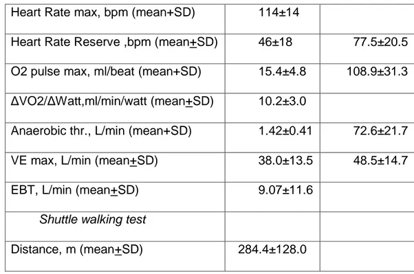
Cardiopulmonary exercise test
VO2peak, L/min (mean+SD) 1.33±0.5 76.3±20.4
VO2peak/Kg, ml/min/Kg (mean+SD) 17.5±5.5 75.4±21.8
Heart Rate max, bpm (mean+SD) 114±14
Heart Rate Reserve ,bpm (mean+SD) 46±18 77.5±20.5
O2 pulse max, ml/beat (mean+SD) 15.4±4.8 108.9±31.3
ΔVO2/ΔWatt,ml/min/watt (mean+SD) 10.2±3.0
Anaerobic thr., L/min (mean+SD) 1.42±0.41 72.6±21.7
VE max, L/min (mean+SD) 38.0±13.5 48.5±14.7
EBT, L/min (mean+SD) 9.07±11.6
Shuttle walking test
Distance, m (mean+SD) 284.4±128.0
Patients travels 284 meters during the shuttle walking test. Borg dyspnoea score was low al rest (0.4+0.8) but became significant at the end of the test (4.7+2.3). The same trend was observed for Borg muscle fatigue score (from 0.4±1.2 to 2.1±2.6).
Oxygen saturation was normal at the beginning of the CPET, at the end was less than 3 point (from 95.2± 1.5 to 92.3±3.4)
Therefore, the results of the two exercise tests confirmed that the major locus for the limitation of the exercise was the ventilatory limitation, with mild support from cardiac and peripheral muscular limitations.
In Table 4 are reported the parameters of the body composition. The majority of these COPD patients were overweight, and also FFM in % pred. was mildly reduced. However, FFMI was in the normal range, suggesting that the reduction in FFM was poorly relevant in these patients.
Table 4. Mean values (and SD) of the nutritional data measured in the whole group of COPD patients
BMI, Kg/m2 (mean+SD) 27.0±4.5
FFM, Kg (mean+SD) 51.0±10.2
FFM, % pred (mean+SD) 67.6±7.2
FFM Index, Kg/height2 (mean+SD) 17.9±2.7
Quality of life measured with Saint George‟s Respiratory Questionnaire (Table 5) showed a major limitation in the activity section, due to the fact the patients had minor capacity to perform the normal activity of daily life (walking, leaving the house) and spent most of time in lying, sitting or standing, probably in order to tolerate the dyspnoea. Dyspnoea according to the Medical Research Council (MRC) scale was 1.7±1.2.
Table 5. Mean values (and SD) of the total score and of the scores obtained in the different sections of the St. George Respiratory Questionnaire, in the whole group of COPD patients
Symptoms, % (mean+SD) 39.7±19.6
Activity, % (mean+SD) 53.6±24.3
Impact, % (mean+SD) 29.8±16.9
Total, % (mean+SD) 38.5±16.8
As regards comorbidities, the Chalson Index was 2.04+1.1, while the CIRS index was 2.77+1.1 and the CIRS severity index was 1.56+0.2, suggesting that few comorbidities were present.
The parameters of daily physical activity measured with Armband are reported in Table 6. The patients reported a low physical activity measured with steps/day according to the classification of physical activity proposed by Tudor-Locke. (55.). According to this classification
of physical activity using different cut-points for healthy adults, our COPD patients were included as a mean in the low active group (5,000 to 7,499 steps/day), with some patients were included in the category of inactive subjects (< 5,000 steps/day). Considering that in 2009 the original sedentary level was further spliced into two additional levels: <2,500 steps/day (basal activity) and 2,500 to 4,999 steps/day (limited activity) (56), these patients were almost all included in the category with limited activity.
Table 6. Mean value (and SD) of the indices derived from the monitoring of the physical activity by the Armband, in the whole group of COPD patients.
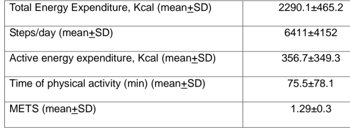
Total Energy Expenditure, Kcal (mean+SD) 2290.1±465.2
Steps/day (mean+SD) 6411±4152
Active energy expenditure, Kcal (mean+SD) 356.7±349.3
Time of physical activity (min) (mean+SD) 75.5±78.1
METS (mean+SD) 1.29±0.3
The results of the biomarkers measured in the peripheral blood, induced sputum and exhaled NO are reported in Table 7 Blood cells counts and hemoglobin were in normal range, while inflammatory biomarkers (ESR and CRP) were higher than normal, suggesting a systemic involvement related to COPD. N-pro-BNP was higher in 50 % of patients. Mean serum TNF-alfa and IL-8 were in the normal range. While microparticles resulted high value (normal value <2.5 nM) in 93% of patients.
Table 7. Mean values (and SD) or median values (and range) of the biomarkers measured in peripheral blood, sputum and exhaled air, in the whole group of COPD patients.
Peripheral blood
Blood Red Cells, cells/ μl (mean+SD) 4976.4±522.2
Haemoglobin, gr/dl (mean+SD) 14.6±1.3
Neutrophils, cells/ μl (mean+SD) 4,619±1,765
Neutrophils, % (mean+SD) 63.1±8.9
Eosinophils, cells/ μl (mean+SD) 276.2±274.8
Eosinophils, % (mean+SD) 3.80±3.1 ESR, 1st hour 28.5 (8-83) CRP, (mg/L) 5.6 (1.4-36.4) Pro-BNP, (pg/ml) 130.0 (0-1335) TNF-alfa, (pg/ml) 58.6 (20-242,7) IL-8, (ng/ml) 10.1 (0.2-24.8) Microparticles, (nM) 7.9 (1.6-34.3) Induced sputum
Neutrophils, % (median, range) 73.8 (37.0-97.1)
Eosinophils % (median, range) 3.1 (0.0-59.6)
TNF-alfa, (pg/ml) (median, range) 122.7 (9.1-904.5)
IL-8, (ng/ml) (median, range) 13.4 (0.2-50.7)
Neutrophil Elastase, μg/ml (median, range) 2.84 (0.1-8.0)
Exhaled breath
FeNo, ppb (median, range) 18.9 (4.2-90.4)