1
INDEX
Abstract p.3 Summary p.6 Introduction p.9Materials and Methods p.16
Statistical analysis p.21 Results p.21 Discussion p.41 Conclusion p.51 References p.52 Scientific Pubblications p.70
2 ABSTRACT
I tumori maligni più abbondanti nella popolazione maschile di età compresa tra i 17 ed i 45 anni, sono i tumori delle cellule germinali (GCTs). Essi comprendono un gruppo eterogeneo di neoplasie in termini istologici, di marker d’espressione ed età di manifestazione. I tumori delle cellule germinali testicolari negli adolescenti e negli adulti (TGCTs) possono essere classificati in tumori seminomatosi (GCT di tipo II) e non seminomatosi. Nel nostro studio prenderemo in considerazione il GCT di tipo II, utilizzando come modello sperimentale la linea cellulare TCam2, ad oggi unica al mondo ampiamente caratterizzata e comprendente tutte le caratteristiche del seminoma umano, originata dalla lesione primaria di un seminoma testicolare sinistro di un paziente di 35 anni. La difficoltà di avere un modello cellulare valido per i tumori seminomatosi è il motivo principale che rende il tumore testicolare uno dei tumori meno studiati. La ricerca sul cancro testicolare continua ad investigare e studiare terapie volte ad indurre la morte nelle cellule tumorali. Recentemente, il metabolismo energetico è considerato un obiettivo innovativo nelle terapie antitumorali, in quanto le alterazioni metaboliche sono una caratteristica comune dei tessuti cancerosi.
Il fenotipo metabolico maggiormente caratterizzante e per prima osservato nelle cellule cancerose è quello conosciuto come Effetto Warburg, che prevede la produzione di ATP attraverso la glicolisi invece che attraverso la fosforilazione ossidativa, anche in presenza di normali concentrazioni di ossigeno (Barger JF et al. 2010).
Tuttavia, la riprogrammazione metabolica nei tumori si estende oltre l'Effetto Warburg. In effetti, la teoria classica sul metabolismo delle
3
cellule tumorali (aumento dell'attività glicolitica e down-regolazione della fosforilazione ossidativa) è ancora oggetto di indagini in quanto numerosi studi hanno dimostrato che le cellule tumorali possono vivere in un ampio spettro di stati bioenergetici che variano dalla predominanza del fenotipo glicolitico, glicolitico parzialmente ossidativo, fino a quello prevalentemente fosforilativo (Smolková K et al. 2011).
Gli estrogeni ed i loro recettori, sono in grado di modulare diversi aspetti del metabolismo cellulare come quello glucidico o lipidico, un’alterazione dei loro pathways trasduzionali è stata correlata infatti allo sviluppo di malattie metaboliche (Faulds Malin Hedengran, 2012). Nel nostro precedente studio abbiamo evidenziato un link tra ERβ/PTEN che attivato dall’estradiolo, induce la morte di tali cellule mediante autofagia e necroptosi (Guido C. et al. 2012). Poiché, morte cellulare e metabolismo energetico sono strettamente correlati, abbiamo ipotizzato che il link E2/ERβ/PTEN possa indurre una alterazione anche nella riprogrammazione metabolica nelle cellule di SE. Il ruolo di PTEN nella sopravvivenza e proliferazione cellulare è stato già riportato, inoltre PTEN è in grado di influenzare alcuni pathways metabolici come il metabolismo del glucosio (Madeline B, 2002), ed il metabolismo lipidico (Qiu W. 2008; Juan Liu, 2012; Ana Ortega-Molina and Manuel Serrano, 2013). Lo scopo di questo studio è quello di investigare un potenziale
cross-talk funzionale tra E2, ERβ e PTEN nell’interferire sulla
riprogrammazione metabolica delle cellule TCam2 di seminoma umano, così da ampliare le nostre conoscenze sul ruolo e sulla regolazione del gene PTEN oltre che sulla biologia di questo tipo di tumore.
I nostri dati evidenziano un nuovo ruolo dell’ERβ come tumor suppressor, indicando che il meccanismo attraverso cui l’E2 induce la
4
morte delle cellule TCam2 avviene anche attraverso l’alterazione della riprogrammazione metabolica in cooperazione con il gene PTEN.
Ad oggi, il metabolismo di questa linea cellulare non è stato ancora investigato e pertanto il nostro lavoro contribuirà a migliorare le conoscenze su questo aspetto della biologia del seminoma umano. Concludendo, i nostri risultati supportano l’idea di una dipendenza estrogenica del tumore testicolare come già riportato in letteratura, indicando l’ERβ come possibile target terapeutico per il trattamento di questa condizione patologica.
5
SUMMARY
Testicular germ cell tumors of adults and adolescents (TGCTs) are the most common tumor in male. TGCTs can be classified into two main histological subtypes, seminoma (SE) and non-seminoma (NS). Here the focus is on SE, by using the TCam2 cell lines, containing typical features of human seminoma and originated from a primary testicular seminoma of a 35-year-old patient (Minzuno et al., 1993). Testicular cancer research continues to investigate and study therapies aimed to induce cell death. Recently, energetic metabolism is considered an innovative target in anticancer therapies since metabolic changes are a common feature of cancerous tissues. The most characterizing metabolic phenotype observed in cancer cells is known as Warburg Effect, according to which ATP production occurs through glycolysis rather than oxidative phosphorylation, even under normal oxygen concentrations. However, the classical theory on the metabolism of cancer cells (increased glycolytic activity and down-regulation of oxidative phosphorylation) is still under investigation since numerous studies have also shown that cancer cells can live in a wide spectrum of states ranging from the predominance of the glycolytic phenotype up to the phosphorylative one.
Estrogen and estrogen receptors (ERs) are well-known regulators of several aspects of metabolism, including glucose and lipid metabolism, and impaired estrogen signaling is associated with the development of metabolic diseases (Faulds Malin Hedengran, 2012). In others and our previous study, we evidenced that the TCam2 cells express the ERβ isoform and not the classical ERα (Mizuno et al., 1993).Upon increasing E2, by western blot and RT-PCR we observed an increase of PTEN both at protein and mRNA levels. From our preliminary data a molecular and
6
functional relationship between ERβ and PTEN in inducing cell death was evidenced on TCam2 seminoma cell line. Since cell survival is closely coupled to the cellular metabolism we hypothesized that the ERβ/PTEN link might be also involved in this issue. Although a regulatory role for PTEN has been always investigated in cell survival and proliferation, it was reported that it influences glucose metabolism by negatively regulating insulin signaling (Madeline B., 2002), and it regulates hepatic lipogenesis (Qiu W, 2008; Juan Liu, 2012; Ana Ortega-Molina and Manuel Serrano, 2013). Furthermore, PTEN appears to be essential for the differentiation of testicular germ cell tumor.
The aim of this study was to investigate a potential functional crosstalk between E2, ERβ and PTEN in altering the metabolic reprogramming of human seminoma cells, to improve our understanding in the biology of testicular tumour and in the regulation and roles of the ERβ and PTEN genes.
To evidence if E2, through ERβ and PTEN, is able to interferes with metabolic reprogramming in TCam2, we have characterized the basal metabolic profile of our cells and evaluated the effect of E2 on different metabolic pathways, in the presence or not of the ERβ and/or PTEN genes, by using an ERβ antisense plasmid (AS/ERβ) and a specific PTEN-silencer.
Our study suggests a novel tumor suppressor role for the ERβ in human seminoma, indicating that the mechanisms through which E2 induces cell death in TCam2 also occurred by altering the metabolic reprogramming. Interestingly, a cooperation between ERβ and PTEN existed. Up to date, no studies have examined energy metabolism management in human seminoma cell line, of consequence our study will greatly improve the knowledge of this aspect in SE.
7
Concluding, these data support estrogen-dependency of human testicular seminoma and candidate the ERβ-ligands for a therapeutic use in the treatment of this pathological condition.
8
INTRODUCTION
The most abundant malignancies among male population between the ages of 17 and 45 years are germ cell tumours (GCTs) and its incidence has increased 3-4 fold in the last 50 years. They comprise a heterogeneous group of neoplasms in terms of their histology, marker expression, and age of manifestation. Testicular germ cell tumors of adolescents and adults (TGCTs) can be classified into seminomatous (SE) and nonseminomatous tumors (NSE). Within the testis, three types of GCTs can be diagnosed: type I (teratomas and yolk-sac tumors of neonates and infants); type II (seminomas and nonseminomas); type III (spermatocytic seminomas). Here the focus is on SE, by using the TCam2 cell lines that is to date, the only one widely characterized and containing all the feature of human seminoma. This type of tumor is one of the less studied because of the difficulty to have a valid model for seminomas, therefore the molecular mechanisms of the disease remain yet to be clarified. To date, TCam2 cells are the unique cells of human seminomas as is reported by Minzuno (Mizuno et al., 1993).
Testicular cancer research continues to modify current therapies aimed to induce cancer cell death. In our previous study we characterized TCam2 cells for the expression of the classic oestrogen receptor isoforms, ERα and ERβ, by which oestrogen hormones act in almost all type of cells. Western Blotting analysis has highlighted that ERβ is mainly expressed in our cellular model (Guido et al., 2012). These data are supported by literature according to which ERβ is the most expressed isoform in all the human male genital tract. Interesting, our data shown for the first time, that E2 is able to up regulate PTEN gene inducing TCam2 to demise through autophagy and necroptosis. Cancer cells survival is
9
strongly linked to metabolic reprogramming of the cells. Disruption of tumour metabolism represents an elegant approach to induce cancer cell death. In fact, energy metabolism has been considered an innovative target in anticancer therapies, since metabolic changes are a common feature of cancerous tissues. However, it may help in understanding how, step by step, the metabolic pathways are arranged in comparison with normal metabolism to characterize a cancer metabolic phenotype.
The most characterizing metabolic phenotype observed in cancer cells is known as the Warburg Effect (Fig.1), which involves the production of ATP through glycolysis instead of oxidative phosphorylation, even in presence of normal oxygen concentrations (Barger JF et al 2010). However, metabolic reprogramming in tumours extends beyond the Warburg Effect. In fact, the classical theory on the metabolism of tumour cells (increased glycolytic activity and down-regulation of oxidative phosphorylation) is still under investigation as numerous studies have shown that cancer cells can live in a wide spectrum of bioenergy states that they range from the predominance of the glycolytic to the phosphorylating phenotype (Smolková K et al., 2011).
10
Estrogen and estrogen receptors (ERs) are well-known regulators of several aspects of metabolism, including glucose and lipid metabolism, and impaired estrogen signaling is associated with the development of metabolic diseases (Faulds Malin Hedengran, 2012). SE resembles primordial germ cells or early gonocytes, the cells from which all TGCTs are thought to be derived. The most widely accepted model of TGCTs development proposes an initial tumorigenic event in utero and the development of a precursor lesion known as intratubular germ cell neoplasia undifferentiated (ITGCNU), also known as carcinoma in situ (Skakkebaek N. E., 1972). This is followed by a period of dormancy until after puberty when TGCTs emerge and this suggests that the TGCTs development is hormone dependent. In addition to the role of androgens, several studies have demonstrated the importance of estrogen in influencing the male reproductive function (Hess et al., 1997, Sharpe, 1998). It is well-known capacity of the testis to convert androgens into estrogens by the enzyme P450 aromatase. The testicular expression of this enzyme has been demonstrated in several species both in somatic (Sertoli cells and Leydig cells) and in germ cells. It was shown that estrogens are able to stimulate proliferation of rat neonatal gonocytes in
vitro, to induce spermatogenesis in the hypogonadal mouse (Li et al.,
1997, Ebling et al., 2000). Interestingly, 17ß-estradiol (E2) appearsto be a potent germ cell survival factor in the human testis since is able to prevent apoptosis of human adult postmeiotic germ cells cultivated in preserved seminiferous tubules (low concentrations of 17ß-estradiol,10-9 and 10-10 mol/L, effectively inhibited male germ cells apoptosis). Conversely, it was shown that E2 is able to inhibit human embryonal carcinoma cell proliferation in vitro through an oestrogen receptor (ER) β-dependent mechanism suggesting that ERβ acts on germ cells as a
11
tumor suppressor (Roger et al., 2005) according to the observations made in ERβ knockout mice by Delbes et al. on neonatal gonocytes (Delbes et al., 2004). In human testis, gonocytes (Gaskell et al., 2003) and most adult germ cells (Mäkinen et al., 2001) express mainly ERβ. However, the precise role of estrogens/ERs (Dupont et al., 2000) and the underlying mechanism(s) in the control and in the biology of testicular tumors remain to be determined.
The best understood biochemical function of PTEN is to counteract the activity of the class I phosphatidylinositol 3-kinases (PI3Ks) (Ana Ortega-Molina and Manuel Serrano, 2013). PTEN negatively regulates the PI3K (Leslie et al., 2002) through the dephosphorylation in position D3 of phosphatidylinositol 3,4,5-triphosphate (PIP3) and then generating inactive PIP2. PIP3 regulates PDK1, a kinase that in turn phosphorylates and activates AKT (Fig. 2).
12
Loss of PTEN function in embryonic stem cells and human cancer cell lines results therefore in PIP3 accumulation and the activation of its downstream signalling molecule, AKT/PKB. Subsequently, activation of the PI3K/AKT pathway by the loss of PTEN stimulates various biological functions, such as cell cycle progression, cell survival and cell migration The tumor suppressor gene PTEN, is frequently mutated in human cancers, including brain, breast, endometrial, prostate, and kidney tumors (Cantley and Neel 1999; Simpson and Parsons 2001) and it is abundantly expressed in germ cells whereas it was virtually absent from 56 % of seminomas as well as from 86% of embryonal carcinomas, leading to an uncontrolled stimulation of growth and survival signals. PTEN appears to be essential for germ cell differentiation and an important factor in testicular germ cell tumor formation and primordial germ cells (PGCs), which are the embryonic precursors of gametes (Kimura et al., 2003). Increased mitotic levels, higher percentages of apoptotic cells, and teratoma formation were observed in vivo for PTEN mutant male gonads. Despite extensive characterization of PTEN mutations in human cancers and relatively good understanding of the molecular roles of PTEN in the control of cellular processes, little is known about the roles that PTEN carry out in the cells. Recently it was reported that mono-ubiquitination regulates nuclear import of PTEN and that in nucleus, PTEN plays an important role in preserving chromosomal integrity. More relevant to this study, it was found that subcellular compartmentalization of PTEN may play a key role in its tumor-suppressive activity, although function of PTEN within the nucleus remains to be defined. Emerging evidence suggests that PTEN also has PI3K/Akt-independent functions (Pingdong, Li, 2014). Other latest studies, highlighted the existence of different translational isoform
13
of this protein, PTENα and PTENβ. The first, seems to be involved in the induction of cytochrome c oxidase activity and ATP production in mitochondria (Liang H. et al., 2014). PTENβ, on the contrary seems to localize in the nucleolus of the cells where seems to inhibits ribosome biogenesis by regulating nucleolin phosphorylation. These results demonstrate the complexity of the PTEN protein family and the diversity of its functions (Liang H. et al., 2017). Till now, the regulation of PTEN expression, localization and function is still unclear especially in testicular tumor cells.
From our preliminary data an outcome of the ERβ/PTEN relationship was evidenced on seminoma cell survival and generally this effect is closely coupled to the cellular metabolism.
Herein, we will test increasing E2 concentrations to evaluate eventual effects on the TCam2 cells metabolic reprogramming as well as the biological significance of this potential link. Three different concentration of E2 will be tested as well as the combination of E2 with ICI 182,780 (ICI) or Fulvestrant, to evaluate the more efficacious dose able to interferes with metabolic reprogramming and if the effect is ER-mediated in human seminoma cell line. After, we analyzed PTEN role by using a PTEN-siRNA. An ERβ antisense plasmid, AS/ERβ, was also used.
Results of this study will begin to determine the regulatory mechanisms involved in SE, expanding our knowledge concerning the actions mediated by ERβ and PTEN. Furthermore, we evidenced their involvement in the alteration of SE metabolic reprogramming.
14
Materials and methods
Mizuno and coworkers reported isolation and characterization of a cell line named TCam2 (Mizuno et al., 1993). This cell line originated from a primary lesion of a left testicular seminoma (typical pure type seminoma) of a 35 aged male patient and was generated initially by in vitro culture, and also propagated as xenografts in SCID mice. Using a multidisciplinary approach, it was concluded that TCam2 is representative for seminoma (de Jong J. et al., 2008).
Cell cultures - Human TCam2 seminoma cell line (a gift from Dr. Leendert H. J. Looijenga Department of Pathology, Erasmus MC-University Medical Center Rotterdam, Josephine Nefkens Institute, TCam2 cells were obtained from Sohei Kitazawa (Division of Molecular Pathology, Kobe University, Japan) were grown in RPMI plus 10% fetal bovine serum (FBS), 1% penicillin/streptomycin, 200 mM glutamine, at 37 °C in a humidified cell culture incubator with 5% carbon dioxide. Chemicals - Estradiol (oestra-1,3,5,(10)-triene-3,17-diol) (E2), and all other chemicals were purchased from Sigma Chemical (Milan, Italy). Acrylamide bisacrylamide was from Labtek Eurobio (Milan, Italy). ICI 182,780 (ICI) were purchased from Tocris chemical (Bristol, UK). Lipofectamine 2000 reagent (Invitrogen, Paisley, UK). ECL system (Amersham Pharmacia, Buckinghamshire, UK). Bradford protein assay was performed using a kit from Bio-Rad Laboratories,Inc. (Milan, Italy). Antibodies used in this study were from Santa Cruz Biotechnology (Santa Cruz, CA, USA). MitoProfile® Total OXPHOS WB Antibody Cocktail was from Abcam (Milan, Italy). Triglycerides, lipase activity, glucose-6-phosphate dehydrogenase (G6PDH) activity and glucose assay kits were from Inter-Medical (Biogemina Italia Srl, Catania, Italy). Molecular Probes' ATP Determination kit (A22066) was from Invitrogen
15
(Milan, Italy). E2 and ICI were dissolved in ethanol (0.02% final concentration) and used as solvent controls did not induce any positive result in all in vitro assays (data not shown).
Glucose assay. Glucose oxidase catalyzes the oxidation of glucose to
gluconic acid. The formed hydrogen peroxide is detected by a chromogenic oxygen acceptor, phenol, 4-aminophenazone in the presence of peroxidase. The intensity of the color formed is proportional to the glucose concentration in the sample (Guido C et al., 2010). Data are presented as nM/mg protein.
Triglyceride assay. Triglycerides were measured in duplicate by a
GPO-POD enzymatic colorimetric method according to the manufacturer's instructions in cell lysates and as previously described (Guido et al., 2011). Data are presented as nM/mg protein.
Lipase activity assay. Lipase activity was evaluated by the method of
Panteghini et al (Panteghini M. et al., 2001) based on the use of 1,2-o-dilauryl-rac-glycero-3-glutaric acid-(6'-methylresorufin) ester as substrate, as previously described (Aquila S. et al., 2006). Data are presented as nM/min/mg protein.
Assay of the G6PDH activity. The conversion of NADP+ to NADPH,
catalyzed by G6PDH, was measured by the increase in absorbance at 340 nm as previously described (Guido et al., 2011; Aquila S. et al., 2006). Data are presented as nM/min/mg protein.
ATP assay. A bioluminescence assay for quantitative determination of
ATP with recombinant firefly luciferase and its substrate D-luciferin (light emission at 560 nm at pH 7.8), was performed as previously described (Pingitore A. et al., 2009). Data are presented as nM/mg protein.
16
Acyl-CoA dehydrogenase activity assay (Fatty Acid Oxidation, FAO) Acyl-CoA dehydrogenases catalyze the initial step in each cycle
of fatty acid-oxidation in the mitochondria of cells. Assay was performed using a modification of a previously described (Lehman TC et al., 1990). In brief, after protein lysis, 50 g of proteins were added to the buffer containing 20 mm Mops, 0.5 mm EDTA and 100 lm FAD at pH 7.2. Reduction of FAD to FADH2 was read at 340 nm upon addition of octanoyl-CoA (100 µg M) every 20 s for 1.5 min. Data are expressed as nmol ⁄ min ⁄g protein. The enzymatic activity was determined with three control media: one without Acyl-CoA as substrate, another without the coenzyme (FAD) and the third without either substrate or coenzyme (data not shown).
Plasmid The ERβ antisense plasmid (AS/ERβ) contains, in reverse orientation, a 1170 bp fragment of the coding sequence of the human ERβ cloned in pIRESpuro2 vector (Clontech).
Transfections and luciferase assays. Transfections were
done as described using Fugene 6 reagent (Promega, E2691). Luciferase activity was measured with the Dual Luciferase kit (Promega, E1500). Lipid-mediated transfection of siRNA duplexes. RNA oligonucleotide directed against PTEN was purchased from Cell Signaling (62515).
Immunoblotting TCam2 cells were grown in 10 cm dishes to 70-80 % confluence and exposed to treatments as indicated in 5% dextran coated charcoal (DCC). Cells were then harvested in cold phosphate-buffered saline (PBS) and resuspendedin lysis buffer containing 20 mM HEPES pH 8, 0.1mM EDTA, 5mM MgCl2, 0.5M NaCl, 20 % glycerol, 1 % NP-40, inhibitors (0.1mM Na3VO4, 1 % PMSF, 20 mg/ml aprotinin). Protein concentration was determined by Bio-Rad Protein Assay (Bio-Rad Laboratories, Hercules, CA USA). A 70μg portion of protein lysates was
17
used for Western Blotting (WB), resolved on a 12 % SDS-polyacrylamide gel, transferred to a nitrocellulose membrane and probed with the indicated Abs. As internal control, all membranes were subsequently stripped (glycine 0.2 M, pH 2.6 for 30 minutes at room temperature) of the first antibody and reprobed with anti-β-actin Ab. The antigen-antibody complex was detected by incubation of the membranes for 1 hour at room temperature with the appropriated secondary antibodies peroxidase-coupled and revealed using the enhanced chemiluminescence system. Blots were then exposed to film. The intensity of bands representing relevant proteins was measured by Scion Image laser densitometry scanning program.
Statistical Analysis
The data obtained (six replicate experiments using duplicate determinations) are presented as the mean±SEM. The differences in mean values were calculated using ANOVA with Newman–Keuls post hoc test. Values of P<0.05 were taken to show a significant difference between means.
18
RESULTS
ERs expression in human seminoma cells
First of all, we characterized TCam2 cell for the presence of ERs. As it concerns the classical ERα, the protein was not detectable in our cells compared with MCF-7, a human breast cancer cell line used as positive control, neither by using an anti-ERα Ab raised against the carboxy-terminal part of ERα (Fig. 3A), nor with an Ab recognizing the amino-terminal part of ERα Fig 3B. On the contrary, an intense band was detected by Western blotting at 60 kDa (Fig. 3C) corresponding to the molecular mass of the ERβ long form.
A B C
FIG.3: Immunoblots of estrogen receptors (ERα, ERβ) from protein extracts of
TCam2. Western blot analysis of proteins showed expression of the estrogen
receptors (ERs). Extracts of TCam2 protein were subjected to electrophoresis on 12% SDS-polyacrylamide gels, blotted onto nitrocellulose membranes and probed with mouse Ab to human ERα (A), rabbit polyclonal Abs to human ERα (B) and ERβ (C). MCF-7 extracts were used as controls. The number on the left corresponds to molecular masses (kilodaltons, kDa) of the marker proteins. The experiments were repeated at least six times, and the autoradiographs of the figure show the results of one representative experiment.
PTEN protein and mRNA increased in response to E2
The precise role of estrogens/ERs and the underlying mechanism(s) in the control and in the biology of testicular tumors remain to be
19
determined (Dupont et al. 2000). Despite a relatively good understanding of the molecular roles of PTEN in the control of cellular processes, little is known about modes of PTEN gene regulation and all the roles that PTEN plays in the cells. In order to evaluate a potential functional cross-talk between E2/ERs and PTEN we studied the effect of increasing concentrations of E2 on PTEN expression. Interestingly, E2 upregulated the PTEN protein expression in a dose-dependent manner from 1 nM to 10 nM, while 100 nM did not give significantly effects (Fig. 4A).
Next, we investigated the mRNA expression of PTEN, and it was induced by E2 in a similar pattern as it was obtained for the protein (Fig. 4B). ICI failed to cause an increase in PTEN protein and mRNA levels, suggesting a direct involvement of the ER in mediating this effect.
A
B
FIG.4: E2 up-regulates PTEN protein and mRNA expression in TCAm2 cells. (A)
20
E2 (1,10,100 nM) concentrations, 1μM ICI alone or in combination with 10 nM E2. β-actin was used as loading control. The side panel shows the quantitative representation of data (mean ± S.E.M.) of six independent experiments including that of A. (B) Semiquantitative RT-PCR evaluation of PTEN mRNA expression. TCam2 cells were treated as in A. 36B4 mRNA levels were determined as control. The side panel shows the quantitative representation of data (mean ± SEM) of six independent experiments including that of B after densitometry and correction for 36B4 expression. *P<0.05 E2-treated vs untreated cells; **P<0.01 E2-treated vs. untreated cells.
E2/ERβ regulated glycolysis inTCam2 cells
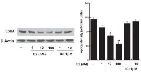
To evaluate the E2 effect on glycolysis in our cells, we first determined the cellular glucose content through spectrophotometric techniques and analyzed the expression levels of various key enzymes involved: 6-phosphofructo1-kinase (PFK1); aldolase; Pyruvate Kinases M1 and LDH expression. Initially, we used increasing concentration of E2 to treat TCam2 cells for 24 h in order to individuate the more efficacious dose able to interfere with metabolic reprogramming in human seminoma cell line. Therefore, we used a specific inhibitor of estrogenic receptor such as Fulvestrant (ICI 182, 780), to test if the effect observed was ER-mediated. From our results E2 treatment induced an increase in the Glucose amount in TCam2 cells at 10 nM E2 (Fig. 5). The effect seemed to be ER-mediated. The expression of PFK-1, aldolase and PKM1 was reduced, particularly at 1 and 10 nM of E2 for PFK1 while the expression of aldolase at 10 and 100 nM and PKM1 was reduced at all E2 concentrations used (Fig. 6). It seems that E2 induced a slowdown of the glycolytic pathway through the ERβ. By testing the LDH we observed a significant reduction in its expression especially at 10 and 100 nM of E2 (Fig. 7-8). The presence of the ER-inhibitor reversed the effect of 10 nM E2.
21
FIG.5: Glucose assay. TCam2 cells treated without (-) or with increasing E2 1nM,
10nM, 100nM, ICI 1µM, ICI 1 µM + E2 10nM. The columns represent the average obteined from at least three indipendent experiments. *P< 0.05 E2-treated vs. untreated cells.
FIG.6: Western blot assay of PFK1, aldolase and PKM1. TCam2 cells treated
without (-) or with increasing E2 1nM, 10nM, 100nM, ICI 1µM, ICI 1 µM+E2 10nM. β-actin was used as loading control. The number on the top of the blot are the mean of bands intensity evaluated in terms of terms of optical density arbitrary units and and expressed as the fold over the control, which was assumed to be 1. For PFK1 was considered the upper band. The sample untreated was considered to be 1. *P<0.05 E2-treated vs. untreated cells. **P<0.01 vs untreated cells.
22
FIG. 7: Western Blot analysis of LDH expression (on the left) and densitometry.
TCam2 cells treated without (-) or with increasing E2 1nM, 10nM, 100nM, ICI 1µM, ICI 1 µM + E2 10nM. β-actin was used as loading control. The columns show the quantitative representation of data (mean ± S.E.M.) of three independent experiments in which the intensity of the band were evaluated in terms of optical density arbitrary units, where each bands have been related to the control. The sample untreated was considered to be 100. *P<0.05 E2-treated vs. untreated cells: **P<0.01 vs untreated cells.
E2/ERβ affected the pentose phosphate pathway (PPP) in TCam2 cells
Glucose can be also metabolized via PPP of which the G6PDH is the first key enzyme. E2 treatment induced a significant reduction in the enzymatic activity (Fig. 8), particularly at 1 and 10 nM of E2, indicating how the decrease in G6PDH in treated cells contributes to the decrease in glucose utilization. Our results indicate that this effect is ERβ mediated and supported the glucose accumulation inside the cells.
23
FIG.8: G6PDH assay. TCam2 cells treated without (-) or with increasing
concentration of E2 at 1nM, 10nM, 100nM, ICI 1µM, ICI 1 µM + E2 10nM. The columns represent the average obtained from at least six independent experiments. *P< 0.05 E2-treated vs. untreated cells.
E2/ERβ effects on the replenishment of the TCA cycle
The refueling of TCA cycle can occur at least through two key points: the PDH and the α-KGD complexes.
In normal cell metabolism, pyruvate maintains the tricarboxylic acid (TCA) cycle flux. The pyruvate, in cancer cells, can be reduced to lactate and shuttled out of the cell or transported in mitochondria and oxidized by the complex the pyruvate dehydrogenase (PDH), which is regulated and blocked by the pyruvate dehydrogenase kinase (PDK), limiting the pyruvate conversion into Acetyl-CoA. (Liem Minh Phan et al., 2014) (Kyle S. McCommis and Brian N. Finck, 2015). As shown in Fig. 9, the PDH (E1 isoform) expression did not undergo to variations after E2 treatment while the PDK expression is slightly but not significantly increased at 10 and 100 nM of E2.
24
FIG. 9: Western Blot analysis of PDH (E1) and PDK expresseion (on the left) and densitometry (on the right). TCam2 cells without (-) or with increasing
concentration of E2 at 1nM, 10nM, 100nM, ICI 1µM, ICI 1 µM+E2 10nM. β-actin was used as loading control. The columns represent the average obteined from three indipendent experiments in which the intensity of each bands were evaluated in terms of optical density arbitrary units where each bands have been related to the control. The sample untreated was considered to be 100.
Altogether, these data suggested us to investigate whether the TCA cycle is refueled at the level of α-KGD complex.
Cancer cells to supply TCA cycle intermediates and support anabolic processes, rely mostly on glutamine, which enters into the cycle as α-ketoglutarate via the α-α-ketoglutarate dehydrogenase (α-KGD) complex. (Liem Minh Phan et al., 2014; Deberardinis RJ, et al., 2008).
As observed in Fig. 10, concentrations of 1 and 10 nM E2 induced the expression of α-KGD in a ERβ-dependent manner. Glutamine, is the most abundant in the plasma and an additional energy source in tumor cells especially when glycolytic energy production is low (Deberardinis RJ et al.; 2008) This aminoacid is converted to α-ketoglutarate in two reaction steps and the second is catalyzed by the glutamate dehydrogenase (GLUD1) which was increased, via ERβ, particularly upon 10 and 100 nM E2 (Fig. 11). Thus, the consequence of E2 action
25
might be that glutaminolysis refuels a ‘truncated’ TCA cycle in human seminoma cancer cells.
FIG. 10: Western Blot analysis of α-KGD and GLUD1. TCam2 cells treated
without (-) or with increasing concentration of E2 at 1 nM, 10 nM, 100 nM, ICI 1µM, ICI 1 µM + E2 10 nM. The autoradiographs show the results of one representative experiment. β-actin was used as loading control. The number on the top the blots represent the average obtained from three independent experiments in which the intensity of each bands was evaluated in terms of arbitrary units of optical density, and expressed as the fold over the control, which was assumed to be 1. *P<0.05 E2-treated vs. untreated cells: **P<0.02 vs untreated cells.
E2 modulated bioenergetic requirements in TCam2 cells
Afterwards, we focused our investigation on an energy sensor, the AMP-activated protein kinase (AMPK) expression and the cellular ATP content as well as the expression of some components of the respiratory chain. AMPK is activated and induced by metabolic stresses which interferes with ATP production (Smolková K, et al., 2011). Under E2 treatment, we observed a significant increase in AMPK expression at all the concentrations used (Fig. 11). Concomitantly, although it seemed contradictory, we also noted a slight but not significant increase in ATP production upon 1 and 10 nM of E2 (Fig. 12).
26
FIG. 11. Western Blot analysis of AMPK expression (on the left)and
densitometry (on the right). TCam2 cells treated without (-) or with increasing E2 1 nM, 10 nM, 100 nM, ICI 1µM, ICI 1 µM + E210 nM. A) The autoradiographs show the results of one representative experiment. β-actin was used as loading control. B) The columns shows the quantitative representation of data (mean ± S.E.M.) of three independent experiments in which the intensity of the band were evaluated in terms of optical density arbitrary units, and expressed as the fold over the control, which was assumed to be 100.*P<0.05 E2-treated vs. untreated cells: **P<0.02 vs untreated cells.
FIG. 12: ATP assay. TCam2 cells without (-) or with increasing concentration of E2
at increasing E2 1 nM, 10 nM, 100 nM, ICI 1µM, ICI 1 µM + E2 10 nM. The columns represent the average obtained from three indipendent experiments.
27
E2 promoted a lipid-lowering effect in TCam2 cells.
Increase in lipid metabolism is another remarkable feature of cancer cells. Similar to glucose and glutamine, fatty acid metabolism supports both the biosynthetic and the bioenergetic requirements for cell proliferation and survival. E2-treated TCam2 cells through ERβ reduced the triglycerides content compared to the untreated cells (Fig. 13). Alongside, the lipase activity and fatty acid oxidation (FAO), both the lipid catabolic processes, increased particularly at 10 nM of E2 (Fig. 14, 15).
FIG. 13. Triglyceride assay. TCam2 cells treated without (-) or with incrasing E2
1nM, 10nM, 100nM, ICI 1µM, ICI 1 µM + E2 10nM. The columns represent the average obteined from three indipendent experiments. **P< 0.05 E2-treated vs. untreated cells.
28
FIG.14: Lipase assay. TCam2 cells treated without (-) or with increasing E2 1nM,
10nM, 100nM, ICI 1µM, ICI 1 µM+E2 10nM. The columns represent the average obteined from three indipendent experiments. *P< 0.05 E2-treated vs. untreated cells. **P< 0.02 E2-treated vs. untreated cells.
FIG. 15: Acyl-coA dehydrogenase assay. TCam2 cells treated without (-) or with
increasing E2 1 nM, 10 nM, 100 nM, ICI 1 µM, ICI 1 µM + E2 10 nM. The columns represent the average obteined from at least three indipendent experiments. *P< 0.05 E2-treated vs. untreated cells. **P< 0.01 E2-treated vs. untreated cells.
E2 effects on de novo fatty acid synthesis
An increased use of a ‘truncated’ TCA cycle, includes diversion of citrate to cytosolic export for lipid synthesis. Cancer cells frequently
29
upregulate de novo fatty acid synthesis to satisfy their demands for lipids and increased fatty acid synthesis has been linked to poor prognosis in breast cancer (Zhang F. and Du G., 2012). It is a multiple step process involving several enzymes such as ATP citrate lyase (ACLY), Acetyl-CoA carboxylase (ACCα), fatty acid synthase (FASN). Interestingly, we observed that all the enzymatic expressions were reduced by E2 (Fig. 16).
FIG. 16: Western Blot analysis of ACLY, ACCα and FASN expression. TCam2
cells treated without (-) or with increasing E2 at 1 nM, 10 nM, 100 nM, ICI 1 µM, ICI 1 µM + E2 10 nM. The autoradiographs show the results of one representative experiment. β-actin was used as loading control. The number on the top the blots represent the average obtained from three independent experiments in which the intensity of each bands was evaluated in terms of arbitrary units of optical density, and expressed as the fold over the control, which was assumed to be 1. For ACLY: *P<0.01 E2-treated vs. untreated cells: **P<0.002 vs untreated cells; for ACCα: *P<0.05 E2-treated vs. untreated cells: **P<0.001 vs untreated cells; for FASN: *P<0.05 E2-treated vs. untreated cells: **P<0.002 vs untreated cells.
E2/ERβ altered the metabolic reprogramming through a mechanism involving PTEN gene in human seminoma cells
Our previous study indicate that E2/ERβ up-regulate PTEN gene phosphatase, which induced a negative modulation of phosphatidylinositol 3-kinase/Akt-dependent cell proliferation, then the
30
cells died (Guido et al., 2012). Further, PTEN has been shown to influence metabolic pathways such as glycolysis, lipolysis and lipogenesis (Garcia-Cao et al., 2012). We hypothesized that ERβ and PTEN may converge in a tumor suppressor activity disorganizing the metabolic reprogramming in human seminoma cells. In this aim we performed ERβ and PTEN silencing studies to evaluate their eventual effects on key metabolic phases. To test whether the two genes were really silenced and whether the absence of one gene interferes with the expression of the other, we blotted the samples without or with 10 nM E2 treatment, first with the anti-PTEN Ab and after having stripped the membrane we incubated it with the anti-ER Ab (Fig.17). The immunoreaction for PTEN and ER was negligible in both silencing conditions. Interestingly, the absence of PTEN strongly reduced the expression of ER and the ERsilencing significantly abated PTEN expression, confirming their molecular link in TCam2 cells.
FIG. 17: Western Blot analysis of PTEN and ERβ expression. TCam2 cells without
(-) or with E2 10 nM, siPTEN control, siPTEN + 10 nM E2, control AS/Rβ, AS/ERβ + 10 nM E2. β-actin was used as loading control. The number on the top of the blots represent the average obtained from three independent experiments in which the intensity of each bands was evaluated in terms of arbitrary units of optical density, and expressed as the fold over the control, which was assumed to be 1. *P<0.05 E2-treated vs. unE2-treated cells: **P<0.02 vs unE2-treated cells.
31
When we examined the knocking of PTEN and ER on some key proteins of the metabolic pathways evaluated, we observed that PTEN specific siRNA and the ER absence both reversed the E2-induced effects observed on PFK1, α-KGDH (E1k or α-KGD), (Fig. 18). Interestingly, their absence increased basal levels of PFK1 and the E2-induced effect was reversed, indicating the protective effect of ER and PTEN on this context. As it concerns the α-KGD, in the same experimental conditions the E2-effect was abolished, indicating the involvement of both ER and PTEN.
FIG. 18: Western Blot analysis of PFK1 and α-KGD expression. TCam2 cells
without (-) or with E2 10nM, siPTEN, siPTEN+E2, AS/ERβ, AS/ERβ+E2. -actin was used as loading control. The number on the top the blots represent the average obtained from three independent experiments in which the intensity of each bands was evaluated in terms of arbitrary units of optical density, and expressed as the fold over the control, which was assumed to be 1. *P<0.05 E2-treated vs. untreated cells: **P<0.02 vs untreated cells.
Next, we analyzed the expression of some OXPHOS components. E2 treatment hesitated even in an alteration of some components of the respiratory chain (CI, CII, CIII, CIV, CV), particularly CIV and CV.
32
When we silenced ER and PTEN and evaluated the expression of some OXPHOS components (Fig. 19), the siPTEN strongly induced CII, CV and CI at basal levels, while AS/ERinduced CV and CIII, reducing CI and CIV.
A B
FIG. 19: Western blot assay of OXPHOS. TCam2 cells without (-) or with E2 at 10
nM, siPTEN, siPEN+E2, AS/ERβ, AS/ERβ+E2. (A)The autoradiographs show the results of one representative experiment repeated at least three times. β-actin was used as a control. For complex CIV: The number on the top the blots represent the average obtained from three independent experiments in which the intensity of each bands was evaluated in terms of arbitrary units of optical density, and expressed as the fold over the control, which was assumed to be 1. (B) Quantitative representation after densitometry for CV, CII, CIII and CI of the OXPHOS components. The columns are the mean of three independent experiments in which band intensities were evaluated in terms of optical density arbitrary units and expressed as a percentage with respect to the respective controls, which were assumed to be 100. *P<0.05, *P<0.02. CIV, CI, CIII, CV, CII indicate OXPHOS components.
Next we evaluated the expression of ACLY, ACCα e FASN in the same experimental conditions. ACLY, ACCα and FASN protein levels significantly diminished upon E2 treatment. Our findings highlighted that the absence of PTEN as well as of ERβ induced the expression of all the proteins considered, concomitantly reversing the effect of E2 treatment (Fig. 20).
33
FIG. 20: Western Blot analysis of ACLY, ACCα and FASN expression. TCam2
cells without (-) or with E2 at 10 nM, siPTEN, siPTEN+E2, AS/ERβ, AS/ERβ+E2. β-actin was used as loading control. The number on the top the blots represent the average obtained from three independent experiments in which the intensity of each bands was evaluated in terms of arbitrary units of optical density, and expressed as the fold over the control, which was assumed to be 1. *P<0.05 E2-treated vs. untreated cells.
Furthermore, we examined the effect of the knocking of PTEN as well as ERβ on: intracellular triglycerides amount, lipase activity and fatty acid β-oxidation. As it concerned triglycerides levels both PTEN and ERβ absence reversed at least in part the E2 effects and interesting the absence of both genes in the untreated sample increased triglycerides amount with respect to the control (Fig. 21).
FIG. 21: Triglyceride assay. TCam2 cells treated without (-) or with E2 10 nM,
34
the average obteined from at least three indipendent experiments. **P< 0.02 E2-treated vs. unE2-treated cells.
As we can observe in Fig. 22, 10 nM E2-induced Lipase activity as we previous highlighted. When we knocked for PTEN or ERβ the E2 effects were not observed.
FIG. 22: Lipase assay. TCam2 cells treated without (-) or with E2 10 nM, siPTEN,
siPTEN + 10 nM E2, AS/ERβ; AS/ERβ + 10 nM E2. The columns represent the average obteined from at least three indipendent experiments. *P< 0.01 E2-treated vs. untreated cells.
As it is shown in Fig. 23, Acyl-coA dehydrogenase is induced by 10 nM of E2. Using siPTEN or AS/ERβ a significant reduction of the enzymatic activity also at basal level was obtained.
35
FIG.23: Acyl-CoA dehydrogenase assay. TCam2 cells treated without (-) or with E2
10 nM, siPTEN, siPTEN+E2, AS/ERβ; AS/ERβ+E2. The columns represent the average obteined from at least three indipendent experiments. *P<0.05 E2-treated vs. untreated cells; **P< 0.01 E2-treated vs. untreated cells; ***P< 0.00.
36
DISCUSSION
Most of the all tumors malignancies (60%) diagnosed in men between 17 and 45 years of age are germ cell tumors (GCT). GCT arise from carcinoma in situ cells, that develop from a transformed fetal germ cell, the gonocytes. In the last years the incidence of testicular GCT (TGCT) has increased annually by 3 % – 6 % in the Caucasian population (Oosterhuis and Looijenga 2005). Cancers are highly heterogeneous diseases with individual metabolic features. Recently, energetic metabolism is considered an innovative target in anticancer therapies since metabolic changes are a common feature of cancerous tissues.
Cancer metabolic programs supply energy and metabolites to support biosynthesis, proliferation and other processes of tumorigenesis. The literature has indicated that the Warburg phenotype is not exclusive and that a decrease of mitochondrial function is not a general feature of cancer cells (Jose C, et al., 2011; Valcarcel-Jimenez L, et al., 2017). Although aerobic glycolysis is often found in malignant tumors, OXPHOS still contributes to energy production in cancers. The contribution ratio of glycolysis versus OXPHOS for the total ATP yield varies in different cells, growth states and microenvironments (Slavov N, et al., 2014). Therefore, carefulness and a multifaceted approach are important to define the metabolic phenotypes of a cancer type.
The effects of oestrogen (E2) are known to be mediated by estrogen receptor-α (ERα) and ERβ subtypes, but mainly the ERβ has been found in human germ cells of normal testis. Interestingly, low estradiol (10-9 and 10-10 mol/L) appears to be a potent germ cell survival factor in the human testis since is able to prevent apoptosis of human adult postmeiotic germ cells cultivated in preserved seminiferous. Conversely, it was also shown that E2 is able to inhibit human embryonal carcinoma
37
cell proliferation in vitro through an ERβ-dependent mechanism suggesting that ERβ acts on germ cells as a tumor suppressor according to the observations made in ERβ knockout mice by Delbes et al., on neonatal gonocytes. Estrogen and the estrogen receptors (ERs) are well-known regulators of several aspects of metabolism, including glucose and lipid metabolism, and impaired estrogen signaling is associated with the development of metabolic diseases (Faulds Malin Hedengran, 2012). Although PTEN initially was discovered as a tumor suppressor with a regulatory role in cell survival and proliferation, particularly in tumor-prone tissues such as the breast and endometrium, more recent studies have highlighted a role for PTEN in metabolism. In fact, it plays a significant role in regulating glucose metabolism in vivo by negatively regulating insulin signaling (Madeline Butler, 2002). Although an extensive characterization of the TCam2 cell line to prove that it is representative for human seminoma has been performed, very few findings have investigated the signaling pathways that govern the biology of seminoma and tested how seminomas respond to environmental cues.
In our previous study we evidenced that a molecular and functional crosstalk between ERβ and PTEN occurs inducing loss of seminoma cell survival by autophagy and necroptosis. Since cell death is strongly related to cell metabolism, we hypothesized that E2/ERβ/PTEN might also act to affect seminoma cell metabolic reprogramming, by using the TCam2 cell line which contain typical features of seminoma.
In our previous study we reported the presence of the ERβ in TCam2 cells. The analysis of Western blotting showed strong expression of the ERβ long isoform at 60 kDa, whereas two different antibodies used in the evaluation of ERα did not show any signal at 67 kDa that is the
38
molecular weight reported for the classical ERα, clearly expressed in the MCF7 breast cancer cells used as positive control. Another study of the JKT-1 cells, embryonic carcinoma cells and testicular non-seminoma as further evidenced by the literature (Roger et al., 2005), showed the total absence of ERα. However, JKT-1, considered by the authors cells of seminoma, through characterization, appeared devoid of genes commonly found in seminoma and therefore do not constitute a suitable cellular model for studying the tumorigenesis in seminoma. Moreover, in human testis, gonocytes (fetal germ cells which differentiate into spermatogonia after birth) (Gaskell et al. 2003) and most adult germ cells (Mäkinen et al. 2001) express mainly the ERβ. The presence of ERβ in TCam2 suggests an estrogen-dependence (or sensitivity to estrogen) and testicular cancer supports a possible involvement of endogenous estrogens and / or environmental carcinogenesis in the testis. It has been reported that estrogen affects all cell types of the male genital tract and that may influence the normal development of the testes.
PTEN, which is a lipid phosphatase, as well as being an important factor
involved in the mechanisms that induce apoptosis, seems to be involved in the differentiation of testicular germ cell tumor.
Our preliminary results also showed that E2 is able to increase the expression of PTEN, both at protein and mRNA levels. This action appears to be mediated by ER since the effect is reversible by ICI, specific antagonist of both ERs isoforms. Furthermore, our data showed that E2 and its receptor are able to modulate the expression of PTEN through a genomic action, involving the Sp1 transcription factor (Guido et al. 2012).
The PTEN gene encodes a lipid phosphatase that negatively regulates the PI3K/AKT pathway, classically involved in cell survival (Haas-Kogan et
39
al., 1998; Stambolic et al., 1998). The activation of E2/ERand PTEN link induced our cells to death through autophagy and necroptosis. To investigate E2 action on metabolic reprogramming in our cell line, we evaluated the expression and/or activity of the main enzymes responsible for the regulation of the major metabolic pathways in TCam2 cells, initially treated with increasing concentrations of E2 and/or ER inhibitor (ICI). Thereafter, we used siPTEN as well as AS/ERto investigate both PTEN and ER role on metabolic reprogramming in our cells.
First, we evaluated glucose content and its metabolism through the glycolytic pathway in TCam2 cells upon increasing E2 treatments. Glucose content augmented significantly at 10 nM E2, while the PFK1, the aldolase and PKM1 expression were eminently decreased. At the same time the LDHA expression was diminished. Collectively these data indicated a shutdown of the glycolysis.
LDHA is a key enzyme in anaerobic glycolysis and it catalyzes the conversion of pyruvate into lactate. In addition, LDHA is a long-lived protein that appears to be degraded mainly or exclusively by autophagy (Kopitz J, et al., 1990). Our data and these observations well fit with our recent findings demonstrating that E2 via ERβ/PTEN axis drives an impaired autophagy determining seminoma cancer cell death (Guido et al. 2012).
PPP is an important pathway of glucose catabolism and it is able to influence several cellular processes, including proliferation, invasion, drug resistance, and metastasis. In this context, our data showed that E2 via ERβ was able to lower the G6PDH activity thus affecting the PPP rate. The low glycolytic flux as well as the strong blockage of the PPP
40
may explain the higher intracellular glucose levels observed during E2 treatment.
Accordingly, our recent study demonstrated that E2/ERβ/PTEN, caused the inhibition PI3K/AKT signal (Guido et al. 2012) and AKT is a well-known stimulator of glycolysis.
Carbon flow derived from pyruvate get into the TCA cycle under the control of PDH complex. (Mullen AR, et al., 2014). Our data showed that PDH1 and PDK1 expression were not influenced by E2, as expected given the decreased production of pyruvate through the glycolytic way. In normal cell metabolism, pyruvate maintains the tricarboxylic acid cycle flux. The pyruvate, in cancer cells, can be reduced to lactate and shuttled out of the cell or transported in mitochondria and oxidized by the complex the pyruvate dehydrogenase (PDH), which is regulated and blocked by the pyruvate dehydrogenase kinase (PDK), limiting the pyruvate conversion into Acetyl-CoA. (Liem Minh Phan et al., 2014; Kyle S. McCommis and Brian N. Finck, 2015). From our data it emerged that the refueling of TCA cycle by PDH is limited, however it can also occur through the α-KGD complex. Therefore, we examined this latter way since the refill of TCA cycle in cancer cells can rely disproportionately on glutamine, which enters into the cycle as α-ketoglutarate via the α-α-ketoglutarate dehydrogenase (α-KGD) complex (Deberardinis RJ, et al., 2008). Glutamine, is the most abundant amino acid in mammals, and it represents an alternative source of energy for cancer cells, mainly when glycolytic pathways is limited (DeBerardinis RJ et al., 2007). The conversion of glutamine to α-ketoglutarate occurs in two different reactions: the first is catalyzed by the glutamate dehydrogenase (GLUD1). According to our data, given the limited
41
alteration of the α-KGD expression. Our results indicated that both
α-KGD and GLUD1 expression were significantly increased, suggesting that E2 encourages the use of ‘truncated’ TCA cycle. Generally, this effect induces a damaging of mitochondria.
In fact, examining the bioenergetics in our cells, our data showed an increased expression of AMPK at all the concentration of E2 used and, at the same time, a slight not significantly increase in ATP production. Thus, E2 induced an increase of energy demand according with both, the increased expression of AMPK and with the activation of autophagic process as we previously demonstrated (Guido et al., 2012).
It appears that both genes are able to alter the respiratory chain components and this generally creates alteration in the ATP and ROS production. Deregulation of the complexes of the respiratory chain may be in agreement with the energetic imbalance as we found. It is known that ROS levels control cell fate toward life or death. Particularly, low production of ROS trigger autophagic process, which may induce cellular death (Panza S. et al., 2017). On the other hand, high levels of ROS lead to cell death when pro-survival attempt fails. These observations well fit with our recent study where E2 determined an increase in ROS production in TCam2 cells by regulating SIRT3 gene (Panza S. et al., 2017).
The bioenergetics and biosynthetic requirements of cancer cells are also balanced by regulating the pathways that metabolize fatty acids. In our study, 10 nM E2 induces a general lipid lowering effect since triglyceride levels decreased and concomitantly the lipase activity and the FAO increase.
Recent studies have demonstrated the important contribution of FAO to tumorigenesis(Camarda R et al., 2016). Fatty acids are an important fuel
42
source that allow cells to obtain more ATP than carbohydrates when required. By increasing FAO cancer cells produce Acetyl CoA, NADH, and FADH2 in each cycle. This may explain the increase in the ATP content as we found in our study since the cells try to recovery energy from the beta-oxidation of fatty acids.
Furthermore, the amplified necessity for cancer cells of biosynthetic intermediates (Lussey-Lepoutre C. et al., 2015) and the use of a 'truncated' TCA cycle includes the diversion of citrate to cytosolic export for use in lipid synthesis. Increased fatty acid de novosynthesis has been linked to poor prognosis in breast cancer (Shurbaji MSet al., 1996). While not coupled to oxidative phosphorylation, TCA cycle guarantees the efflux of biosynthetic intermediates for lipid and amino acid synthesis. Citrate is for instance transferred from the mitochondrial matrix to the cytosol to be cleaved by ACLY into OAA and Acetyl-CoA. The latter, may be used for lipid synthesis de novo which is a multiple step process involving several enzymes such as ACCα and FASN. FASN elevation is observed in breast, prostate and other types of cancer (Menendez JA, 2007). ACCα is also very important for tumorigenesis as inhibition of ACCα stops cancer growth and induces apoptosis of prostate cancer cells (Brusselmans K et al., 2005). In our finding, E2 reduced ACLY, ACCα and FASN expression inhibiting de novo lipid synthesis. These data agreed with the idea that AMPK, that was induced by E2, is a potent inhibitor of fatty acid synthesis in cancer cells (Mihaylova et al., 2008). In addition, the energy sensor AMPK, activated by ATP depletion or glucose starvation, has been shown to activate autophagy (Mihaylova et al., 2008). In agreement with the reduced content of triglycerides and the increased lipase activity, we showed that E2 by ERβ induced the activity of this enzyme, supporting the idea that
43
TCam2 cells are induced by E2 treatment to attempt an energy recovery by exploiting lipid catabolism. Then, we also examined the FAO by testing the activity of Acyl-coA dehydrogenase, a key enzyme of β-oxidation. Anyway, energy recovery is insufficient for cell survival, as shown in our previous study (Guido et al., 2012).
From our previous data, we discovered a molecular and functional link between E2/ERβ and PTEN which was able to induce Tcam2 cell death. As cell death is closely associated with cell metabolism we evaluated whether this cross-talk is also able to interfere in the metabolic reprogramming of our cells. Therefore, in the main key biochemical pathways examined, we knocked TCam2 cells for PTEN as well as ERβ. Summarizing, the PTEN absence at basal levels showed an increased expression of PFK1, CII, CV, CI (in the OXPHOS complex) and of ACLY, ACCα, FASN expression. On the other side it reduced the expression of α-KGD and CIV (in the OXPHOS) complex as well as of lipase and Acyl-CoA dehydrogenase activities. Collectively, PTEN reduced glycolysis flux, altered some OXPHOS components, reduced both the production of lipids de novo as well as their catabolism, addressing a protective effect.
ERβ absence increased PFK1 expression, altered CV, CIII, CI and CIV complexes (in the OXPHOS), induced ACLY, ACCα, FASN, while it reduced lipase and Acyl-CoA dehydrogenase activities. Interestingly, it appears that both genes more strictly interact on lipid metabolism with respect to glucose metabolism. Altogether, our results confirmed the onco-suppressor role for both PTEN and ERβ in human seminoma since they are able to interfere in the metabolic reprogramming of our cells indicating a new mechanism to inhibit tumorigenesis.
44
We retain that ERβ/PTEN represent an important point of the metabolic shift in seminoma tumors which influence several aspects of the metabolic reprogramming, determining a protective effect against cancer.
CONCLUSION
Concluding these data implicated a decline on TCam2 cell metabolism which results insufficient for survival. In fact, according to our previous study the seminoma cells died by autophagy and necroptosis. Interestingly, the results obtained from this project indicated for the first time that E2/ERβ /PTEN cooperate to interfere in the metabolic reprogramming of the Human Seminoma cells.
45
REFERENCE
Ana Ortega-Molina Al, Serrano M. 2013. PTEN in cancer,
metabolism, and aging. Trends Endocrinol Metab. 24(4):184-9. doi: 10.1016/j.tem.2012.11.002.
Aquila S, Bonofiglio D, Gentile M, Middea E, Gabriele S, Belmonte M, Catalano S, Pellegrino M and Andò S. (2006). Peroxisome
proliferator-activated receptor (PPAR)gamma is expressed by human spermatozoa: Its potential role on the sperm physiology. J Cell Physiol 209: 977-986.
Arico S, Petiot A, Bauvy C, Dubbelhuis PF, Meijer AJ, Codogno P, Ogier-Denis E. (2001) The tumor suppressor PTEN positively regulates
macroautophagy by inhibiting the phosphatidylinositol 3-kinase/protein kinase B pathway. Biol Chem. ; 276(38):35243-6.
Barger J.F. and Plas D.R. (2010). Balancing biosynthesis and
bioenergetics: metabolic programs in oncogenesis. Endocr Relat Cancer 17: R287-304.
Behrens D, Gill JH, Fichtner I. (2007) Loss of tumourigenicity of
stably ERbeta-transfected MCF-7 breast cancer cells. Mol Cell Endocrinol. ; 274(1-2):19-29.
Biederbick A, Kern HF, Elsässer HP. (1995) Monodansylcadaverine
(MDC) is a specific in vivo marker for autophagic vacuoles. Eur J Cell
46
Black AR, Black JD, Azizkhan-Clifford J. (2003). Sp1 and
kruppel-like factor family of transcription factors in cell growth regulation and cancer. J Cell Physiol 188:143–160.
Blume SW, Snyder RC, Ray R, Thomas S, Koller CA, Miller DM.
(1991) Mithramycin inhibits SP1 binding and selectively inhibits transcriptional activity of the dihydrofolate reductase gene in vitro and in vivo. J Clin Invest.;88(5):1613-21.
Bonofiglio D, Qi H, Gabriele S, Catalano S, Aquila S, Belmonte M, Andò S. (2008) Peroxisome proliferator-activated receptor gamma
inhibits follicular and anaplastic thyroid 9 carcinoma cells growth by upregulating p21Cip1/WAF1 gene in a Sp1-dependent 10 manner.
Endocrine-Related Cancer.; 15:545-557. doi: 10.1677/ERC-07-0272.
Brusselmans K, De Schrijver E, Verhoeven G, Swinnen JV. (2005).
RNA interference-mediated silencing of the acetyl-CoA-carboxylase-alpha gene induces growth inhibition and apoptosis of prostate cancer cells. Cancer Res.; 65:6719-25. doi:10.1158/0008-14 5472.CAN-05-0571.
Camarda R, Zhou AY, Kohnz RA, Balakrishnan S, Mahieu C, Anderton B, Eyob H, Kajimura S, Tward A, Krings G, Nomura DK, Goga A. (2016). Inhibition of fatty acid oxidation as a therapy for MYC-overexpressing triple-negative breast cancer. Nat Med.; 22:427-32. doi: 10.1038/nm.4055.
47
Cantley LC, Neel BG. (1999) New insights into tumor suppression:
PTEN suppresses tumor formation by restraining the phosphoinositide 3-kinase/AKT pathway. Proc Natl Acad Sci U S A.; 13;96:4240-5.
Carreau S, Bilinska B, Levallet J. (1998) Male germ cells. A new
source of estrogens in the mammalian testis Ann Endocrinol .;59(2):79-92.
Christos E. Zois et al. (2014). Alterations of metabolic pathways as
therapeutic targets Glycogen metabolism in cancer. Biochem Pharmac.
1;92(1):3-11. doi: 10.1016/j.bcp.2014.09.001.
De Amicis F, Russo A, Avena P, Santoro M, Vivacqua A, Aquila S, Tramontano D, Fuqua SAW, Andò S. (2013). In vitro mechanism of action for down-regulation of ERalpha expression by epigallocatechin gallate in ER+/PR+ human breast cancer cells. Mol Nutr Food Res.;
57:840-53. doi: 10.1002/mnfr.201200560.
De Amicis F, Guido C, Santoro M, Lanzino M, Panza S, Avena P, Panno ML, Perrotta I, 1 Aquila S, Andò S. (2014). A novel functional
interplay between Progesterone Receptor-B and PTEN, via AKT, modulates autophagy in breast cancer cells. J Cell Mol Med.; 18:2252-65. doi: 10.1111/jcmm.12363
De Jong J, Stoop H, Gillis AJ, Hersmus R, van Gurp RJ, van de Geijn GJ, van Drunen E, Beverloo HB, Schneider DT, Sherlock JK, Baeten J, Kitazawa S, van Zoelen EJ, van Roozendaal K, Oosterhuis JW, Looijenga LH. (2008) Further characterization of the first
48
Deberardinis RJ, Lum JJ, Hatzivassiliou G, Thompson CB. The
biology of cancer: metabolic reprogramming fuels cell growth and proliferation. Cell Metab. 2008; 7:11–17 20. doi: 10.1016/j.cmet.2007.10.002).
DeBerardinis RJ, Mancuso A, Daikhin E, Nissim I, Yudkoff M, Wehrli S, Thompson CB. (2007). Beyond aerobic glycolysis:
transformed cells can engage in glutamine metabolism that exceeds the requirement for protein and nucleotide synthesis. Proceedings of the National Academy of Sciences of the United States of America.; 104:19345–17 19350. doi:10.1073/pnas.0709747104.].
Delbes G, Levacher C, Pairault C, Racine C, Duquesne C, Krust A, Habert R. (2004) ERbeta-mediated inhibition of male germ cell line
development in mice by endogenous estrogens during perinatal life.
Endocrinology; 145:3395–3403.
Downes CP, Ross S, Maccario H, Perera N, Davidson L, Leslie NR.
(2007) Stimulation of PI 3-kinase signaling via inhibition of the tumor suppressor phosphatase, PTEN. Adv Enzyme Regul.;47:184-94.
Dupont S, Krust A, Gansmuller A, Dierich A, Chambon P, Mark M.
(2000) Effect of single and compound knockouts of estrogen receptors alpha (ERalpha) and beta (ERbeta) on mouse reproductive phenotypes.
Development; 127:4277-4291.
Ebling FJ, Brooks AN, Cronin AS, Ford H, Kerr JB. (2000).
Estrogenic induction of spermatogenesis in the hypogonadal mouse.