CONTENTS
Abstract ... 3 Riassunto ... 5 Chapter Ⅰ ... 9 Introduction ... 91.1. Biological membranes and transport activity ...10
1.2. Membrane transport proteins ...10
1.3. Amino acid metabolism and transport ...11
1.4. Mammalian amino acid transporters ...12
1.5. The APC superfamily...13
1.6. The solute carrier SLC7 family ...13
1.7. The heteromeric amino acid transporters HATs ...14
1.7.1. HATs and their heavy subunits ... 15
1.7.2. HATs and their light subunits ... 17
1.8. 4F2hc (SLC7A2) ...18
1.9. LAT1 (SLC7A5) ...18
1.10. Amino acids, LAT1 and tumours cells ...19
1.11. SLC7A5 genetic variants in Autism Spectrum Disorder ...22
1.12. The intriguing role of 4F2hc in the intrinsic transport activity of LAT1 ...22
1.13. Pharmacological approaches for LAT1 ...23
1.14. X-ray crystallography state for transmembrane protein and bioinformatics approaches ...24
1.15. The study of transport proteins through a multidisciplinary approach ...24
1.16. Experimental methods to study transport proteins ...25
1.16.1. Intact cell systems... 25
1.16.2. Proteoliposomes ... 25
1.17. Proteoliposomes as method to reveal xenobiotic-transporter interaction mechanisms ...26
Chapter Ⅱ ... 28
Materials and Methods ... 28
2.1. Materials ...29
2.1.1. RIPA Buffer 1X ... 29
2.1.2. Buffers for h4F2hc purification ... 29
2.1.3. Washing buffer for hLAT1 pellets ... 29
2.1.4. FPLC buffers for hLAT1 purification... 30
2.1.5. Desalt buffer for purified hLAT1... 30
2.1.6. Running buffer for PAGE 10X ... 31
2.1.7. Loading dye for PAGE 3X ... 31
2
2.1.9. Destaining solution ... 31
2.1.10. Washing buffer for western blot analysis ... 31
2.1.11. Lowry’s solution ... 31
2.2. Experimental procedures ...32
2.2.1. Protein purification by affinity chromatography ... 32
2.2.2. Gel Filtration Chromatography ... 32
2.2.3. Reconstitution into liposomes ... 32
2.2.4. Ultracentrifugation of proteoliposomes ... 33
2.2.5. Cross-link ... 34
2.2.6. Polyacrylamide gel electrophoresis (PAGE) ... 34
2.2.7. Western blot... 35
2.3. Extraction of 4F2hc/LAT1 complex from SiHa cells ...36
2.4. Recombinant GST-h4F2hc purification ...36
2.5. Recombinant 6His-hLAT1 purification ...36
2.6. Reconstitution into liposomes of extracted 4F2hc/LAT1 complex ...37
2.7. Reconstitution into liposomes of purified h4F2hc or hLAT1 ...37
2.8. Transport assay...37
2.8.1. Uptake experiments ... 37
2.8.2. Efflux experiments ... 38
2.9. Elaboration of experimental data ...38
Chapter Ⅲ ... 40
Results ... 40
3.1. Functional characterization of LAT1 mediated transport ...41
3.1.1. 4F2hc and LAT1 are linked through a disulphide bridge in cell membrane ... 41
3.1.2. Purification of recombinant h4F2hc and hLAT1 ... 41
3.1.3. Transport assay of native (4F2hc)/LAT1 in proteoliposomes ... 43
3.1.4. Transport assay of purified hLAT1 and h4F2hc in proteoliposomes ... 44
3.1.5. Involvement of 4F2hc in transport specificity... 45
3.1.6. hLAT1 functional asimmetry ... 47
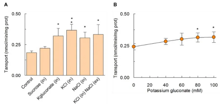
3.1.7. Effect of cations on LAT1 transport activity... 50
3.2. Kinetic characterization of hLAT1 ...50
3.2.1. Oligomeric structure of hLAT1 ... 53
3.3. Characterization of substrate-binding site of hLAT1 ...54
3.3.1. Identification of critical amino acid residues ... 54
3.3.2. Characterization of hLAT1 mutants ... 56
3.4. Role of Cys residues in protein stability and reactivity ...59
Chapter Ⅳ ... 63
Conclusions ... 63
References ... 65
3
4
Amino acids transport in mammalian cells is mediated by different amino acid transporters whose activity allow the flow of an important source for metabolic need of cells. Moreover, some amino acids such as Gln, Arg and Leu work as signalling molecules and their availability and concentration represent key factors in the regulation of intracellular signalling pathways responsible of cellular growth. Thus, amino acids flow, which is important under physiological condition, becomes particularly relevant under pathological conditions such as in tumours cells to satisfy their unique metabolic and proliferative needs. Therefore, since in tumours upregulation of amino acids transporters is an important step to satisfy the increased demand for these nutrients, the same transporters are potential drug targets for cancer therapy. However, the certainty that a specific transporter could be a target in human therapy requires its functional characterization and the knowledge of the enchanting structure/function relationships. In this context, an important transporter that became of particular interest for its overexpression in many tumours is LAT1, and the aim of this work has been that to shed light on still unclear aspects of its function.
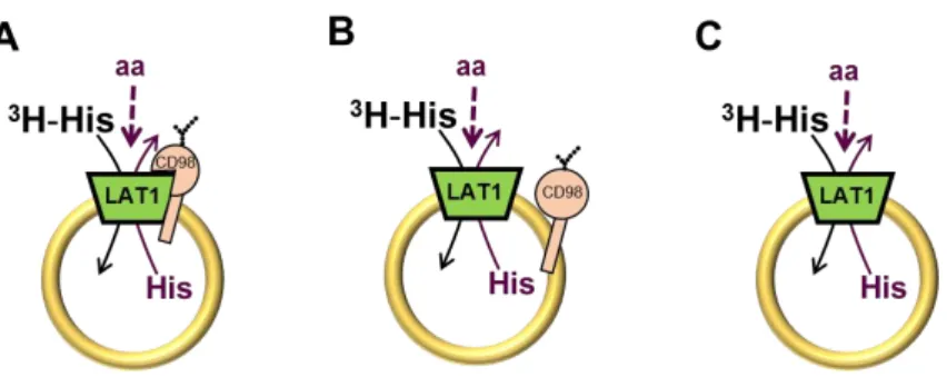
hLAT1 belongs to SLC7 family and into the plasma membrane forms heterodimers with the glycoprotein 4F2hc (also known as CD98 in mice), member of SLC3 family. Studies conducted in intact cells showed that 4F2hc/LAT1 complex catalyses amino acids transport; however, in this experimental model it was not possible to clarify whether one or both subunits are competent for transport activity and substrate recognition. Thus, aimed to unravel the dark side of 4F2hc/LAT1 mediated transport, different experimental strategies were adopted allowing to demonstrate that LAT1 is the sole transport competent unit of the heterodimer. Indeed, using western blot analyses and transport assays in liposomes reconstituted with proteins extracted from SiHa cells and in liposomes reconstituted with recombinant LAT1, it has been demonstrated that neither the covalent interaction nor the association of 4F2hc with LAT1 influence transport and specificity of LAT1. Moreover, the suitability of proteoliposome model used for reconstitution of recombinant LAT1, allowed to identify a functional asymmetry of this transporter which, on a physiological point of view, exhaustively elucidates the reciprocal correlation between the transport activity of LAT1 and that of another important amino acids transporter overexpressed in tumours cells, ASCT2. To the same extent, proteoliposome tool together with bioinformatics and site-directed mutagenesis have been useful to probe critical residues of the substrate binding site of LAT1. These results laid the groundwork for deciphering molecular mechanism of LAT1 function and for setting up studies aimed to identify new potent and specific inhibitors great for human health.
5
6
Il trasporto di amminoacidi nelle cellule umane è mediato da diverse proteine di trasporto la cui attività consente il flusso, attraverso la membrana, di un’importante classe di nutrienti necessari a soddisfare le richieste metaboliche delle cellule stesse. Alcuni amminoacidi, quali Gln, Arg and Leu, fungono anche da molecole segnale perciò la loro disponibilità e concentrazione rappresentano fattori chiave per il controllo di vie di segnalazione intracellulari responsabili della crescita cellulare. Per tale ragione, il flusso di amminoacidi è rilevante sia in contesti fisiologici che patologici. Le cellule tumorali, ad esempio, presentano un incremento del flusso di amminoacidi necessari a sostenere la loro proliferazione. Per soddisfare l’aumentato flusso di nutrienti, le cellule tumorali over-esprimono sistemi di trasporto di amminoacidi che, quindi, divengono potenziali target nella terapia anticancro. Tuttavia, perché una specifica proteina di trasporto possa essere studiata come target farmacologico, è necessaria una profonda conoscenza delle sue caratteristiche funzionali e cinetiche nonché del rapporto struttura funzione. Per raggiungere tale scopo, nel presente lavoro di dottorato è stato studiato uno specifico trasportatore umano, LAT1, over-espresso in molti tumori.
hLAT1 appartiene alla famiglia SLC7 e forma, nella membrana plasmatica, eterodimeri con la glicoproteina 4F2hc (anche nota come CD98) appartenente alla famiglia SLC3. L’attività del complesso 4F2hc/LAT1, come riportato in letteratura, è stata studiata in cellule intatte, tuttavia, tale modello sperimentale non ha consentito di chiarire se una o entrambe le subunità del complesso fossero necessarie per l’attività di trasporto e per il riconoscimento del substrato. Per tale ragione, al fine di rivelare gli aspetti poco chiari del trasporto mediato dal complesso 4F2hc/LAT1, sono state adottate differenti strategie sperimentali che hanno consentito di dimostrare che LAT1 è l’unica unità competente del complesso. Analisi di western blot e studi di trasporto condotti in proteoliposomi hanno consentito di dimostrare che né il trasporto né la specificità del trasportatore LAT1 sono influenzate dall’interazione covalente o dall’associazione con la glicoproteina 4F2hc. Questi risultati sono stati ottenuti sia usando le proteine estratte da cellule SiHa, sia la proteina ricombinate LAT1. La corrispondenza di risultati ottenuti con la proteina nativa e quella ricombinante ha permesso di dimostrare la validità del modello sperimentale di ricostituzione in liposomi del trasportatore LAT1. Il sistema LAT1 è un antiport di amminoacidi Na+ e pH indipendente che mostra una preferenza per grandi amminoacidi quali Trp, Phe Tyr ed His, ma anche amminoacidi più piccoli quali Met, Val, Leu, Ile risultano essere trasportati. Nel modello sperimentale della ricostituzione in liposomi è stata dimostrata anche l’asimmetria funzionale e cinetica di LAT1 che, da un punto di vista fisiologico, chiarisce la reciproca correlazione tra l’attività di trasporto mediata da LAT1 e quella mediata da ASCT2, un altro trasportatore di amminoacidi ampiamente espresso nelle cellule tumorali. Allo stesso modo, la ricostituzione in liposomi della proteina ricombinante LAT1, in associazione a studi di bioinformatica e all’uso della mutazione sito-diretta per ottenere proteine ricombinanti mutate, hanno consentito di verificare il coinvolgimento di specifici residui amminoacidici nel riconoscimento, legame e traslocazione del substrato. In particolare è stato dimostrato che F252, S342 e C335 sono cruciali nel riconoscimento del substrato, mentre C407 svolge un ruolo marginale. I risultati ottenuti, rappresentano una base solida per decifrare il meccanismo molecolare del trasportatore oggetto di studio che
7
è di tipo random simultaneo in cui i due substrati legano nello stesso momento il sito interno ed esterno del trasportatore. I risultati complessivamente ottenuti nel presente lavoro di dottorato costituiscono le basi per effettuare studi volti all’identificazione di nuovi potenti e specifici inibitori importanti per la salute umana.
8
Abbreviations
AdiC L-arginine/agmatine antiporter
APC Amino acid-polyamine-organocation (APC) superfamily ApcT Proton-coupled broad-specificity amino acid transporter BBB Blood brain barrier
BCH 2-aminobicyclo-(2,2,1)-heptane-2-carboxylic acid C12E8 Octaethylene glycol monododecyl ether
CD98 Cluster of differentiation 98 DDM N-Dodecyl β-D-maltopyranoside DTE Dithioerythritol
E. coli Escherichia coli
GST Glutathione-S-transferase
LATs Light subunits of amino acid transporters LeuT Na+-coupled leucine transporter
mTOR Mammalian target of rapamycin
MTSEA 2-Aminoethyl methanethiosulfonate hydrobromide NEM N-ethylmaleimide
SLC Solute carrier
TEMED Tetramethylethylenediamine TMD Transmembrane domain TX-100 Triton X-100
9
Chapter Ⅰ
10
1.1 . Biological membranes and transport activity
Biological membranes represent a permeability barrier that divide the cytoplasmic from the extracellular space in all cells, establishing and discerning the biochemical identity of a single cell from the external environment. Therefore, biological membranes govern substance traffic between the internal and the external environments of cells, safeguarding nutrient assumption, metabolic intermediates retention and scrap product ejection. Biological membranes consist of a continuous phospholipids bilayer in which many membrane proteins are included, indeed, a pure phospholipidic bilayer would impede the access of ions, amino acids, sugars and other hydro soluble molecules. In this contest transport proteins guarantee selective transport of molecules. In the past, it was believed that a large number of molecules could diffuse through membranes. It is now well established that transmembrane proteins are necessary for translocating virtually all the molecules with the exception of very few molecules such as oxygen [1, 2].
In particular, transport proteins mediate entry of nutrients into the cytoplasmic compartment, allowing metabolism of exogenous sources of carbon, nitrogen, sulfur, phosphorus, ions, micronutrients and, then, elimination of end products of metabolic pathways. Transporters allow also uptake and efflux of drugs and other toxic compounds. Active transport of ion species is important to maintain a membrane potential given by ion concentration gradients. Transporters are also directly involved in the elimination of many physiological molecules produced by the cell metabolism, such as lipids, proteins, and complex carbohydrates into and beyond the cytoplasmic membrane. Sometimes, transport proteins work in conjunction with extra cytoplasmic receptors or with cytoplasmic energy-coupling and regulatory proteins forming protein complex [3].
Transporters are also important for metabolite exchange among different sub-cellular compartments, allowing completion of biochemical pathways.
1.2. Membrane transport proteins
Given their essential role for uptake, elimination, and intracellular traffic of all nutrients and metabolites, transport proteins are fundamental for life, indeed they represent a significant fraction of all proteins encoded in the genomes of both simple and complex organisms.
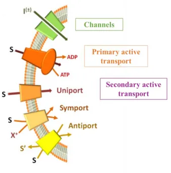
Transport proteins can be classified using different criteria based on functional, molecular or evolutionary aspects. On a functional point of view, in both prokaryotes and eukaryotes, transport systems can be classified in two main groups: channels and permeases.
I. Channels are transmembrane proteins, which catalyse ion transport with an high turnover rates. The
11
II. Permeases catalyse transport of different compounds with a lower turnover than channels and are
active transporters, which utilize diverse energy-coupling mechanisms [4]. In particular, on the basis of the origin of the transport driving force, permeases can be subdivided into primary and secondary active transporters.
Primary active transporters contain ATPase domains or subunits which generate free energy from ATP hydrolysis coupled to the transport process.
Secondary active transporters, which constitute the largest group, can be classified in three categories (uniporters, symporters and antiporters) on the basis of the number of substrates and of transport direction. The driving force for this group of transporters is generated, by concentration gradients of the transported substrates (uniporters), coupling to a co-transported ion such as Na+ or H+
(symporters), or coupling to a counter substrate, which is transported in the opposite direction (antiporters) (Fig. 1) [1].
Fig. 1: Functional classification of membrane proteins. (Adapted from Scalise et al., Proteoliposomes as Tool for Assaying Membrane Transporter
Functions and Interactions with Xenobiotics, 2013).
1.3. Amino acid metabolism and transport
Protein’s share through diet is about 30% in a typical western diet. Protein degradation, in their constituent amino acids, takes place in the gastrointestinal segment. Indeed, after their digestion, the resulting dipeptides and amino acids are efficiently absorbed by the enterocytes of the small intestine where dipeptides are metabolized. Most of globular proteins from animal sources are almost completely degraded in free amino acids [2].
12
Amino acids derived from intracellular or diet proteins degradation, are distributed to tissues and metabolized in liver. Amino acids are an important biological molecule class whose oxidation gives a significant, but variable, contribution to metabolic energetic request of cells. In particular oxidative degradation of amino acids require a phase in which the amino group is separated from carbon skeleton and sent to specialized pathway for amino group metabolism, whereas, carbon skeletons are channelled into citric acid cycle to obtain energy from oxidation. Amino acids are also necessary for synthesis of proteins and bioactive molecules and they are delivered to all tissues through the blood [2, 5]. The flow of these important nutrients, across the plasma membrane, is mediated and strictly controlled by specific transport proteins.
1.4. Mammalian amino acid transporters
Researches on mammalian amino acid transporters have been introduced in 1960 by Christensen’s group, to identify functional characteristics, which allow to discern different transporter. Indeed, using radiolabelled amino acids and amino acid analogues, Christensen and colleagues observed that amino acid transporters accept groups of amino acid rather than single amino acids. Functional characteristics, such as substrate specificity, kinetic and regulatory properties, ion dependence and pH sensitivity, distinguish specific transporters [6]. On the basis of the differential characteristics emerged from these studies, the main criteria adopted to classify amino acid transporters has been the type of amino acid (acidic, zwitterionic or hydrophobic)and the thermodynamic properties of the transport. Moreover, amino acids transport activity was commonly defined “system”, term used to indicate a transport activity functionally identified and very similar in different cell types.
In particular, Christensen’s work identified:
System L which includes amino acid transporters that prefer leucine and other large hydrophobic neutral amino acids;
System A for alanine and other small neutral amino acids; System ASC for alanine, serine, and cysteine;
System y+ for cationic amino acids; System X-AG for anionic amino acids;
Allamino acid transporters are divided into two categories, Na+-dependent and Na+-independent.
Na+-dependent amino acid transporters utilize the energy present across the membrane established by Na+ electrochemical gradient which is mainly maintained by the Na+/K+ -ATPase, to drive the uptake of amino acids across the membrane.
Na+-independent amino acid transporters drive the selective movement of amino acids across the plasma membrane independently of Na+.
13
The nomenclature used for mammalian amino acid transporters terms Na+-dependent systems in uppercase letters and Na+-independent systems in lowercase letters. Only the Na+-independent transporter System L has maintained its uppercase designation for historical reasons [7].
Even though the above described classification is still considered effective on a functional point of view, the growing knowledge of structural data obtained on transporters, together with bioinformatics, introduced a classification of amino acid transporters based on the sequence homology. In the latest two decade, in silico methodologies gave additional information on eukaryote amino acid transporters deriving from homology modelling based on prokaryotic template structures [8]. These studies start from the outset that similar amino acid sequences in proteins reflect similar three-dimensional structures and mechanisms of action; thus by determining the structure and function of at least one member of each protein family, it is possible obtain information about structures, substrate specificities and function of other proteins belonging to the same family [2, 54].
1.5. The APC superfamily
One of the major transporter groups is the amino acid/polyamine/organocation (APC) superfamily, which is represented in each of the life domains (eukaryotes, bacteria and archea). The APC superfamily includes10 families for which literature data report phylogenetic/sequence analyses defining the evolutionary relationships of the proteins to each other as well as the phylogeny of each of the 10 families within the APC superfamily [9-11]. The members of these 10 families function as solute:cation symporters and solute:solute antiporters. The families belonging to APC superfamily are: EAT (ethanolamine transporter) family; AAT (amino acid transporter) family; YAT (yeast amino acid transporter); LAT (l-type amino acid transporter) family; CAT (cationic amino acid transporter) family; APA (basic amino acid/polyamine transporter) family; ACT (amino acid/choline transporter) family; ABT (archaeal/bacterial transporter) family; GGA (Glutamate:GABA antiporter) family; SGP (Spore germination protein) family. The substrate specificities of some APC superfamily transporters have been carefully studied revealing that while some have exceptionally broad specificity for amino acids, others are restricted to just one or a few amino acids.
The majority of homologous integral membrane APC permease polypeptide chains, vary in size from about 400 to 800 amino acid residues. According to hydropathy profile analysis and biochemically established topological features of most prokaryotic and eukaryotic APC superfamily members, both the N- and C-termini of the proteins are located in the cytoplasm with a 12-transmembrane segment topology, with some exception [9-11].
1.6. The solute carrier SLC7 family
One family of the APC transporter superfamily have human members involved in relevant physiological functions. This family is the solute carrier SLC7 family whose members belong to two subfamily: the cationic
14
amino acid transporters (CATs, SLC7A1–4 and SLC7A14) and the L-type amino acid transporters (LATs, SLC7A5-13 and SLC7A15). The latter are also called light chains or catalytic subunits of the heteromeric amino acid transporters (HATs) or glycoprotein-associated amino acid transporters (the gpaAT family) (Fig.2).
Fig. 2: Phylogenetic tree of SLC7 family members. The SLC7 family is composed of the CATs and the light subunits of HATs. (Adapted from
Fotiadis D. et al., The SLC3 and SLC7 families of amino acid transporters, 2013).
CATs are the principal entry path for cationic amino acids, playing an important role in nitric oxide synthesis by delivering L-arginine for nitric oxide synthase. These transporters are N-glycosylated and have 14 putative transmembrane domains (TMDs) with cytosolic N- and C-termini in line with transmembrane topology prediction and experimental evidence [12].
LATs are not N-glycosylated and the peculiar property of this group of proteins consists in forming covalent heterodimer with larger polypeptides belonging to the SLC3 family, a small group of type II membrane glycoproteins. LATs only have 12 TMDs, which show significant similarity to the first 12 TMDs of CATs. As indicated from sequence analyses both, CATs and LATs, originate from an ancestral 12 TMD protein and the duplication of the last two TMDs of this ancestral protein is the origin of the 14 TMD CAT structure. An estimation indicates that this duplication happened about 2.6 billion years ago [13]. Homologous CAT and LAT proteins are found in prokaryotes, but the cysteine residue of the LATs involved in the disulphide bridge with polypeptides belonging to the SLC3 family is not conserved [14]. SLC3 proteins and HATs are only found in metazoans [12].
1.7. The heteromeric amino acid transporters HATs
The heteromeric amino acid transporters, HATs, are composed of a light and a heavy subunit linked by a disulphide bridge. The heavy subunits are the SLC3 members, whereas the light subunits are the eukaryotic LATs from the SLC7 family of amino acid transporters. For SLC3 members, the cysteine residue participating in the disulphide bridge with the corresponding light subunit is four to five amino acids away from the TMD,
15
whereas for LATs the cysteine residue involved in the disulphide bridge with the heavy subunit is located between TMD III and IV (Fig. 3).
Fig. 3: Model of human heterodimer 4F2hc/LAT1 proteins. (Adapted from Fotiadis D. et al., The SLC3 and SLC7 families of amino acid
transporters, 2013).
1.7.1. HATs and their heavy subunits
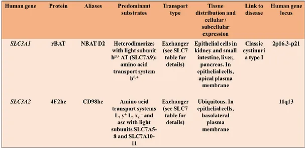
The SLC3 family includes two members: rBAT (SLC3A1) and 4F2hc (SLC3A2, also named CD98) (Table 1).
Table 1: SLC3 family (Adapted from Palacin, M., Kanai, Y., 2004. The ancillary proteins of HATs: SLC3 family of amino acid transporters).
The two proteins share about 20% amino acid sequence identity and both are N-glycosylated with a molecular mass of ~94 and ~85 kDa for rBAT and 4F2hc, respectively [12]. In particular, SLC3 proteins are type II membrane glycoproteins with a single transmembrane domain and the C-terminal located outside the cell. The large extracellular domain of SLC3 members has sequence and structural homology with bacterial α-glucosidases and the insect maltases. In this regard, a bioinformatics study describing a comparison of all
16
available rBAT and 4F2hc sequences with GH13 enzyme family is reported in literature [15]. The crystal structure of the extracellular domain of human 4F2hc has been solved at 2.1 Å resolution, and it has been observed that the protein has the characteristic fold of these enzymes, a (βα)8 barrel and a C-terminal,
anti-parallel β8 sandwich [16]. However, despite this structural similarity, 4F2hc lacks the key catalytic residues
necessary for glucosidase activity [16]. The atomic structure of the rBAT extracellular domain has not been solved.
rBAT was identified by expression in Xenopus oocytes and in 1999, b0,+AT (SLC7A9) was identified as the light subunit that co-expresses with rBAT. This complex mediates the antiport of cystine and dibasic amino acids with neutral amino acids. The rBAT/b0,+AT heterodimer is defective in cystinuria, a recessive disease, and the most common primary inherited aminoaciduria, characterized by hyperexcretion of dibasic amino acids and cystine in urine [17].
4F2hc was originally identified as a lymphocyte activation antigen through a monoclonal antibody 4F2 and is a ubiquitous multifunctional protein [17]. 4F2hc, indeed, could associate with different transporters (see Table 1 and below) over-expressed in many tumours and in activated lymphocytes suggesting a role of 4F2hc and the associated transporters in cell growth. Furthermore, 4F2hc mediates β-integrin signalling and cell fusion. Thus, given the dual function exerted by 4F2hc, it is possible to hypothesize that this protein plays a key role in integrating integrin signalling and amino acid transport [12].
In agreement with the different roles of both heavy subunits, rBAT/b0,+AT expression is restricted to the apical
domain of the plasma membrane of epithelial cells of the small intestine and of the renal proximal tubule, whereas 4F2hc-associated transporters are almost ubiquitously expressed. Moreover, 4F2hc is expressed in the basolateral plasma membrane of epithelial cells (Fig. 4) [18].
17
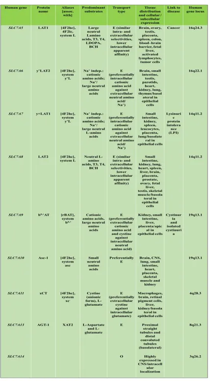
1.7.2. HATs and their light subunits
The eukaryotic LATs from the SLC7 family of amino acid transporters that represent the light subunit of HATs are seven transporter, which differ each other for substrate selectivity, ion coupling and tissue distribution (Fig. 4 and Table 2). As above described, only the amino acid transporter b0,+AT (SLC7A9) forms heterodimers with rBAT, whereas six of these transporters form heterodimers with 4F2hc: LAT1 (SLC7A5), LAT2 (SLC7A8), y+LAT1 (SLC7A7), y+LAT1-2 (SLC7A6) and the cystine/glutamate antiporter xCT (SLC7A11) and ASC-1 (SLC7A10) (Table 2). (The same table contain also SLC7A13 associated with an unknown protein and SLC7A14 an orphan transporter).
E: exchanger; O: orphan transporter
18
A characteristic of HATs is that they function as obligatory antiporters with the exception of system Asc that also mediate facilitated diffusion [19]. The differing tissue localizations of HATs appear to complete them each other [20] but at the same time, they are quite diverse in terms of substrate selectivity [21]. Their selectivity ranges from large neutral amino acids (LAT1-2) to small neutral amino acids (ala, ser, cys-preferring, Asc-1), negatively charged amino acid (xc-) and cationic amino acids plus neutral amino acids (y+L and b0,+-like). The (obligatory) antiporters equilibrate the concentrations of their substrate amino acids across membranes. In particular, these transporters uses the driving force provided by a transmembrane gradient of one amino acid, accumulated by a parallel transport process. Nevertheless, the transport of specific amino acids is guaranteed by the intrinsic asymmetry of these antiporters. The fact that genetic defects of the epithelial b0,+AT and y+LAT1 cause non-type I cystinuria and lysinuric protein intolerance, respectively, demonstrates that these HATs perform transport of specific amino acids in vivo [20-22].
1.8. 4F2hc (SLC7A2)
The 4F2hc heavy chain of HATs (also called CD98 in mice) has been originally identified in 1981 by a murine monoclonal antibody (mAb4F2) raised against the human T-cell tumor line HSB-2 [23]. Initial studies utilizing anti-4F2 antibodies revealed that the antigen is present on all established human cell lines and the majority of malignant human cells [24]. The gene has been identified on chromosome 11 and seems to be more ubiquitously expressed than the other human heavy chain rBAT. 4F2hc gene codified a protein constituted by 630 amino acids with a theoretical mass of ~85 kDa [12, 17, 25].
1.9. LAT1 (SLC7A5)
LAT1 is the first member of HATs that has been identified in 1998 [26, 27]. The LAT1 subunit in humans is a 507 amino acid long hydrophobic polypeptide with a molecular mass of 55.0 kDa. The protein is predicted to be constituted by 12 transmembrane segments. The high hydrophobicity that characterize this protein is responsible of its apparent molecular mass in SDS-PAGE of ~40 kDa. The higher electrophoretic mobility is probably due to a more compact form of the protein induced by partial oxidation and formation of disulphides among some of the 12 Cys residues present in hLAT1 amino acidic sequence [25].
As member of HATs LAT1 forms heterodimers with 4F2hc stabilized by a conserved disulphide between residues C164 of LAT1 and C109 of 4F2hc (Fig. 3). The role of LAT1/4F2hc heterodimer in mediating amino acid transport across the plasma membrane has been extensively studied in cell systems. Available literature data describe LAT1/4F2hc heterodimer mediated transport as a sodium independent amino acid antiport with preference for neutral amino acids with large, branched or aromatic side chains such as Phe, Tyr, His and Trp, and smaller amino acids such as Met, Val, Leu, Ile [27, 28]. LAT1/4F2hc has been also described as a transporter of non amino acid substrates, such as L-DOPA, gabapentin and thyroid hormones [29, 30]. As
19
other member of LATs subfamily, i.e. LAT2, LAT1 is inhibited by BCH commonly used to discriminate its activity from that of other amino acid transporters in cells [31].
LAT1 is broadly expressed and mainly localized in basolateral membranes of polarized epithelia [7, 28, 32]. Important exceptions are the luminal and abluminal membranes of BBB (blood–brain barrier) and the brush border membranes of placenta, i.e. maternal side [29]. More specifically SLC7A5 gene coding for hLAT1 is expressed in the placenta > the brain > the spleen > the testes and the colon [27]. Literature data suggest that in the inner blood-retinal barrier LAT1 plays an important role in transporting large neutral amino acids and neurotransmitters [33], as well as in placental membranes nourish fetus and placenta with thyroid hormones and amino acids [12, 29]. Moreover, an intracellular localization of LAT1/4F2hc heterodimer in lysosomes has been described [34].
1.10. Amino acids, LAT1 and tumours cells
The distinctive traits of tumour cells are rapid growth and proliferation that could be achieved by facilitation of cell cycle and resistance to apoptosis. The first step to enhance cell proliferation is an increased demand for nutrients used as building blocks for the synthesis of macromolecules and as carbon source for generation of metabolic energy. Thus, different nutrients are necessary, i.e. glucose, amino acids, vitamins, fatty acids, micronutrients, and, tumours cells adopt particular mechanisms to satisfy their increased demand for them. One of these is the vasculogenesis to increase the availability of nutrients, whereas upregulation of specific transporters allow entry of the nutrients into tumour cells. In some instances, the same signalling events that promote vascularization participate also in the upregulation of nutrient transporters, thus coordinating the availability of nutrients with their entry into tumour cells [35].
Nevertheless all nutrient are important to support growth and proliferation of tumour cells, the latter have an unique metabolic need for amino acids. Amino acids, indeed, are essential for protein synthesis but also as carbon and nitrogen source for purine and pyrimidine nucleotides, amino sugars, and glutathione synthesis. Moreover, the regulation of intracellular signalling pathways responsible of cellular growth involves upstream sensing of concentration of certain amino acids. The consequent scenario is that although the primary function of the amino acids is to serve as the building blocks for protein synthesis, some amino acids have specific signalling functions. Glutamine, glycine, and aspartate are needed for nucleotide biosynthesis, a process critical for proliferation of tumour cells. Serine plays an important role as a one-carbon source that is critical in nucleotide synthesis and DNA methylation. Leucine, glutamine, and arginine serve as signalling molecules [36].
The two best-studied nutrient signalling cascades in higher eukaryotes, important under physiological conditions, are the GCN (general control non-derepressible) and mTOR (mammalian target of rapamycin) pathways, both of which are regulated by mechanisms that include upstream sensing of intracellular amino acids concentrations.
20
One protein involved in the first nutrient signalling cascade is the GCN2 protein kinase. GCN2 appears to monitor intracellular amino acids concentration from the level of tRNA charging. In particular, uncharged tRNA accumulation correlated to a low concentration of amino activates GCN2, which phosphorylates and inactivates the initiation factor eIF2α (eukaryotic initiation factor 2α). As consequence, the global mRNA translation is suppressed, but occur the translation of the transcription factor ATF4 (activating transcription factor 4) which allow the induction of genes for amino acids biosynthesis and transport [37].
The mTOR pathway, in contrast, is stimulated by cellular amino acids supplementation and activates mRNA translation, promoting cell growth. This pathway regulates many major cellular processes thus it became particular relevant in tumours cells. mTOR is the target of a molecule named rapamycin which is a macrolide produced by Streptomyces Hygroscopicus bacteria that kept attention because of its antiproliferative properties. In the early 1990s, genetic screens in budding yeast identified TOR1 and TOR2 as mediators of the toxic effects of rapamycin on yeast.Successively biochemical approaches in mammals led to purification of mTOR and its discovery as the physical target of rapamycin. mTOR is an atypical serine/threonine protein kinase belonging to the phosphoinositide 3-kinase (PI3K)-related kinase family and interacts with several proteins to form two distinct complexes named mTOR complex 1 (mTORC1) and 2 (mTORC2) (Fig. 5) [38].
Fig. 5: mTORC1 and mTORC2 Complexes. (Adapted fromMathieu Laplante and David M. Sabatini, mTOR Signaling in Growth Control and Disease, 2012).
Amino acids are required for the activation of mTORC1, but not mTORC2 [39]. Although not yet fully resolved, the field of mTORC1 activation by amino acids has rapidly progressed [40]. mTORC1 activation occurs at the lysosomal membrane through an inside-out model of amino acid sensing, in which amino acids accumulate in the lysosomal lumen and initiate signalling through a mechanism that requires the vacuolar H+
-adenoside triphosphate ATPase (V-ATPase). At the surface of the lysosome, the V-ATPase directly interacts with the Ragulator, a pentameric complex important for amino acids regulation of mTORC1. The Ragulator serves as a scaffold protein complex and possess guanine nucleotide exchange factor (GEF) activity linked to Rag GTPases a group of four GTPases (RagA, RagB, RagC and RagD) that can recruit and activate mTORC1
21
at the lysosomal surface. Upon amino acid stimulation, the GEF activity of the Regulator promotes the loading of RagA or RagB with GTP in a V-ATPase-dependent manner allowing RagA or RagB to interact with RAPTOR component of mTORC1. This interaction results in the recruitment of mTORC1 at the lysosomal surface, where its endogenous activator Rheb resides [40].
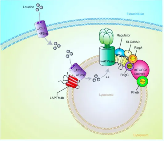
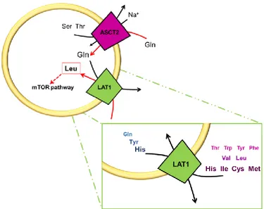
In particular, a relevant role in activating mTORC1 is fulfilled by Leu and leucine-sensing mechanisms. Therefore, it is not unexpected that LAT1, which is predominantly expressed in tumours cells, is of particular interest in the contest of mTORC1 signalling [37, 40]. Indeed, on one hand the overexpression of LAT1 at the plasma membrane of tumours cells together with other amino acid transporters, such as ASCT2, allow to supply cell with Leu. On the other hand, the recent work by Milkereit and colleagues, which identified that LAPTM4b recruits LAT1/4F2hc to lysosomes, suggested a novel mechanism based on leucine lysosomal sensing (fig. 6) [34].
Fig. 6: Proposed role of LAPTM4b in activation of mTORC1 via recruitment of the Leu transporter to lysosomes. (Adapted fromMilkereit et al., LAPTM4b recruits the LAT1-4F2hc Leu transporter to lysosomes and promotes mTORC1 activation, 2015).
LAPTM4b recruits the Leu transporter LAT1/4F2hc to lysosomes, enhances Leu uptake into lysosomes and stimulates mTORC1 activation via V-ATPase. These findings not only identify a functional Leu transporter at the lysosomal membrane, but also help solve the puzzle of how mTORC1 is activated by amino acids in the lysosome by an inside-out mechanism (intra-lysosomal stimulation of V-ATPase) originally noted by Sabatini et al. as described above. Being LAT1/4F2hc an antiport intra-lysosomal neutral amino acids would be
22
necessary as exchange substrates to sustain LAT1/4F2hc antiport function. Possible sources of lysosomal amino acids include uptake of large neutral (non-essential) amino acids such as Gln into lysosomes via directional/accumulative amino-acid transporters. Candidate transporters could be in particular SLC38A3 and SLC38A5 [34]. Another transporter presents in lysosomal membrane is SLC38A9 that transport Gln and Arg with low-affinity, indeed its peculiar role is that to function as a transceptor to sense Arg which allow to bind directly to Ragulator and Rag proteins. Thus, SLC38A9 participates in mTORC1 activation at the lysosome rather than contributing to the import of extracellular amino acids at the plasma membrane [41].
1.11. SLC7A5 genetic variants in Autism Spectrum Disorder
Beyond the vast literature data that give to LAT1/4F2hc heterodimer a prominent role in tumour survival, very recently, inherited mutations of SLC7A5 have been described as responsible of Autism Spectrum Disorder (ASD) a group of disorders often overlapping with other neurological conditions. This interesting study evidenced that in mice, deletion of SLC7A5 from the endothelial cells of the BBB leads to atypical brain amino acid profile. Furthermore, the same study has identified several patients with autistic traits and motor delay carrying homozygous mutations in the SLC7A5 gene. In particular, two genetic variants have been identified and mapped onto a homology model of SLC7A5. The first, A246V mutation, changes a highly conserved alanine affecting the transporter’s structure. A246 is located in transmembrane helix 6 in proximity to the extracellular side and to the channel, and its substitution with the slightly larger residue valine has been experimentally demonstrated that impair transport activity by disrupting helix-helix packing and ligand transport. The second mutation identified, P375L, instead, leading to the change of the conserved proline in position 375 to a leucine. P375 is located in transmembrane helix 9 in close proximity to the cytoplasmic side. Proline often plays a key role in allowing conformational changes important for transporter function. Thus, mutation of this residue to leucine is likely to disrupt the flexibility required for transport by SLC7A5. Also for this mutant, this possible effect has been evaluated measuring transport activity, and it has been observed that the mutation P375L impairs the driving force for taking up SLC7A5 substrates. Thus, this study demonstrated that the substitution of the SLC7A5 alanine 246 into a valine, or of the proline 375 into a leucine, is sufficient to significantly reduce the SLC7A5-mediated BCAA uptake. This is an important feature because the fine-tuning of brain BCAA concentrations is critical for normal brain function, and mutations affecting genes contributing to BCAA homeostasis and the downstream signalling cascade may underlie a larger subgroup of ASD [42].
1.12. The intriguing role of 4F2hc in the intrinsic transport activity of LAT1
Although there are many literature data about transport properties of LAT1/4F2hc heterodimer, the precise role of 4F2hc in the intrinsic transport activity of LAT1 is still an open issue. The heavy subunit may be involved in trafficking the light chain to the plasma membrane or, together with the light subunit may catalyse
23
the transporter function [17, 21]. The two proteins as well as HATs proteins form a heterodimer interacting via a disulphide bridge. However, if there were other possible interaction between the proteins of the heterodimers and if both these and the disulphide bridge are critical and/or essential for the stability of the heterodimer and/or for the transport are intriguing aspects, thus many literature data are present on this issue. Some authors declaim that the absence of disulphide bond does not preclude, but only decreases transport surface expression and/or function. The heterodimer interaction takes place also in absence of disulphide bridge formation and this interaction is stable enough to allow cell surface expression of the light chain [43]. Other authors suggest that despite the disulphide bridge is not necessary for the functional association, in absence of the covalent bridge, the C-terminal of the heavy chain is necessary for a functional transport competence [44]. Moreover, seemingly conflicting results published on 4F2hc/LAT1 interaction, declaimed the importance of the N-terminal part of 4F2hc or alternatively, the role of the extracellular part of 4F2hc [45, 46]. Whereas, other authors deduced that the interaction of the heavy chain 4F2hc with the light chain LAT1 involves all domains of 4F2hc [47].
Studies performed in intact cells considered 4F2hc crucial for LAT1 transport activity [27, 48]. There again, other authors, claimed that involvement of 4F2hc in transport is linked to maturation and trafficking of LAT1 protein in plasma membrane [26, 47, 49, 50]. In particular, it is reported that rat LAT1 remains in Golgi area in absence of the heavy subunit [49], thus the heavy chain is necessary for trafficking LAT1 to the plasma membrane. Moreover, in mouse hepatocarcinoma cells, over-expressed LAT1 has been showed to mediate leucine transport without involvement of 4F2hc [51].
Therefore, considering that in intact cell, which is the only system used until now, 4F2hc is always present, it is difficult to understand if 4F2hc is only necessary for maturation, trafficking and/or surface residence of LAT1 or it is essential for LAT1 transport function. Thus, this aspect remain unclear.
1.13. Pharmacological approaches for LAT1
The over-expression of LAT1 in tumour cells made this human transporter an interesting pharmacological target. Indeed, as for other amino acids transporters over-expressed in tumour cells compared to normal cells, compounds with the ability to inhibit the cellular pathways responsible for their induction or to block the function of the induced transporters would have potential effects as chemotherapeutic agents. Indeed, since tumour cells induce these transporters specifically for their unique metabolic needs, normal cells are expected to be relatively resistant to the therapeutic actions of such compounds, thus reducing undesirable side effects [35]. In this regard, many authors are involved in the research of inhibitors of this transporter useful as potential anticancer drugs. More specifically literature data report two approaches for anticancer therapies involving LAT1. The first regard the capacity of this transporter to mediate the uptake of several amino acid-derived anticancer drugs, thus this approach is based on the notion that LAT1 could be involved in the cellular internalization of these anticancer drugs. The second approach is a novel strategy based on the inhibition of
24
LAT1 activity to reduce tumour proliferation and progression but the targeting of amino acid transporters in cancer is under development and there are few specific inhibitors available. Thus, the development of more effective inhibitors of LAT1 will change the scenario. However, identification or design of potent inhibitors to be proposed as potential drugs, requires a deep knowledge of the target structure and action mechanism that, in the case of LAT1, are still missing [52-55].
The described roles employ by LAT1 justify the growing number of literature works regard this fascinating protein. Nevertheless, the study of transport proteins is not of simple realization. Given their hydrophobic nature, transport proteins require different devices for their study.
1.14. X-ray crystallography state for transmembrane protein and bioinformatics approaches
As described above, the three-dimensional structure of the C-terminal of the heavy chain has been solved [16], while crystals of LAT1 are not available, as for all the mammalian amino acid transporters. The lateness in the possibility to obtain stable crystals for transmembrane proteins is due to their hydrophobicity; moreover, resolution of the structure needs large-scale purified transport proteins. The lack of crystal structure of transport proteins makes difficult the understanding of the structure/function relationships and the mechanisms of interaction with xenobiotics, a very important issue connected with drug discovery and toxicity.
Nevertheless, the increasing study of atomic structure of homologues prokaryotic transporter allowed unravelling their molecular mechanisms. In particular, the prokaryotic amino acid transporters AdiC, whose structure has been solved by x-ray crystallography, is the closest homologue of the mammalian LAT transporters (amino acid sequence identity ranging from 14% to 20%)[56-60]. These transporters share the protein fold named 5+5 inverted repeat fold (also known as LeuT-fold). Proteins with the LeuT-fold, although differing each other in the total number of TM helices, are characterized by two structurally similar repeats, each containing a core of five consecutive TMs. The feature of this fold is that the first TM in each of the two inverted repeats (TM1 and 6) is discontinuous and consists of two short alpha helices connected by a highly conserved unwound segment. This structural feature is important for the transport mechanism of transporters with the LeuT-fold as it is part of the substrate coordination site in crystallized amino acid transporters with this fold [59, 60]. Indeed, in absence of crystal structure for LATs transporter, elucidation of the structure of closely related prokaryotic homologues of human LAT1 transporters and the generation of structural models from these crystal structures through a bioinformatics approach could be useful to make a step forward in the understanding of the structure/function relationship. Nevertheless, the silico methodologies represent only a tile in the complex mosaic of the structure/function relationship of LAT1.
1.15. The study of transport proteins through a multidisciplinary approach
Even though the building of structural model using in silico methodologies could represent an important feature for advancing the knowledge of a specific transport protein, on the other hand, transport measurements
25
are essential to validate, experimentally, the in silico predictions. Nevertheless, to measure transport activity the best way is to use different system that could confirm each other the results obtained. Indeed, measuring transport activity both in intact cells and in an isolated environment such as that offered by proteoliposomal tool, is possible to obtain more clear information. However, the use of proteoliposomal tool requires the heterologous expression of the protein object of study, an important methodology that, together with site direct mutagenesis, allowing to obtain also mutants of the protein of interest with consequent possibility of better understanding of the involvement of specific residues in transport activity.
Thus, the better way to obtain trustworthy results is develop a multidisciplinary approach, which confirm each other the made observation allowing to shedding light in the complex mosaic of the structure/function relationship of the studied transport protein.
1.16. Experimental methods to study transport proteins
Two different methods could be used for measuring transport activity of a protein of interest.
1.16.1. Intact cell systems
The study on transport proteins started in 1970’ and the first model adopted was that of intact cells. In particular, transport functions was studied following the flux of labelled compounds through native membranes of intact cells, isolated organelles (such as mitochondria) or microsomes, derived from endoplasmic reticulum. Intact cell models are still broadly used as tools for studying the functional properties of transport systems. Different cells lines are used, expressing endogenous transporters or over-expressing specific transporters after transient transfection by appropriate plasmids. X. laevis oocytes over-expressing a specific transporter are also used in some studies. The over-expression procedure offers the advantage of increasing the amount of the protein of interest. In this system, the activity of the over-expressed protein should overcome that of the endogenous ones, leading to a better resolution of the transport activity measurement.
Nevertheless, the use of intact cell systems, disclose some limits. Indeed the impossibility to control the intracellular compartment limits the kinetic characterization of the transporter especially on the internal side. Moreover, intracellular enzyme pathways can readily metabolize the substrates used for measuring the transport activity and the presence of many similar transporters in the same membrane is responsible of interferences. Therefore, the use of intact cell systems as unique method is not suitable for a comprehensive functional characterization of a transporter and even less for revealing structure/function relationships.
1.16.2. Proteoliposomes
An alternative model for studying transport proteins is the proteoliposomes. Liposomes were initially produced for encapsulating and delivering enzymes or pharmaceutical compounds [61-63], and then, used as
26
a tool for studying membrane transporters [64, 65], which are inserted into the membrane bilayer to obtain active proteoliposomes. Transport proteins could be extracted from biological samples in non-ionic detergent, but a more recent and effective method is that of producing recombinant proteins by over-expression in heterologous systems and purification by affinity chromatography. Production of recombinant proteins offer the advantage of excluding any kind of interference and, in addition, to obtain human proteins that, obviously, cannot be extracted from tissues. This method offers several advantages with respect to intact cells: 1) the possibility to control internal and external compartments as well as the internal and the external site of the transport protein allowing detailed kinetic analysis; 2) the possibility to construct specific mutants in which selected amino acid residues are substituted, thus allowing the study of structure/function relationships; 3) the study of interaction with chemicals and potential drugs, that can be well characterized with respect to their mechanism of action [1].
1.17. Proteoliposomes as method to reveal xenobiotic-transporter interaction mechanisms
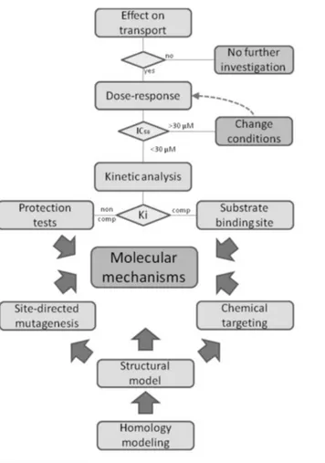
When the interest is that of characterizing the interaction between a transport protein and xenobiotic compounds such as environmental contaminants or drugs, IC50 or Ki parameters have to be measured with a good confidence. The threshold fixed for considering an interaction of interest is 30µM [1].
Using both intact cell system and proteoliposomes different and complementary information can be obtained. Intact cell system, including the case of cell lines over-expressing specific transporters, gives information on vitality of the cells after exposition to a specific compound and only a general alteration of the transport activity. On the contrary, the suitability of the proteoliposomal method allow the description of the mechanism of the interaction at the molecular level. Indeed the possibility to make extensive kinetic analysis of the transporter-xenobiotic interaction gives information both on the constants, i.e. Ki, and on the type of interaction competitive, non-competitive or mixed inhibition. Moreover, when a homology structural model is available and possibly validated by site-directed mutagenesis, information on the involvement of amino acid residues could be obtained.
Thus, the use of proteoliposomes together with intact cells as method to study transporter function, kinetics and interactions is an example of how a multidisciplinary approach, including bioinformatics allow obtaining detailed and trustworthy results on the structure/function relationship of a transport protein (Fig. 7).
27
Fig. 7: Work plan for xenobiotic-transporter interaction studies in proteoliposomes. (Adapted from Scalise et al., Proteoliposomes as Tool for
28
Chapter Ⅱ
29
2.1. Materials
2.1.1. RIPA Buffer 1X
20 mM Tris-HCl (pH 7.5)
150 mM NaCl
1 mM Na2EDTA
1 mM EGTA
1% NP-40
1% sodium deoxycholate
2.5 mM sodium pyrophosphate
1 mM β-glycerophosphate
1 mM Na3VO4
1 µg/ml leupeptin2.1.2. Buffers for h4F2hc purification
2.1.2.1. Binding buffer for h4F2hc purification
5% Glycerol 1% Triton X-100 PBS pH 7.4 5 mM DTE
2.1.2.2. Washing buffer for h4F2hc purification
0.1% Triton X-100
PBS pH 7.42.1.3. Washing buffer for hLAT1 pellets
30
2.1.4. FPLC buffers for hLAT1 purification
2.1.4.1. Equilibration buffer
20 mM Tris-HCl pH 8
200 mM NaCl
10% Glycerol
0.1% Sarkosyl2.1.4.2. Washing buffer
20 mM Tris-HCl pH 8 200 mM NaCl 10% Glycerol 0.05% DDM 3 mM DTE2.1.4.3. Elution buffer
20 mM Tris-HCl pH 8 200 mM NaCl 10% Glycerol 0.05% DDM 3 mM DTE 400 mM Imidazole2.1.5. Desalt buffer for purified hLAT1
20 mM Tris-HCl pH 8 0.05% DDM
31
2.1.6. Running buffer for PAGE 10X
250 mM Tris 14.4 % Glycine
1% SDS or 1% Sarkosyl
2.1.7. Loading dye for PAGE 3X
0.2 M Tris-HCl pH 6.8 7.5% SDS or 7.5% Sarkosyl 3% Glycerol
0.01% Bromophenol blu 100 mM DTE (when indicated)
2.1.8. Coomassie Brilliant Blue
0.25 g Coomassie Brilliant Blue 45 mL Methanol 45 mL Distilled H2O 10 mL Acetic acid
2.1.9. Destaining solution
10% Acetic acid 40% Methanol 50% H2O2.1.10. Washing buffer for western blot analysis
50 mM Tris-HCl pH 7 150 mM NaCl
2.1.11. Lowry’s solution
4 mg/mL NaOH and 20 mg/mL Na2CO3 in water
32 5 mg/ mL CuSO4
1% SDS
2.2. Experimental procedures
2.2.1. Protein purification by affinity chromatography
Affinity chromatography is a separation method based on a specific binding interaction between an immobilized ligand and its binding partner. This technique is commonly used for purification of recombinant proteins genetically modified in order to harbour specific tag(s) for affinity binding. In the case of the proteins studied in this work, glutathione-S-transferase (GST) or hexahistidine (6His) were used as tags. GST has an affinity for glutathione, which is commercially available immobilized as glutathione agarose. Histidine tags have an affinity for nickel or cobalt ions, which have been immobilized by forming coordinate covalent bonds with a chelator, incorporated in the stationary phase. The elution of the protein with a 6-His tag is performed using an excess amount of a compound able to act as a metal ion ligand, such as imidazole.
2.2.2. Gel Filtration Chromatography
Gel filtration chromatography is commonly used to separate molecules in a mixture on the basis of their sizes. The molecules move through a bed of porous beads and diffuse into the beads to greater or lesser degrees. In particular, smaller molecules diffuse more slowly than larger molecules, which do not enter or enter the beads to a lesser extent. Gel filtration chromatography may be used for different purposes. In our case, this approach has been used to separate proteoliposomes from the external substrate both after reconstitution procedure and transport assay, or for salt removal from the protein.
2.2.3. Reconstitution into liposomes
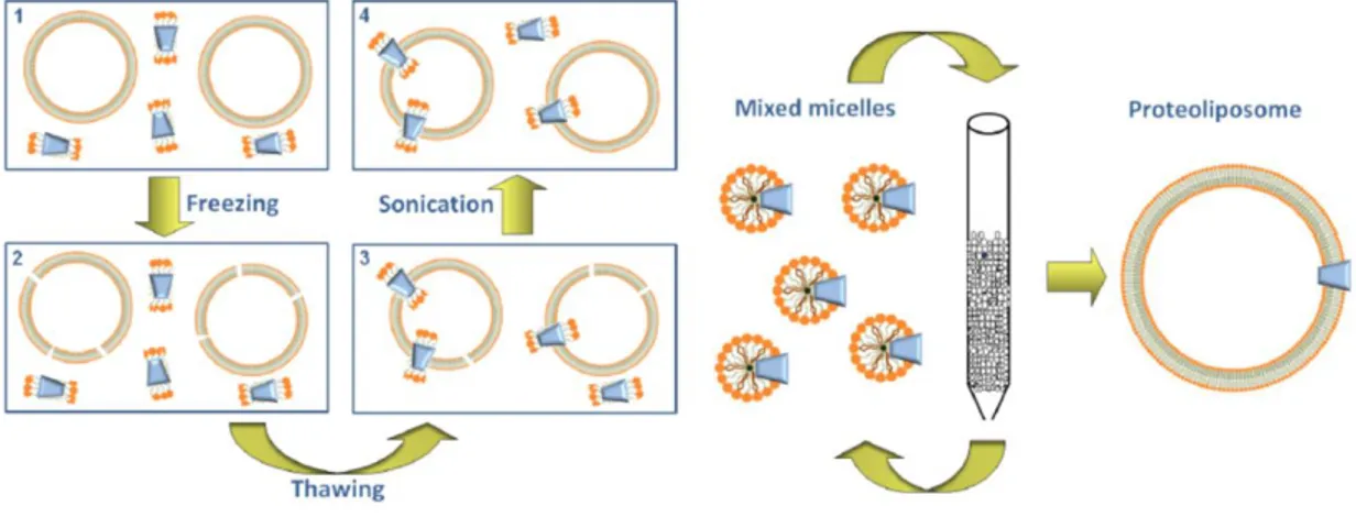
The reconstitution into liposomes of a transport protein can be performed with two main procedures, which differ for the efficiency. The first is the freeze-thaw-sonication procedure that consists of three steps: i) a fast freezing of a mixture of liposomes, transport protein and non-ionic detergent; ii) a slow thawing of this sample; iii) a final sonication for sealing formed proteoliposomes. After freezing, liposomes are broken due to ice formation and, during the slow thawing, the protein inserts into the phospholipid bilayer. This procedure has the disadvantage that the detergent is not removed and can impair the complete sealing of proteoliposomes. The second procedure, instead, is based on the removing of detergent from mixed micelles of protein, detergent and phospholipids. In particular, the remove of detergent from mixed micelles can be performed by cyclic chromatography of the reconstitution mixture on hydrophobic column for 10–20 cycles, or by the batch-wise procedure. In the latter, the mixed micelles are incubated with an hydrophobic resin for a time sufficient to remove virtually all the detergent. The main advantage of detergent removal is that the proteoliposomes are more sealed, unilamellar and, hence, virtually impermeable to hydrophilic compounds. Moreover, by mean of
33
detergent removal procedure the protein is inserted with an unidirectional orientation, which in most cases, corresponds to that of the transporter in the native membrane. This is due to the feature of the starting micelles that have a relatively small radius: this property forces the asymmetric protein to insert into the micelles with a right side-out orientation (Fig. A).
Fig. A: Sketch of methodologies for proteoliposome preparation. (Adapted from Scalise et al., Proteoliposomes as Tool for Assaying Membrane
Transporter Functions and Interactions with Xenobiotics, 2013).
Given the suitability of the detergent removal method, it has been adopted in this work using the batch-wise procedure. This initial mixture contains primarily the protein of interest, which has been previously solubilized from cell extracts (in our case from SiHa cells) or purified through affinity chromatography (recombinant proteins overexpressed in E.Coli). Subsequently, non-ionic detergent (octaethylene glycol monododecyl ether C12E8), egg yolk phospholipids (w/v) (in the form of liposomes prepared through sonication), substrate, buffer
and water in a final volume of 700 µL are added to complete the mixture. This mixed mixture is then incubated at room temperature with a hydrophobic resin (Amberlite XAD-4) for a time sufficient to remove virtually all the detergent and obtain active proteoliposomes.
2.2.4. Ultracentrifugation of proteoliposomes
To evaluate the insertion of the protein into liposome membranes, after reconstitution the ultracentrifugation of the proteoliposomes can be performed. After gel filtration chromatography, 500 µL of obtained samples are subjected to two consecutive ultracentrifugation steps for 90 min 110,000 × g at 4◦C. The first one is performed in order to separate the fraction containing proteoliposomes from that of empty liposomes, i.e. vesicles which did not incorporate the protein during reconstitution. The separation is achieved because proteoliposomes are heavier than empty liposomes and, after the first ultracentrifugation, are present in the pellet. The same pellet is then washed with a saline buffer and the sample obtained is subjected to a second ultracentrifugation. The pellet obtained after this second ultracentrifugation, is solubilized with 3% of detergent such as Sarkosyl or SDS, according to experimental needs, and can be used for PAGE or western blot analyses.
34
2.2.5. Cross-link
Cross-link strategy is used to bind one polymer chain to another. These bonds can be covalent or ionic and the polymer chains can be synthetic or natural polymers such as proteins. In our case, the covalent bond of interest is a disulphide bridge between two cysteine residues of the proteins and this strategy has been adopted to evaluate the possible formation of a dimeric structure of the protein. The formation of a disulphide bridge from two thiol groups (-SH) is a two-electron reaction that require an oxidizing agent or electron acceptor. These disulphide bridges can form spontaneously in vitro downstream the loss of electrons by two cysteine residues together with the acquisition of the same electrons by an electron acceptor such as the molecular oxygen. If the electron acceptor is the molecular oxygen, a catalyst is necessary, such as a transition metal, to overtake the slow association of the oxygen with the thiol groups of the proteins. Thus, the formation of the disulphide bridge between the two proteins could be performed in vitro placing the purified proteins in the presence of molecular oxygen and of a catalyst for an interval of time. In particular, in the cross-link strategy adopted to our purpose 1 or 2 mM copper phenanthroline (Cu++-phenanthroline) has been used as catalyser and the formation of the disulphide bridge has been evaluated on PAGE under non-reducing conditions (in absence of reducing DTE during gel run).
2.2.6. Polyacrylamide gel electrophoresis (PAGE)
To separate biological macromolecules, such as proteins, according to their mobility, a technique diffusely used is polyacrylamide gel electrophoresis (PAGE). The mobility of the protein depends on their molecular weight, conformation and charge and on PAGE the proteins can be run either in their native or denatured state. Sodium dodecyl sulfate (SDS) is the most common ionic detergent applied to protein samples to linearize them and to confer a negative charge triggering proteins to be separates on the basis of their mass/charge ratio. This application is called SDS-PAGE. Moreover, using the ionic detergent N-Lauroylsarcosine sodium salt (Sarkosyl), with lower denaturation power respect to SDS, a gel under mild-denaturating condition can be performed. The polyacrylamide gels typically consist of acrylamide, bisacrylamide, the denaturant (SDS or Sarkosyl), and a buffer with adjusted pHs. A source of free radicals and a stabilizer, i.e. ammonium persulfate and TEMED are added to initiate polymerization. TEMED accelerates the rate of formation of free radicals from persulfate and these, in turn, catalyze polymerization. The polymerization reaction creates a gel because of the bisacrylamide, which can form cross-links between two polyacrylamide molecules. The acrylamide concentration of the gel can also be varied, generally in the range from 5% to 25%. In our case, the concentration was 10% or 12%. Lower percentage gels are better to separate higher molecular weight molecules, while much higher percentages are needed to resolve smaller proteins. The protein samples were solubilized with loading dye (Materials, section 2.1.7.) and then ran using running buffer (Materials, section 2.1.6.). The gels were stained using Coomassie staining (Materials, section 2.1.8.) or Silver staining. In the Coomassie staining, gel was incubated with Coomassie solution for 30 min and then washed with destaining
35
solution (Materials, section 2.1.9.) to remove the unbound Coomassie and detect the protein as blue bands. In the Silver staining gel was incubated 20’ with destaining solution (Materials, section 2.1.9.), 15’ with 10% glutaraldehyde, 15’ with water, 15’ with 0.16 mM DTE, 20’ with 0.1% silver nitrate and the dection of proteins was performed with 30 mM sodium carbonate plus 0.03% formaldehyde.
2.2.7. Western blot
To identify specific proteins in sample derived from tissue extract, homogenate or protein extracts, a diffusely technique used is the western blot also known as immunoblot. In our case, the proteins derived from cell extracts (SiHa cells) or obtained through affinity chromatography (recombinant purified proteins). For western blot analysis, different steps are required. First of all the proteins present in the sample of interest are separated through PAGE (SDS-PAGE or Sarkosyl-PAGE). In a second step, the proteins are transferred to a nitrocellulose membrane where the same proteins are stained using specific antibodies for the target proteins. The proteins subjected to an electric field move from the gel onto the membrane maintaining the organization that they had within the gel. Then, the incubation with the specific antibody is performed. During the incubation, to prevent the non-specific binding of the antibody, a solution of 3% Bovine serum albumin (BSA) and a minute percentage (0.1%) of detergent such as Tween 20 is used. The membrane is then washed with washing buffer (Materials, section 2.1.10.) to remove the unbound antibody. A further incubation with a secondary antibody, linked to a reporter enzyme (peroxidase), is necessary for the detection of reaction. This is performed by using Electro Chemi Luminescence (ECL) (Fig B).
More specifically, in our purpose western blot analyses were performed using anti-4F2hc antibody 1:2000 to immuno-detect h4F2hc and anti-LAT1 antibody 1:2000 or anti-His antiserum 1:1000 to immuno-detect hLAT1. The reaction has been performed over night at 4°C for anti-4F2hc and anti-LAT1 antibody and detected by Electro Chemi Luminescence (ECL) assay after an incubation of 1 h with secondary antibody anti-rabbit 1:5000. For anti-His HRP conjugate the reaction has been performed for 1 h at room temperature and detected by Electro Chemi Luminescence (ECL) at the end of incubation.