UNIVERSITA’ DEGLI STUDI DI NAPOLI FEDERICO II
DIPARTIMENTO DI SANITA’ PUBBLICA
DOTTORATO DI RICERCA IN SANITA’ PUBBLICA E MEDICINA
PREVENTIVA - XXXI CICLO
“
Application of Next Generation Technologies in liquid
biopsy: focus on Non Small Cell Lung Cancer and
Metastatic Colorectal Cancer
”
Tutor: Candidato:
Ch.mo Prof. Giancarlo Troncone Dott. Francesco Pepe
Coordinatore:
Ch.mo Prof. Stefania Montagnani
Abstract………4
Introduction ... 6
Chapter 1 1.1 : Liquid biopsy based ColoscapeTMassay evaluation in triage of fit+ patients ………..9
1.1.1 : Material and Methods………10
1.1.2 : Results ... ..13
1.1.3 :Discussion... 15
Chapter 2 2.1 : Development of a gene panel for next generation sequencing of clinically relevant mutations in cell-free DNA from cancer patients... 17
2.1.1 : Material and Methods ... 18
2.1.2 : Results ... 25
2.1.3 :Discussion... 31
Chapter 3 3.1 : Cell free DNA analysis by SiRe® next generation sequencing panel in non small cell lung cancer patients: focus on basal setting ... 35
3.1.2: Results ... 38
3.1.3:Discussion ... 43
Background: In the precision medicine era, the increasing request of clinical relevant biomarkers
to improve the patients management lead to the need of most biological source. To address this
issue, also if tissue represents the gold standard for the assessment of clinical relevant biomarkers
mutational status, some alternative approaches, based on the analysis of circulating free DNA
(cfDNA) extracted from “liquid biopsies”, are under evaluation. The aims of this thesis were to
investigate the role of liquid biopsy in two specific settings: analysis of EGFR mutational status in
Non Small Cell Lung Cancer (NSCLC) patients treated with EGFR Tyrosine Kinase Inhibitors
(TKIs) and as a screening tool for Colorectal Cancer (CRC) in comparison of Fetal
Immunochemical Test (FIT).
Methods: Regarding EGFR mutational status assessment in NSCLC patients, the analytical
sensitivity of SiRe panel, which covers 568 mutations in six genes (EGFR, KRAS, NRAS, BRAF,
cKIT and PDGFRα) was validated on cell line DNA and cfDNA derived from cancer patients at presentation (n=42), treatment response (n=12) and tumor progression (n=11) were analyzed; all
patients had paired tumor tissue and cfDNA previously genotyped with a Taqman-derived assay
(TDA). In addition, we tested blood samples prospectively collected from NSCLC patients (n=79)
to assess the performance of SiRe in clinical practice.
In relation to CRC patients, employing the analytical validated Real Time PCR-based ColoScape
assay kit, mutations in the APC, KRAS, BRAF and CTNNB1 genes were assessed on 52
prospectively collected whole-blood samples obtained from FIT+ patients enrolled in the CRC
screening program of ASL NAPOLI 3 SUD, using colonoscopy as confirmation.
Results: In relation to the analysis of EGFR mutational status in NSCLC patients treated with
EGFR TKIs, SiRe showed high analytical performance and a 0.01% lower limit of detection.
Regarding the results obtained in the retrospective series, SiRe was able to detect 40 EGFR, 11
KRAS, 1 NRAS and 5 BRAF mutations (96.8% concordance with TDA). In the baseline sample
tissue. In the prospective series, SiRe detected 8.7% (4/46) of EGFR mutations at baseline and
42.9% (9/21) of EGFR p.T790M in patients at tumor progression.
Regarding the application of Real Time PCR based ColoScape assay kit as a screening tool for
CRC patients, the assay's sensitivity for advanced adenomas was 53.8% and the specificity was
92.3%. The Positive Predictive Value was 70.0% and negative predictive value was 85.7%. Of
note, four of the six positive cases missed by ColoScape had a less than suboptimal DNA input.
Had they been ruled out as inadequate, sensitivity would have increased from 53.8% to 69%.
Conclusions: In the landscape of EGFR mutated NSCLC patients treated with TKIs, SiRe
represents a feasible NGS panel for cfDNA analysis in clinical practice, while in CRC patients
setting, ColoScape is a promising tool for screening program aims to evaluate the triage of FIT+
Introduction
In the precision medicine era, the increasing request of clinical relevant biomarkers improves the
importance of patients management giving the opportunity to realize a “tailed therapy” based on
molecular features of neoplastic disease for each tumor patients.(1) Several type of specimens are
adopted to provide mutational assessment of clinical relevant biomarkers for each patient but
independently from sample type (cytological, histological) and sample preparation (FNA, liquid
based cytology, cell block, FFPE) increasing number of clinical biomarkers revealed the need of a
biological source characterized by high quality and quantity to perform molecular tests.(2) Several
limitations affect the use of tissue specimen in clinical setting: the discomfort suffered by the
patient, clinical risks, tumor heterogeneity, potential surgical complications and economic
considerations meaning that multiple or serial biopsies are often impractical.(3) To address this
issue, also if tissue represents the gold standard for the assessment of clinical relevant biomarkers
mutational status, some alternative approaches, based on the analysis of circulating free DNA
(cfDNA) extracted from “liquid biopsies”, are under evaluation. Indeed, the specific detection of tumor-derived cfDNA has been shown to correlate with tumor burden, to a change in response to
treatment or surgery, to indicate that subpopulations of tumor cells acquired resistance to a
specific treatment and to represent a prognostic tool in relation to selected molecular features.(4)
The aims of this thesis were to investigate the role of liquid biopsy in two specific settings:
analysis of EGFR mutational status in Non Small Cell Lung Cancer (NSCLC) patients treated
with EGFR Tyrosine Kinase Inhibitors (TKIs) and as a screening tool for Colorectal Cancer
patients (CRC) in comparison of Fetal Immunochemical Test (FIT). In the first chapter of this
thesis the prognostic role of liquid biopsy in CRC patients was evaluated. Today Screening
programs for colorectal cancer in Europe are based on FIT test as a primary screener. FIT+
patients are referred to immediate colonoscopy and the PPV is usually 25%. Liquid biopsy may be
to reduce patients with adenoma who referred to colonoscopy. (5-6) In 2017, DiaCarta Inc., a
company based in Richmond (California), developed ColoScapeTM, a RT-qPCR based assay that
exploits wild-type clamping probe technology to amplify selectively mutated DNA, was
investigated in order to evaluate technical performance in a retrospective sample setting of FIT+
patients.
The second chapter focused on predictive role of liquid biopsy about clinical relevant mutation
detection in lung cancer patients. In addition to EGFR, EMEA and AIFA approved analysis of
other relevant biomarkers (ALK, ROS-1, RET, and PDL-1), in clinical practice for lung cancer
patients to offer a complete molecular profile of genetic alterations sensitizing to small molecules
thyrosine kinase inhibitors (TKIs) Gefitinib (Iressa®, AstraZeneca, London, UK) and Erlotinib
(Tarceva®, F. Hoffmann-La Roche, Basel, Switzerland), or the second-generation TKI Afatinib
(Giotrif®, Boehringer Ingelheim, Ingelheim, Germany).(7) In a wide range of lung cancer patients
the biological source is represented by a unique cytological slide on which morphological
diagnosis and molecular tests could be performed. In 16% of cytological specimens nucleic acids
quantity extracted can not reached DNA input required to perform molecular tests; in this case
liquid biopsy may be applied to detect sensitizing mutation in EGFR and predict clinical response
to TKIs in NSLC patients according to histological type ( adenocarcinoma), sex, smoking history,
histological grade or other clinical risk factors. (8) To evaluate circulating free DNA (cfDNA) is
necessary the implementation in clinical setting of next generation technologies characterized by
high sensitivity and specificity in order to detect also <1% clinical relevant mutations.(9) As it is
detailed in chapter 2, this thesis describes the implementation of a custom NGS panel able to
detect 568 clinical relevant mutations in six genes which play a predictive role in four solid
tumors and analyzes the performance of this panel in a prospective clinical group following
clinical parameters, progression free survival (PFS) and overall survive (OS), in a specific
The third chapter addressed the implementation of liquid biopsy in clinical practice of Predictive
Molecular Lab of University Federico II to evaluate clinical relevant mutations in predictive
biomarkers of NSCLC patients by NGS platform. Cell-free DNA (cfDNA) can be used as a
surrogate for EGFR mutational testing, whenever tissue is unavailable. However, the detection of
gene mutations on cfDNA is challenging; in fact, the extremely low concentration of circulating
tumor DNA requires the implementation of highly sensitive and validated next generation
Chapter 1
1.1 Liquid biopsy based ColoscapeTMassay evaluation in triage of fit+ patients.
Colorectal cancer is the third most frequent cancer in the world. Approximately 1.7 million new
cases were diagnosed in 2015, with about 832,000 deaths. The progression from pre-cancer to
cancer and metastasis is relatively slow, averaging 15 years. This creates an opportunity for early
detection and successful treatment. In Europe, the test of choice in most screening programs is the
fecal immunochemical test for the detection of blood in the stool (FIT).(5) Patients who test
positive at FIT are referred to colonoscopy, where, however, about 75% of them turn out to be
negative.(6) An intermediate test with good sensitivity and specificity could help select FIT+
patients at greater risk to be positive at colonoscopy. Researchers all over the world have focused
their attention on mutational analysis with a view to identifying biomarkers that could aid in the
early detection of CRC and/or its recurrences. Some important results have been obtained in late
stage and metastatic cancer, where mutational analysis is now routinely used prior to prescribing
some novel biological therapies.(11) The assessment of wild-type status in the RAS gene is a
prerequisite to the use of cetuximab and panitumumab, to give an example.(12) On the other hand,
not much experience and literature exist on molecular analysis in early detection of CRC. An
article published in the NEJM in 2014 (13) described an FDA-approved stool-DNA test
(Cologuard, Exact Sciences, Madison, WI) and reported sensitivity of 42% for advanced
blood samples.(14) In 2017, DiaCarta Inc., a company based in Richmond (California), developed
ColoScapeTM, an assay that combines a multiplex gene biomarker panel developed by Dr Bettina
Scholka at the University of Postdam (Germany)(15-16) with proprietary xenonucleic acid (XNA)
wild-type clamping probe technology. XNA allows the selective DNA polymerase amplification
of only target nucleic acid templates that contain mutations, while blocking wild-type templates,
thus maximizing analytical sensitivity. In this preliminary pilot study the sensitivity and
specificity of the ColoScapeTM assay were investigated in order to collect some initial
performance parameters as a basis to design a follow-on study of adequate power and sample size
that will provide information for the assay’s potential use in the triage of FIT+ patients.
1.1.2 Material and Methods
Patient and sample collection
Sixty patients referred to colonoscopy for a FIT+ test were enrolled by the Gastroenterology
Department of ASL Napoli 3 Sud – Hospital S. Maresca of Torre del Greco. Informed consents
were obtained and 20 ml of blood were drawn from each patient and stored in Cell-Free DNA
BCT® Streck tubes.
Plasma separation and DNA extraction
Whole-blood samples were transferred to the processing laboratory (Predictive Molecular
Pathology Laboratory, Department of Public Health, University Federico II of Naples), where the
plasma was separated using the previously described double-spin.(17) Approximately 10 ml of
plasma were obtained from each sample and frozen for later use. Cell-free DNA (cfDNA) was
manufacturer’s instructions. Evaluation of DNA quality and quantity was performed on TapeStation 4200 (Agilent, Santa Clara, California, USA).
ColoscapeTMassay test
The ColoScapeTM kit (DiaCarta Inc., Richmond, CA), is a real-time PCR based in vitro diagnostic
assay for the detection of colorectal cancer associated mutations in genes including APC (codons
1309,1367,1450,) KRAS (codons 12 and 13), BRAF (codon 600) and CTNNB1 (codons 41 and
45).(15) The assay can be performed on DNA extracted from either formalin-fixed
paraffin-embedded (FFPE) or plasma samples to identify the presence or absence of mutations in the
targeted regions but does not specify the exact nature of the mutation. The QClamp technology
used by the ColoScapeTM assay is based on XNA mediated PCR clamping technology. XNA is a
synthetic DNA analog in which the phosphodiester backbone has been replaced by a novel
synthetic backbone chemistry. XNAs hybridize tightly to complementary DNA target sequences
only if the sequence is a complete match. Binding of XNA to its target sequence blocks strand
elongation by the DNA polymerase. When there is a mutation in the target site, and therefore a
mismatch, the XNA-DNA duplex is unstable, allowing strand elongation by the
DNA-polymerase. Addition of an XNA, whose sequence is a complete match to the wild-type DNA, to
a PCR reaction, blocks amplification of wild-type DNA allowing selective amplification of
mutant DNA(18). XNA oligomers are not recognized by DNA-polymerases and cannot be
Figure 1.The QClamp technology used by the ColoScapeTM assay. XNA is a synthetic DNA analog that hybridizes
tightly to complementary DNA target sequences only if the sequence is a complete match. When there is a mutation in the target site, and therefore a mismatch, the XNA-DNA duplexis unstable, allowing strand elongation by the DNA-polymerase.
The test was performed on ABI QuantStudio 5 instrument according to DiaCarta’s instructions and the cycling parameters were presented in Table 1.
Step Temperature (°C) Time (Seconds) Ramp Rate (°C/s) Cycle s
Data Collection
Pre incubation 95 300 1.6 1 OFF
Denaturation 95 20 1.6 OFF
XNA Annealing 70 40 1.6 X50 OFF
PrimerAnnealing 66 30 1 OFF
Extension 72 30 1 FAM and VIC
1.1.3 Results
cfDNA was successfully extracted from all the samples and no genomic DNA contamination was
observed based on TapeStation analysis (data not shown). The estimated cfDNA concentrations
varied largely ranging from 0.4 to 9.0 ng/µL and, as expected, the extracted cfDNA
concentrations from 10 mL plasma were much higher than those from 5 mL plasma (median 2.9
vs 1.6 ng/µL). There were 52 valid samples and 8 samples were excluded from analysis due to
either a missing colonoscopy report or technical reasons. Advanced precancerous lesions (AA)
include all advanced adenomas and sessile serrated polyps measuring 1 cm or more in size. No
cancers were found in this sample set. Colonoscopy was used as the truth throughout to calculate
performance indicators. Out of 52 valid samples, 13 showed positive colonoscopy results among
which 7 were tested positive by ColoScapeTM assay with a sensitivity being 53.8%. Among 39
samples with negative colonoscopy results, 36 samples were tested as negative by ColoScapeTM
assay with a specificity being 92.3% (Tables 2 ).
Colonoscopy positive Colonoscopy negative Total
ColoScapeTM positive 7 3 10
ColoScapeTM negative 6 36 42
Total 13 39 52
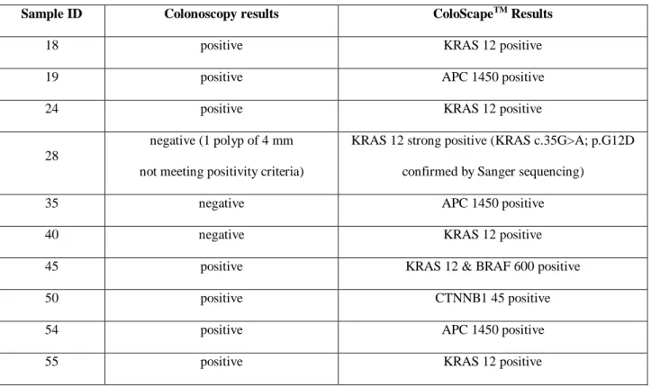
Table 2. Summary of Colonoscopy and ColoScapeTM results
Sample ID Colonoscopy results ColoScapeTM Results
18 positive KRAS 12 positive 19 positive APC 1450 positive 24 positive KRAS 12 positive
28
negative (1 polyp of 4 mm not meeting positivity criteria)
KRAS 12 strong positive (KRAS c.35G>A; p.G12D confirmed by Sanger sequencing)
35 negative APC 1450 positive 40 negative KRAS 12 positive 45 positive KRAS 12 & BRAF 600 positive 50 positive CTNNB1 45 positive 54 positive APC 1450 positive 55 positive KRAS 12 positive
1.1.4 Discussion
Liquid-biopsy is a challenging type of sample for mutational analysis. We look for cfDNA and
ctDNA. Estimates for ctDNA range from 1 to 10% of cfDNA. On top of this, mutations can occur
at different allelic frequencies, which may be in some cases as low as 0.1%.(19) XNA aims to
maximize analytical sensitivity due to its ability to selectively amplify only, or predominantly,
mutant forms and block wild-types. The manufacturer recommends a minimum of 5 ng of DNA
per reaction, although there are evidences that it could work with a 2.5 ng DNA input as well. In
this pilot study, it was aimed to assess the limits of the assay considerably, and determined to
accept even samples with a sub-optimal DNA input, for the goal was to establish the best
work-flow for advanced adenomas. Of note, 4 of the 6 positive cases missed by ColoScapeTM had a less
than suboptimal DNA input (data not shown). Had they been ruled out as inadequate, sensitivity
would have increased from 53.8 to 69%. However, as stated previously , this is not a clinical trial,
but rather an initial, preliminary technical evaluation. The most prevalent mutation was found in
the KRAS gene (4 cases). Other mutations were APC (2 cases) and CTNNB1 (1 case), and BRAF
in one case of dual positivity with KRAS. Interestingly, a case (#28) of a polyp with size of 4 mm,
which did not meet the positivity criteria, showed a KRAS positivity and Sanger sequencing
confirmed the mutation being KRAS c.35G>A; p.G12D. One case that was excluded due to
inadequate bowel preparation, was negative and showed no relevant genetic variations.
Given the small sample size, sensitivity, specificity and resulting predictive values, must be
considered only estimates that will help design and power a future clinical trial. However, it is of
considerable interest to consider that detection of advanced adenomas is a real challenge for
screening programs that are based on the FIT test, and for the other clinically approved molecular
tests, such as Cologuard and Septin 9. One has to also consider specificity that should ideally
exceed 90% in order to rule out a significant number of FIT+ patients that now turn out negative
most important result obtained from this study was the identification of a clinically relevant
work-flow that can optimize performance and allows an estimation of the test sensitivity and specificity
that will be a crucial focus of the future trial. Other interesting aspects to be investigated will be:
management of FIT+, triage – patients, management of FIT+, triage + and colonoscopy – patients,
management of patients with inadequate bowel preparation. Based on the results from this study,
it
further studies are warranted in order to validate the use of liquid biopsy – based ColoScapeTM
Chapter 2
2.1. Development of a gene panel for next generation sequencing of clinically relevant mutations in cell-free DNA from cancer patients.
Precision medicine, coupled with the tissue-based assessment of biomarkers predictive of
treatment outcome, has transformed pathology practice. (20) RAS and BRAF mutation testing
in colorectal cancer (21,22), EGFR in non-small-cell lung cancer (NSCLC, 23) BRAF in
melanoma (24) and cKIT and PDGFRa in gastrointestinal stromal tumours (25) has added a
genotypic element to the phenotypic diagnostics of solid tumours. However, tumour tissue is
not always available or may be insufficient for molecular testing. In this setting serum- and
plasma-derived cfDNA represents a complementary biological source to evaluate molecular
assessment of clinical relevant biomarkers by adopting next generation sequencing platforms
(NGS), which can be multiplexed across several genes to cover less common and even novel
variants (26, 27). Large gene panels or whole-exome approaches to screen for a large number
of genomic regions may not be easily implemented in cfDNA analysis , in fact circulating
tumour DNA represents only a small fraction (<0.5%) of the total cfDNA and a ‘ultra-deep
sequencing’ strategy, based on implementation of small NGS panels that tailored to target a limited number of actionable genes, can significantly increase analytic sensitivity reducing the
number of samples classified as “inadequate”. Following this concept Molecular pathology lab of University Federico II designed and developed a narrow gene panel that targets 568
clinically relevant mutations in six genes (EGFR, KRAS, NRAS, BRAF, cKIT and PDGFRa)
tissue sample or liquid biopsy with high sensitivity and specificity. Moreover, its clinical
performance in a clinical setting was evaluated.
2.1.1 Materials and Methods
Design of the SiRe panel.
The Ion AmpliSeq Designer suite v5.3.1 with hg19 was used as reference genome to develop a
customised panel targeting six genes (EGFR, KRAS, NRAS, BRAF, cKIT and PDGFRa) that
are associated with treatment outcome in NSCLC, GIST, CRC and metastatic melanoma
(21-24). A single primer pool leading to the selection of 42 amplicons (ranging from 125 to 175bp)
enabled us to cover all COSMIC annotated mutations (n=568) in the selected exons of the
target genes. The amplicon design covering 5.2kb of genomic DNA was optimized for the
simultaneous analysis of 16 samples with the 316v2 chip (Thermofisher, Foster City, CA,
USA) on a Personal Genome Machine Torrent (Thermofisher).
Study design, patients and samples.
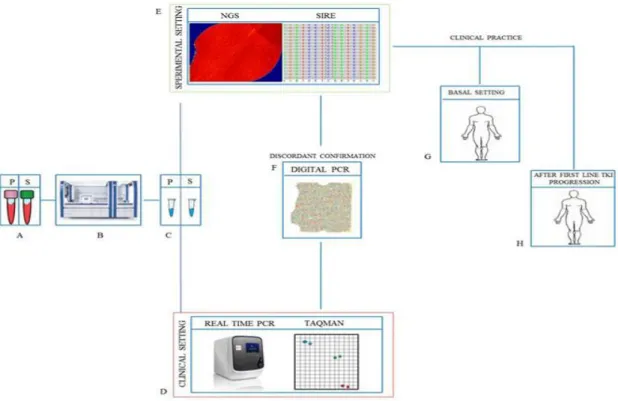
The panel performance was evaluated in three steps (Figure 2). First, the analytical sensitivity
of the assay was assessed on DNA from two cell lines and by using an artificial reference
standard with multiple mutations in different genes. Second, clinical sensitivity and specificity
was determined using archival cfDNA from 63 cancer patients with paired tumour tissue,
previously genotyped with a Taqman derived assay(TDA). As exploratory analysis, to confirm
that our NGS approach cover the mutations in cKit and PDGFRa genes, two GIST samples
(bloods and tissues) were tested with SiRe and the relative data are not reported only in this
thesis. Third, the performance of the panel in daily clinical practice was assessed using blood
consent was obtained from all patients and documented in accordance with the general
authorization to process personal data for scientific research purposes from ‘The Italian Data
Protection
Authority’(http://www.garanteprivacy.it/web/guest/home/docweb/docwebdisplay/export/24853 92). All information regarding human material was managed using anonymous numerical
codes, and all samples were handled in compliance with the Helsinki Declaration
(http://www.wma.net/ en/30publications/10policies/b3/). DNA purification. DNA from the two
cell lines was isolated using the QIAamp Mini Kit (Qiagen, Hilden, Germany) according to the
manufacturer’s instructions. Circulating-free DNA was purified as follows: 15ml blood was withdrawn from patients and collected in Vacutainer tubes (BD, Plymouth, UK). Plasma and
serum were isolated by centrifugation twice at 2300 r.p.m. for 10min. The supernatant (serum
or plasma) was aliquoted and used immediately for cfDNA isolation or stored at - 80ºC.
Cell-free DNA was purified from serum and plasma for each patient (1.2ml). In the rare instances
that the volume of the serum and plasma sample obtained from a patient was between 1 and
1.2ml, PBS up to 1.2ml was added to the samples, which were then purified using the
QIAsymphony robot (Qiagen) and the QIAsymphonyDSPVirus/ Pathogen Midi Kit, according
to the manufacturer’s instructions, and cfDNA was eluted in a final volume of 30ml. Since correct pre-analytical handling of blood specimens is crucial to maintain the sample
informative, the process was standardised (in terms of blood collection, sample centrifugation
and cfDNA extraction) in the Department of Public Health of the University of Naples
Federico II, and all procedures were performed in-house by a nurse belonging to the laboratory
Sample sequencing.
We analysed the serum and plasma cfDNAs of each patient enrolled in the study. Libraries
were constructed and purified on the Ion Chef (Thermofisher), and eight samples
(corresponding to 4 patients) were added per run. Library generation was as follows: 15µ of
cfDNA were dispensed on Ion Code plates and amplified using Ion AmpliSeq DL8
(Thermofisher). We used 22 cycles for cfDNA amplification and 6 cycles for library
reamplification after barcoding, under the thermal conditions defined by the manufacturer.
Purified libraries derived from eight cfDNA samples were diluted to 60pM and combined with
eight additional cfDNA-derived libraries to obtain a 16 Ion Code pooled library. The
two-pooled libraries were re-loaded into the Ion Chef instrument, and templates were prepared
using the Ion PGM Hi-Q IC Kit (Thermofisher). Finally, templates were loaded into the 316v2
chip and sequenced on PGM. Data analysis. Signal processing and base calling were carried
out using the default base-caller parameters on Torrent Suite [v.5.0.2] and coverage analysis
was performed using SiRe designed bed files with coverage plug-in (v.5.0.2.0). BAM files
were visually inspected with the Golden Helix Genome Browser v.2.0.7 (Bozeman,MT, USA).
Variants were automatically annotated using variant caller plug-in (v.5.0.2.1) at specific
optimised parameters of the SiRe panel. In particular, only variants with ≥5X allele coverage
and a quality score ≥20, within an amplicon that covered at least 1000X alleles, were called,
Figure 2. Study design .cfDNAs (A) extracted with the QIAsymphony virus/pathogen kit (B) from paired (P)
plasma and (S) serum (C) samples were analyzed by quantitative 50-nuclease TaqMan PCR (D) and by the NGS SiRe panel (E). Any discordance between the two techniques was evaluated by dPCR (F). After preclinical validation, the SiRe panel was applied in clinical practice in cases in which tissues were not available to select patients for TKI treatment, at baseline (G), and to evaluate the selection of resistant clones after disease progression (H). (ref. 17)
Preclinical assessment.
Genomic DNA from the HCC827 (EGFR p.E746-A750del; KRAS wt) and A549 (EGFR wt;
KRAS p.G12S) cell lines was used to assess analytical performance. Both cell lines were
obtained from the National Research Council/Institute of Experimental Endocrinology and
Oncology on courtesy of Dr Pierlorenzo Pallante (Naples, Italy). The analytical sensitivity of
the assay for point mutation and indel detection was determined by diluting DNA from the
A549 for indels). DNA dilutions ranged between 1:10 and 1:10000, which correspond to allelic
fractions from 1:20 to 1:20000 of the mutated allele (both cell lines are heterozygous). Each
dilution was analyzed in duplicate to estimate inter-run assay reproducibility, and the library
obtained from each dilution was sequenced twice to evaluate intra-run assay reproducibility. In
addition, customized Horizon Diagnostics Multiplex gDNA reference standard, with mutation
in EGFR (p.E746_A750del and p.G719S), KRAS (p.G12D), NRAS (p.Q61L) and BRAF
(p.V600E), each of them at three different dilution points (1, 0.5 and 0.1%), were assessed to
provide stronger evidence on SiRe analytical performance.
Clinical validation.
We determined the specificity and sensitivity of our assay by analyzing archival serum and
plasma cfDNA from 40 cancer patients at presentation attending the QuironDexeus University
Hospital (33 NSCLC, 2 CRC and 5 metastatic melanoma) with paired tumour tissue. In
addition, we tested archival serum and plasma cfDNAs from 12 responder patients and 11
patients at the time of tumour progression after treatment (18 NSCLC, 2 CRC and 3 metastatic
melanoma; Table 4). All of the 63 cfDNA samples and tumour tissues had previously been
genotyped for EGFR, KRAS, NRAS and BRAF mutations using a TDA (28, 29). In the case of
tumour tissues, genotyping had been confirmed by standard PCR followed by Sanger
sequencing. Cases showing discordance between the NGS SiRe panel and the TDA were
further investigated by digital PCR (dPCR) on a QuantStudio 3D Digital PCR System platform
(Thermofisher) as previously described. (30)
Performance of the SiRe panel in prospective clinical samples.
genotyped 79 advanced NSCLC patients (37 men and 42 women; mean age: 65 years) using
blood samples collected at the Department of Public Health of the University of Naples
Federico II (Table 4). According to the European Medicines Agency guidelines, mutations
related to EGFR disease were tested in patients when tissue was not available at presentation
(n=46), or at tumour progression (n=33) in patients previously treated with erlotinib (n=14),
gefitinib (n=14) or afatinib (n=5) in the attempt to detect the emergence of resistance secondary
mutations. In 21 of the 33 cases with tumour progression, first-line TKI administration had
been based on the demonstration of an EGFR mutation in tissue, whereas in the remaining
12/33 cases, TKI treatment had been administrated in second line without evidence of EGFR
mutations.
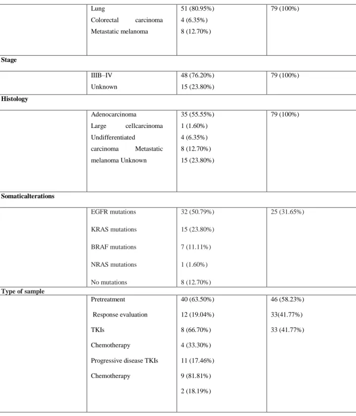
Clinical characteristics Retrospective validation (N=63) Prospective validation (N=79) Age ≥29–60 <61–80 Unknown 22 (34.92%) 25 (39.68%) 16 (25.40%) 22 (27.85%) 57 (72.15%) Sex Male Female Unknown 24 (38.10%) 24 (38.10%) 15 (23.80%) 37 (46.84%) 42 (53.16%) Smoking status Never smokers Ex-smokers Smokers Unknown 11 (17.46%) 9 (14.30%) 5 (7.93%) 38 (60.31%) 38 (48.10%) 29 (36.70%) 6 (7.60%) 6 (7.60%)
Type of tumour Lung Colorectal carcinoma Metastatic melanoma 51 (80.95%) 4 (6.35%) 8 (12.70%) 79 (100%) Stage IIIB–IV Unknown 48 (76.20%) 15 (23.80%) 79 (100%) Histology Adenocarcinoma Large cellcarcinoma Undifferentiated carcinoma Metastatic melanoma Unknown 35 (55.55%) 1 (1.60%) 4 (6.35%) 8 (12.70%) 15 (23.80%) 79 (100%) Somaticalterations EGFR mutations KRAS mutations BRAF mutations NRAS mutations No mutations 32 (50.79%) 15 (23.80%) 7 (11.11%) 1 (1.60%) 8 (12.70%) 25 (31.65%) Type of sample Pretreatment Response evaluation TKIs Chemotherapy
Progressive disease TKIs
Chemotherapy 40 (63.50%) 12 (19.04%) 8 (66.70%) 4 (33.30%) 11 (17.46%) 9 (81.81%) 2 (18.19%) 46 (58.23%) 33(41.77%) 33 (41.77%)
Table 4. Characteristics of the patients included in the retrospective (left) and prospective (right)
2.1.2 Results
Panel design and preclinical performance evaluation.
The SiRe panel was designed to cover 568 clinically relevant mutations in six genes (EGFR,
KRAS, NRAS, BRAF, cKIT and PDGFRa) involved in NSCLC, GIST, CRC and
metastatic. In Preclinical evaluation, in On cell line derived DNA, the SiRe panel detected
the EGFR deletion p.E746_A750del and the KRAS point mutation p.G12S at a level as low
as one copy of the mutated allele in a background of 20000 copies of wild-type alleles
(0.005% mutated allele fraction), with 100% of intra- and inter-run reproducibility. In
addition, regarding the results obtained on multiplex gDNA reference standard (Horizon
Diagnostics), p.E746_A750del and p.G719S point mutation in EGFR, p.G12D mutation in
KRAS exon 2, p.Q61L mutation in NRAS exon 3 and p.V600E mutation in BRAF exon 15
were correctly identified for each different dilution point. This high analytical performance
was achieved thanks to the use of optimised parameters set in variant caller plug-in
(v.5.0.2.1) which detected low abundant mutated alleles with a specificity of 100%.
Clinical sensitivity and specificity of the SiRe panel in cfDNA samples.
The retrospective series of cfDNAs was constituted by 126 paired serum and plasma
samples from 63 patients. In each run, up to 16 paired serum and plasma samples from eight
patients on 316v2 were processed. Run median output was 257 Mb (Mega bases), median
read length was 124bp, mean read depth was 2821x and coverage uniformity was 97%.
55 patients with EGFR, KRAS, NRAS or BRAF mutations in tumour tissue, the SiRe panel
detected the same mutation in the serum and/or plasma cfDNA in 46 cases (sensitivity
83.6%, CI 67.3–94.3%;). (Table 5.)
TDA (cfDNA) SiRepanel
(cfDNA)
Mut + Mut - Total
Mut + 42 4 46
Mut - 0 17 17
Total 42 21 63
Table 5.Concordance of Taqman-derived assay (TDA) and the SiRe panel NGS in retrospective serum and
plasma cfDNA samples.
Comparison of the SiRe panel with a TDA in cfDNA samples.
We compared the performance of the SiRe panel for mutation analysis in cfDNA with that of a
previously reported TDA (28, 29) in 63 samples: (i) the 40 cfDNA samples obtained at
presentation mentioned above; (ii) archival serum and plasma cfDNAs from 12 patients in
response to different types of antitumor drugs; and 11 patients mutations in the cfDNA of 46 of
63 patients. The test was positive in both serum and plasma cfDNA in 35 patients (76.1%),
positive in plasma but not in serum in 5 patients (10.9%), and positive in serum but not in
plasma in 6 patients (13%). An EGFR sensitising mutation and the p.T790M resistance
mutation were detected simultaneously in 10 patients at progression to EGFR TKIs. As
reported in Table 5, there was a high concordance (Cohen’s Kappa 0.85) between the results
obtained with the NGS SiRe panel and the TDA, although the performance of the SiRe was
the SiRe panel, and the 17 negative samples with the panel were also negative at TDA. In
addition, NGS detected mutations in the cfDNA of four patients, whereas TDA did not. The
mutations in these four patients appeared also in paired tumour tissue. One was a p.L597R
mutation in BRAF not covered by the TDA, and was confirmed by dPCR. The remaining three
mutations were a p.L861Q mutation in EGFR and two KRAS mutations, p.G12C and p.G12A.
Both TDA and NGS using the SiRe panel enable quantification of the mutated alleles..There
was a significant correlation in the levels of serum cfDNA between the two techniques
(r=0.64). In contrast, correlation was lower in the case of plasma (r=0.35), but improved
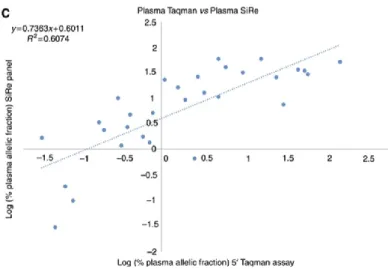
significantly when three outlier samples were removed (r=0.61). (Figure 3.)
Figure 3. Quantification of mutated allele fractions.Comparison of the quantification of mutated allele
fractions by Taqman Derived Assay vs SiRe NGS in serum (A) and plasma (B) cfDNA. In the case of plasma, three outliers were removed and results re-plotted (C). (Ref. 17)
Evaluation of the SiRe panel for prospective analysis of clinical samples.
The performance of the SiRe panel in the clinical setting was evaluated by prospectively testing
the serum and plasma cfDNA of patients with advanced NSCLC for whom no tissue was
available in order to select them for TKI treatment. Seventy-nine patients were tested, 46 at
presentation and 33 at the time of tumour progression after first-line TKI treatment (Table 4).
The NGS procedure was adequate for variant calling in the 79 cfDNA paired serum and plasma
samples. The run metrics parameters were not dissimilar from those of the retrospective
samples. In fact, in prospective cfDNA samples, the median output was 210Mbases, the
median read length 125.57bp, the mean read depth 3385.45 and coverage uniformity 97.49%.
Among the 46 patients analysed at baseline, we detected four EGFR mutations (8.7%), one
point mutation in exon 18 (p.G719A), two deletions in exon 19 (both p.E746_A750delELREA)
and one insertion in exon 20 (p.H773-V774insH). In all four patients, the mutant alleles were
shown). Regarding samples at progression, the SiRe panel did not detect mutations in 12
patients, whose tissues had been identified as EGFR wild type in biopsies at presentation. In
contrast, among the 21 patients EGFR positive in baseline tissue, the SiRe panel confirmed the
same mutation in cfDNA in 19 cases (Table 6).
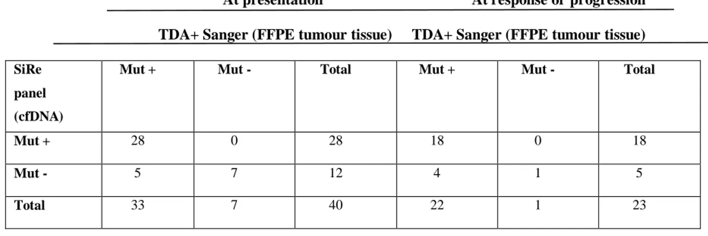
At presentation At response or progression TDA+ Sanger (FFPE tumour tissue) TDA+ Sanger (FFPE tumour tissue)
SiRe panel (cfDNA)
Mut + Mut - Total Mut + Mut - Total
Mut + 28 0 28 18 0 18
Mut - 5 7 12 4 1 5
Total 33 7 40 22 1 23
Table 6. Comparison of the mutational status in FFPE tumor tissue at presentation with the results of the SiRe
panel in archival cfDNA purified from serum and plasma baseline (n=42, left) and at response or after tumor progression (n=23, right)
Thus, sensitivity and specificity in this cohort of patients at progression were within the
range of those observed in the retrospective cohort. Interestingly, in 9 of those 19 cases
(47%), we observed the emergence of the EGFR p.T790M mutation in addition to the
original EGFR activating mutation. The appearance of EGFR p.T790M mutation in relation
to TKIs treatment regimen was reported in figure 4. Of the 28 mutations (sensitising
+p.T790M) detected, 10 (35.70%) were present in both serum and plasma, 7 (25%) in
plasma alone and 11 (39.3%) in serum alone. All mutations detected by the SiRe panel at
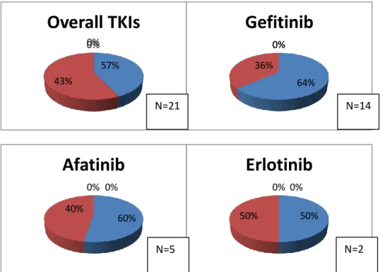
Figure 4. Frequency of the EGFR p.T790M mutation (green: T790M− red T790M+) after progression to
thyrosine kinase inhibitors (TKIs) in the serum and plasma cfDNA of EGFR-mutated patients evaluated with SiRe panel NGS. 57% 43% 0% 0%
Overall TKIs
64% 36% 0% 0%Gefitinib
60% 40% 0% 0%Afatinib
50% 50% 0% 0%Erlotinib
N=21 N=14 N=5 N=22.1.3 Discussion
In this chapter, we analyzed the performance of ultra-deep sequencing using a narrow NGS
panel on Ion Torrent PGM is excellent, and how this procedure can be used for the routine
testing of relevant tumour mutations in cfDNA. The high sensitivity (90.5%) and analytical
specificity (100%) of this panel equal or even surpass those of such other procedures as real
time PCR-based methods. Unlike earlier NGS applications that cover large genomic regions
(27), our small gene panel (5.2kb) focuses on biomarkers that are currently used in the clinical
setting. The ultra-deep sequencing procedure reported herein has various advantages. In fact,
using a single panel, we were able to detect up to 568 relevant mutations in six genes (EGFR,
KRAS, NRAS, BRAF, cKIT and PDGFRa). These mutations included less common, but
actionable variants such as the BRAF p.L597R mutation in melanoma. Sequencing with the
SiRe panel was more efficient than real-time PCR target techniques in detecting deletions
(n=2) and point mutations (n=6) on cfDNA samples. In addition, NGS per se is a time-effective
procedure for analysing large numbers of samples, thereby optimising the work flow in
molecular pathology laboratories.(26) With our procedure, different types of samples (DNA
from tumour tissues and cfDNAs from biological fluids) from patients affected by different
types of diseases (e.g., NSCLC, GIST, CRC and melanoma) can be processed simultaneously.
Consequently, sample batching is more effective and does not require a minimum number of a
given tumour type. As a result, turnaround time (TAT) can be as short as three working days,
as recommended by international guidelines.(31) The recently developed Ion Chef automated
library preparation station, which has a better procedure reproducibility and standardisation
than manual procedures, also contributes to the short TAT (26). The Ion Torrent PGM
cfDNA. Firstly, we reduced the number of genes and exons vs commercially available tests,
and we modified the thresholds for variant calling, in particular all the variants with ≥5X allele coverage and a quality score ≥20, within an amplicon that covered at least 1000X alleles, were called. We also adapted the Ion Chef template preparation protocol by pooling two 16-sample
libraries in each run. Thus, using this well standardized procedure, we were able to sequence
simultaneously up to 32 paired plasma/serum samples in less than 3h on the PGM, with a
consequent reduction in the total consumable cost. In a previous study (26) we showed that by
using a commercially available 22 gene panel (AmpliSeq Colon and Lung Cancer Panel) on the
Ion Torrent PGM, the consumable cost was euro 196 per sample. Using the modified protocol
that we developed in this current study the cost per sample was lowered to 98 euro for
simultaneously analysis of six different genes. This is comparable with the cost of the most
commercially available Real Time PCR based kits. The simultaneous analysis of paired
plasma/serum samples is a crucial feature of this new procedure since the sensitivity of somatic
mutation analysis in cfDNA increases when serum and plasma are analysed together (28, 29).
Our results are in agreement with this finding. In fact, of the 89 patients found to carry
mutations in cfDNA, 58 (65.17%) were positive in both serum and plasma, 15 (16.85%) in
plasma alone and 16 (17.98%) in serum alone. From the technical point of view, even when
sequencing 16 samples simultaneously in a run, the SiRe panel had optimal run metrics in our
daily clinical practice in terms of both mean depth reads and uniformity of coverage, which
resulted in a high assay sensitivity in cfDNA vs tumour tissue (90.5%) and a specificity of
100%. This is a very high degree of concordance, particularly given the 91.7% concordance
between paired surgical resection and cytological samples (32). Thanks to the high sensitivity
of our assay, the EGFR mutational rate of 8.7% that we identified in NSCLC patients
prospectively tested on cfDNA at baseline is in keeping with previous data on tissue samples.
(33) Similarly, the frequency of the EGFR p.T790M mutation, which was detected in the
n=1 erlotinib), is in line with data obtained on tissues samples collected after disease
progression (29). The performance of our methodology compares favorably with that of NGS
for mutational analysis in the blood of cancer patients. An Ion Torrent-derived sequencing of
five genes in cfDNA purified from never smoking lung cancer patients achieved a modest 58%
sensitivity and 87% specificity (34). An analysis of 23 amplicons in five genes using cfDNA
from breast cancer patients identified 10 mutations but missed 6 identified by droplet digital
PCR. (35) When restricted to EGFR, deep sequencing achieved 61–80% sensitivity and 94–
98% specificity in advanced NSCLC.(36) The 90.5% sensitivity of our assay also exceeds the
77% recently reported when NSCLC plasma-derived cfDNA was analysed on an Illumina NGS
platform with a panel covering amplicons of 11 clinically relevant genes.(37) Despite the
variations inherent to the platforms used, such as the library preparation and the longer TAT (6
days), the Illumina-based NGS approach featured similar run metrics and analytical parameters
as our assay, which supports the use of ultra-deep sequencing in the clinical setting.(37) It is
conceivable that the higher sensitivity achieved by our panel is due not only to technical
differences but also to the simultaneous testing of serum and plasma in each patient. Besides
being an alternative to molecular diagnosis at presentation when tumour tissue is not available,
liquid biopsy is also a non invasive test with which to monitor response to targeted therapy and
to detect the emergence of resistance mutations in genes such as EGFR (38) and ESR1 (Chu et
al, 2016). Monitoring would consist in quantifying the mutant allelic fractions in cfDNA over
time, which can be reliably assessed by our NGS assay. The SiRe panel detects the appearance
of resistance mutations such as EGFR p.T790M. Finally, the non-synonymous mutation burden
correlates with a good response to immunotherapy in NSCLC (39) and other tumours, and
NGS has been proposed as a tool with which to design customised immunotherapies that target
common driver mutations.(40) Our panel, which covers several exons in frequently mutated
mutations in cfDNA purified from the serum and plasma of patients with the tumours most
commonly tested for molecular alterations (such as NSCLC, CRC and metastatic melanoma).
The SiRe panel has excellent sensitivity and specificity, and is hence suitable for testing blood
samples in the clinical setting. Finally, it enables the application of NGS on a prospective basis
in daily molecular predictive pathology practice, particularly when tumour tissue is not
Chapter 3
3.1.1 Cell free DNA analysis by SiRe® panel in a basal setting of NSCLC patients.
Non small cell lung cancer (NSCLC) is diagnosed in most cases at advanced stages of
disease. Diagnostic samples are frequently scarcely cellular, being represented by
either cytological specimens or small tissue endoscopic biopsies; these limited tissue
samples often may be not sufficient for epidermal growth factor receptor (EGFR) and
other clinical relevant biomarkers, such as ALK translocation and PD-L1 expression,
whose assessment is required to select patients for first line treatment administration
(40, 41). In particular for EGFR tyrosine kinase inhibitors (TKIs), such as gefitinib,
erlotinib and afatinib the identification of activating EGFR mutations in exon 18, 19
and 21 is mandatory before the first line treatment (33, 42- 46). To date according to
the European Medicines Agency guidelines, in patients without tissue availability,
only for EGFR TKIs treatment decision making, cell-free DNA (cfDNA) can be used
as a fast and non-invasive surrogate for EGFR mutational testing (17, 29, 47- 49).
However, the assessment of gene mutations in cfDNA is challenging, in particular in
basal setting, for the detection of first and second TKIs generation EGFR sensitizing
mutations, due to the very low concentration of circulating tumor DNA, that represent
only a small fraction of the total cfDNA. (17,48,49,50-52) Thus, the clinical
implementation of next generation techniques, such as next generation sequencing
(NGS) or digital PCR (dPCR) based assay is crucial (17,48,49,50,53,54,). In a recent
mutation detection in EGFR, KRAS, NRAS, BRAF, cKIT and PDGFR starting from
cfDNA retrieved from patients with different solid tumors (NSCLC, metastatic
colo-rectal cancer, melanoma and gastrointestinal stromal tumor).(17) SiRe® showed a
lower limit of detection (0.01%) and an higher analytical performance respect to a
very sensitive modified TaqMan probe real time PCR based approach.(17) The
analysis of cfDNA gene mutations was carried out using as gold standard the
mutational status obtained on matched tissue derived DNA, but little is known
regarding the application of this approach in clinical setting, in particular in baseline
setting of NSCLC patients, prior to EGFR TKIs administration, without a referent
DNA derived tissue to confirm the mutational data obtained on cfDNA.(55) The aim
of the present study was to review the NGS data obtained by using SiRe® NGS
panel starting from cfDNA collected in routine NSCLC baseline setting to
prospectively select patients, without tissue availability, for first and second
generation EGFR TKIs treatment administration.
3.1.2 Material and Methods
From January 2017 to March 2017 , n=64 liquid biopsy analysis was requested from
the oncologists of different South Italy institutions (n=14), following the European
Medicines Agency guidelines, for the analysis of EGFR mutations on cfDNA in
NSCLC patients without tissues availability at presentation, to assess the eligibility to
first and second generation EGFR TKIs (Table 7). On the overall n=39 men and
n=25 women were analyzed with a mean age of 66 years (range, 36–89 years). For
each patient, 10 mL of blood was collected in-house by using EDTA Vacutainer
of the University of Naples Federico II. The protocols adopted in this study were
previously validated (13). Briefly, before cfDNA extraction, two centrifugation steps
(2,300 rpm for 10 min) were carried out to obtain at least 1.2 mL of plasma for each
patient. cfDNA was extracted by using the QIAsymphonyDSPVirus/Pathogen Midi
Kit on the QIAsymphony robot (Qiagen, Venlo Limburg) accordingly with the
manufacturer instructions. By using SiRe panel, following the previously validated
protocol, libraries were automated constructed and purified using Ion AmpliSeq DL8
Kit (Thermofisher) on the Ion Chef instrument (Thermofisher) and, after barcoding,
purified libraries derived from eight cfDNA plasma samples were diluted and
combined with eight additional cfDNA-derived libraries to obtain a 16 Ion Code
pooled library, re-loaded into the Ion Chef instrument for template preparation by
using the Ion PGM Hi-Q IC Kit (Thermofisher). Finally, templates were loaded into
the 316v2 chip and sequenced on Personal Genome Machine (PGM). Signal
processing and base calling were carried out using the default base caller parameters
on Torrent Suite (v.5.0.2) and coverage analysis was performed using SiRe specific
bed files with coverage plug-in (v.5.0.2.0). In addition to automatic variant calling
analysis, by using SiRe panel specific optimized variant caller plug-in (v.5.0.2.1)
parameters, BAM files were visually inspected with the Golden Helix Genome
Browser v.2.0.7 (Bozeman, MT, USA). Only variants with >5× allele coverage and a
quality score >20, within an amplicon coverage at least 1,000× alleles, were reported
and the relative mutated allele frequency was annotated, considering not only EGFR,
but also KRAS, BRAF and NRAS gene hotspots region, relevant for NSCLC and
covered by the SiRe panel. Written informed consent was obtained from all patients
and documented in accordance with the general authorization to process personal
docwebdisplay/export/2485392) and all samples were handled in compliance with
the Helsinki Declaration (http://www.wma.net/en/30publications/10policies/b3/).
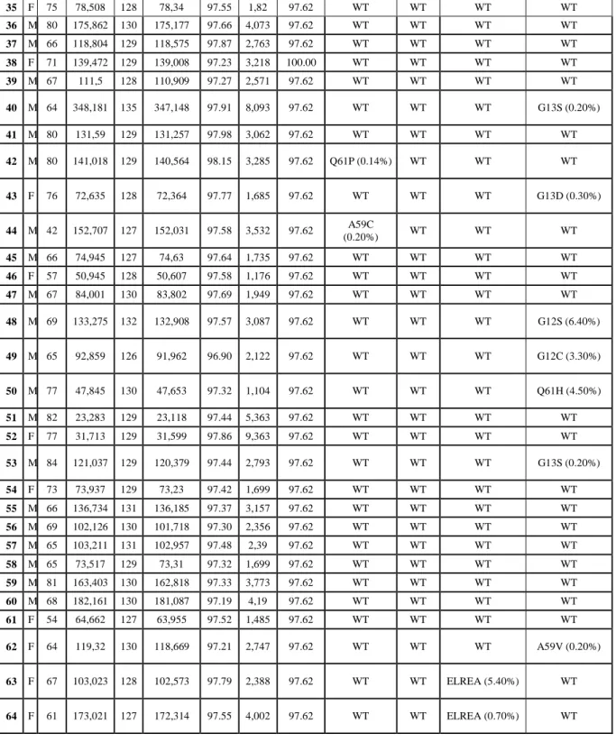
3.1.3 Results
The SiRe NGS analysis results were adequate in 98.4% of cases (63/64) accordingly to the
quality parameters reported in the methods section and previously validated; only one cases
(#30) failed to reach the quality thresholds for data analysis. Regarding the run metrics
parameters (Table 7), the median number of reads for sample was 120,960, the median
number of read length was 127 bp, the median number of mapped reads was 120,498, the
mean percentage of reads on target was 97%, the average reads for amplicon was 2,894 and
the uniformity of coverage was 98%, in accordance with the data obtained in our previous
validation study (17). On the overall, considering EGFR, KRAS, NRAS and BRAF genes, 24
patients (38%) showed at least one mutation. Only one patient (#7) showed two concomitant
mutations (NRAS p.G13D and KRAS p.Q61H). In particular, 5 EGFR mutations (8%) were
detected [n=2, exon 19 deletions (both p.E746_A750delELREA); n=2, exon 20 insertions
(p.H773_ V774insH and V769_D770insASV); and n=1, p.L858R exon 21 point mutation]; 14
KRAS point mutations (22%) [n=11, exon 2 mutations (n=4 p.G12C, n=3 p.G12D, n=1
p.G12S, n=1 p.G13D and n=2 p.G13S); and n=3, exon 3 point mutations (n=1 p.A59V and
n=2 p.Q61H)]; n=4 NRAS point mutations (6%) [n=2, exon 2 mutations (n=1 p.G12Sand n=1
p.G13D); and n=2, exon 3 point mutations (n=1 p.A59C and n=1 Q61P)]; 2 (3%) BRAF point
mutations [n=1 exon 11 p.G469A mutation and n=1 exon 15 p.V600E mutation]. The mutated
only the EGFR detected mutations by the SiRe panel were also confirmed by digital PCR
P a ti e n ts S e x A g e R e a d s M e a n R e a d Le n g th N u m b e r o f m a p p e d r e a d s % r e a d s o n ta r g e t (%) A v e r a g e r e a d s p e r a m p li c o n U n ifo r m ity o f a m p li c o n c o v e r a g e (%) N R A S (a ll e li c fr e q u e n c y ) BR A F (a ll e li c fr e q u e n c y ) EG F R (a ll e li c fr e q u e n c y ) K R A S (a ll e li c fr e q u e n c y ) 1 M 76 325,098 127 324,684 97.31 7,523 100.00 WT WT WT WT 2 F 77 223,871 130 223,209 97.71 5,187 100.00 WT WT WT WT 3 M 68 288,543 127 287,41 97.60 6,679 100.00 WT WT WT WT 4 F 74 26,017 125 25,666 96.70 590.9 97.62 G12S (1.00%) WT WT WT 5 F 36 163,218 128 162,752 98.06 3,8 97.62 WT WT WT G12D (1.50%) 6 F 50 146,656 127 145,906 97.25 3,378 100.00 WT V600A (0.20% ) WT WT 7 M 70 173,021 127 172,314 97.55 4,002 97.62 (0.34%) G13D WT WT Q61H (0.20%) 8 M 64 151,445 127 150,37 97.03 3,474 97.62 WT WT WT G12C (1.30%) 9 M 61 75,353 127 74,972 97.68 1,744 97.62 WT WT WT WT 10 M 45 190,62 126 189,708 97.49 4,403 100 WT WT H773_V774insH (37.0%) WT 11 M 48 103,023 128 102,573 97.79 2,388 97.62 WT WT WT G12C (0.60%) 12 F 61 163,363 128 162,841 97.75 3,79 97.62 WT WT WT WT 13 F 57 130,596 128 130,25 97.72 3,031 97.62 WT WT WT WT 14 F 51 155,551 128 155,018 97.40 3,595 97.62 WT WT WT WT 15 M 62 60,927 128 60,611 97.77 1,411 97.62 WT WT V769_D770insA SV (12.30%) WT 16 F 87 67,571 128 67,404 97.85 1,57 97.62 WT WT WT WT 17 F 77 181,295 129 180,633 98.06 4,217 95.24 WT WT WT WT 18 M 75 104,653 127 104,925 97.79 2,429 97.62 WT WT WT WT 19 M 58 262,771 130 261,202 97.94 6,091 97.62 WT WT WT WT 20 F 38 248,897 128 248,287 97.68 5,775 97.62 WT WT WT WT 21 F 51 169,989 128 169,581 97.44 9,734 97.62 WT WT WT WT 22 M 64 36,774 128 36,693 97.84 854.7 97.62 WT WT WT WT 23 M 74 195,245 128 194,624 97.50 4,518 100.00 WT WT L858R (3.20%) WT 24 F 54 82,48 128 82,303 97.80 1,916 97.62 WT WT WT WT 25 M 84 12,499 127 12,439 97.79 289.6 97.62 WT WT WT WT 26 F 59 71,556 129 71,36 97.54 1,657 97.62 WT WT WT G12D (1.30%) 27 M 68 62,909 128 62,723 97.61 1,458 97.62 WT WT WT WT 28 M 56 51,993 128 51,453 97.44 1,194 100.00 WT WT WT WT 29 M 89 21,306 127 21,126 97.12 488.5 97.62 WT WT WT G12D
30 M 53 76 60 66 39.39 0.69 91.45 Failed Failed Failed Failed
31 F 67 49,539 129 49,435 97.50 1,148 97.62 WT G469A (5.00% ) WT WT 32 M 62 113,202 130 112,93 98.01 2,635 97.62 WT WT WT G12C (5.63%) 33 M 70 72,936 129 72,752 97.77 1,694 97.62 WT WT WT WT 34 F 71 133,127 129 132,803 97.93 3,096 97.62 WT WT WT WT
35 F 75 78,508 128 78,34 97.55 1,82 97.62 WT WT WT WT 36 M 80 175,862 130 175,177 97.66 4,073 97.62 WT WT WT WT 37 M 66 118,804 129 118,575 97.87 2,763 97.62 WT WT WT WT 38 F 71 139,472 129 139,008 97.23 3,218 100.00 WT WT WT WT 39 M 67 111,5 128 110,909 97.27 2,571 97.62 WT WT WT WT 40 M 64 348,181 135 347,148 97.91 8,093 97.62 WT WT WT G13S (0.20%) 41 M 80 131,59 129 131,257 97.98 3,062 97.62 WT WT WT WT 42 M 80 141,018 129 140,564 98.15 3,285 97.62 Q61P (0.14%) WT WT WT 43 F 76 72,635 128 72,364 97.77 1,685 97.62 WT WT WT G13D (0.30%) 44 M 42 152,707 127 152,031 97.58 3,532 97.62 A59C (0.20%) WT WT WT 45 M 66 74,945 127 74,63 97.64 1,735 97.62 WT WT WT WT 46 F 57 50,945 128 50,607 97.58 1,176 97.62 WT WT WT WT 47 M 67 84,001 130 83,802 97.69 1,949 97.62 WT WT WT WT 48 M 69 133,275 132 132,908 97.57 3,087 97.62 WT WT WT G12S (6.40%) 49 M 65 92,859 126 91,962 96.90 2,122 97.62 WT WT WT G12C (3.30%) 50 M 77 47,845 130 47,653 97.32 1,104 97.62 WT WT WT Q61H (4.50%) 51 M 82 23,283 129 23,118 97.44 5,363 97.62 WT WT WT WT 52 F 77 31,713 129 31,599 97.86 9,363 97.62 WT WT WT WT 53 M 84 121,037 129 120,379 97.44 2,793 97.62 WT WT WT G13S (0.20%) 54 F 73 73,937 129 73,23 97.42 1,699 97.62 WT WT WT WT 55 M 66 136,734 131 136,185 97.37 3,157 97.62 WT WT WT WT 56 M 69 102,126 130 101,718 97.30 2,356 97.62 WT WT WT WT 57 M 65 103,211 131 102,957 97.48 2,39 97.62 WT WT WT WT 58 M 65 73,517 129 73,31 97.32 1,699 97.62 WT WT WT WT 59 M 81 163,403 130 162,818 97.33 3,773 97.62 WT WT WT WT 60 M 68 182,161 130 181,087 97.19 4,19 97.62 WT WT WT WT 61 F 54 64,662 127 63,955 97.52 1,485 97.62 WT WT WT WT 62 F 64 119,32 130 118,669 97.21 2,747 97.62 WT WT WT A59V (0.20%) 63 F 67 103,023 128 102,573 97.79 2,388 97.62 WT WT ELREA (5.40%) WT 64 F 61 173,021 127 172,314 97.55 4,002 97.62 WT WT ELREA (0.70%) WT
Table 7. Patients characteristics, SiRe next generation sequencing (NGS) panel run metric parameters (reads, mean
read length in base pair, number of mapped reads, percentage of read on target, average reads per amplicon, uniformity of amplicon coverage) and genes mutational status with relative mutated allele frequency are reported for each sample.
Figure 5. Case n.64 is reported. Digital PCR Quant Studio 3D cloud software (Thermofisher) was used to analyze
the scatter plot (A) and the copies of mutated and wild type alleles detected in one μl of the extracted cell-free DNA (cfDNA) (B). In the panel (C), the SiRe panel next generation sequencing (NGS) result is reported obtained on the same extracted cfDNA and analyzed by using Golden Helix Genome Browser v.2.0.7 (Bozeman, MT, USA) and showing an epidermal growth factor receptor (EGFR) exon 19 deletion (p.E746_ A750delELREA).
3.1.4 Discussion
Data, generated by the SiRe NGS panel on cfDNA, prospectively collected from NSCLC
patients, without tissue availability, examined for first and second generation EGFR TKIs
treatment administration, are here reported; the performance of this NGS panel designed to
cover only the current clinical relevant mutations, was more than excellent. Our data confirm
previous validation data. Preliminary, we had prospectively analyzed a total of 79 NSCLC
patients on cfDNA. In 46 instances, cfDNA had been derived from NSCLC patients at
presentation; in this subset, we detected four EGFR mutations (8.7%); more in details, these
were one point mutation in exon 18 (p.G719A), two deletions in exon 19 (both
p.E746_A750delELREA) and one insertion in exon 20 (p.H773-V774insH) (17). Here, in this
current subsequent study, we detected two exon 19 deletions (both p.E746_A750delELREA),
two exon 20 insertions (p.H773_ V774insH; V769_D770insASV) and one p.L858R exon 21
point mutation. Thus, we confirm an overall EGFR mutation rate of 8.0%. In all instances, the
EGFR mutations were always confirmed by an independent orthogonal dPCR based assay
(Figure 5). In addition, in the present study we have also sequenced, in the same sample set,
KRAS, NRAS and BRAF NSCLC relevant hot-spot regions, reporting an overall mutation
rate of 38%. In particular, we detected 22% KRAS, 6% NRAS and 3% BRAF mutated
samples, with only one patient that showed two concurrent mutations (NRAS p.G13D and
KRAS p.Q61H). It is remarkable to note that the mutation distribution in cfDNA of this
NSCLC baseline patient series was very similar to that reported on tissues derived DNA by
previous studies exploiting a multi-gene assay in NSCLC (55-58). As a general rule, in the
clinical trial settings the analysis of cfDNA had as a reference the mutational status obtained
on tissue derived DNA (55-58). Conversely, following the European Medicines Agency
NRAS and BRAF genes, offer an internal control in patients that do not show alterations in
EGFR, considering that in the most part of the cases these mutations in these genes are
mutually exclusive. In conclusion, our data update and confirm that SiRe NGS panel
represents a robust analytical tool for a centralized laboratory enabling the possibility to test
cfDNA mutational status in basal setting of NSCLC patients when no tissue samples are
References
1. Ahmadzada T, Kao S, Reid G, et al. An Update on Predictive Biomarkers for Treatment
Selection in Non-Small Cell Lung Cancer J Clin Med. 2018 Jun; 7: 153.
2. Cree I, Deans Z, Marjolijn J et al. Guidance for laboratories performing molecular
pathology for cancer patients J ClinPathol. 2014 Nov; 67: 923–931.
3. Elizabeth McPherson Genetic Diagnosis and Testing in Clinical Practice Clin Med Res.
2006 Jun; 4: 123–129.
4. Zhang R, Wenjun Yang, Zhang R, Yang W et al . Circulating Tumor
DNA as a Liquid Biopsy in Cancer. ClinOncol. 2017; 2: 1265.
5. Robertson DJ, Lee JK, Boland CR et al.Recommendations on Fecal Immunochemical
Testing to Screen for Colorectal Neoplasia: A Consensus Statement by the US
Multi-Society Task Force on Colorectal Cancer. Gastroenterology. 2017;152:1217-1237.
6. GISCOR Survey CCR 2014 (www.giscor.it)
7. Bubendorf L, Büttner R, Al-Dayel Fet al. Testing for ROS1 in non-small cell lung cancer:
a review with recommendations Virchows Arch. 2016; 469: 489–503.
8. Treece A, Montgomery N,Patel Net al. Fine-Needle Aspiration Smears as a Potential
Source of DNA for Targeted Next-generation Sequencing of Lung Adenocarcinomas
Cancer Cytopathol. 2016 Jun; 124: 406–414.
9. Volik S, Alcaide M, Morin Ret al. Cell-free DNA (cfDNA): Clinical Significance and
Utility in Cancer Shaped By Emerging Technologies Mol Cancer Res. 2016 Oct;14:
898-908.
10. Rolfo C, Mack P, Scagliotti GV et al. Liquid Biopsy for Advanced Non-Small Cell Lung