UNIVERSITÀ DEGLI STUDI DI CATANIA
DOTTORATODIRICERCAINBIOLOGIA,GENETICAUMANAEBIOINFORMATICA: BASICELLULARIEMOLECOLARIDELFENOTIPO-XXVIIICICLO
DIPARTIMENTODISCIENZEBIOMEDICHEEBIOTECNOLOGICHE SEZIONE DI BIOLOGIA E GENETICA “GIOVANNI SICHEL”
Rosalia Battaglia
MicroRNA signature changes in human ovarian follicle
in relation to reproductive aging
Tesi di Dottorato
Coordinatore:
Chiar.mo Prof. MICHELE PURRELLO Tutor:
Chiar.ma Prof.ssa CINZIA DI PIETRO
TABLE OF CONTENTS
1. Abstract 1
2. Introduction 4
From Germline Stem Cells toward fertilization-competent Oocyte 4
Human Primordial Germ Cells 4
Female Gametogenesis 9
Folliculogenesis 11
The role of the oocyte–cumulus cell dialogue in oocyte developmental competence 16
Transcriptional activity in oocytes 20
The new world of RNAs 23
microRNAs: genomics, biogenesis and function 25
Modulation of miRNAs 29
Vesicular and non-vesicular trafficking of miRNAs 30
miRNAs and ovarian function 34
miRNAs and Oocyte aging 38
Circulating miRNAs as biomarkers of reproductive disorders 42
3. Materials And Methods 45
Experimental design and sample classification 45
Women enrolled in the studies 47
Sample collection 48
Human metaphase II oocytes 48
Follicular fluid 49
Plasma 49
Mouse oocyte collection 50
Sample preparation 50
Exosome characterization 51
miRNA profiling in follicular fluid, plasma and exosomes 52 miRNA Isolation, Reverse Transcription, Preamplification, and miRNA expression
profile 52
miRNA profiling in human MII oocytes 55
Total RNA isolation, miRNA Reverse Transcription and miRNA
expression profile. 55
Expression Data Analysis 56
Study I 56
Study II 57
Study III 59
miRNA Target Prediction, Gene Ontology (GO) and Pathway Analysis 60
Study I 60
Study II 61
Study III 62
Network analysis 63
miRNA and Gene expression analysis 64
Statistical analysis 65
4. Results 66
Study I: Molecular characterization of exosomes and their microRNA cargo in human follicular fluid: bioinformatic analysis reveals that
exosomal microRNAs control pathways involved in follicular maturation. 66
Exosome Characterization and miRNA Isolation 66
Expression Profile of miRNAs 68
Genomic Analysis 70
GO and Pathway Analysis 71
Study II: The miRNome changes in Exosomes from human follicular
fluid in relation to aging. 74
Expression Profile of miRNAs 74
Study III: MiRNAs in human MII oocyte and their expression profile
changes in relation to aging 79
miRNA identification 79
mRNA targets, Gene Ontology and pathway analysis 82 Identification of miRNAs for validation in aging mouse model 84
Network analysis and mRNA selection 84
Validation with Single assays 87
5. Discussion 89
6. Conclusions and future perspective 94
Pag. 1
1. ABSTRACT
Small non-coding class of RNAs includes different types of molecules involved in different steps of RNA synthesis, processing, translation, as well as RNA modulation of transcription initiation, RNA degradation or protein synthesis block. Among these, microRNAs (miRNAs) represent the most studied and better characterized class of ncRNAs in terms of function and impact in human health and disease. miRNAs are short non-coding RNAs involved in the control of gene expression in different species. Specifically, miRNAs play a major role as master regulators of protein-coding genes. They target specific mRNAs for cleavage or translational repression. miRNA role in ovarian follicles has been characterized and the existence of miRNAs in human follicular fluid has been recently demonstrated. It was also reported that altered regulation of miRNA expression affects crucial pathways for follicle growth and oocyte maturation in several reproductive diseases. In addition, global miRNA analysis, on different tissues and organs and animal models has also shown that aging can influence mirna expression. Decreased female fertility with advanced maternal age has been widely documented. Although it is widely recognized that a key aspect that explains infertility in reproductive aging is the decline in oocyte quality, which also associates with higher risk of birth defects, genetic disorders and miscarriage, the molecular mechanisms underlying reproductive aging in female mammals are poorly understood. This thesis has aimed at evaluating impact of maternal age on miRNA expression profiles in human ovarian follicle and characterizing the pathways significantly affected by female ageing, identifying their regulator miRNAs. The manuscript includes three studies that for concision, they will be referred in the text, by Roman numerals, as Study I, II and III respectively. The Study I has been focused on the characterization of
Pag. 2
microRNAs in human follicular fluid (FF), the Study II on the miRNome changes in Follicular fluid exosomes from women of two different age groups; while miRNA identification in human MII oocyte and their expression profile changes in relation to reproductive aging have been shown in the Study III. Firstly, it was ascertained whether miRNAs are cargo of FF exosomes and whether they are involved in the regulation of follicle maturation. At a later stage, TaqMan Human microRNAs cards were performed to verify differently expressed miRNAs in FF respect with plasma samples, collected from 15 healthy women who underwent to intracytoplasmic sperm injections (ICSI). 37 miRNAs had significantly higher expression levels in human FF. 32 are carried by exosomes and involved in critically important pathways for follicle growth and oocyte maturation. Specifically, nine of them target and negatively regulate mRNAs expressed in the follicular microenvironment encoding inhibitors of follicle maturation and meiosis resumption. In order to reveal the contribution of miRNAs to female reproduction aging, we examined their expression changes during aging in human follicular fluid, using TLDA technology. Different miRNA distribution was found in FF exosomes from old and younger women. We detected about 50 miRNAs in FF exosomes which showed highly significant differences related to aging and were predicted to regulate ECM-receptor interaction, PI3K-Akt, p53, TGF-beta, HIF-1 and mTOR signaling pathways. Finally, we identified 57 miRNAs constantly expressed in 12 MII oocytes and 12 miRNAs displaying altered regulation in women of advanced reproductive age. By computational approach we explored the possible functions of differentially expressed miRNAs inside human germ cells, and confirmed experimentally miRNA differential expression in murine MII oocyte. Finally, a significant negative correlation miRNA-targets was found for miR-29a, miR-203 and specific mRNAs, that have been ascribed responsible for mechanisms modulating
Pag. 3
epigenetic changes underlying compromised oocyte quality caused by advanced maternal age. Data shown in this thesis were published in 2014 in Fertility and Sterility [Santonocito M, Vento M, Guglielmino MR, Battaglia R, Wahlgren J, Ragusa M, Barbagallo D, Borzì P, Rizzari S, Maugeri M, Scollo P, Tatone C, Valadi H, Purrello M, Di Pietro C. 2014. Molecular characterization of exosomes and their microRNA cargo in human follicular fluid: bioinformatic analysis reveals that exosomal microRNAs control pathways involved in follicular maturation. Fertil Steril;102(6):1751-61.e1] and also presented at the 30th Annual Meeting of the European Society of Human Reproduction and Embriology, held in Munich, Germany [R. Battaglia, M. Santonocito, M. Vento, M.R. Guglielmino, J. Wahlgren, M. Ragusa, D. Barbagallo, P. Borzì, S. Rizzari, M. Maugeri, P. Scollo, C. Tatone, H. Valadi, M. Purrello, C. Di Pietro. “MicroRNAs upregulated in follicular fluid are carried by exosomes: new actors in the communication between oocyte and somatic follicular cells?” 30th Annual Meeting of the European Society of Human Reproduction and Embriology, Munich, Germany from 29 June to 2 July 2014. Oral communication] and 16th World Congress on Human Reproduction held in Berlin, Germany [Rosalia Battaglia, Marilena Vento, Placido Borzì, Marco Ragusa, Davide Barbagallo, Paolo Giovanni Artini, Paolo Scollo, Carla Tatone, Michele Purrello, Cinzia Di Pietro. The miRNome landscape changes in human ovarian follicles in relation to aging. 16th World Congress on Human Reproduction held in Berlin, Germany on March 18-21 2015. Oral communication].
Pag. 4
2. INTRODUCTION
From Germline Stem Cells toward fertilization-competent Oocyte
Human Primordial Germ Cells
In most multicellular organisms, including mammals, germ cells are the origin of new organisms and ensure the perpetuation of the genetic and epigenetic information across the generations. They prepare for totipotency during their ontogeny through genetic and epigenetic regulations of their genome function. Specification of the germ cell lineage is fundamental in development and the correct transmission of the genome to the next generation [Saitou and Yamaji, 2012]. There appear to exist at least two pathways for the specification of germ cell fate. In one pathway, which is called ‘preformation’, embryonic cells that inherit maternal determinants from the egg proceed to form the germ cell lineage, whereas in the other, which is called ‘epigenesis’, pluripotent cells formed early in development are induced by signals from adjacent tissues to form the germ cell lineage. In mice, and presumably in all mammals, germ cell fate is induced by ‘epigenesis’ [Saitou and Yamaji, 2010], thus itis not an inherited trait from the egg, but is induced in the in pluripotent epiblast cells by signaling molecules [Saitou, 2009]. In many animals, primordial germ cells (PGCs) are the first germline cell population which colonize the developing gonads by active migration [Richardson and Lehmann, 2010]. The precursors of PGCs are early committed and specified in the epiblast before gastrulation and rapidly moved into an extraembryonic region where PGCs are determined. PGCs reenter into the embryo proper during early gastrulation to reach the developing gonads, the gonadal ridges (GRs). During this journey, while undergoing proliferation, PGCs begin extensive nuclear reprogramming (activation of genes for pluripotency and epigenetic changes of the genome involving DNA demethylation and
Pag. 5
histone code) to regain differentiation totipotency and reset the genomic imprinting. These processes are completed after their arrival into the GRs [Coticchio et al., 2013]. After some cycles of proliferation, PGCs initiate to differentiate either toward the spermatogenic or the oogenic pathway, into gonocytes or oogonia within testes and ovaries, respectively (Figure 1).
Figure 1: Schematic representation of germ cell development in mice. (Left) PGC development (from
E6.25 to ∼E12.5); (upper right) male germ cell development (from ∼E13.5); (lower right) female germ cell development (from ∼E13.5). [from Saitou and Yamaji, 2012 ].
The spermatogenic pathway most typically involves the establishment of spermatogonial stem cells (or male germline stem cells [GSCs]) with an enormous mitotic potential, whose homeostasis is likely accomplished by a stochastic balance of self-renewal and differentiation and not by regulated asymmetric cell division, whereas
Pag. 6
it appears that female GSCs in adults are rare across the phylogenetic spectrum [Spradling et al., 2011]. However, it is still generally accepted that the oogenesis is dependent on PGSs set established in human ovaries during fetal period of life and does not increase following birth [Virant-Klun, 2015], although several recent research studies have identified functional oogonial stem cells in the postnatal ovary of several different species including humans [Oatley and Hunt, 2012; White et al., 2012]. In the absence of the testicular determining factor, the differentiation of PGCs into oogonia occur at 6–8 weeks of gestation, reflected in a rapid mitosis of the germ cells that peaks around 20 weeks of gestation to a total of about 7 millions of oogonia [Djahanbakhch et al., 2007] (Figure 2).
Figure 2: In human females, all germ cells are formed during fetal life. The total number of oocytes is at maximum at 5 months of gestation and drops to about 2 million (in both ovaries) by the time of birth. [from Djahanbakhch et al., 2007]
The mitotic proliferation of oogonia lasts several weeks and overlaps the period of their entry into meiosis (10–11 weeks) [Kurilo, 1981]. By entering the prophase of meiosis,
Pag. 7
oogonia become primary oocytes and their development is soon arrested at the diplotene stage of the first meiotic prophase. They remain at this stage until puberty, when a surge of luteinizing hormone (LH) induces the resumption of meiosis and ovulation of eggs arrested at metaphase II (MII). When oocytes enter the diplotene stage of the meiotic prophase I, they must be furnished with several flattened somatic cells, termed pregranulosa cells, to form the so-called primordial follicle or they undergo atresia [Nilsson et al., 2013]. At this stage, primordial follicles have one of two fates: (1) recruitment into the growth phase, with the possibility of ovulation or (2) death. The fate of each follicle is controlled by endocrine as well as paracrine factors [Richards et al., 1995]. At later stages, growth and differentiation and the selection of the cohort are largely dependent on FSH activity [Fauser and van Heusden, 1997]. Two important regulation steps can be identified during “the life history of ovarian follicles”: the initiation of growth of follicles from the primordial pool (initial recruitment) and rescue of the antral follicles from atresia (cyclic recruitment), which is the result of the increase in circulating FSH during each reproductive cycle (Figure 3).
Figure 3: Life history of ovarian follicles: endowment and maintenance, initial recruitment, maturation,
Pag. 8
In this way, the follicles develop through primordial, primary, and secondary stages before acquiring an antral cavity. At the antral stage, most follicles undergo atretic degeneration, whereas a few of them, under the cyclic gonadotropin stimulation that occurs after puberty, reach the preovulatory stage. Among this cohort, a single leading follicle, the Graafian follicle, eventually emerges as dominant by secreting high levels of estrogens and inhibins to suppress pituitary FSH release. This event results in a negative selection of the remaining cohort, leading to its ultimate demise and concomitantly in an increase of local growth factors and vasculature allowing a positive selection of the dominant follicle, thus ensuring its final growth and eventual ovulation. Eventually, depletion of the pool of resting follicles leads to ovarian follicle exhaustion and senescence [McGee and Hsueh, 2000; Woodruff and Shea, 2011]. Throughout life, the number of primordial follicles drastically declines, so the supply of follicles decreases to 1 000 000/ovary at birth and 300 000/ovary by puberty until complete depletion at the menopause or reproductive senescence begins [Faddy, 2000] (Figure 2). The decline in oocyte number from the peak of 7 000 000 oogonia at 20 weeks of gestation to 300 000/ovary at puberty can be attributed to several mechanisms: germ cells in the cortical area migrating to the surface of the ovary and becoming incorporated within the surface epithelium or being eliminated in the peritoneal cavity, regression during meiosis and failure to become encapsulated with granulosa cells to become primordial follicles. Once all the primordial follicles have been formed, continuous loss of oocytes occurs through the physiological process of follicular growth and atresia. This process continues throughout the woman's life, during fetal life, infancy, childhood, puberty, pregnancy and periods of anovulation [Djahanbakhch et al., 2007].
Pag. 9
Female Gametogenesis
Oogenesis is defined as the formation, development, and maturation of an oocyte accompanied by re-initiation and completion of the first meiotic division and the subsequent progression to MII that entails the completion of nuclear and cytoplasmic processes that are essential for fertilization and the subsequent early embryo development [Küpker et al., 1998]. In mammals, it is a process that is initiated during fetal development but completed many years later at the time of fertilization. The precursors to oocytes are the primordial germ cells (PGCs), which are specified early in embryogenesis outside the embryo proper. Subsequently, PGCs migrate and colonize the urogenital ridge to establish the undifferentiated gonad [Coticchio et al.,2013]. Human oocytes are amongst the most long-lived cells in the body. The meiotic division of oocytes begins prenatally in the in the foetal ovary when primary oocytes from oogonial mitotic divisions enter meiotic S-phase and initiate prophase of meiosis I (Fig. 4). For this, the replicated homologous chromosomes each containing two chromatids connected physically by cohesion complexes condense chromatin, pair and recombine with the aid of a synaptonemal complex, a unique meiotic pairing structure during leptonene, zygotene and pachytene until diplotene stage of prophase I of meiosis. Unlike in male meiosis in which there is a continuous progression from prophase I to completion of first and second meiotic divisions in the testis of the sexually mature male, oocytes become meiotically arrested for long periods of time at prophase/G2 phase of meiosis I (termed dictyate stage), in the human for up to five decades, until they complete meiosis. At this stage the oocyte nucleus is called the germinal vesicle (GV), as this stage refers to the GV stage of maturity [Virant-Klun et al., 2013]. Primordial follicles containing the dictyate stage blocked, prophase I meiotically arrested primary oocytes are formed prior to or shortly after birth from nests of oocytes
Pag. 10
(nest breakdown) and recruit granulosa cells. These primordial follicles first have to be recruited into the growing stage, many become atretic and die, and a fraction undergoes an extensive growth phase from the primary to the large antral stage of folliculogenesis before the oocytes reach full growth and acquire the competence to resume maturation to metaphase II and emit a first polar body containing one set of dyads/metaphase II chromosomes with two sister chromatids [Eichenlaub-Ritter, 2012] (Figure 4).
Pag. 11
Since the follicle pool is restricted in size, the recruitment of primordial follicles and follicular atresia eventually lead to ovarian depletion, when cycles eventually become irregular and finally pool size reaches a critical size, ovulation seizes and menopause occurs. Apart from dictyate stage, there is a second meiotic arrest in oocytes, in most mammals at the metaphase of the second meiotic division (MII) [Ikeda and Yamada, 2014]. Although chromosomes are aligned and attached to spindle fibres, a condition that leads to anaphase progression in mitosis, metaphase II arrest occurs. The MII-stage oocyte has the potential to be fertilized. The fusion between a sperm and the oocyte plasma membrane at fertilization triggers the resumption of meiosis II whereby sister chromatids segregate to either the oocyte or the second haploid polar body (Figure 4). Once the second meiotic division is completed, a haploid male and female pronucleus are subsequently formed in the zygote [Nicholas et al., 2009; Eichenlaub-Ritter, 2012; Virant-Klun et al., 2013].
Folliculogenesis
In our species folliculogenesis begins when the oocyte is surrounded by somatic cells to form primordial follicles, which are generally believed to represent the stock of oocytes available to the female during her reproductive years. Folliculogenesis, or development of ovarian follicle, accompanies the oocyte development, preparing a single oocyte from a primordial follicle for ovulation [Hawkins and Matzuk, 2008]. At the beginning a primordial follicle emerges from the surrounding cohort to become a primary follicle (figure 5). This transition is known as primordial follicle activation. It is characterized by dramatic growth of the oocyte itself, accompanied by proliferation and differentiation of the surrounding pregranulosa cells [McGee and Hsueh, 2000]. Furthermore, the oocyte begins deposition of the zona pellucida, and stromal cells become organized into theca cell layers outside the basement membrane following a
Pag. 12
highly co-ordinated process that involves a number of autocrine and paracrine factors that have been demonstrated to arrest or induce the recruitment of primordial ovarian follicle. Among these, Nobox (newborn ovary homeobox-encoding gene), Sohlh I and Lhx8, are oocyte-specific genes found correlated with the transition from primordial to primary follicles in mice [Rajkovic et al., 2004] [Pangas et al., 2006].
Figure 5: Stages of ovarian follicle development and anatomy of antral ovarian follicle [from Hawkins
and Matzuk, 2008].
Primary follicles develop into secondary follicles with continued growth of the oocyte and the somatic granulosa and thecal cells. From multilayer secondary follicle, GCs become FSH responsive in most mammal species. In the presence of FSH, these follicles begin to grow even more and are competent to develop into antral follicles. Without FSH, the follicles become atretic [Hawkins and Matzuk, 2008]. The transition from the pre-antral to the antral stage is under the control of both FSH and paracrine factors secreted by the oocyte. In 2008, Diaz et al. suggested that this transition is controlled by TGF-β ligands, which might be processed differently depending on the
Pag. 13
presence of the convertase protein PCSK6 in granulosa cells [Diaz et al., 2008]. Reached the antral stage, the appearance of islets filled with follicular fluid (FF), within the GC intercellular space, begins. The fusion of FF pockets leads to the formation of the antrum cavity, synonym of tertiary follicle. The appearance of the antral cavity establishes the morphological and functional separation of granulosa cells into mural granulosa cells (MGCs), localized close to the basal lamina of the ovarian follicle, and the cumulus cells (CCs), which surround the oocyte [Coticchio et al.,2013] (Figure 5).
Although formation of the antrum is not fundamental for the acquisition of full developmental potential, the follicular fluid represents an essential microenvironment for the oocyte, enriched in nutritional and regulatory molecules as well apoptotic factors. It is well known that high concentrations of estradiol and low concentrations of insulin-like growth-factor binding proteins (IGFBP-2, -4, and -5) in the follicular fluid are the hallmark of dominant and pre-ovulatory follicles [Fortune et al., 2004]. As mentioned earlier, the antral phase of follicular development is characterized by dependency on FSH and LH, which are cyclically secreted by the pituitary gland. FSH, binding to its receptor, activates the cAMP/protein kinase A pathway, thus promoting cell proliferation, the differentiation of follicle cells into cumulus and mural granulosa cells, and the acquisition of meiotic competence. The layer of theca cells with exposure to low levels of LH, produces androgens in humans that are converted to estrogen via a member of the cytochrome P450 superfamily, CYP19 (aromatase) in the granulosa cells. FSH induces granulosa cell proliferation, induction of aromatase, and increased FSH receptors on the granulosa cells, thus leading to a very high estrogen microenvironment [Hawkins and Matzuk, 2008]. This high estrogen down regulates FSH from the anterior pituitary and begins the process of selecting for a single dominant follicle. Follicles that are not at the appropriate stage and are not able to maintain a high
Pag. 14
estrogen microenvironment without stimulation from FSH degenerate and become atretic follicles. The very high estrogen levels feed back to the anterior pituitary to induce the LH surge, which ultimately leads to ovulation[Hawkins and Matzuk, 2008] During ovulation, the oocyte is expulsed from the follicle with cumulus granulosa cells surrounding it. The physical expulsion of the mature oocyte from the follicle appears to be the result of an LH-induced increase in collagenase and prostaglandins. The prostaglandins may cause localized contractions in the smooth muscles of the ovary and may also increase the flow from the ovarian capillaries, increasing fluid pressure in the antrum [Diaz-Infante et al., 1974; Koos et al., 1983]. The final phase of folliculogenesis that leads to meiotic resumption and germinal vesicle break down is triggered by a surge of LH and results from the release from the inhibitory action exerted by the follicle cells surrounding the oocyte and the interruption of the action of cAMP or other inhibitory molecules on the oocyte [Mehlmann, 2005]. Following the LH surge, phosphodiesterase type 3A (PDE3A) is activated, the level of cAMP falls and protein kinase A is inactivated. As a consequence, Cdc25 phosphatase is activated and removes inhibitory phosphatases from the Cdk1 subunit of the MPF, chromosomes start to condense and germinal vesicle break down occurs [Zuccotti et al., 2011]. At this stage, the remaining follicular cells in the ovary become luteinized as part of the corpus luteum which secretes progesterone. Corpus luteum play a central role in the reproductive events associated with pregnancy establishment and maintenance [Eppig, 2001]. During the luteal phase, the granulosa cells within the corpus luteum also produce inhibin A, an α:βA heterodimeric member of the transforming growth factor β (TGFβ) superfamily, which acts as an endocrine hormone to suppresses pituitary FSH, inhibiting growth of other ovarian follicles. With no fertilization or implantation of the embryo, the corpus luteum degenerates in corpus albican, possibly in response to activin, homodimers
Pag. 15
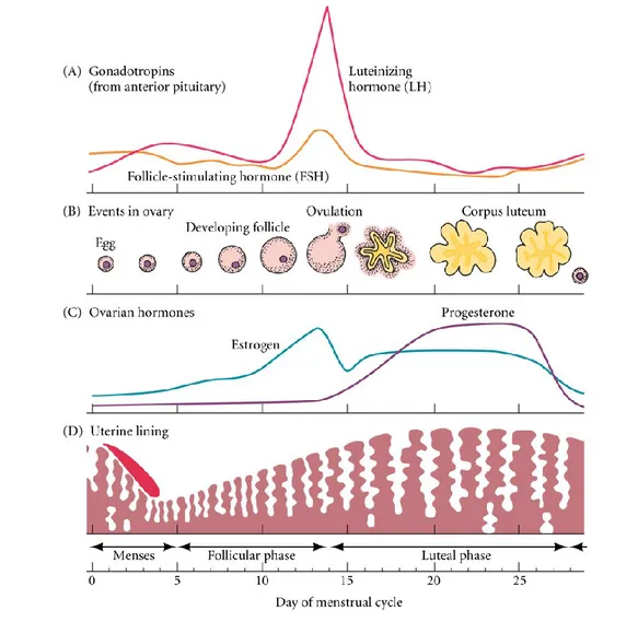
(βA:βA or βB:βB) or heterodimers (βA:βB) that share the β subunits with inhibin A and inhibin B (α:βB). When inhibin and progesterone levels fall with regression of the corpus luteum, suppression of FSH is released. At the luteal-follicular transition, FSH levels increase, and the next cycle begins [Hawkins and Matzuk, 2008] (Figure 6).
Figure 6: The coordination of ovarian and uterine cycles controlled by the pituitary and the ovarian
Pag. 16
The role of the oocyte–cumulus cell dialogue in oocyte developmental competence
Ovarian folliculogenesis requires complex regulatory mechanisms involving both extrinsic (endocrine) and intrinsic signalling pathways. In the case of intrinsic signalling pathways, several intra- ovarian peptides, which are members of several growth factor families appear to influence follicle growth and maturation through a paracrine signalling [Kidder and Mhawi, 2002]. Albertini et al. reviewed evidence that such signalling is facilitated by transzonal projections or follicle cell extensions that traverse the zona pellucida and terminate on the oocyte cell [Albertini et al., 2001]. The cumulus cells and oocyte form a closely connected complex called the cumulus oocyte complex (COC); the COC is connected by gap junctions between cumulus cells, as well as between cumulus cells and oocytes [Kidder and Mhawi, 2002]. Moreover, the
establishment of this bidirectional communication allows the production of developmentally competent oocytes [Cecconi et al., 2004; Gilchrist et al., 2008]. Oocyte-follicular cell contacts are not permanent structures, but rather function as specific ‘devices’ continuously adapting their morphology in response to the activity of both oocyte and cumulus cells [Zuccotti et al., 2011]. In ovarian follicle, the physical contact between somatic cells and between somatic cells and oocyte is mediated by the presence of connexins (Cx), which are expressed from the early stage of development [Gittens and Kidder, 2005; Gittens et al., 2005]. In particular, Cx43 and Cx45 have been identified between granulosa cells. The number of Cx43 gap junctions per granulosa cell increases concomitantly with follicle development and, in particular, during the transition from the preantral to the antral stage. In the absence of Cx43, gap junctions between somatic cells do not form and folliculogenesis arrests at the unilaminar stage [Gittens and Kidder, 2005]. On the contrary, Cx37 is found in gap junction between the oocyte and granulosa cells at the primary follicle stage and is required for fertility
Pag. 17
[Simon et al., 1997]. Mice lacking Cx37 do not ovulate and oocytes are meiotically incompetent [Carabatsos et al., 2000]. Bevens and Harris first reported differential permeabilities of connexins to biological signaling molecules [Bevans et al., 1998]. Cx43 and Cx37 form channels that have different permeability properties and Cx43-positive and Cx37-Cx43-positive plaques do not overlap suggesting that each Cx could play a specific physiological role, e.g. the transfer of different signals between the different compartments of the developing follicles. Molecules that pass via gap junctions include ions, metabolites and amino acids that are necessary for oocyte growth, as well as small regulatory molecules such as cAMP that control oocyte nuclear maturation, and gap-junctional signaling is a key means of disseminating local and endocrine signals to the oocyte via CCs [Albertini et al., 2001]. Gap junctions had long been thought to control meiotic arrest delivering meiosis-arresting substances like cAMP to the oocyte and just recently gap junctional closure has been uncovered as a key regulatory component of this pathway [Norris et al., 2008]. Moreover in regulating of meiotic events central is the role of cGMP that appears to decrease in the oocyte in response to LH and therefore releases the oocyte from meiotic arrest, as a consequence of EGF-mediated closure of connexin 43 gap junctions that may be manifest at both the level of communication between the oocyte and granulosa as well as the lateral integration of metabolism between the cumulus cells themselves [Norris et al., 2009; Norris et al., 2010]. It has recently become evident that the oocyte in fact is a central regulator of follicular cell function and thereby plays a critical role in the regulation of oogenesis, ovulation rate and fecundity [Eppig, 2001; McNatty et al., 2004; Gilchrist and Thompson, 2007]. The oocyte achieves this by secreting soluble growth factors, oocyte-secreted factors (OSFs), which act on neighboring follicular cells to regulate a broad range of GC and CC functions [Nekola and Nalbandov, 1971; Buccione et al., 1990; Vanderhyden et al.,
Pag. 18
1990]. More recently, there was a great deal of scientific interest in GDF9 and BMP15 biology. These molecules are members of the transforming growth factor β (TGFβ) superfamily, and apart from being required for early folliculogenesis, are central regulators of GC/CC differentiation and may be associated with the pathogenesis of ovarian dysfunction [Shimasaki et al., 2004; Juengel and McNatty, 2005; McNatty et al., 2007]. More recently, Sugiura et al. demonstrated that GDF-9 and BMP-15, together with 17β-estradiol, co-ordinate cumulus cell development and expansion [Sugiura et al., 2010]. It was proposed that Oocytes secrete soluble growth factors, notably GDF9 and BMP15 and probably others, that lead to the activation of SMAD2/3 [Dragovic et al., 2007] and MAPK signaling in CCs, which in turn regulate a multitude of CC gene expression and key CC functions (Figure 7). Appropriate CC function and maintenance of the COC microenvironment is dependent on OSFs.In conjunction with maternal signals such as FSH and EGF, CCs pass regulatory growth factors and small metabolites back to the oocyte via paracrine and gap-junctional signaling. This bidirectional CC–oocyte communication loop appears to regulate unknown processes in the oocyte that improves its quality, embryo development and fetal viability [Gilchrist et
al., 2008] (Figure 7). FSH and EGF signalling contribute to the acquisition of oocyte developmental competence by prolonging gap-junctional communication, which mediates the exchange of factors necessary for optimal oocyte developmental competence and subsequent fetal development [Yeo et al., 2009]. In this specific context, it is not surprising that also addition of recombinant GDF-9 during mouse oocyte IVM significantly increases blastocyst quality and fetal survival [Yeo et al., 2008].
Pag. 19 Figure 7: OSF regulation of CC function and oocyte quality [from Gilchrist et al., 2008].
Among the different mechanisms of autocrine and paracrine communication inside the ovarian follicle, over the last few years, an alternative mechanism has come to light. Follicular Fluid (FF), accumulated inside the antral follicle, represents an additional means of communication between oocyte and somatic cells. FF is the product of both the transfer of blood plasma constituents, that cross the blood-follicular barrier, and of the secretory activity of granulosa and thecal cells [Fortune JE, 1994]. FF consists of a complex mixture of nucleic acids, proteins, metabolites, and ions, which are secreted by the oocyte, granulose and thecal cells, combined with plasma components that cross the blood–follicular barrier via thecal capillaries [Revelli et al., 2009; Rodgers and Irving-Rodgers, 2010]. It represents a very important microenvironment for the development of the oocyte and its biochemical composition reflects the physiological status of the follicle. It is reasonable to think that the analysis of FF components may provide useful information on oocyte quality, and the subsequent potential to achieve fertilization and
Pag. 20
embryo development [Leroy et al., 2004; Revelli et al., 2009]. In fact, hormones, growth factors, cytokines, and chemokines secreted by follicular cells in FF are able to promote oocyte maturation and an alteration of biochemical composition of FF is related to low quality oocytes. By the FF the oocyte and somatic follicular cells could interchange nutrients, as well as molecular signals. However the main molecular actors of this alternative communication tool will be discussed in detail as follows.
Transcriptional activity in oocytes
During oocyte maturation, fertilization and early embryo development until zygotic genome activation, which occurs at the 2-cell stage in mice and 4- to 8-cell stage in humans, transcription is suppressed and gene expression is dependent upon the timely activation of stored mRNAs [Braude et al., 1988; Schultz, 1993]. Fully-grown and meiotically competent murine oocytes have been estimated to contain ~6 ng of total RNA which is almost ~200 times the amount of RNA found in a typical somatic cell [Sánchez and Smitz, 2012]. Synthesis of transcripts is highest in the earliest phases of development,which coincides with active proliferation of follicular cells. However, maternal mRNAs are degraded in a well-defined temporal, transcript-specific pattern [Evsikov et al., 2006; Su et al., 2007., Chen et al., 2011] in synchrony with maturation; as a result, mRNA content is decreased by 30% in MII oocytes. Infact, almost half of the 85 pg mRNA stored during oocyte growth is degraded during meiosis resumption and by the MII stage the oocyte carries about 35 pg mRNA; of this, about half undergoes stabilization through selective deadenylation of the poly(A) tail at the 3′ region [Paynton et al., 1988]. In this way, a vast number of transcript important for germinal vesicle block and for the following stages of meiosis resumption are eliminated while transcripts associated with the maintenance of MII oocyte features or
Pag. 21
playing important roles during the early stages of development are kept stabilized through deadenylation [Su et al., 2007] [Thélie et al., 2007]. Soon after fertilization and by the time the embryonic genome is first expressed, translation of maternal mRNAs, necessary for the early phases of development, began through the polyadenylation of the 3′ untranslated region operated by embryonic cis regulatory cytoplasmic polyadenylation elements [Mendez and Richter, 2001; Racki and Richter, 2006]. Moreover it is important to pinpoint that during this quiescent period, fundamental changes in nuclear function also take place as the differentiated cells, oocyte and sperm, unite to give rise to an embryonic genome. These changes, known as “genome reprogramming,” transform the genome to the state of totipotency, a critical event for the successful development of an embryo [Dean et al., 2003]. However, transcriptional quiescence of the genome requires post-transcriptional and post-translational mechanisms to orchestrate the multitude of processes participating in meiotic maturation, fertilization, and reprogramming of the nascent embryonic genome [Solter et al., 2004]. Recent advances include identification of RNA-binding proteins that function as specificity factors to direct the maternal degradation machinery to its target mRNAs; the action of small silencing RNAs (ssRNAs) and signaling pathways that trigger production and/or activation of the clearance mechanism in early embryos; and mechanisms for spatial control of transcript clearance [Walser and Lipshitz, 2011]. Little is known about the role played by ssRNAs during oocyte growth and preimplantation development. The few studies that have analyzed the presence of ssRNAs in oocytes have showed that their average amount does not vary during maturation, although single miRNAs may vary considerably [Tang et al., 2007]. Moreover, these maternal ssRNAs have been presumed to represent the only contribution to the zygote, since the paternal miRNAs, brought into the zygote by the
Pag. 22
sperm, do not seem to contribute significantly to the total miRNAs in the zygote [Amanai et al., 2006]. At 1-cell and 2-cell stage of development, the amount of these decreases by 60 %, suggesting an active process of degradation that coincides with a global RNA degradation occurring at this time of development [Zuccotti et al., 2011].. Moreover emerging evidences indicate important the role of miRNAs, a class of ssRNAs, during early development as supported by recent studies that have demonstrated their crucial involvement in regulating ES cell pluripotency and differentiation [Gangaraju and Lin, 2009]. Clearly, further understanding and deciphering of changes that the oocyte transcriptome and epigenome undergoes may expand our knowledge on their potential role in the context of oocyte maturation.
Pag. 23
The new world of RNAs
Although Jacob and Monod had suggested in 1961 the centrality of RNA in the flow of genetic information, establishing the concept of messenger RNA in their paper “Genetic Regulatory Mechanisms”, for many decades proteins represented not only the primary structural and functional components of cells, but also the main regulatory components of the genome in both simple and complex organisms [Jacob and Monod, 1961]. This protein-centred view was, in fact, considered as rather simplistic and misleading when applied to higher organisms [Ragusa et al., 2015]. In this way, recent advances in the fields of RNA biology and genome research have reassessed the old assumption and provided significant evidence of the importance of RNAs as “riboregulators” outside of their more conventional role as accessory molecules [Prasanth and Spector, 2007]. The number of protein-coding genes that structure the human genome has long been a source of discussion. With the publication of the final draft of the Human Genome Project, the number of protein-coding genes was revised to between 20 000 and 25 000. Most recently, Clamp and co-workers used evolutionary comparisons to suggest that the most likely figure for the protein-coding genes would be at the lower end of this continuum, just 20 500 genes. This number is lower than expected and very close to the number of protein-coding genes in C. elegans, suggesting that maybe these genes alone are not sufficient to appropriately and completely explain the complexity of higher eukaryotes [International Human Genome Sequencing Consortium, 2004; Szymanski and Barciszewski, 2006; Ezkurdia et al., 2014]. Recent high-throughput studies of the human transcriptome have revealed that about 85%-90% of our genome is dynamically and pervasively transcribed and that ∼98% of the transcriptional output of the human genome represents non-protein-coding RNAs (ncRNAs) [Mattick, 2005; Prasanth and Spector, 2007]. Many observations have, only recently, suggested convincingly that
Pag. 24
ncRNAs significantly contribute to the complex molecular signalling needed to regulate structures and functions in different cells and developmental programming [Amaral et al., 2008]. Hence, dysregulation of these ncRNAs is being found to have relevance not only to tumorigenesis, but also to neurological, cardiovascular, developmental and other many pathological conditions [Taft et al., 2010; Esteller, 2011]. It is not precisely known how many ncRNA genes are present in the human genome. The number of known functional ncRNA genes has risen dramatically in recent years and over 800 ncRNAs (excluding tRNAs, rRNAs and snRNAs) have been catalogued in mammals, at least some of which are alternatively spliced [Pang et al., 2005]. ncRNA genes are difficult to identify because of their structural heterogeneity: extreme length variation from 20 nucleotides to > 100 kb; absence of Open Reading Frames (ORFs); no or low evolutionary conservation in many cases; no preferential localization within the genome; and relative tolerance to point mutations [Ragusa et al., 2015]. ncRNAs are divided into several different categories according to size and function. ncRNAs are commonly classified, according to their length, in two different classes: (1) long coding RNAs (lncRNAs), which are longer than 200 nucleotides (nt); and (2) small non-coding RNAs, whose length is equal to or less than 200 nt [i.e., microRNAs (miRNAs), small interfering RNAs (siRNAs), PIWI-interacting RNAs (piRNAs) (Table I). Other classifications have been proposed to categorize ncRNAs according to functional features into two main classes: (1) housekeeping ncRNAs; and (2) regulatory ncRNAs. Housekeeping ncRNAs are constitutively expressed and required for normal function and viability of cell. They include transfer RNAs (tRNAs), ribosomal RNAs (rRNAs), small nuclear RNAs (snRNAs), small nucleolar RNAs (snoRNAs), and telomerase RNAs [Zhu et al., 2013]. Conversely, regulatory ncRNAs can be expressed in a cell-specific way, or during defined stages of development and cell differentiation, and
Pag. 25
finally in response to external stimuli [Brosnan and Voinnet, 2009]. Regulatory ncRNAs
can affect the expression of other genes at the level of transcription or translation. This category comprises miRNAs, siRNAs, lncRNAs (i.e., the RNA molecules more closely involved in cancer biology), and piRNAs [Adams et al., 2014].
Table I. Class of non-coding RNA in mammals [from Taft et al., 2010].
microRNAs: genomics, biogenesis and function
Small non-coding class of RNAs includes different types of molecules involved in different steps of RNA synthesis, processing, translation, as well as modulation of transcription initiation [i.e., piRNAs, promoter associated small RNAs (PASRs)], RNA
Pag. 26
degradation or protein synthesis block (e.g., miRNAs, siRNAs), RNA maturation (e.g., snoRNAs) [Dogini et al., 2014]. Among these, microRNAs (miRNAs) represent the most studied and better characterized class of ncRNAs in terms of function and impact in human health and disease. Specifically, miRNAs are small endogenous 18-25 nucleotides (nt) long, that regulate gene expression, in animals and plants, post- transcriptionally in a sequence-specific manner [Bartel, 2004]. miRNAs had been discovered for the first time in 1993 by Ambros and colleagueswhen two independent studies identified the real nature of the C. elegans heterochronic gene lin-4 as small non-coding RNA (ncRNA) [Lee et al., 1993]. At that time, these findings were not adequately appreciated by the scientific community, because this small ncRNA was considered a specific tool, employed by the worms, to control somehow the expression of the heterochronic coding genes. After seven years, Reinhart et al. identified another C. elegans 22 nt non-coding RNA, the let-7 [Reinhart et al., 2000]. Surprisingly, it was also found to be complementary to the 3’UTR of another gene, lin-41, promoting its translational knockdown. This discovery triggered scientist curiosity to look for other small ncRNAs and, later, independent groups have identified these small ncRNAs also in other different organisms and cellular systems providing evidence for the existence of a large class of small ncRNAs with potential regulatory roles that was named microRNAs [Pasquinelli et al., 2000; Lagos-Quintana et al., 2001; Lau et al., 2001; Lee and Ambros, 2001]. Currently, 2603 human and 1920 murine mature miR entries are reported in the miRbase sequence repository (mirbase.org; November 2015). Animal miRNAs are phylogenetically conserved (~55% of C. elegans miRNAs have homologues in humans) and mammalian miRNA genes have also multiple isoforms (paralogues), which are probably generated by duplication (human let-7 gene accounts for 8 different isoforms distributed in 11 genomic loci) [Di Leva et al., 2014].
MiR-Pag. 27
encoding genes are present in the genome as intergenic clusters or individual transcriptional units. In both cases, their expression is under the control of promoters and enhancers with similar characteristics and regulation to those for protein-coding genes [Liu et al., 2013]. Several miRNAs, however, are embedded in the intronic sequences of other genes (miRtrons) and are then co-transcribed accordingly with the encompassing genes [Curtis et al., 2012]. miRNAs are canonically produced through a multistep process that, starting in the nucleus and ending into the cytoplasm, is composed by three main events: cropping, export and dicing[Kim et al., 2009] (Figure 8). Following the canonical pathway, miRNA genes are generally transcribed by RNA polymerase II into primary transcripts of several hundred nucleotides (pri-miRNAs), bearing secondary hairpin structures and undergoing 5′ capping and 3′ polyadenylation [Ha and Kim, 2014]. In few cases, miRNA genes are transcribed by RNA polymerase III [Diebel et al., 2014]. While still inside the nucleus, the microprocessor complex, formed by the RNase-III Drosha and its co-factor Dgcr-8, cleaves the pri-miRNAs into ~ 70 nt-long precursor molecules (pre-miRNAs) [Liz et al., 2014]. Dgcr-8 is a dsRNA binding protein that recognizes the proximal ~10 bp of stem of the pri-miRNA hairpin, positioning the catalytic sites of the RNase III enzyme Drosha [Yeom et al., 2006]. Pre-miRNAs are, then, transported to the cytoplasm by exportin-5 and the cofactor Ran-GTP [Yi et al., 2003; Lund et al., 2004]. Once in the cytoplasm, pre-miRNAs are further cleaved, near the terminal loop, releasing the ~ 22 nt-long double-stranded molecules by a complex that includes another RNase-III, Dicer, in combination with its RNA-binding cofactor, Tbrp [Winter et al., 2009]. Whereas mammals typically encode a single DICER that can generate several classes of small RNAs, Drosophila and C. elegans have two types of Dicer [Lee et al., 2004]. Human Dicer interacts with two related proteins TRBP (TAR RNA-binding protein) and PACT (also known as PRKRA)
Pag. 28
that, as miRNA duplexes are produced, associate with Argonaute (Ago) proteins, forming the RNA-induced silencing complex (RISC) [Schwarz et al., 2003; Chendrimadaet al., 2005; Lee et al., 2006]. After Dicer-mediated maturation, miRNAs are loaded on the RISC as single strands. Mature miRNA duplexes are in fact separated in a guide strand (mature miRNA) and a passenger strands (miRNA*). The guide strand usually presents the lower free energy and weakest base pair at 5′ and is preferentially loaded on the RISC complex, whereas the complementary passenger strand is preferentially degraded [Khvorova et al., 2003]. The RISC complex includes Argonaute proteins, such as Ago-2, and targets the miRNA to its messenger RNA (mRNA) targets. Also, Ago-2-binding protein, Gw-182, shuttles the miR-loaded, active RISC complex to discrete foci in the cytoplasm, called processing bodies (P-bodies), where several enzymes are available for mRNA decapping, deadenylation and degradation [Leung and Sharp, 2013]. The seed sequences of the miRNA (7 nt sequences mapping to positions 2–8 at the molecule's 5′ end) are considered essential for the selection of the target mRNAs. According to the binding complementarity of the seed sequence, miRNAs repress gene expression by targeting the 3′ untranslated region (3′-UTR) of mRNA. If the pairing is complete (100% sequence complementarity between the miRNA and mRNA), the mRNA will be degraded; however, if the sequence is only partially complementary, this will result in translational inhibition of the mRNA [Cannell et al.,
2008]. The latter is achieved through various mechanisms, including RISC-mediated destabilization of ribosomal assembly or translation continuation [Kim et al.,2014]. Non-canonical processing of miRNA transcripts, typical for miRtrons, relies on direct processing of the pre-miRNAs during intron splicing of the harboring gene mRNA. Other microprocessor-independent sources of pre-miRNAs include small nucleolar
Pag. 29
RNAs (snoRNAs), transfer RNA precursors (tRNAs) and short hairpin RNAs (shRNAs) [Yang and Lai, 2011].
Figure 8: Mirna Biogenesis. miRNA genes are transcribed as primary miRNAs (pri-miRNAs) by RNA
polymerase II (Pol II) in the nucleus. The long pri-miRNAs are cleaved by Microprocessor, which includes DROSHA and DiGeorge syndrome critical region 8 (DGCR8), to produce the 60–70-nucleotide precursor miRNAs (pre-miRNAs). The pre-miRNAs are then exported from the nucleus to the cytoplasm by exportin 5 (XPO5) and further processed by DICER1, a ribonuclease III (RIII) enzyme that produces the mature miRNAs. One strand of the mature miRNA (the guide strand) is loaded into the miRNA-induced silencing complex (miRISC), which contains DICER1 and Argonaute (AGO) proteins, directs the miRISC to target mRNAs by sequence complementary binding and mediates gene suppression by targeted mRNA degradation and translational repression in processing bodies (P-bodies). TRBP, transactivation-responsive RNA-binding protein [from Lin and Gregory, 2015].
Modulation of miRNAs
Many factors regulate miRNA biogenesis, hence affect the downstream miRNA-mediated gene repression. Both miRNA precursors and mature miRNAs undergo A-to-I RNA editing by deaminases, affecting the miRNA maturation process and activity [Tomaselli et al., 2013], or 3′ uridylation that can influence Drosha- or Dicer-dependent
Pag. 30
processing steps. Also, abundance of Ago-2, which has intrinsic endonuclease activity in mammals, or agonist proteins directly affects RISC activity levels [Slezak-Prochazka et al., 2010]. Such regulation of miRNA processing fine-tunes their intracellular levels, and modulates their biological activity. Indeed, miRNA intracellular concentration directly relates to their ability to affect mRNA translation. Another important aspect of intrinsic miRNA regulation, consists of turnover and degradation. After mRNA targeting. and in the presence of still unidentified signals, the miRNA guide strand is released and degraded. Processes underlying average half-life of miRNAs have been supposed involve exoribonucleases, although still remain largely unknown [Gantier et al., 2011; Großhans and Chatterjee, 2011].
Vesicular and non-vesicular trafficking of miRNAs
Besides operating intracellularly, miRNAs can be released from the producing cells in the surrounding areas or in the circulation. Intriguingly, circulating miRNAs are traceable in plasma or serum and other biological fluids and appear resistant to harsh conditions such as RNase activity, pH changes, boiling, freeze-thawing and storage at room temperature [Chen et al., 2008; Weber et al., 2010; Cortez et al., 2011]. In these fluids, miRNAs display remarkable stability and resistance to degradation, possibly because of their association with carriers. Non-vesicle carriers encompass protein complexes, including Ago-2 and Nucleophosmine-1, and lipoprotein complexes, such as high-density lipoproteins (HDLs). Among the vesicle-based carriers, exosomes are emerging as important regulators of long-range miRNA shuttling [Valadiet al., 2007; Atay and Godwin, 2014]. Exosomes are small vesicles (30-100 nm diameter) that enclose their cargo with a lipidic bi-layer and are generated through inward budding of
Pag. 31
endosomal membranes, which gives rise to intracellular multivesicular bodies (MVB). In addition to exosomes, cells can also shed other types of extracellular membrane vesicles, namely, microvesicles, microparticles and apoptotic bodies, after various biological stimuli, including induction of programmed cell death [Majno and Joris, 1995; Aupeix et al., 1997]. Exosomes are different from the other extracellular vesicles because the latter are the result of a direct budding process of the plasma membraneand seem to have properties distinct from those of exosomes. Exosomes mature though a still unknown process, most likely at the interface with the Golgi reticulum and later fuse with the plasma membrane releasing the intraluminal vesicles into the extracellular environment. This process completes the exocytosis event [Waldenström and Ronquist, 2014] (Figure 9).
Figure 9: Biogenesis and composition of Exosomes [from Waldenström and Ronquist, 2014].
However, exosome biogenesis and secretion are multifaceted mechanisms and they are different depending on the cell type and cargo sequestered. Exosomes can be formed at
Pag. 32
least via two distinct pathways: the first utilizes the Endosomal Sorting Complexes Required for Transport (ESCRT) machinery, while the second, independent from ESCRT complexes, involves lipids or tetraspanins [Van Niel et al., 2011; Mayers and Audhya, 2012]. In the extracellular space, exosome interaction with recipient cells can take place in different ways: by ligand-receptor interactions, by fusion, or by internalization via receptor-mediated endocytosis [Urbanelli et al., 2013]. Therefore, exosomes can act as paracrine or autocrine mediators and also as endocrine mediators that through the blood stream are able to target cells located at distant sites in the body [Cortez et al., 2011]. The first evidence of exosome secretion was demonstrated in reticulocytes undergoing maturation into red blood cells [Johnstone et al., 1987]. In 1996, Raposo et al. demonstrated in B-lymphocytes that both the limiting membrane and the internal vesicles contain major histocompatibility complex class II, and the released exosomes were perceived to have a role in antigen presentation in vivo. Subsequently, it was realized that many cell types release exosomes [Raposo et al.,
1996]. Exosomes are released by a variety of “normal” cells including mast cells (MC), dendritic cells, reticulocytes, epithelial cells, B-cells, trophoblastic cells, and neural cells, as well as a variety of tumor cells. In addition, exosomes are found in various biological fluids including bronchoalveolar lavage, blood, ascites, urine, pregnancy associated sera, breast milk, saliva, follicular fluid and malignant effusions [Sang et al., 2013; Atay and Godwin, 2014; Santonocito et al, 2014]. Several studies have demonstrated that exosomes from different cellular origins share some common characteristics. Such characteristics are the lipid bilayer with exceptionally high cholesterol/phospholipid ratio, size, density, and a basic collection of lipid and protein composition. Over the last few years, proteomic analysis of exosomes has identified common characteristic marker proteins on their surface and in their lumen [Mathivanan
Pag. 33
et al., 2010] (Figure 9). Because of their endosomal origin, exosomes contain several proteins involved in the ESCRT complex (e.g., TSG101, Alix) and in transport and fusion (e.g., Rab11, Rab7, Rab2 and various annexins) [Atay and Godwin, 2014]. Further markers expressed in or on exosomes include tetraspanins (CD9, CD63, CD81, CD82) and heat shock proteins (HSC70 and HSP90) [Mathivanan et al., 2010]. Moreover, in 2007, Valadi et al. demonstrated that exosomes contain both mRNAs and miRNAs, which can be delivered to another cell, and can be functional in this new location. They proposed to call this RNA "exosomal shuttle RNA" [Valadi et al., 2007]. Therefore, exosomes are able to transfer macromolecules to target cells, but the mechanisms that allow the selection of the correct have not yet been characterized, however, it has been demonstrated that the cells are able to modify the number and the cargo of exosomes depending on specific conditions [Lo Cicero et al., 2015]. Moreover, it has been shown that there is a specific repertoire of miRNAs selectively exported to exosomes, whereas others are usually excluded, indicating that an active sorting mechanism occurs at the RNA level [Van Niel et al., 2011]. However, at present, the relative abundance of vesicular or carrier-based complexes for circulating miRNAs in the bloodstream is still debated. Some reports suggest that the majority of circulating miRNAs are exosome-borne whereas others suggest that miRNAs in body fluids are predominantly vesicle-free [Turchinovich et al., 2011; Gallo et al., 2012]. These discrepancies are probably due to still different and rather incomparable methodologies of investigation. While our understanding of intercellular communication has been mainly based on the secretion of soluble factors, such as growth factors and neurotransmitters, cytokines, and chemokines, and their specific recognition by cell-surface receptors, evidence indicating that cells also communicate via the direct exchange of DNA and RNA has been growing over the past several years [Deregibus et
Pag. 34
al., 2007; Ehnfors et al., 2009]. Additionally, following the notion of exosomes as miRNA carriers, notwithstanding the lack of details concerning putative receptors and intracellular processing, pre- or mature miRNAs are delivered to other cell where they exert their specific regulation [Salido-Guadarrama et al., 2014]. In this way, miR-based cross-communication is emerging as unconventional mechanism regulating intercellular communication.
miRNAs and ovarian function
Currently, over 2000 human miRNAs have been identified by cloning and sequencing approaches. It is predicted that miRNA genes account for 1%-2% of the human genome and control the expression of at least 30-50% of all protein coding genes [Krol et al., 2010]. By modulating the expression of target transcripts, miRNAs may, therefore, affect many different signaling pathways and cellular processes, such as cell proliferation, differentiation, migration, angiogenesis and apoptosis: accordingly, they are considered potential oncogenes or tumor suppressors in cancer development and progression [Bueno and Malumbres, 2011]. The miRNA repertoires are cell type specific and change markedly during development [Carthew and Sontheimer, 2009]. Crucial and time-dependent events such as degradation of the LH receptor transcripts or translational repression of Connexin 43, after LH surge, in ovarian granulosa cells have suggested that gene expression may, also, be under control of miRNAs in reproductive tissues [Carletti and Christenson, 2009]. miRNAs constitute the most abundant class of
small RNAs in the ovary. By using microarrays, high-throughput quantitative polymerase chain reaction, and next-generation sequencing techniques miRNA population in the ovaries was identified in human and other species. Regardless of
Pag. 35
species, let-7 family, miR-21, miR-99a, miR-125b, miR-126, miR-143, miR-145, and miR-199b were found to be the most commonly abundant miRNA populations [Li et al., 2015]. Many studies demonstrate also that miRNAs are essential for follicle development in different species [Abramov et al., 2013; Yang et al., 2013; Zhang et al., 2014]. miRNAs are involved in the entire process of ovarian follicle development, including follicle growth, atresia and ovulation in which different growth factors contribute to stage-specific functions in different cell types [Li et al., 2015]. Studies of the role of miRNAs in ovarian function have been highlighted primarily through Dicer, a ribonuclease that is required for the synthesis and processing of mature functional miRNAs. As the function of Dicer and its products (miRNAs and siRNAs) were studied in the female reproductive tract, the essential role for these post-transcriptional gene regulators in female fertility was revealed [Carletti and Christenson, 2009]. Dicer is expressed in both oocytes and granulosa cells of the mouse ovarian follicle. Oocytes contain 10–15-fold higher levels of Dicer transcripts than any other cells and/or tissues [Imbar and Eisenberg, 2014]. In mice, knockout (KO) of Dicer results in postimplantation embryonic lethality [Bernstein et al., 2003]. In the germ cell-specific Dicer1 knockout mouse model, in which Cre recombinase is driven by the zona pellucida 3 (Zp3) promoter, Dicer1-deficient oocytes are unable to complete meiosis, and arrest with defects in meiotic spindle organization and chromosome congression [Murchison et al., 2007]. Moreover mouse mutant with a hypomorphic Dicer allele (Dicer d/d) resulted in Dicer1 deficiency with subsequent female infertility due in part to the insufficiency of corpus luteum (CL) function [Otsuka et al., 2008]. Likewise, in ovarian granulose cells, Dicer1 cKO in mouse led to an increased primordial follicle pool endowment, accelerated early follicle recruitment, and more follicle degeneration in the cKO ovaries [Lei et al., 2010]. Dicer1 regulates follicle development by
Pag. 36
downregulating miR-503, an ovary-specific miRNA, as well as miR-503 target genes, such as anti-Müllerian hormone (AMH); inhibin beta A subunit (INHBA); cytochrome P450, family 17, subfamily a, polypeptide 1 (Cyp17a1); cytochrome P450, family 19, subfamily a, polypeptide 1 (Cyp19a1); zona pellucida glycoproteins (ZPs); growth differentiation factor 9 (GDF9) and bone morphogenetic protein 15 (BMP15) [Li et al., 2015]. All these studies demonstrate that Dicer plays important roles in follicle growth and oocyte maturation. Moreover, a study of miRNA expression in mouse mural granulosa cells identified miR-132 and miR-212 upregulation following LH/hCG induction. Interestingly, knockdown of both miR-212 and miR-132 resulted in decreased protein levels, but not of mRNA levels, of C-terminal binding protein 1 (CTBP1) which is a known target of miR-132, and whose gene product acts as a corepressor of nuclear receptor genes [Fiedler et al., 2008]. Further studies to determine how these miRNAs cause these changes in CTBP1 expression would be useful to establish the precise relationship. Sirotkin et al. investigating the role of miRNAs in granulosa cells, by using 80 individual artificial miRNA precursors, that mimic endogenous miRNAs, to transfect cultured primary ovarian granulosa cells, demonstrated the direct involvement of miRNAs in controlling both proliferation and apoptosis in these cells [Sirotkin et al., 2010]. Another study, in rodents, revealed a biphase regulation of miRNAs by FSH. The expression of 31 miRNAs was altered during FSH-mediated progesterone secretion of cultured granulosa cells. Specifically, 12 h after FSH treatment, miRNAs mir-29a and mir-30d were significantly down-regulated. However, their expression increased two and threefold respectively, 48 h after FSH exposure [Yao et al., 2010]. These two miRNAs could be, hence, involved in the fine-tuning of FSH-mediated granulose cell function. Moreover using a deep-sequencing approach specific miRNAs that are abundant in MII oocyte (MIR184,
Pag. 37
MIR100 and MIR10A) or CCs (MIR29a, MIR30d, MIR21, MIR93, MIR320a, MIR125a and the LET7 family) were identified in women who underwent IVF procedures[Assou et al., 2013]. In the same study, predicted target genes of the oocyte miRNAs were associated with the regulation of transcription and cell cycle, whereas genes targeted by CC miRNAs were involved in extracellular matrix and apoptosis. Comparison of the predicted miRNA target genes and mRNA microarray data resulted in a list of 224 target genes that were differentially expressed in MII oocytes and CCs, including PTGS2, CTGF and BMPR1B, important for cumulus–oocyte communication. Functional analysis using primary CC cultures revealed that BCL2 and CYP19A1 mRNA levels were decreased upon MIR23a overexpression, suggesting an important role of MIR23a in controlling transcripts that are involved in the ovulatory follicle apoptosis [Assou et al., 2013]. Given their important roles in normal physiology, it is not surprising that miRNAs have been shown to play contributory roles in ovarian diseases such as ovarian cancer, polycystic ovarian syndrome (PCOS) and premature ovarian failure (POF) [Yang et al., 2012; Zheng et al., 2013]. Sang et al. assessed miRNA expression in human follicular fluid of PCOS patients and identified numerous miRNAs that play important roles in steroidogenesis. miR-132 and miR-320 are expressed at a significantly reduced level in the follicular fluid of polycystic ovary patients compared with healthy controls. In addition, miR-132, miR-320, miR-520c-3p, miR-24 and miR-222 regulate estradiol concentrations, and miR-24, miR-193b, and miR-483-5p regulate progesterone concentrations in PCOS patients [Sang et al., 2013]. Recent studies indicate also that miRNA single-nucleotide polymorphisms (SNPs) are associated with disease susceptibility. A study related to miRNA polymorphism analysis identified the association between combined genotypes and haplotypes of miR-146aC>G, miR-196a2T>C, and miR-499A>G and POF risk in Korean women; the


![Table I. Class of non-coding RNA in mammals [from Taft et al., 2010].](https://thumb-eu.123doks.com/thumbv2/123dokorg/4518177.34810/29.892.148.760.374.950/table-i-class-non-coding-rna-mammals-taft.webp)

![Figure 9: Biogenesis and composition of Exosomes [from Waldenström and Ronquist, 2014]](https://thumb-eu.123doks.com/thumbv2/123dokorg/4518177.34810/35.892.157.796.690.953/figure-biogenesis-composition-exosomes-waldenström-ronquist.webp)

