Altered gene transcription linked to astrocytes and oligodendrocytes in frontal
cortex in Creutzfeldt-Jakob disease
Pol Andrés-Benito a, Mayelin Domínguez-Gonzáleza, and Isidro Ferrer a,b,c,d aDepartment of Pathology and Experimental Therapeutics, University of Barcelona
AQ1 �
AQ2
� AQ3
�;bBiomedical Research Centre of Neurodegenerative 5 Diseases (CIBERNED), Institute of Health Carlos III, Ministry of Economy, Innovation and Competitiveness, Hospitalet de Llobregat;cSenior
consultant, Service of Pathologic Anatomy, Bellvitge University Hospital (IDIBELL);dInstitute of Neurosciences, University of Barcelona, Barcelona, Spain
ABSTRACT
Targeted expression of genes coding for proteins specific to astrocytes, oligodendrocytes and myelin 10 was performed in frontal cortex area 8 of Creutzfeldt-Jakob disease methionine/methionine and valine/valine (CJD MM1 and VV2, respectively) compared with controls. GFAP (glial fibrillary acidic protein) mRNA was up-regulated whereas SLC1A2 (solute carrier family 1 member 2, coding for glutamate transporter 1: GLT1), AQ4 (aquaporin 4), MPC1 (mitochondrial pyruvate carrier 1) and UCP5 (mitochondrial uncoupled protein 5) mRNAs were significantly down-regulated in CJD MM1 15 and CJD VV2, and GJA1 (connexin 43) in CJD VV2. OLIG1 and OLIG2 (oligodendocyte transcription factor 1 and 2, respectively), SOX10 (SRY-Box10) and oligodendroglial precursor cell (OPC) marker NG2 (neuronal/glial antigen) 2 were preserved, but GALC (coding for galactosylceramidase), SLC2A1 (solute carrier family 2 member 1: glucose transporter member 1: GLUT1) and MCT1 (monocarboxylic acid transporter 1) mRNA expression levels were significantly reduced in CJD MM1 and CJD VV2. 20 Expression levels of most genes linked to myelin were not altered in the cerebral cortex in CJD. Immunohistochemistry to selected proteins disclosed individual variations but GFAP, Olig-2, AQ4 and GLUT1 correlated with mRNA levels, whereas GLT1 was subjected to individual variations. However, MPC1, UCP5 and MCT1 decrease was more closely related to the respective reduced neuronal immunostaining. These observations support the idea that molecular deficits linked to energy 25 metabolism and solute transport in astrocytes and oligodendrocytes, in addition to neurons, are
relevant in the pathogenesis of cortical lesions in CJD.
ARTICLE HISTORY Received 16 March 2018 Revised 27 May 2018 Accepted 5 July 2018 KEYWORDS Creutzfeldt-Jakob disease; prion diseases; astrocytes; oligodendrocytes; myelin; energy metabolism; astrogliopathy; oligodendrogliopathy
Introduction
Prion diseases are a group of transmissible encephalo-30 pathies linked to the prion protein (PrPC) which is converted into an abnormally conformed proteinase K-resistant protein named prion (PrPSC). The human prion diseases are sporadic, iatrogenic and genetic Creutzfeld-Jakob disease (sCJD, iCJD and gCJD, 35 respectively), Gerstmann-Straüsler-Scheinker disease (GSS) disease, and fatal familial insomnia (FFI). gCJD, GSS and FFI are due to mutations in prion protein gene (PRNP) [1–7]. The main prionopathies in animals are scrapie (typical and atypical) in sheep and goats; 40 chronic wasting disease in deer, elk, moose and rein-deer; and bovine spongiform encephalopathy (BSE) in cattle [8].Variant CJD (vCJD) is considered a form of BSE transmitted to humans [9]. CJD is not homoge-neous; different subtypes are distinguished in sCJD 45 depending on the prion type (I and 2) and the compo-sition of codon 129 in PRNP (methionine/methionine:
MM, valine/valine: VV, and methionine/valine: MV), each one with singular clinical phenotypes [10,11]. The most frequent subtypes are sCJD MM1 and sCJD VV2.
50 Common neuropathological lesions in CJD are neu-ron loss, spongiform change and deposition of PrPSC. Additional characteristics of sCJD MM1 are microva-cuolar spongiform change in the frontal and occipital cortices, and molecular layer of the cerebellum, and
55 synaptic-like PrPSC deposition. Particular features of sCJD VV2 are microvacuolar or confluent spongiform change, major involvement of the cerebellum, and synaptic, perineuronal and plaque-like PrPSC deposits [4–7]. Reactive astrocytosis and microgliosis,
accompa-60 nied by a robust molecular inflammatory response, are important accompanying features [12–15].
Besides their contribution to astrocytic gliosis and inflammation, little is known about the defects of astro-cytes in CJD. In addition to neuronal bodies, dendrites,
65 synapses and axons, PrPSCcan be present in astrocytes
CONTACTIsidro Ferrer [email protected] Department of Pathology and Experimental Therapeutics, University of Barcelona, c/Feixa Llarga sn, 08907 Hospitalet de Llobregat, Spain
AQ4 �
2018, VOL. 00, NO. 00, 1–10
https://doi.org/10.1080/19336896.2018.1500076
and microglia [16]. Aquaporin 1 and 4 immunoreactiv-ity is increased [17,18], thus suggesting adaptation to water transport. Moreover, recent studies have shown that human astrocytes have the capacity to take up and 70 degrade normal and protease-resistant prion protein [19] and that they can transfer PrPSC to neurons via nanotubules [20] thereby contributing to prion disease progression.
Regarding oligodendrocytes, less information is 75 available in CJD. Oligodendrocytes are apparently resis-tant to PrPSCinfectivity [21]. However, axon and mye-lin damage occurs in prion diseases [22], abnormal interactions between oligodendroglia and astrocytes are found in experimental CJD and scrapie [23], and 80 engulfment of oligodendrocytes by hypertrophic astro-cytes has been reported in the white matter in CJD [24]. PRPSC is also localized as arrays adjacent to myelin fibers in the cerebrum and cerebellum in CJD [25].
Based on these data, the present study was under-85 taken to gain understanding about transcription altera-tion of genes encoding proteins specifically expressed in astrocytes and oligodendrocytes in frontal cortex area 8 of CJD MM1 and CJD VV2. Selected genes include those encoding structural proteins of astrocytes and 90 myelin, those involved in energy metabolism and axon maintenance, and genes of connexins of the gap junctions between oligodendrocytes and astrocytes. Protein expression of altered has been assessed by immunohistochemistry.
95 Results
Astrocytic markers
Covariance analysis (ANCOVA) of GFAP (coding for glial fibrillary acidic protein) mRNA expression levels revealed a significant group effect [F(2,19) = 4.383, P = 0.027] but did 100 not show RIN effect [F(1,19) = 0.337, P = 0.568]. Subsequent one-way ANOVA analysis [F(2,20) = 7.918, P = 0.0029] revealed significant increase in CJD MM1 and CJD VV2 when compared with controls (P = 0.012 and P = 0.005, respectively). In contrast, ANCOVA of 105 ALDH1L1 (coding for aldehyde dehydrogenase 1, family member L1) mRNA, used as marker of total astrocytes, had non-significant group [F(2,22) = 1.087, P = 0.355] and RIN [F(1,22) = 0.021, P = 0.887] effects; no differences of ALDH1L1 mRNA were observed between CJD and con-110 trols. ANCOVA of AQP4 (coding for aquaporin 4) mRNA showed significant group effect [F(2,21) = 4.307, P = 0.027] but not RIN effect [F(1,21) = 0.334, P = 0.569]. Subsequent one-way ANOVA analysis [F(2,22) = 8.596, P = 0.0017] showed significant decreased expression in CJD MM1 and 115 CJD VV2 when compared with controls (P = 0.26 and
P = 0.002, respectively). ANCOVA analysis of SLC1A2 (coding for glutamate transporter 1) mRNA expression showed significant group effect [F(2,21) = 6.031, P = 0.009] but not RIN effect [F(3,25) = 0.754, P = 0.395].
120 One-way ANOVA analysis [F(1,21) = 8.865, P = 0.0015] revealed significant down-regulation reduction of SLC1A2 in CJD VV2 when compared with controls (P = 0.001) and with CJD MM1 (P = 0.028).
ANCOVA of MCT4 (coding for solute carrier family 125 member 16: monocarboxylic acid transporter member 4) mRNA expression revealed no significant changes with respect to group [F(2,20) = 2.110, P = 0.147] and RIN [F (1,20) = 0.195, P = 0.664] effect. Conversely, ANCOVA of MPC1 (coding for mitochondria pyruvate carrier 1) and
130 UCP5 (coding for mitochondrial anion carrier, mitochon-drial uncoupling protein 5) showed significant group effect [F(2,21) = 6.282, P = 0.007] and [F(2,21) = 9.103, P = 0.001], respectively, as well as RIN effect [F(1,21) = 4.972, P = 0.037] and [F(1,21) = 8.734, P = 0.008], respectively.
135 Next, ANCOVA revealed significantly decreased expres-sion of MPC1 and UCP5 in CJD MM1 (P = 0.019 and P = 0.008 respectively) and CJD VV2 (P = 0.002 and P = 0.00�1, respectively) when compared with controls. ANCOVA of UCP4 mRNA (coding for mitochondrial
140 uncoupling protein 4) did not reveal significant alterations related to group [F(2,20) = 0.009, P = 0.991] but to RIN effects [F(1,20) = 11.258, P = 0.003].
Finally, the expression of GJB6 (coding for connexin 30), which was preserved (although with a trend to
145 decrease in CJD VV2), was not dependent on group [F(2,19) = 2.418, P = 0.113] or RIN [F(1,19) = 1.158, P = 0.294] effect. ANCOVA of GJA1 (coding for connexin 43) mRNA expression indicated significant group effect [F(2,21) = 8.218, P = 0.002] but not RIN
150 effect [F(1,21) = 4.105, P = 0.056]. Subsequent one-way ANOVA analysis [F(2,22) = 8.224, P = 0.002] revealed significantly reduced GJA1 expression in CJD VV2 when compared with controls (P = 0.007) and with CJD MM1 (P = 0.004) (Figure 1(a)).
155
Oligodendrocytic markers
ANCOVA showed no group or RIN effect on mRNA expression levels of the oligodendroglial markers OLIG1 [F(2,23) = 0.221, P = 0.804] and [F(1,23) = 0.336, P = 0.568], OLIG2 [F(2,23) = 0.801, P = 0.461] and [F
160 (1,23) = 0.276, P = 0.604], and SOX10 [F(2,23) = 1.369, P = 0.274] and [F(1,23) = 3.930, P = 0.059]; and the OPC marker NG2 [F(2,23) = 0.412, P = 0.667] and [F (1,23) = 0.001, P = 0.978]. Expression levels of OLIG1, OLIG2 and NG2 were similar in CJD and controls.
165 Likewise, ANCOVA showed no alterations in the expression levels of genes coding for different myelin
proteins, such as MYRF (coding for myelin regula-tory factor), MBP (coding for myelin basic protein), PLP1 (coding for proteolipid protein 1), MAL 170 (coding for Mal), MOG (myelin oligodendrocyte gly-coprotein) and MOBP (myelin-associated oligoden-drocyte basic protein) due to group effect [F (2,23) = 2.692, P = 0.0.89], [F(2,22) = 2.214, P = 0.133], [F(2,23) = 2.356, P = 0.117], [F 175 (2,23) = 3.373, P = 0.052], [F(2,23) = 3.146, P = 0.062] and [F(2,23) = 2.409, P = 0.112], respec-tively, or due to RIN effect [F(1,23) = 3.664, P = 0.068], [F(1,22) = 0.918, P = 0.349], [F (1,23) = 0.991, P = 0.330], [F(1,23) = 0.311, 180 P = 0.582], [F(1,23) = 1.968, P = 0.174] and [F (1,23) = 3.916, P = 0.060], respectively. However, ANCOVA analysis revealed significant group effect
on CNP (coding for 2ʹ,3ʹ-cyclic nucleotide 3ʹ phos-phodiesterase) and MAG (myelin associated
glyco-185 protein) mRNA expression [F(2,21) = 3.970, P = 0.034] and [F(2,22) = 3.786, P = 0.039], respec-tively; but not RIN effect [F(1,21) = 1.697, P = 0.207], [F(1,22) = 3.307, P = 0.083], respectively. Subsequent analysis revealed no differences in CNP [F
190
(2,22) = 2.328, P = 0.12] and MAG [KG
(2,23) = 5.391, P = 0.0675] mRNA expression levels between CJD and controls. Regarding GALC (coding for galactosylceramidase), covariance analysis showed a significant effect of group [F(2,21) = 12.680,
195 P = 0.000] and RIN [F(1,21) = 10.171, P = 0.004]. ANCOVA’s contrast analysis revealed significant GALC mRNA down-regulation in CJD MM1 and CJD VV2 (P = 0.001 and P = 0.000, respectively). Figure 1.Expression levels, as revealed by RT-qPCR, of genes coding for specific proteins of astrocytes (a) and oligodendrocytes (b) in frontal cortex area 8 of CJD MM1 and CJD VV2 compared with expression levels in controls. Statistical analysis of the expression data between groups uses one-way analysis of variance (ANOVA) followed by Tukey post-test for GFAP, AQP4, SLC1A2, MPC1, UCP4, GJB6, GJA1, OLIG1, OLIG2, NG2, CNP, MAL, GALC, MCT1, GJB1 and GJC2. Kruskal-Wallis test followed by Dunns post-hoc test was used for ALDH1L1, MCT4, UCP5, SOX10, MYRF, MBP, PLP1, MAG, MOG, MOBP, SLC2A1 and CLDN11 (SPSS software. IBM SPSS Statistics for Windows, Version 21.0). All data are expressed as mean ± SEM. Differences between groups are considered statistically significant at * P < 0.05, ** P < 0.01, *** P < 0.001 when comparing CJD cases with controls; and set at $ P < 0.05 and $$ P < 0.01 when comparing CJD MM1 with CJD VV2.
ANCOVA applied to expression levels of SLC2A1 200 (coding for solute carrier family 2: glucose transporter, member 1) and MCT1 (coding for solute carrier family 16: monocarboxylic acid transporter, member 1) revealed significant group effect [F(2,22) = 9.304, P = 0.001] and [F(2,21) = 3.468, P = 0.050], respec-205 tively; but no RIN effect [F(1,22) = 0.034, P = 0.855] and [F(1,21) = 3.924, P = 0.061], respectively. Post-variance analysis of SLC2A1 and MCT1 ([KG (2,23) = 15.26, P = 0.0005] and [F(2,22) = 11.333, P = 0.000], respectively) showed significant SLC2A1 210 and MCT1 decrease in CJD MM1 (P = 0.01 and P = 0.04, respectively) and CJD VV2 (P = 0.01 and P = 0.001, respectively) when compared with control group.
Finally, ANCOVA of expression of genes coding for 215 connexins 32 and 47 (GJB1 and GJC2), and claudin 11 (CLDN11) did not reveal significant group [F (2,22) = 3.271, P = 0.057], [F(2,23) = 2.461, P = 0.107] and [F(2,23) = 2.608, P = 0.095], and RIN [F(1,22) = 1.326, P = 0.262], [F(1,23) = 0.730, 220 P = 0.402] and [F(1,23) = 0.719, P = 0.405] effects, respectively. GJB1, GJC2 and CLDN11 expression was not modified in CJD, although there was a trend to decrease in CJD MM1, when compared with controls (Figure 1(b)).
225
Immunohistochemistry
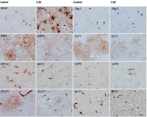
Paraffin sections processed for immunohistochemistry showed marked increase in GFAP immunoreactivity in CJD when compared with controls, as expected. GFAP-immunoreacytive astrocytes were increased in number,
230 and the amount of GFAP was increased per astrocyte with robust immunostaining of astrocyte processes (Figure 2). In contrast, OLig-2 immunostaining in the cerebral cortex did not reveal major differences between CJD and controls (Figure 2). Aquaporin 4
235 (AQP4) immunoreactive localized in astrocytes deli-neating fine and varicose network processes in control cerebral cortex. AQP4 immunoreactivity in CJD was variable from one case to another yet preserving the same morphological profile (Figure 2). In advanced
240 cases, AQP4 immunoreactivity was increased and blurred as reported elsewhere. GLT1 immunoreactivity in control cases was seen as delicate meshwork of fine and varicose processes around the nuclei of astrocytes. This pattern was preserved in CJD, although with
245 marked differences from one case to another; in some cases GLT1 immunoreactivity practically disappeared in areas with severe spongiform change while it was practically preserved in others; Moreover variations were also observed in different areas in the same tissue
250 section (Figure 2). MPC1 was mainly localized in the
Figure 2.Representative images of glial fibrillary acidic protein (GFAP), Olig-2, aquaporin 4 (AQP4), solute carrier family 1, member 1 (SLC1A2: glial affinity glutamate transporter: GLT1), mitochondrial pyruvate carrier 1 (MPC1), mitochondrial uncoupling protein 5 (UCP5), solute carrier family 2 member 1 (glucose transporter 1: GLUT1) and solute carrier family 16 member 1 (monocarboxylic acid transporter 1: MCT1) immunoreactivity in the frontal cortex area 8 of controls and cases with CJD MM1. Paraffin sections processed for immunohistochemistry and slightly stained with hematoxylin, bar = 25 μm.
cytoplasm of neurons in the cerebral cortex of control cases as a punctuate immunostaining consistent with a mitochondrial localization. MPC1 immunoreactivity was dramatically reduced in neurons in CJD but 255 increased in a few cortical glial cells (Figure 2). Similarly, UPC5 immunostaining mainly decorated neurons in controls. However, UPC5 immunoreactivity decreased in neurons but increased in glial cells, mainly in cells with astrocyte morphology, in CJD (Figure 2). 260 GLUT1 was mainly found in the walls of small blood
vessels consistent with endothelial localization as expected. In addition, GLUT1 immunoractivity was found as blurred patches in the cortical neuropil in control cases; these patches were markedly reduced in 265 size and intensity in CJD (Figure 2). Finally, MCT1 was mainly localized in endothelial cells and to a lesser degree in neurons and glial cells in controls. Marked reduction in MCT1 immunoractivity was found in neurons and glial cells, but not in endothelial cells, in 270 CJD (Figure 2).
Discussion
The present study is focused on astrocytes and oligo-dendrocytes of the frontal cortex area 8 in CJD MM1 and VV2 compared to controls. In spite of the opti-275 cally-monitored separation of the cerebral cortex from the underlying white matter, contamination by subcor-tical U fibers cannot be ruled out in our study. This is convenient remark as astrocytes and oligodendrocytes in grey matter differ from astrocytes and oligodendro-280 cytes in white matter, and from one region to another. Astrogliosis is one of the characteristic neuropatho-logical lesions in cerebral cortex CJD which is here manifested by increased significant GFAP mRNA expression, as previously reported [15]. However, no 285 significant differences are seen in the expression levels of the gene coding for aldehyde dehydrogenase 1 family member L1 (ALDH1L1) used as a marker of the total astrocyte population. Aquaporin 4 protein levels, as revealed by gel electrophoresis and western blotting, 290 are markedly increased in CJD [17]. This is in contrast with the down-regulation of AQP4 mRNA in CJD MM1 and CJD VV2, here observed. Yet, AQP4 immu-noreactivity is variable from one case to another:
AQP4 immunorecativity is apparently decreased in 295 some cases, increased in others, and even increased and blurred in cases with severe spongiform change. We do not have an explanation for this point; enhanced trans-lation and/or reduced degradation can account for dis-crepancies between mRNA and protein expression, and 300 for case-to-case and area differences in protein immu-noreactivity. Expression of solute carrier family 1A2
(SLC1A2: GLT1) is reduced in CJD VV2, pointing to the possibility of unbalanced glutamate concentrations between neurons and astrocytes in certain forms of
305 CJD. However, immunohistochemistry reveals indivi-dual variations and areal variations in GLT1 expression in the same tissue section. Therefore, further studies in animal models are needed to evaluate GLT1 mRNA and protein expression in prion diseases.
310 In addition to these aspects, new information has been obtained in this study. Present quantitative data show altered expression of several genes coding for key specific proteins of astrocytes and oligodendrocytes in frontal cortex area 8 in CJD which may have functional
315 implications in the pathogenesis of the disease. Reduced expression of mitochondrial pyruvate carrier 1 (MPC1) and mitochondrial uncoupling protein 5 (UCP5, SLC25A14, BMCP1: brain mitochondrial uncoupling protein 1) suggest altered mitochondrial
320 function in CJD MM1 and CJD VV2. Reduced expres-sion of solute carrier family 2A1 (SLC2A1 or GLUT1) points to altered glucose transport, whereas reduced expression of solute carrier family 16 (MCT1) suggests impaired monocarboxylic acid transport. These are
325 important aspects as blood-derived glucose through GLUT1 is metabolized via glycolysis to produce piru-vate and lactate which are delivered to the axons through specific solute carriers, the monocarboxylase transporters (MCTs) located in cell membranes [26,27].
330 In glia, MCT1 is mainly expressed in developing oligo-dendrocytes and MCT4 in astrocytes [28,29]. Inhibition of MCT1 in organotypic cultures of the spinal cord in glucose-deprived media is toxic to neurons; the effects on neurons are reversed with the addition of lactate
335 into the medium [29]; moreover, reduction of MCT1 activity in vivo results in axonal damage [29].
These changes are not accompanied by altered expres-sion of oligodendroglial markers OLIG1, OLIG2 and SOX10 [30]; and adult oligodendroglial precursors
340 (NG2) [31,32]. The expression of genes encoding main myelin proteins is also preserved in frontal cortex in CJD, including myelin basic protein (MBP), proteolipid protein (PLP), 2ʹ,3ʹ-cyclic nucleotide-3ʹphosphodiesterase (CNP), myelin-associated glycoprotein (MAG), myelin
oligoden-345 drocyte glycoprotein (MOG) and myelin/oligodendrocyte basic protein (MOBP) [33–35].The expression of CLDN11, the gene encoding for claudin 11, principal component of tight junctions of myelin connections at their edges [36,37], is not modified in CJD. The
expres-350 sion of MYRF, encoding myelin regulatory factor which triggers myelination following binding to several promo-ters of genes coding for myelin proteins [38], is main-tained in CJD. In contrast, the expression of the gene coding for galactocerebrosidase (GALC) is reduced in
355 CJD MM1 and VV2, thus indicating possible impairment in glycosphingolipid metabolism. To our knowledge, most studies of brain lipids in CJD were carried out many years ago and the main focus was on ganglioside alterations [39–42]. Since the present studies are focused 360 on the cerebral cortex, further investigation is needed to get information about brain lipid alterations in the white matter in prion diseases.
Oligodendrocytes are connected with each other and with astrocytes through gap junctions thus forming a 365 glial syncytium in the white matter tracts [43,44]. Gap junctions are composed of connexins (Cx); Cx32 and Cx47 are synthesized by oligodendrocytes, and Cx30 and Cx43 by astrocytes [45]. The expression of genes coding for Cx32 (GJB1) and Cx47 (GJC2), and Cx30 (GJB6) is 370 similar in CJD and in control cases, but GJA1 (encoding Cx43) mRNA expression is reduced in CJD VV2 when compared with controls and CJD MM1. Whether these changes have implications in the normal function of the glial syncitium deserves further attention.
375 Interpretation of some of the present data must be taken with caution. MPC1 is expressed in brain [46] principally in neurons in controls. MPC1 immunoreac-tivity is reduced in neurons but increased in glial cells in CJD. Therefore, the origin of decreased MPC1 mRNA 380 expression in CJD here observed may be due to damaged neurons, whereas MPC1 protein is increased in glial cells. Similarly, UC5P in the brain is mainly expressed in neu-rons [47]; reduced UCP5 mRNA expression here observed in CJD is accompanied by reduced UPC5 385 immunoreactivity in neurons, but this accompanied by increased immunoreactivity in glial cells with the mor-phology of astrocytes.
GLUT1 is largely expressed in red blood cells and endothelial cells, including brain microvessels [48,49]. No 390 alterations in GLUT1 immunoreactivity is found in CJD microvessels; reduction of GLUT1 mRNA correlates with decreased GLUT1 immunoreactivity in the neuropil in CJD. Finally, MCT1 is widely expressed in endothelial cells in brain [50–52], but also in glial cells especially during 395 development [53,54]. MCT1 immunoreactivity in the adult brain is also observed in neurons. MCT1 immunoractivity is preserved in endothelial cells in CJD when compared with controls; reduced MCT1 mRNA and protein in CJD can be ascribed to reduced expression in neurons. 400 The concepts of astrocytopathy and
oligodendrocyto-pathy, referring to molecular alterations in astrocytes and oligodendrocytes, are appropriate in neurodegenerative diseases with abnormal protein aggregates [55–60]. This study supports the idea that molecular flaws in astrocytes 405 and oligodendrocytes, in addition to neurons, are relevant
in the pathogenesis of cortical alterations in CJD.
Material and methods
Human cases
CJD cases were clinically diagnosed on the basis of rapid 410 dementia together with variable accompanying symp-toms, neuroimaging alterations and characteristic CSF biomarkers; all of them were tested for homozygosity of codon 129 in PRNP. Verification of clinical diagnosis was obtained after post-mortem neuropathological study
415 of the brain. Controls did not show neurological symp-toms; moreover, cases with metabolic syndrome, auto-immune diseases, fever and prolonged agonal state were not included in this series. Brain samples were obtained from the Brain Banks of the Institute of Neuropathology
420 HUB-ICO-IDIBELL Biobank and the Hospital Clinic-IDIBAPS Biobank following the guidelines of the Spanish legislation on this matter and the approval of the local ethics committees. The post-mortem interval between death and tissue processing was between 2h
425 45min and 22h 40min. One hemisphere was fixed by immersion in 4% buffered formalin for 3 weeks. The neuropathological study in control and CJD cases was carried out on, at a minimum, twenty selected 4μm-thick de-waxed paraffin sections of representative regions of
430 the frontal, temporal, parietal, motor, primary visual, anterior cingulate and entorhinal cortices, hippocampus, amygdala, basal forebrain, caudate, putamen, globus pal-lidus, thalamus, midbrain, pons, medulla oblongata, cer-ebellar vermis, hilus and cerebral white matter. Sections
435 were stained with haematoxylin and eosin, Klüver-Barrera, or processed for immunohistochemistry for microglia (Iba-1, Wako, Richmond, VA, USA), glial acidic protein (GFAP, Dako, Gostrup, Denmark), β-amyloid (Dako, clone 6F/3D), phospho-tau (Thermo
440 Scientific, Rockford, USA, clone AT8), α-synuclein (Novocastra, Newcastle, UK, clone KM51), TDP-43 (Abnova, Taipei, Taiwan, clone 2E2-D3), PrP (clon 3F4, Millipore, Darmstadt, GE) following proteinase K incubation, ubiquitin (Dako, Polyclonal Rabbit) and p62
445 (BD Biosciences, San Jose, USA, Purified Mouse Anti-p62 LCK ligand) using EnVision+ System peroxidase (Dako), and diaminobenzidine and H2O2.
Samples of the frontal cortex and cerebellum of the other hemisphere were rapidly dissected, frozen on metal
450 plates over dry ice and stored at −80ºC until use. Part of this material was used for gel electrophoresis and western blotting for identification of protease-resistant PrP type. CJD was diagnosed following well-established neuro-pathological criteria, codon 129 genotype and
character-455 istics of PrP pattern on western blots [61]. None of the cases had suffered from panencephalopathic CJD [62]. Additional brain pathology consisting of neurofibrillary
tangles at stages I-II of Braak and Braak, argyrophilic grain disease stage 2, mild small blood vessel disease, 460 and Lewy bodies in the brain stem in some individuals were expected with the age of the patients. Similar lesions were found in the control group and, therefore, they were not contemplated as discriminating variables.
The whole series included 7 CJD MM1 (4 men and 3 465 women; age: 70.5 ± 5.5), 10 CJD VV2 (4 men and 6 women; age 66.5 ± 6.85) and 10 controls (6 men and 4 women, age 67.1 ± 7.6). A summary of cases is shown inTable 1.
Biochemical studies were carried out in the frontal cortex area 8 after our best optically-monitored dissec-470 tion of the cerebral cortex and white matter. However, inclusion of white matter subcortical U fibers cannot be ruled out in the present study.
RNA purification
RNA from frozen frontal cortex area 8 was extracted fol-475 lowing the instructions of the supplier (RNeasy Mini Kit, Qiagen® GmbH, Hilden, Germany). RNA integrity and 28S/18S ratios were determined with the Agilent Bioanalyzer (Agilent Technologies Inc, Santa Clara, CA, USA). RIN values are shown inTable 1: control 6.76 ± 0.5; 480 CJD MM1 5.68 ± 0.8 and CJD VV2 5.84 ± 0.6). Samples were treated with DNase digestion, and RNA concentra-tion was evaluated using a NanoDrop™ Spectrophotometer (Thermo Fisher Scientific, Waltham, MA, USA).
Rt-qPCR
485 TaqMan RT-qPCR assays were performed in duplicate for each gene on cDNA samples in 384-well optical plates using an
AQ9
�ABI Prism 7900 Sequence Detection system (Applied Biosystems, Life Technologies, Waltham, MA, USA).�Probes are listed in Table II.
490 For each 10μL TaqMan reaction, 4.5μL cDNA was mixed with 0.5μL 20x TaqMan Gene Expression Assays and 5μL of 2x TaqMan Universal PCR Master Mix (Applied Biosystems). Values of GUS-β were used as internal controls for normalization [63].
495 The parameters of the reactions were 50°C for 2min, 95°C for 10min, and 40 cycles of 95°C for 15sec and 60°C for 1min. Finally, capture of all TaqMan PCR data used the Sequence Detection Software (SDS ver-sion 2.2.2, Applied Biosystems). For the data analysis,
500 threshold cycle (CT) values for each sample were processed to obtain the double delta CT (ΔΔCT) values. First, delta CT (ΔCT) values were calculated as the normalized CT values of each target gene in relation to the CT of endogenous controls GUS-β.
505 Then, ΔΔCT values were obtained from the ΔCT of Table 1.Summary of cases M: man; W: woman; Age in years; CJD:
Creutzfeldt-Jakob disease, subtypes MM1 and VV2; PMD: post-mor-tem delay; RIN: RNA integrity number.
Case Sex Age Diagnosis PMD RIN
1 M 66 Control 18 h 00 min 6.4 2 M 61 Control 03 h 40 min 7.0 3 M 74 Control 06 h 40 min 7.2 4 M 65 Control 05 h 15 min 6.8 5 M 63 Control 08 h 05 min 7,1 6 W 79 Control 03 h 35 min 6.8 7 W 67 Control 05 h 20 min 6,2 8 M 70 Control 03 h 45 min 7.2 9 M 52 Control 04 h 40 min 7.2 10 W 74 Control 02 h 45 min 5.7 11 M 69 CJD MM1 15 h 00 min 6.4 12 M 70 CJD MM1 N/A 4.7 13 W 64 CJD MM1 14 h 00 min 5.6 14 M 64 CJD MM1 14 h 00 min 4.7 15 M 77 CJD MM1 07 h 10 min 6,0 16 W 72 CJD MM1 08 h 00 min 7.0 17 M 78 CJD MM1 22 h 40 min 5.1 18 W 76 CJD VV2 05 h 00 min 5.5 19 M 65 CJD VV2 06 h 00 min 5.3 20 W 65 CJD VV2 07 h 15 min 5.4 21 W 72 CJD VV2 06 h 00 min 6.2 22 W 62 CJD VV2 08 h 45 min 4.7 23 M 71 CJD VV2 09 h 00 min 5.8 24 M 66 CJD VV2 05 h 00 min 6.8 25 M 52 CJD VV2 06 h 30 min 6.5 26 W 59 CJD VV2 09 h 00 min 6,7 27 W 67 CJD VV2 12 h 30 min 5.5
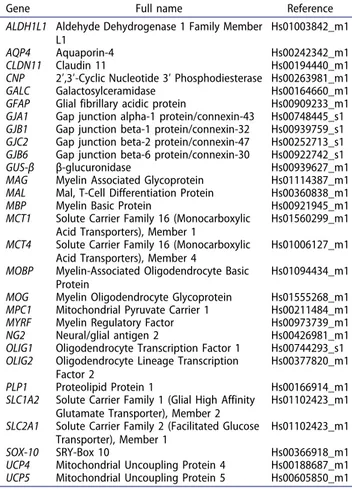
Table 2.TaqMan probes: gene identification, full name and reference.
Gene Full name Reference
ALDH1L1 Aldehyde Dehydrogenase 1 Family Member L1
Hs01003842_m1
AQP4 Aquaporin-4 Hs00242342_m1
CLDN11 Claudin 11 Hs00194440_m1 CNP 2ʹ,3ʹ-Cyclic Nucleotide 3ʹ Phosphodiesterase Hs00263981_m1 GALC Galactosylceramidase Hs00164660_m1 GFAP Glial fibrillary acidic protein Hs00909233_m1 GJA1 Gap junction alpha-1 protein/connexin-43 Hs00748445_s1 GJB1 Gap junction beta-1 protein/connexin-32 Hs00939759_s1 GJC2 Gap junction beta-2 protein/connexin-47 Hs00252713_s1 GJB6 Gap junction beta-6 protein/connexin-30 Hs00922742_s1 GUS-β β-glucuronidase Hs00939627_m1 MAG Myelin Associated Glycoprotein Hs01114387_m1 MAL Mal, T-Cell Differentiation Protein Hs00360838_m1
MBP Myelin Basic Protein Hs00921945_m1
MCT1 Solute Carrier Family 16 (Monocarboxylic Acid Transporters), Member 1
Hs01560299_m1 MCT4 Solute Carrier Family 16 (Monocarboxylic
Acid Transporters), Member 4
Hs01006127_m1 MOBP Myelin-Associated Oligodendrocyte Basic
Protein
Hs01094434_m1 MOG Myelin Oligodendrocyte Glycoprotein Hs01555268_m1 MPC1 Mitochondrial Pyruvate Carrier 1 Hs00211484_m1 MYRF Myelin Regulatory Factor Hs00973739_m1 NG2 Neural/glial antigen 2 Hs00426981_m1 OLIG1 Oligodendrocyte Transcription Factor 1 Hs00744293_s1 OLIG2 Oligodendrocyte Lineage Transcription
Factor 2
Hs00377820_m1 PLP1 Proteolipid Protein 1 Hs00166914_m1 SLC1A2 Solute Carrier Family 1 (Glial High Affinity
Glutamate Transporter), Member 2
Hs01102423_m1 SLC2A1 Solute Carrier Family 2 (Facilitated Glucose
Transporter), Member 1
Hs01102423_m1 SOX-10 SRY-Box 10 Hs00366918_m1 UCP4 Mitochondrial Uncoupling Protein 4 Hs00188687_m1 UCP5 Mitochondrial Uncoupling Protein 5 Hs00605850_m1
each sample minus the mean ΔCT of the population of control samples.
Statistical analysis
The normality of distribution of fold change values was 510 analyzed with the Kolmogorov–Smirnov test. Pearson’s correlation coefficient was used to assess a possible linear association between two continuous quantitative vari-ables. To determine the relationship between gene expres-sion and RIN values according to pathologic variables, we 515 used the analysis of covariance (ANCOVA). Statistical analysis of the expression data between groups was per-formed using one-way analysis of variance (ANOVA) followed by Tukey post-test or Kruskal-Wallis test fol-lowed by Dunns post-hoc test when required using the 520 SPSS software (IBM Corp. Released 2013. IBM SPSS Statistics for Windows, Version 21.0. Armonk, NY: IBM Corp.). Outliers were detected using the GraphPad soft-ware QuickCalcs (P < 0.05). All data were expressed as mean values ± SEM. Differences between controls and 525 CJD MM1 or CJD VV2 were considered statistically sig-nificant at * P < 0.05, ** P < 0.01, *** P < 0.001; and set at $ P < 0.05 and $$ P < 0.01when comparing CJD MM1 and CJD VV2.
Immunohistochemistry
530 De-waxed sections, 4 microns thick, were processed for immunohistochemistry (control n = 8; CJD MM1 n = 6; CJD VV2 n = 2). The sections were boiled in citrate buffer (20min) to retrieve tau antigenicity. Endogenous perox-idases were blocked by incubation in 10% methanol-1% 535 H2O2 solution (15min) followed by 3% normal horse serum solution. Then the sections were incubated at 4ºC overnight with one of the primary antibodies against glial fibrillary acidic protein (GFAP) (rabbit polyclonal, used at 1:500, Dako, Glostrup, DK), Olig-2 (rabbit polyclonal, 540 used at 1:500, Abcam, Cambridge, UK), aquaporine 4 (AQP4) (monoclonal, used at 1:400, Sigma, St Louis, Missouri, USA), solute carrier family 1 member 2 (GLT1) (guinea pig used at 1:100, Merck-Millipore, Billerica, MA, USA), mitochondrial pyruvate carrier 1 545 (MPC1) MPC1) (polyclonal, used at 1:100, Cell Signaling Technology, Danvers, MA, USA), mitochon-drial uncoupled protein 5 (UCP5) (polyclonal, used at 1:10, Novus Biological, Littleton, CO, USA), solute carrier family 2 member 1 (GLUT1) (polyclonal, used at 1:100, 550 Abcam, Cambridge, CB, UK) and solute carrier family 16 member 1 (MCT1) (monoclonal, used at 1:50, Sigma, St Louis, Missouri, USA). Following incubation with Following incubation with the primary antibody, the sec-tions were incubated with EnVision + system peroxidase
555 (Dako, DK) for 30min at room temperature. The perox-idase reaction was visualized with diaminobenzidine and H2O2. Control of the immunostaining included omission of the primary antibody; no signal was obtained following incubation with only the secondary antibody. Peptides for
560 pre-absorption studies were not available.
Acknowledgments
We wish to thank T. Yohannan for editorial assistance
Disclosure statement
No potential conflict of interest was reported by the authors.� AQ5
565
Funding
Part of this work was supported by the Ministry of Economy and Competitiveness, Institute of Health Carlos III (co-funded by European Regional Development Fund, ERDF, a way to build Europe): FIS PI17/00809, IFI15/00035 fellowship
570 to PA-B and co-financed by ERDF under the program Interreg Poctefa: RedPrion 148/16;Insti�tute of Health Carlos
III�[FIS PI17/00809,]. AQ6
ORCID
Pol Andrés-Benito http://orcid.org/0000-0003-3000-0338
575 Isidro Ferrer http://orcid.org/0000-0001-9888-8754
References
[1] Prusiner SB. An introduction to prion biology and diseases. In: Prusiner SB, editor. Prion biology and diseases, 2nd ed. New York (NY): Cold Spring
580 Harbor Laboratory;2004. p. 1–87.� AQ7 [2] Aguzzi A, Sigurdson C, Heikenwaelder M. Molecular
mechanisms of prion pathogenesis. Annu Rev Pathol.
2008;3:11–40.
[3] Gambetti P, Cali I, Notari S, et al. Molecular biology 585 and pathology of prion strains in sporadic human prion diseases. Acta Neuropathol.2011;121:79–90. [4] Budka H, Head MW, Ironside JW. Sporadic
Creutzfeldt-Jakob disease. Dickson DW, Weller RO, et al., editors. Neurodegeneration: the molecular pathology of dementia
590 and movement disorders. 2nd ed. Chichester, West Sussex:Willey-Blackwell;2011. p.322–335.
[5] Parchi P, Gambetti P, Capellari S. Genetic Creutzfeldt-Jakob disease. In: Dickson DW, Weller RO, editors. Neurodegeneration: the molecular pathology of dementia
595 and movement disorders. 2nd ed. Chichester, West Sussex: Willey-Blackwell;2011. p. 336–345.
[6] Ghetti B, Tagliavini F, Kovacs GG. Gerstmann-Straüssler-Scheinker. Dickson DW, Weller RO, et al., editors. Neurodegeneration: the molecular pathology of
600 dementia and movement disorders. 2nd ed. Chichester, West Sussex:Willey-Blackwell;2011. p.364–377.
[7] Head MW, Ironside JW, Ghetti B. Prion diseases. Love S, Budka H, Ironside J, et al., editors. Greenfield’s neuropathology. 9th ed. Boca Raton, Florida:CRC 605 Press;2015. p.1016–1086.
[8] Colby DW, Prusiner SB. Prions. Cold Spring Harb Perspect Biol.2011;3:a006833.
[9] Ironside JW, Head MW, Will RG. Variant Creutzfeldt-Jakob disease. In: Dickson DW, Weller RO, editors. 610 Neurodegeneration: the molecular pathology of dementia and movement disorders. 2nd ed. Chichester, West Sussex: Willey-Blackwell;2011. p. 354–363.
[10] Parchi P, Giese A, Capellari S, et al. Classification of sporadic Creutzfeldt-Jakob disease based on molecular 615 and phenotypic analysis of 300 subjects. Ann Neurol.
1999;46:224–233.
[11] Parchi P, Strammiello R, Notari S, et al. Incidence and spectrum of sporadic Creutzfeldt-Jakob disease variants with mixed phenotype and co-occurrence 620 of PrPSC types: an updated classification. Acta
Neuropathol.2009;118:659–671.
[12] Giese A, Brown DR, Groschup MH, et al. Role of microglia in neuronal cell death in prion disease. Brain Pathol.1998;8:449–457.
625 [13] Van EB, Dewulf E, Pals P, et al. The role of cytokines, astrocytes, microglia and apoptosis in Creutzfeldt-Jakob disease. Neurobiol Aging.2002;23:59–64. [14] Szpak GM, Lewandowska E, Lechowicz W, et al. The brain
immune response in human prion diseases. Microglial 630 activation and microglial disease. I. Sporadic
Creutzfeldt-Jakob disease. Folia Neuropathol.2006;44:202–213. [15] Llorens F, López-González I, Thüne K, et al. Subtype
and regional-specific neuroinflammation in sporadic Creutzfeldt-Jakob disease. Front Aging Neurosci. 635 2014;6:198.
[16] Kovacs GG, Preusser M, Strohschneider M, et al. Subcellular localization of disease-associated prion pro-tein in the human brain. Am J Pathol.2005;166:287–294. [17] Rodríguez A, Pérez-Gracia E, Espinosa JC, et al. 640 Increased expression of water channel aquaporin 1 and aquaporin 4 in Creutzfeldt-Jakob disease and in bovine spongiform encephalopathy-infected bovine-PrP trans-genic mice. Acta Neuropathol.2006;112:573–585. [18] Iwasaki Y, Mimuro M, Yoshida M, et al. Enhanced 645 aquaporin-4 immunoreactivity in sporadic
Creutzfeldt-Jakob disease. Neuropathology.2007;27:314–323. [19] Choi YP, Head MW, Ironside JW, et al. Uptake and
degradation of protease-sensitive and -resistant forms of abnormal human prion protein aggregates by 650 human astrocytes. Am J Pathol.2014;184:3299–3307.
[20] Victoria GS, Arkhipenko A, Zhu S, et al. Astrocyte-to-neuron intercellular prion transfer is mediated by cell-cell contact. Sci Rep.2016;6:20762.
[21] Prinz M, Montrasio F, Furukawa H, et al. Intrinsic 655 resistance of oligodendrocytes to prion infection. J
Neurosci.2004;24:5974–5981.
[22] Liberski PP, Yanagihara R, Wells GA, et al. Ultrastructural pathology of axons and myelin in experimental scrapie in hamsters and bovine spongi-660 form encephalopathy in cattle and a comparison with the panencephalopathic type of Creutzfeldt-Jakob dis-ease. J Comp Pathol.1992;106:383–398.
[23] Liberski PP, Brown P, Cervenakova L, et al. Interactions between astrocytes and oligodendroglia
665 in human and experimental Creutzfeldt-Jakob disease and scrapie. Exp Neurol.1997;144:227–234.
[24] Shintaku M, Yutani C. Oligodendrocytes within astrocytes (“emperipolesis”) in the white matter in Creutzfeldt-Jakob disease. Acta Neuropathol.2004;108:201–206.
670 [25] El Hachimi KH, Chaunu MP, Brown P, et al.
Modifications of oligodendroglial cells in spongiform encephalopathies. Exp Neurol.1998;154:23–30. [26] Saab AS, Tzvetanova ID, Nave K-A. The role of myelin
and oligodendrocytes in axonal energy metabolism. 675 Curr Opin Neurobiol.2013;23:1065–1072.
[27] Pierre K, Pellerin L. Monocarboxylate transporters in the central nervous system: distribution, regulation and function. J Neurochem.2005;94:1–14.
[28] Rinholm JE, Hamilton NB, Kessaris N, et al. Regulation 680 of oligodendrocyte development and myelination by glucose and lactate. J Neurosci.2011;31:538–548. [29] Lee Y, Morrison BM, Li Y, et al. Oligodendroglia
metabolically support axons and contribute to neuro-degeneration. Nature.2012;487:443–448.
685 [30] Marinelli C, Bertalot T, Zusso M, et al. Systematic
review of pharmacological properties of the oligoden-drocyte lineage. Front Cell Neurosci.2016;10:27. [31] Dawson MR, Polito A, Levine JM, et al.
NG2-expres-sing glial progenitor cells: an abundant and widespread 690 population of cycling cells in the adult rat CNS. Mol Cell Neurosci.2003;24:476–488.
[32] Peters A. A fourth type of neuroglial cell in the adult central nervous system. J Neurocytol.2004;33:345–357. [33] Kursula P. The current status of structural studies on
695 proteins of the myelin sheath. Int J Mol Med.
2001;8:475–479.
[34] Harauz G, Ladizhansky V, Boggs JM. Structural poly-morphism and multifunctionality of myelin basic pro-tein. Biochemistry.2009;48:8094–8104.
700 [35] Jahn O, Tenzer S, Werner HB. Myelin proteomics:
molecular anatomy of an insulating sheath. Mol Neurobiol.2009;40:55–72.
[36] Gow A, Southwood CM, Li JS, et al. CNS myelin and sertoli cell tight junction strands are absent in Osp/
705 claudin-11 null mice. Cell.1999;99:649–659.
[37] Morita K, Sasaki H, Fujimoto K, et al. Claudin-11/ OSP-based tight junctions of myelin sheaths in brain and Sertoli cells in testis. J Cell Biol.1999;145:579–588. [38] Cahoy JD, Emery B, Kaushal A, et al. A transcriptome
710 database for astrocytes, neurons, and oligodendrocytes: a new resource for understanding brain development and function. J Neurosci.2008;28:264–278.
[39] Suzuki K, Chen G. Chemical studies on Jakob-Creutzfeldt disease. J Neuropathol Exp Neurol.1966;25:396–408.
715 [40] Bass NH, Hess HH, Pope A. Altered cell membranes in
Creutzfeldt-Jakob disease. Microchemical Studies. Arch Neurol.1974;31:174–182.
[41] Tamai Y, Kojima H, Ikuta F, et al. Alterations in the composition of brain lipids in patients with
720 Creutzfeldt-Jakob disease. J Neurol Sci.1978;35:59–76. [42] Yu RK, Manuelidis EE. Ganglioside alterations in gui-nea pig brains at end stages of experimental Creutzfeldt-Jakob disease. J Neurol Sci.1978;35:15–23.
[43] Bedner P, Steinhauser C, Theis M. Functional redun-725 dancy and compensation among members of gap junc-tion protein families? Biochim Biophys Acta.�2012; 1818:
1971-1984.�—�Remove:�-AQ8
�
[44] Nualart-Marti A, Solsona C, Fields RD. Gap junction communication in myelinating glia. Biochim Biophys 730 Acta.2013;1828:69–78.
[45] Orthmann-Murphy JL, Freidin M, Fischer E, et al. Two distinct heterotypic channels mediate gap junction coupling between astrocyte and oligodendrocyte con-nexins. J Neurosci.2007;27:13949–13957.
735 [46] Li Y, Han G, Ji Y, et al. Establishment of mitochondrial pyruvate carrier 1 (MPC1) gene knockout mice with preliminary gene function analyses. Oncotarget.
2016;7:79981–79994.
[47] Kim-Han JS, Reichert SA, Quick KL, et al. BMCP1: a 740 mitochondrial uncoupling protein in neurons which regulates mitochondrial function and oxidant produc-tion. J Neurochem.2001;79:658–668.
[48] Shawahna R, Uchida Y, Declèves X, et al. Transcriptomic and quantitative proteomic analysis of transporters and 745 drug metabolizing enzymes in freshly isolated human
brain microvessels. Mol Pharm.2011;8:1332–1341. [49] Nakamura S, Muramatsu SI, Takino N, et al. Gene
therapy for Glut1-deficient mouse using an adeno-associated virus vector with the human intrinsic 750 GLUT1 promoter. J Gene Med.2018;20:e3013.
[50] Smith JP, Drewes LR. Modulation of monocarboxylic acid transporter-1 kinetic function by the cAMP sig-naling pathway in rat brain endothelial cells. J Biol Chem.2006;281:2053–2060.
755 [51] Daneman R, Zhou L, Agalliu D, et al. The mouse blood-brain barrier transcriptome: a new resource for understanding the development and function of brain endothelial cells. PloS ONE.2010;5:e13741.
[52] Liu Z, Sneve M, Haroldson TA, et al. Regulation of 760 monocarboxylic acid transporter 1 trafficking by the canonical Wnt/β-catenin pathway in rat brain endothe-lial cells requires cross-talk with Notch signaling. J Biol Chem.2016;291:8059–8069.
[53] Gerhart DZ, Enerson BE, Zhdankina OY, et al. 765 Expression of monocarboxylate transporter MCT1 by brain endothelium and glia in adult and suckling rats. Am J Physiol.1997;273:E207–213.
[54] Zhang M, Ma Z, Qin H, et al. Monocarboxylate transporter 1 in the medial prefrontal cortex
devel-770 opmentally expresses in oligodendrocytes and associates with neuronal amounts. Mol Neurobiol.
2017;54:2315–2326.
[55] Seifert G, Schilling K, Steinhauser C. Astrocyte dys-function in neurological disorders: a molecular
per-775 spective. Nat Rev Neurosci.2006;7:194–206.
[56] Pekny M, Pekna M, Messing A, et al. Astrocytes: a central element in neurological diseases. Acta Neuropathol.2016;131:323–345.
[57] Tognata R, Miller RH. Contribution of the oligoden-780 drocyte lineage to CNS repair and neurodegenerative pathologies. Neuropharmacology.2016;110:539–547. [58] Ettle B, Schlachetzki JCM, Winkler J. Oligodendroglia
and myelin in neurodegenerative diseases: more than just bystanders? Mol Neurobiol.2016;53:3046–3062.
785 [59] Ferrer I. Diversity of astroglial responses across human
neurodegenerative disorders and brain aging. Brain Pathol.2017;27:645–674.
[60] Verkhratsky A, Zorec R, Parpura V. Stratification of astrocytes in healthy and diseased brain. Brain Pathol.
790
2017;27:629–644.
[61] Parchi P, de Boni L, Saverioni D, et al. Consensus classification of human prion disease histotypes allows reliable identification of molecular subtypes: an inter-rater study among surveillance centres in Europe and
795 USA. Acta Neuropathol.2012;124:517–529.
[62] Jansen G, Head MW, Rozemuller AJ, et al. Panencephalopathic Creutzfeldt-Jakob disease in the Netherlands and the UK: clinical and pathological characteristics of nine patients. Neuropathol Appl
800 Neurobiol.2009;35:272–282.
[63] Barrachina M, Castaño E, Ferrer I. TaqMan PCR assay in the control of RNA normalization in human post-mortem brain tissue. Neurochem Int.2006;49:276–284.