UNIVERSITA’ POLITECNICA DELLE MARCHE
FACOLTA’ DI MEDICINA E CHIRURGIA
Corso di Dottorato in Salute dell’Uomo
Ciclo XXXI
Ageing-related expression of
Twinfilin-1 regulates cholangiocyte
biological response
to injury
Ph.D. dissertation of:
Advisor:
Debora Maria Giordano
Prof. Antonio Benedetti
Co-advisor:
INDEX
1. INTRODUCTION 1 1.1 Biliary tree 1 1.1.1 Architecture 1 1.1.2 Vascularization 2 1.1.3 Innervation 3 1.2 Cholangiocyte 41.2.1 Morphological and functional heterogeneity 5 1.2.2 Functional role in health state: bile production 6 1.2.3 Functional role in disease state 9
1.2.3.1 Proliferative response 11
1.2.3.2 Ductopenia 11
1.2.3.4 Fibrosis 13
1.2.3.5 Releasing of proinflammatory mediators 13 1.2.3.6 Upregulation of adhesion molecules 16 1.2.3.7 Immunoglobulins and peptides releasing 16
1.3 Cholangiopathies 17
1.3.1 Primary sclerosing cholangitis and Primary biliary cholangitis: an
overview 19
1.3.1.1 Therapy 21
1.4 Aging 22
1.4.1 The hallmarks of aging 23
1.4.1.1 Primary hallmarks 23
1.4.1.2 Antagonistic hallmarks 24
1.4.1.3 Integrative hallmarks 27
1.5 Aging, senescence and biliary tree disorders 28
2. AIM OF THE THESIS 31
3. MATERIALS AND METHODS 32
3.1 Materials 32
3.2 Methods 33
3.2.1 MicroRNAs selection 33
3.2.2 MicroRNAs testing in cholangiocyte: isolation, reverse transcription and
3.2.3 In silico analysis of microRNAs putative targets and intracellular
pathways 34
3.2.4 Expression of Twf1 in cholangiocyte 34
3.2.5 Cell lines and in vitro experiments 35
3.2.5.1 Evaluation of Twf1 role in cholangiocytes proliferation 35
3.2.5.2 Evaluation of Twf1 role in cholangiocyte senescence 36
3.2.6 Animal models 37
3.2.6.1 SAMR1 and SAMP8 38
3.2.6.2 Twf1+/+ and Twf1-/- 40
3.2.7 RNA extraction, reverse transcription and real-time PCR 42
3.2.8 ELISA 44
3.2.9 Western Blotting 44
3.2.10 Immunohistochemistry and histochemistry analysis 46 3.2.11 Liver hydroxyproline content quantification 47
3.2.12 Statistical analysis 47
4. RESULTS 48
4.1 Age-related miRs are upregulated in diseased cholangiocytes 48 4.2 Twf1 is the molecular target of upregulated miRs 50 4.3 Twf1 and related miRs are upregulated in old and diseased
cholangiocytes 52
4.4 Evaluation of TWF1 expression in human samples 54 4.5 Twf1 is involved in cholangiocytes proliferation 56 4.6 Twf1 is involved in cholangiocytes senescence 58 4.7 Accelerated aging exacerbates biliary injury in vivo 64 4.8 The absence of Twf1 reduces biliary response to injury in vivo 67
5. CONCLUSIONS 69
6. BIBLIOGRAPHY 76
7. TABLES INDEX 96
ABSTRACT
Cholangiocytes are the epithelial cells which line the bile ducts and represent the targets of a heterogeneous group of chronic biliary diseases termed cholangiopathies, with different aetiology. The two most common cholangiopathies are the Primary sclerosing cholangitis (PSC) and the Primary biliary cholangitis (PBC) whose pathogenesis have to be fully elucidated. Despite several lines of evidence suggest the potential link between ageing and hepatobiliary diseases onset and progression, it is not yet known whether ageing process modulates cholangiocyte biology during bile duct injury. The aim of the current study is to unveil molecular pathways related to ageing process and to evaluate their possible effect in the pathophysiology of cholangiocytes. A panel of microRNAs (miRs) known to be involved in ageing process, was evaluated in cholangiocytes isolated by young and old mice (2-month and 22-month old, respectively) subjected or not to DDC diet, as a model of sclerosing cholangitis. By in silico analysis, it was possible to identify Twinfilin-1 (Twf1), an actin - binding protein involved in motile and morphological cellular processes, as putative molecular target commonly regulated by more than one miR taken into account. TWF1 expression was evaluated in human samples collected by PSC and PBC patients. In vitro, cholangiocyte proliferation and senescence were evaluated in NRC silenced for Twf1 expression. In vivo, bile ducts proliferation and collagen deposition were evaluated in a mouse model of accelerated senescence (SAMP8) and in Twf1 -/- mice. Twf1 lack of expression
reduces cell proliferation in vitro. Knock-down of Twf1 increased senescence and SASP markers expression upon pro-proliferative stimulation as well as in
biliary proliferation and fibrosis, whereas Twf1-/- had a tendency to reduce biliary proliferation and collagen deposition upon DDC administration compared to control animals. In conclusion, it was possible to verify the role of Twf1 in cholangiocyte adaptation to injury, in terms of proliferation and senescence establishment. We identified Twf1 as a potential mediator of cholangiocyte adaption to injury and ageing process. Our findings suggest that cholangiopathies and aging might share common molecular pathways. A deeper understanding of intracellular pathways involved in the modulation of cholangiocyte biology (proliferation, senescence or apoptosis, releasing of proinflammatory mediators and cross-talk with other liver cells) is essential to devise novel effective therapies, for cholangiopathies treatment which are currently lacking. Taken together our findings allow the identification of a possible molecular target which is able to modulate both in vitro and in vivo, different aspects of cholangiocyte pathophysiology.
1. INTRODUCTION
1.1 Biliary tree
1.1.1 Architecture
The biliary tree is a network of ducts arising from the canals of Hering up to the terminus of common bile duct that progressively increase in size. The biliary system drains the hepatic-derived bile from the bile canaliculi into the gallbladder or directly into the lumen of intestine. It includes intrahepatic and extrahepatic ducts which are encircled by epithelial cells, the cholangiocytes [1], [2]. According to biliary lumen diameter and proximity, human intrahepatic bile ducts (IHBDs) could be divided into ductules (diameter < 15 μm) arising from the canals of Hering, interlobular ducts (diameter comprises between 15-100 μm) that originate from the convergence of ductules, septal ducts (diameter comprises between 100-300 μm) consisting of at least two interlobular ducts, area ducts (diameter comprises between 300-400 μm) which converge to form the segmental ducts (diameter comprises between 400 -800 μm) and the hepatic ducts (diameter > 800 μm) which underlie the passage towards the extrahepatic bile ducts. Extrahepatic bile ducts (EHBDs) include the common hepatic duct, originating from the union of right and left hepatic ducts, the cystic duct, the gallbladder and the common bile duct which transports the bile directly into the duodenum [3] (Figure 1). In cross section, small intrahepatic bile ducts are lined by 4-5 cholangiocytes while about 40 cholangiocytes could be found in large
In rodents, the architecture of the biliary tree is more simplified. It could be divided by size into small (diameter < 15 μm) and large (diameter > 15 μm) intrahepatic bile ducts. Small bile ducts are lined by 4 to 5 cholangiocytes, as observed in humans, while large bile ducts are constituted by 8 up to 15 cholangiocytes [4], [6], [7].
Figure 1. Biliary tree architecture (From de Jong et al. 2018 [8]).
1.1.2 Vascularization
Blood supply of biliary tree is ensured by intrahepatic and extrahepatic biliary plexus (PBP). It is a vascular system which originates from hepatic artery branches and flows either directly (lobular branch) or indirectly, through the portal vein (prelobular branch), into the hepatic sinusoids. The PBP supports the transport of bile substances reabsorbed from
cholangiocytes to hepatocytes [9]. PBP encircles large bile ducts, becoming less ramified and thinner around the small bile ducts.
The importance of PBP has been evidenced in animal models subjected to bile duct ligation (BDL) as a model of liver fibrosis, taken into account when it is intended to study cholangiocytes proliferation. Therefore, BDL is associated to cholangiocytes proliferation, noticeable since the 2 days after treatment. The microvasculature arrangement follows cholangiocyte proliferation to support newly formed bile ducts in terms of nutrients. In particular, between PBP and bile duct expansion there is a lag time suggesting that, in the first phases of biliary obstruction, cholangiocytes are more susceptible to ischemic and toxic reabsorbed substances-related damage [9].
1.1.3 Innervation
The liver is characterized by two autonomic nervous fibers. The sympathetic fibers which originate from the celiac ganglion and the parasympathetic fibers which originate from the vagus nerve [10]. These fibers extend from the hilar of the hepatic portal and then run along with the hepatic arteries and portal veins. The nervous fibers could release a number of neuropetides such as catecholamines and acetylcholine (Ach) but also other neurotransmitters (e.g. neuropeptide Y), calcitonin gene-related peptide or CGRP and somatostatin). These molecules regulate cholangiocytes absorptive/secretory activities and proliferation through the expression of relative specific receptors on cholangiocytes membrane. An elegant review of Marzioni and his colleagues focused on the neuropeptides/hormonal regulation in cholangiocytes adaptive response to injury [11].
1.2 Cholangiocytes
Cholangiocytes are the epithelial cells which line the bile ducts and are the central target of a group of diseases with different aetiology, termed cholangiopathies. The main physiological function of cholangiocytes is to modify the canalicular bile volume and composition through both absorptive and secretory processes, tightly regulated by molecules of different nature (e.g. neurotransmitters, hormones or peptides) [12]. Cholangiocytes are polarized cells which possess an apical or luminal membrane and a basolateral membrane. At the apical membrane level, biliary epithelial cells are supplied by tight junctions that join adjacent cells [13] and maintain cell polarity [14]. Cell-cell communication between adjacent cells is also ensured by the presence of gap junctions [15]. Along the apical membrane, cholangiocytes possess a number of microvilli protruding in the bile duct lumen, which increase of 5-fold cell surface area [6]. Moreover, biliary cells are provided with a primary cilium which exerts sensory functions, transmitting extracellular-derived informations inside the cells thus modulating different intracellular pathways involved in cellular biological functions (e.g. differentiation, proliferation or secretion) [16]. The primary cilium is a non-motile structure composed by a microtubule-based core surrounded by axoneme, which extends from the basal body up to cell membrane [3], [16]. At the primary cilium level it is possible to find a number of proteins and receptors whose activation results in the upregulation or downregulation of different signalling pathways [16].
As well as other cell types, cholangiocytes’ plasma, possess an actin- cytoskeleton which plays a key role as structural support of cell membrane, in conferring and maintaining cell polarity, in vesicle-trafficking and in the modulation of protein distribution [17]. Cholangiocytes exocytic capabilities lie in multivesicular bodies (MVBs) and exosomes releasing, processes that take place in the apical domain of plasma membrane [18]. MVBs are intracellular organelles, also known as late endosomes, which have a diameter > 1 μm and are part of the lysosomal system. Some MBVs are fused with lysosomes for protein recycling others instead, are released as exosomes in the extracellular space following MVBs membrane and cell plasma membrane fusion [19]. On the other hand, the exosomes are small cell-derived vesicles (diameter comprises between 30–100 nm) that seem to be involved in physiological biological processes. They deliver several macromolecules (e.g. lipids and proteins) and nucleic acids (e.g. mRNAs and miRs) to closer or even distant cells, activating molecular pathways in the target cells [20].
1.2.1 Morphological and functional heterogeneity
According to morphology, cholangiocytes could be divided into small and large cholangiocytes which line small and large intrahepatic bile ducts, respectively. Small cholangiocytes possess a cuboidal shape and a high nucleus to cytoplasm ratio. Thus small cholangiocytes are likely able to modify their biology in pathological conditions, due to a less cellular differentiation (functional plasticity). They possess a rich Golgi while the endoplasmatic reticulum is not abundant and although it appears to be
Unlike small cholangiocytes, large cholangiocytes have a columnar morphology and a small nucleus to cytoplasm ratio [6].
The morphological heterogeneity of small and large cholangiocytes reflects the functional heterogeneity existing between the two cells populations. Large cholangiocytes lining interlobular, septal and larger bile ducts actively participate to modification of bile trough secretory and absorptive mechanisms, due to the presence of proper ion transport systems and hormone receptors at the plasma membrane and supported by the presence of microvilli in the apical domain of cell membrane [1].
On the other hand, small cholangiocytes are able to modify their phenotype in response to exogenous/endogenous noxious stimuli thus participating to the inflammatory response during biliary tree damage, and serves as liver progenitor cells under certain conditions [21].
1.2.2 Functional role in health state: bile production
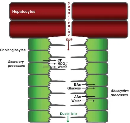
Physiologically, cholangiocytes are implicated in the modification of hepatic-derived bile. To accomplish this task cholangiocytes are equipped of different transporters and channels localized at both apical and basolateral cell membrane which ultimately lead to secretion into the bile of water, Cl- and
HCO3-, and extraction of glucose, bile acids and amino acids [22]. The
releasing of HCO3- is functional to bicarbonate umbrella composition. This
mechanism protects cholangiocytes against bile acids-dependent injury, by reducing the passive absorption of bile acids through cholangiocytes [3], [22]. Bile is an aqueous secretion produced by hepatocytes and composed of water for the 95% and for the remaining 5% by organic (e.g. bile acids), inorganic solutes and lipids [23].
The hepatic bile is transported by bile canaliculi, formed by the apical membrane of neighbour hepatocytes kept together by tight junctions (zona occludens), in the opposite direction to that of portal blood toward the canals of Hering in the bile ducts and, after modification by cholangiocytes, into the gallbladder or into the duodenum [23]. In the ileum the majority of bile acids (95%) is absorbed and then recycled in the liver via enterohepatic circulation [22]. Differently, through the cholehepatic shunt, a portion of bile acids (unconjugated) secreted by hepatocytes, after being passively absorbed by cholangiocytes, return to hepatocytes [3]. Subsequently, these unconjugated bile acids are secreted again by hepatocytes stimulating once again HCO3
-secretion.
One of the most important molecule which regulates biliary secretion is the secretin (STC) hormone, whose specific receptor is expressed at the basolateral domain of large cholangiocytes membrane [24]. Briefly, in response to peptides and acidic pH produced after a meal, duodenal and jejunal S cells release the STC into the portal blood [3]. Once reached the liver via enterohepatic circulation, STC binds to secretin receptor (SR) thus initiating an intracellular molecular cascade culminating in 3′,5′-cyclic monophosphate (cAMP) production. The increased levels of cAMP in turns activate the protein kinase A (PKA) that catalyses the phosphorylation of cystic fibrosis transmembrane conductance regulator (CFTR). The CFTR is a chloride channel which upon activation determines the extrusion of Cl- ions
in the bile lumen [12]. The existing Cl- gradient across the two sides of plasma
membrane activates the anion exchanger 2 (AE2) and, as a consequence, the net releasing of HCO3- associated to the osmotic influx of H2O through the
3
An alternate mechanism though which the efflux of Cl- ions is
ensured, depends on acetylcholine (ACh) binding to muscarinic receptor M3, expressed on the basolateral side of plasma membrane [26]. In response to ligand binding to its specific receptor, the levels of Inositol trisphosphate (IP3) increase and the release of Ca2+ evidenced following IP3 binding to its
receptors, results in the apical secretion of Cl- [27]. Besides the well-studied
secretory functions of cholangiocytes mainly dependent on STC/SR axis, other channels and proteins located in the apical membrane as well as at the basolateral level, are involved in physiological secretory functions of biliary epithelial cells (e.g. Na+-independent Cl−/HCO − exchanger, K+ channel
(SK2)) [3]. The modification of bile is achieved not only by secretory functions but also by absorptive capabilities of cholangiocytes which rely on the expression of transporters on both apical and basolateral cellular side (e.g. Na+-dependent bile acid transporter or ASBT, sodium-dependent glucose
cotransporter 1, water channel or AQP1) [3]. As mentioned above, bile contains organic cations excreted by hepatocytes which could be noxious for cholangiocytes [23]. The passive diffusion and accumulation of such molecules inside cholangiocytes could lead to deleterious effects. With this regard, cholangiocytes possess different protective mechanisms. As an example, the multidrug resistance 1 (MDR1) is an ATP-dependent transmembrane efflux pump localized in the apical pole, which is able to excrete again into the bile, exogenous or endogenous lipophilic compounds [27],[28]. At the basolateral pole of cholangiocytes membrane, it is possible to find transporters which allow organic anions to be released from cholangiocytes into the PBP (e.g. multidrug-associated protein 3 or MPR3 and MRP4) [29].
Figure 2. Hepatic bile production and modification, an overview. Hepatocytes synthetize the canalicular or primary bile which, as it flows into the bile duct lumen, is modified by cholangiocytes through either secretory (Cl-, HCO3- and water extrusion) or absorptive processes (glucose, bile acids, water and amino acids absorption). BAs: bile acids; AAs: amino acids (From Tabibian et al. [3]).
1.2.3 Functional role in disease state
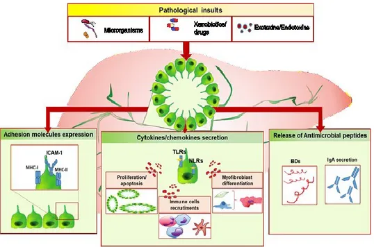
In physiological conditions cholangiocytes are mitotically dormant, until biliary tree damage occurs. In response to exogenous/endogenous stimuli (e.g. microorganisms, toxins/drugs or hormones), cholangiocytes become reactive and orchestrate a compensatory response. This includes the releasing of a wide range of proinflammatory and fibrogenic mediators which act in autocrine/paracrine fashion mediating: 1) cholangiocyte proliferation [30] so as to overcome biliary mass loss and to preserve the secretory/absorptive activities; 2) senescence and apoptosis processes which lead to ductopenia when the balance between proliferation/cell death come
the injured tissue and remodel the biliary tree. Thus, cholangiocyte response to injury is not merely characterized by enhanced proliferation, but it also relies on the activation of immune response through the secretion of proinflammatory and chemotactic mediators, on the upregulation of adhesion molecules expression and on antimicrobial molecules releasing through which cholangiocytes directly exert host defence activities (Figure 3).
Figure 3. Overview of cholangiocytes immune biology. Once activated by noxious stimuli of different nature (microorganisms, xenobiotics, drugs or exotoxins/endotoxins), reactive cholangiocytes respond by releasing cytokines and chemokines which in turns determine cholangiocytes proliferation and apoptosis, when the balance between cell growth/date come less, immune cell recruitments and myofibroblast differentiation. Moreover, activated cholangiocytes upregulate the expression of surface proteins such as MHC-I, MHC-II and ICAM-1) and start to release antimicrobial molecules such as beta-defensins (BDs) or IgA, following epithelial transcytosis. If the biliary damage persists, these compensatory processes may lead to chronic inflammation and fibrosis establishment. PF, portal fibroblast; HSC, hepatic stellate cell. (From Giordano et al. [31]).
1.2.3.1 Proliferative response
The ability of cholangiocyte to undergo proliferation has been evidenced in both experimental studies and in different human pathological conditions [32], [33]. Cholangiocyte proliferation is the main contribution of the so called “ductular reaction”. This term has been coined by Popper in 1957 and refers to proliferation of either pre-existing ductules, progenitor cell activation and appearance of intermediate hepatocytes at the interface between biliary tree and hepatocytes [34].
Based on in vivo experimental studies, four types of ductular reaction have been identified and summarized in a comprehensive review of Svegliati-Baroni and his colleagues [35].
In course of biliary damage, cholangiocytes proliferation is achieved through interleukin-6 (IL-6) upregulation. It plays a crucial in biliary mass maintenance and in functional activities enhancement [11], [36]. The importance of IL-6 in cholangiocytes population support, has been confirmed by an in vivo study carried out on mice lacking IL-6 gene expression. Transgenic mice subjected to BDL showed a worsening of pathological conditions (e.g. increase of serum total bilirubin, development of advanced stage fibrosis and an high mortality rate) compared to controls [37].
1.2.3.2 Ductopenia
Ductopenia occurs in the setting of chronic biliary inflammation, when the balance between proliferation and cell death phenomena come less. In normal conditions, senescent or damaged cholangiocytes undergo programmed cell-death to be replaced by newly formed cholangiocytes [38].
In pathological conditions, the homeostasis between cholangiocyte proliferation and death is lost, and disease progresses toward ductopenia.
Apoptosis has been evaluated in several experimental studies [39], [40]. As an example an in vivo study carried out on BDL rats, has shown that isolated cholangiocytes incubated with both TNFα and Actinomycin D, undergo cell programmed death which in turns lead to loss of cholangiocytes secretion capabilities. These data suggest that in course of cholestasis, cholangiocytes are more susceptible to TNFα-mediated cell death [41].
The establishment of senescent phenotype leads to progressive ductopenia, as a result of impaired proliferative and regenerative compensatory processes, which make non-replicative cells prone to undergo subsequently injuries [42]. Interestingly, it has been shown that cholangiocytes of PSC patients upregulate both senescence markers (p16INK4A
and p21CIP/WAF1) and SASP secretome components [42]. These findings
suggest a possible role of cholangiocytes in the modulation of both organ homeostasis and microenvironment [42], [43].
According to human data previously discussed, multi-drug resistance 2 knockout (mdr2-/-), a genetic mouse model of sclerosing cholangitis [44],
showed the accumulation of senescent cholangiocytes in the liver [42]. In line with these data, the prolonged exposure to LPS (10 days) determines a persistent TLR4 activation in cultured normal human cholangiocytes (NHC) and as a consequence the upregulation of proteins which regulate cell-cycle and SASP components [42].
1.2.3.4 Fibrosis
As a readout of chronic liver injury, it could be observed hepatic fibrosis development. It has been demonstrated that the bile ducts of fibrotic livers express high levels of transforming growth factor- β2 (TGF-β2) transcripts [45]. TGF-β acts by downregulating adhesion molecules expression on cell surfaces, by promoting the myofibroblastic differentiation (by activating portal fibroblast and hepatic stellate cells [46] and by inhibiting cholangiocytes proliferation [47].
On the other hand, TGF-β stimulates cholangiocytes secretion of endothelin-1 and modulates extracellular matrix protein deposition by mesenchymal liver cells [45].
Injured cholangiocytes are also able to secrete MCP-1, a well-known chemotactic factor, which could induce portal fibroblasts proliferation and transdifferentiation in myofibroblasts [48].
1.2.3.5 Releasing of proinflammatory mediators
Cholangiocytes represent the first line of defence of biliary system against potentially noxious stimuli of different nature (against microorganism, endotoxin/esotoxins, drugs) that reach the hepatic compartment through the portal blood circulation. For this purpose, cholangiocytes are endowed with pathogen recognition receptors (PRRs) that bind to either microbial components (e.g. bacterial DNA, product or cell wall components) identified as pathogen-associated molecular patterns (PAMPs) and molecules released from damaged cells known as damage- associated molecular patterns (DAMPs) [49].
includes several type 1 transmembrane glycoprotein receptors (10 members in human and 13 in mammalian cells) that upon recognition of conserved microbial components, activate a cascade of intracellular signals which culminate in the activation of nuclear factor kappa-b (Nf-kB) or activator protein-1 (AP-1) and, as a consequence in the biosynthesis of proinflammatory cytokines [50].
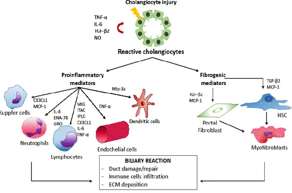
On the other hand, the nucleotide-binding oligomerization domain (NOD)-like receptor (NLR) family consists of soluble PRRs, which could recognize intracellular pathogens and endogenous noxious stimuli through the DAMPs [51]. Depends on the triggering stimulus, cholangiocytes release a different pattern of soluble mediators (Figure 4).
Figure 4. Crosstalk between activated cholangiocytes and resident/non-resident cells in response to biliary injury. Biliary epithelial cells are able to release a huge amount of proinflammatory cytokines and chemokines acting in autocrine/paracrine manner. The downstream events are cholangiocytes proliferation, immune cells recruitment and myofibroblast differentiation of portal fibroblast and HSC. HSC: hepatic stellate cell (From Pinto et al. [52]).
As an example, enteric-derived bacterial products (e.g. lipopolysaccharide or LPS, lipoteichoic acid), IL-1 and TNFα stimulus, trigger the release of IL-8, epithelial cell derived neutrophil activating protein (ENA-78) and growth-related oncogene (GRO), which are chemotactic factor acting on neutrophils [53]. On the contrary, INFy stimulus induces the releasing of a set of inflammatory mediators which characterize a chronic inflammatory process. With this regards, it has been shown that INFy treatment downregulates IL-8 releasing and, on the other hand, enhances the secretion of the monocyte chemoattractant protein-1 (MCP-1) and other mediators typical of a prolonged inflammatory process (e.g. monokine induced by INFy or Mig, interferon inducible T cell alpha chemoattractant or ITAC and interferon gamma inducible protein 10 or IP10) [53]. IL-1β, TNF-α, IL-17 stimuli or TLRs activation, culminate in cholangiocytes mediated-dendritic cells chemotaxis via macrophage inflammatory protein-3α (Mip-3a) releasing [54]. Among others, reactive cholangiocytes are able to release the fractalkine known also as chemokine (C-XC-C motif) ligand 1 (CX3CL1), that belongs to CX3C family. Fractalkine is a chemoattractant molecules acting on monocytes and T lymphocytes in its soluble form, and mediating leukocytes adhesion to CX3CR1 receptor-expressing cells in the cell-bound form [55]. High levels of fractalkine have been detected in sera of PBC-patients, suggesting its possible role in PBC pathogenesis. Moreover, patients affected by PBC showed an high infiltrate of CX3CR1-positive lymphocytes at the portal tracts and cholangiocytes level [56].
1.2.3.6 Upregulation of adhesion molecules
Several epithelial-derived inflammatory cytokines are able to modulate the expression of adhesion molecules (i.e. ICAM-1, MHC-I and MHC-II) on cholangiocytes surface, which are involved in cell-mediated immune response. In vitro experiments have shown that TNFα, IL-1 and INFγ drive the upregulation of the adhesion molecules MHC-I, MHC-II and ICAM-1 on cholangiocyte membrane [57], [58]. Conversely, TGFβ decreases the expression of these surface proteins [59]. Data collected from human specimens have shown the overexpression of MHC-II in the bile ducts of PBC- or PSC-patients [58], [60].
1.2.3.7 Immunoglobulins and peptides releasing
Cholangiocyte immune response is also achieved through secretory immunoglobulin A (sIgA) transcytosis into the bile to preserve mucosal integrity during pathogens infection and prevent microbial attachment to cholangiocyte surface [61].
Cholangiocytes are able to synthesize and release antimicrobial peptides, in particular Defensins. They are small proteins rich in cysteine able to bind to microbial surface and induce membrane disruption, intracellular ion release and ultimately cell death. The family of defensins consists of two submfamilies, α and β-defensins. The latter plays a role in mucosal defense in course of local infections. Six isoforms (Hbd-1 to Hbd-6) have been identified in humans. The isoform Hbd1 is expressed in the cytoplasm of cholangiocytes, lining intrahepatic bile ducts, constitutively. By contrast, cultured biliary epithelial cells subjected to LPS stimulus, to E.coli infection [62] or stimulated with inflammatory cytokines whose levels are increased in
cholangiophies [63] (i.e. TNFα and IL-1β), show the upregulation of hbd2 expression.
1.3 Cholangiopathies
Cholangiopathies are a group of progressive liver diseases targeting cholangiocytes, which evolve into end-stage liver disease, with limited therapeutic strategies. Nowadays, due to the high rate of mortality and morbidity, the need for liver transplantation and the overall cost to society, cholangiopathies represent an important moiety of liver diseases.
Cholangiopathies possess a unique presentation and course but they share cholangiocytes-mediated processes involved in disease pathogenesis (e.g. proinflammatory cascade activation, innate immune responses, cholangiocyte proliferation and tissue repair).
According to aetiology, cholangiopathies could be classified into immune-mediated, infectious, genetic, idiopathic, malignant, and secondary sclerosing cholangitis [64].
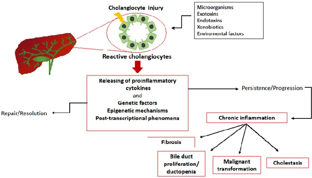
Besides local cholangiocytes-associated events, other factors could determine cholangiopathies onset and perpetuation. These factors include genetic variants, epigenetic mechanisms, post-transcriptional events and protein expression modulation by miRs (Figure 5). They are short non- coding RNAs which have been recently described as important regulators of gene expression. However, the regulatory circuits of miRs are complex and still unknown. Thus, the knowledge of the relation between miR and its targets is not simple. The function of miRs rely on negative regulation of target or it
The influence of genetic factors in the pathophysiology of cholangiopathies is not yet fully elucidated and the environmental factors contributing to disease perpetuation are largely unknown, thus limiting our current understanding. Taken together these variables influence disease progression or resolution.
Figure 5. Schematic representation of cholangiopathies pathogenesis. Following an insult, cholangiocytes become activated and start to release inflammatory mediators (cholangiocytes-dependent events). The resolution of the disease process or alternatively the perpetuation of inflammatory state depends on either cholangiocytes-dependent events or genetic factors, epigenetic modifications and post-transcriptional phenomena. The persistence of the insult determine chronic inflammation establishment which could result in fibrosis, ductopenia, cholestasis and malignant transformation of the biliary epithelium (From Pinto et al. [52]).
1.3.1 Primary sclerosing cholangitis and Primary biliary cholangitis: an overview
Primary sclerosing cholangitis (PSC) and Primary biliary cholangitis (PBC) are the two most common cholangiopathies, characterized by progressive vanishing of bile ducts. Despite they possess commonalities (i.e. portal inflammation with lymphocytes infiltration, bile duct injury, fibrosis development and progression to cirrhosis which leads to end-stage liver failure), PSC targets medium to large extrahepatic and intrahepatic bile ducts, while PBC occurs in small intrahepatic bile ducts [65], [66]. Therefore, recognition of biliary epithelium heterogeneity is necessary to unveil the molecular mechanisms underlying cholangiopathies development and progression, and possibly to devise novel therapeutic targets.
PSC is an idiopathic disease whit a mean age of diagnosis of 40 years which predominantly affect men. However, as the population get old, the patient age at diagnosis becomes older [67]. Despite the presence in the 80% of patients of autoantibodies anti p-ANCA, PSC differs from the others autoimmune disease because of the absence of female prevalence and the low responsiveness to immunosuppressive therapy [68]. The characteristic histologic features includes portal inflammation, onion-skin fibrotic lesions and obliterative cholangitis [69]. Bile ducts inflammation and fibrosis lead to bile synthesis and flow impairment and a progressive liver failure. Approximately, 75% of PSC patients are affected by inflammatory bowel disease (IBD), mainly ulcerative colitis (UC), whose pathogenesis have been supposed to be related to gut dysbiosis [70], [71]. Clinical data have shown that PSC patients display portal bacteremia and bacteria-bilia [72] with
More interesting, bowel resection after or during liver transplantation, reduced PSC recurrence rate (which hints the 37% of PSC-transplanted patients), suggesting the potential role of gut microbiota in disease relapse [74], [75]. Further evidences supporting the involvement of gut microbiota in PSC development derive from the beneficial effects of oral antibiotics documented in PSC patient treatment [76].
Another hypothesis proposed to explain PSC development is the aberrant mucosal T-cells homing in the liver. Briefly, T-cell memory originally activated in the gut, are aberrantly recruited in the liver and recognize self-antigens of the liver or gut antigens that reach the hepatic compartment via portal blood circulation, triggering an exacerbated inflammatory response [68].
PBC is considered as an autoimmune disease due to the presence of anti-mitochondrial antibody (AMA) in sera of 95% of patients. PBC is a middle-age and older females disease (the average of diagnosis is 55 years), even if some cases of paediatric onset have been reported [77].
The characteristic histological findings includes bile duct lesion with segmental degeneration of ducts and formation of epithelioid granulomas [64]. Different triggers have been linked to the immunological breakdown evidenced in PBC; in particular self/non-self mimicry has been identified as a driving mechanism [78]. Among environmental triggers xenobiotics, microorganisms (N. aromaticivorans or E. coli which share the amino acidic sequence with the immunodominant epitope of pyruvate dehydrogenase complex-E2 or PDC-E2, the key autoantigen of PBC [79] or hormones, have been identified. Several features evidenced in an aged immune system has been reported in patients affected by PBC.
For example, the modification of T-cell regulatory response [80], the telomere length attrition [81], the DNA damage, the apoptosis (e.g. increasing of the apoptotic markers Bcl-1 and Fas in cholangiocytes), senescence (e.g. increased expression of p16 and p21) [81], downregulation of Ki67 [82] (in human cholangiocytes) and autophagy (e.g. increased expression of LC3) in liver samples of PBC patients (90.5% inflamed bile duct and 25% of non- inflamed bile ducts) with respect to controls [83].
A relevant role in shaping cholangiocyte response to injury is played by genetic variants, which among others, determine whether reactive cholangiocytes regress to a normal phenotype or lead to chronic inflammation of the bile duct, with progression of cholangiopathy. Data collected by 4 different genome wide-association studies (GWASs) highlight 30 susceptibility loci associated with PBC involved in immune cells functions and processes. As for PBC, a number of 31 risk loci related to immune cells responses, have been identified in PSC [84].
1.3.1.1 Therapy
The lack of knowledge on the pathophysiological mechanisms of PSC and PBC development and progression has a severe impact on the availability of therapeutic strategies to treat affected patients. Awareness of the exact molecular pathways characterizing cholangiocytes biology in physiological and pathological conditions may help the identification of specific molecular targets for novel drugs discovery. Up to the present, ursodeoxycholic acid (UDCA) treatment represents the first therapeutic option for PBC patients.
Its effects rely on the promotion of choleresis by upregulation of both cholangiocytes and hepatocellular transporters, thus protecting biliary epithelial cell by toxic bile acids-mediated injury [85]. Intolerant or non-responding PBC patients to UDCA (about 40%) are subjected to obeticholic acid (OCA) administration. Therefore, the combination of the two strategies does not cover all PBC patients [86]. By contrast, no approved therapies is nowadays available for PSC treatment. The administration of high doses of UDCA (28–30 mg/kg) have been associated to serious side effects, such as the increasing risk of liver transplantation and esophageal varices development [87]. The only effective therapies for PSC patients is liver transplantation. A number of clinical trials based on drugs targeting specific molecular pathways (i.e. agonists of nuclear receptors and membrane transporters involved in bile acids homeostasis and metabolisms) related to cholangiocyte biology are currently being investigated for PSC and PBC treatment [86].
1.4 Aging
Aging is the process of functional decline of multiple cells and tissue which ultimately lead to body deterioration and increased susceptibility to death [88], [89]. Several disorders such as cardiovascular diseases [90], pathologies of nervous system [91] or cancer [92] have been related to aging. Few decades ago, it has been possible to identify cellular characteristics and molecular pathways associated to aging process [89].
1.4.1 The hallmarks of aging
The hallmarks of ageing have been identified based on specific criteria, that are: a) the hallmark have to manifest during normal gaining; b) the exacerbation of the hallmark accelerate the aging process; c) the blockage of the hallmark results in retarded aging and, as a consequence, the increase of lifespan.
The aging hallmarks could be divided into three main categories: - Primary, which are the cause of age-related damage;
- Antagonistic, identified as the response to age-related damage;
- Integrative, that are the consequence of responses and responsible of ageing phenotype;
1.4.1.1 Primary hallmarks
The causes of damage related to ageing are multiple. The accumulation of DNA damage (e.g. mutations, telomeres shortening or gene disruption) is one of the features characterising the aging process [93] which (both genomic and mitochondrial DNA) depends on both exogenous stressors (physical, chemical or biological triggers) and/or endogenous events (DNA replication errors or ROS production) [94]. Therefore, the organisms possess DNA repair machineries which repair damaged nuclear DNA and with lower efficiency mitochondrial DNA [95]. As an example, telomerases are specialized DNA polymerases which preserve telomeres length and function by replicating the terminal sequences of chromosomes. The link between telomeres shortening and aging has been demonstrated in different animal models [96].
Epigenetic alterations such as histone modification, DNA methylation, chromatin remodelling or non-coding RNA, are known to be involved in tissue aging. For instance, experimental mice model lacking sirtuine-6 (SIRT6), a gene belonging to the NAD-dependent protein deacetylases family involved in chromatin function modulation, show an accelerated aging [97]. Accordingly, the overexpression of SIRT6 transgene in mice results in a prolonged life-span [98].
Also the impairment of proteostasis, term which refers to protein homeostasis, has been correlated with aging process [99]. This cellular process plays a critical role in the stabilization of protein exhibiting a correct folding and in mechanisms of protein degradation by proteasome and lysosomes. Thus, the deregulation of proteostasis (due to toxins or free radicals) could lead to the chronic expression of unfolded or misfolded protein or to the accumulation of protein aggregates. This process has been linked to different age-related pathologies of nervous system [100].
1.4.1.2 Antagonistic hallmarks
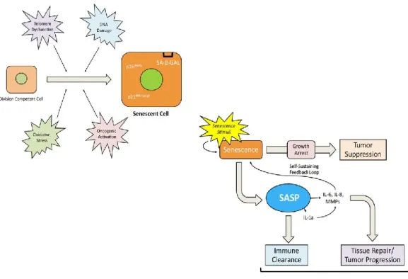
As a result of primary hallmarks, cell activates compensatory processes known as antagonistic hallmarks. Cellular senescence represents the main response to age-related damage. In vivo data have shown the accumulation of senescent cells in aged tissues. Senescence is defined as the irreversible process of cell growth arrest associated to complex cellular changes such as chromatin organization, metabolic reprogramming, increasing of autophagy and the release of a number of proinflammatory mediators and growth factors,which are included in the senescence associated secretory phenotype (SASP) [88], [101]. Senescence process was firstly described by Hayflick and
his colleague [102], [103], around 50 years ago. They observed in cultured normal fibroblast the exhaustion of cells proliferative capacity after serial replicative cycles. Further, it has been shown that the phenomenon observed
in vitro was due to telomere shortening [104]. Apart from telomere damage,
different others stimuli are known to triggers senescence process [105]. The stable replicative arrest of cells is a physiologic process which impedes damaged or cancer cells to divide and expand, thus suppressing tumorigenesis [106]. This compensatory process requires the homeostasis between clearance of senescent cell and mobilization of progenitor cells. The lack of balance, determines the accumulation of senescent cells which contributes to aging.
The growth arrest, which is the intrinsic characteristic of senescent cell, results in the absence of proliferation markers and the acquisition of peculiar morphological changes (e.g. doubling of cell volume and flattened morphology). Cellular senescence is driven by the upregulation of two tumour suppressive pathways, the p16INK4A/pRB and p21CIP/WAF1/p53 [105], [107].
Along with replicative arrest, senescent cells are characterized by the “secretome” enrichment in proinflammatory cytokines and metalloproteases, known as component of senescence-associated secretory phenotype (SASP) which act in autocrine/paracrine manner mediating angiogenesis (e.g. vascular endothelial growth factors or VEGF), cell growth (e.g. growth-related oncogenes or GROs), chemo resistance, stimulation of epithelial to mesenchymal transition (e.g. IL-6, IL-8 and MMPs), chronic inflammation (e.g. IL-1α , IL-6 , IL-8, MCP or MIP),alteration of stem cells renewal [108] and differentiation, and tissue remodelling [101].
Finally, the role of SASP mediators is to recruit immune cells to eliminate senescent cells [109]. An important feature which allows the identification of senescent cells both in culture conditions and in vivo, is the lysosomial β-galactosidase enzyme. The activity of this enzyme results increased in course of replicative senescence, and confers histochemical positivity for the senescent associated β-galactosidase (SA-β- GAL) staining [110].
Figure 6. Cellular Senescence: an overview. Different triggers could induce cell growth arrest such as telomeres dysfunction, DNA damage and so on. Once cells become senescent, modify their phenotype. The nucleus becomes wider and increases the expression of two nuclear proteins which suppress cell proliferation (p16 and p21) and of the senescence marker, SA-β-GAL). On one hand, senescence establishment prevents cell transformation and thus tumour development. On the other hand, senescent cells secrete a variety of proinflammatory cytokines and other soluble factors which allow tissue repair and the immune clearance of senescent cells. These events lead to tissue aging (From Meng et al. 2015 [111]).
Either nutrients sensing deregulation or mitochondrial dysfunction are comprises in the antagonistic hallmarks of ageing. The main physiologic pathway which regulates metabolism is the growth hormone (GH)/insulin- like growth factor (IGF-1) axis. In particular IGF-1 induces insulin releasing in response to glucose sensing. The insulin/IGF-1 signalling pathway targets the transcription factors family FOXO and the mTOR complexes. In both humans and model organisms it has been shown that genetic polymorphisms or mutation which reduces GH, IGF-1 or insulin receptor or downstream effectors, determine an increased lifespan. According with these findings, dietary restrictions increase lifespan of both unicellular and multicellular organisms [112], [113], [114]. Mitochondrial dysfunction has been linked to aging. Different causes have been associated to a reduced efficiency of mitochondria which determines a reduced mitochondria biogenesis such as: deletion of mtDNA, oxidation of mitochondrial proteins, destabilization of the macromolecular organization of respiratory chain complexes, alteration of lipid composition of mitochondrial membranes, loss of mitochondrial homeostasis (as a result of imbalance between fission and fusion events) and defective mitophagy (which physiologically allows the proteolytic degradation of deficient mitochondria) [115], [116].
1.4.1.3 Integrative hallmarks
The decline of regenerative capacity of a tissue is an important characteristic of ageing. Although loss of stem cells proliferation could be detrimental for organisms maintenance, the exacerbation of stem cell growth could led to exhaustion of stem cells niches. A compelling evidence derives
from Drosophila intestinal cells. The excessive stem cells proliferation was linked to premature aging.
Changes in cell—cell communication (i.e. endocrine, neuroendocrine or neuronal) have been linked to aging process. Inflammaging, decline of immune system efficiency, accumulation of senescent cells and defective autophagy underlie several age-associated diseases such as diabetes, arthritis, metabolic syndrome and cardiovascular disease [117].
1.5 Aging, senescence and biliary tree disorders
Up to the present, it is not fully understood the influence of ageing in the pathophysiology of liver and biliary diseases. Despite cholangiopathies are not specific of elderly, several data suggest the role of ageing in the clinical course of disease and in the modulation of cholangiocyte pathophysiology.
Previous studies documented age-related alteration of the liver (e.g. hepatic volume reduction, increasing of lipofuscin body or reduction of hepatobiliary functions) as well as molecular changes (e.g. altered response to oxidative stress, a reduced efficiency of DNA repair mechanisms or telomere shortening) which could contribute to decline of liver regenerative capability and to shortening survival rate after liver transplantation [118].
From a clinical point of view, the progression of cholangiopathies seems to be influenced by advanced age. Accordingly, the clinical course of PSC is influenced by patient age at diagnosis with an increasing occurrence of cholangiocarcinoma (CCA) and poor outcome [119].
In addition, donor age has been implicated in the development of biliary complication after liver transplantation [120].
From a molecular point of view, important clues support the role of aging in shaping cholangiocyte biology in course of biliary injury. As an example, analysis of liver samples collected by PSC and PBC patients have shown the increased expression of senescent markers and SASP components in diseased cholangiocytes [42]. Another study of Sasaki, et al. demonstrates that small bile ducts of liver tissue collected by early stage PBC-patients, possess elevated positivity to SA-β-GAL staining and increased expression of senescence markers p16 and p21 [82]. As well as in PBC, isolated cholangiocytes from PSC patients show a strong positivity to SA-β-GAL staining and, in addition, a decrease of cholangiocytes proliferation, and the increase of IL-6 and IL-8 levels as compared to control normal human [121]. These intriguingly findings achieved in PSC and PBC patients are corroborated by data collected in animal models of cholestatic liver injury. As an example, isolated cholangiocytes from multi- drug resistance 2 knockout (mdr2 -/-) mice, a model of PSC, undergo senescence [122]. The mdr2 -/- mice
lack the expression of the canalicular phospholipid flippase mdr2, which serves for bile acids assembly in mixed micelles thus protecting biliary epithelium by toxic bile acids-mediated injury [44]. As a direct consequence of mdr2 lack of expression, toxic bile acids accumulates in the bile triggering bile duct damage.
Cholangiocyte senescence has been recently related to deregulation of cholangiocyte immune response in course of cholangiopathies.
show the increased expression of IL-6 to face cellular damage and cholangiocyte loss. In turns, persistent LPS stimulation induces cholangiocytes senescence as evidenced by either upregulation of protein involved in cell cycle arrest (p16 and p21) and SASP components [42].
Despite these findings demonstrate a link between senescence establishment and disease presentation, it is unknown whether senescence is the trigger of disease or if it is a consequence of chronic damage [81], [123]. Certainly, non-replicative cholangiocytes play a role in the modulation of biliary microenvironment and organ homeostasis [43]. The loss of proliferative capabilities increases cholangiocytes susceptibility to subsequent injuries, leading to a worsening of inflammation process.
2. AIM OF THE THESIS
My thesis project was focused on the identification of molecular pathways which are activated both during the ageing process and in response to damage of the bile ducts. Cholangiocytes, the cell lining the bile ducts, represent the unique target of cholangiopathies, a group of chronic cholestatic diseases which evolve to end-stage liver disease with liver transplantation as the only curative strategy. Due to the progressive nature of the disease and the lack of effective therapies, a deeper understanding of cholangiocytes pathobiology is needed in order to device novel effective therapeutic strategies. Bile duct injury during the course of cholangiopathies is influenced by a number of factors such as genetic susceptibility, epigenetic events or protein expression modulation by miRs. Several clinical and experimental data have recently identified cellular senescence, the age-related damage response, as an important player in the disease development and progression. The aim of this work is to discover new molecular targets involved in the modulation of cholangiocyte biology in pathophysiological conditions. To reach this goal, bioinformatics tools have been exploited to select intracellular pathways and putative molecular targets to be investigated both in in vitro and in vivo model as well as in human samples of cholangiopathies.
3. MATERIALS AND METHODS
3.1 Materials
Reagents and antibodies, where not specifically indicated, were purchased respectively by Sigma-Aldrich (St. Louise, MO) and by Santa Cruz Biotechnologies Inc. (Santa Cruz, CA). 3,5-Diethoxycarbonyl-1,4- dihydrocollidine (DDC) was purchased by Sniff GmbH (Soest, Germany). Anti-Ck-19 antibody was purchased by Abcam (Cambridge, United Kingdom). Anti-actin antibody HRP conjugated was provided by Cell signaling (Danvers, MA). Normal rat cultured cholangiocytes (NRC), a murine intrahepatic bile duct cell line, were a generous gift of Prof. Gianfranco Alpini, Texas A&M University, Temple, TX, USA. siRNA oligonucleotides were purchased from Ambion® (Carlsbad, CA, USA). The
transfection reagent INTERFERin® was from Polyplus-transfection (Illkirch,
France). The senescence accelerated mouse prone 8 (SAMP8) mice, and the control strain SAM resistant 1 (SAMR1) were bred in house at the Experimental Animal Models for Aging Unit, Scientific Technological Area, IRCCS-INRCA, Ancona, Italy. Twf1 knockout (C57BL/6NTac Twf1-/-) mice, and their respective controls were purchased from the European Mouse Mutant Archive (EMMA), National Centre for Biotechnology (Campus de Cantoblanco, Madrid, Spain).
3.2 Methods
3.2.1 MicroRNAs selection
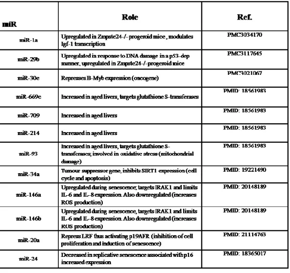
Biomedical literature search on Medline has been carried out in order to find relevant studies about miRs involvement in aging process, irrespective of organ or system. It was possible to identify a total of 12 miRs which have been subsequently investigated in our project in line with the aim of the study.
3.2.2 MicroRNAs testing in cholangiocyte: isolation, reverse transcription and real-time PCR
Total RNA was extracted from cholangiocytes isolated by 8-week DDC-fed mice and control animals, and by young and old mice subjected or not to DDC administration. Cholangiocyte isolation was performed by immune-beads purification as previously reported Following RNA purification (miRNeasy mini Kit, QIAGEN, Hilden, Germany). MiRs expression levels were assessed through the SYBR® Green PCR Kit miR-
specific miScript Primer Assay (forward primer) and the miScript SYBR®
Green PCR Kit (QIAGEN, Hilden, Germany), containing the miScript Universal Primer (reverse primer) were used for specific miRs expression evaluation. Relative abundance of miRs expression was normalized to U6 expression as internal control.
3.2.3 In silico analysis of microRNAs putative targets and intracellular pathways
To predict putative targets and molecular pathways shared among miRs resulted upregulated in cholangiocytes isolated from DDC-fed mice, the software simple String IDentifier (SID1.0) was used [124]. This program is based on an exhaustive search strategy and is specifically designed to screen shared data (target genes, microRNAs and pathways) available from the TargetScan Human 6.2 and DIANA-MicroT 3.0 target prediction databases (http://diana.cslab.ece.ntua.gr/microT/). The actin- binding protein, Twinfilin-1 (TWF1) was selected for the downstream evaluations.
3.2.4 Expression of Twf1 in cholangiocyte
TWF1 expression was assessed in liver tissues of patients affected by PSC (n=10) and PBC (n=10) by real-time PCR. Normal liver tissue surrounding colorectal hepatic metastases (n=17) was used as internal controls. The isolation of cholangiocyte was carried out as previously [125]. In addition, TWF1 expression was evaluated by immunohistochemistry staining in liver sections of PSC patients (n=5) compared to control livers (n=3 alcoholic liver disease and n=2 cryptogenic liver disease). Ethical approval was obtained from the research ethics committees at each participating medical centre in accordance with the declaration of Helsinki (S-08872b). After acquisition of informed consent, samples were collected at the time of liver biopsy, surgical liver resection or liver transplantation. Expression levels of Twf1 were evaluated in cholangiocytes isolated from
both control mice (young and old) and mice (young and old) fed with 0.1% DDC, by real-time PCR.
3.2.5 Cell lines and in vitro experiments
NRC were cultured on rat tail collagen type I coated flasks and maintained in Dulbecco’s modified Eagle medium: nutrient mixture-F12 (DMEM/F12) supplemented with 5% of fetal bovine serum (FBS), 0.01 ml/ml minimum essential media non-essential amino acids, 0.01 ml/ml of chemically defined lipid concentrate, 0.01 ml/ml insulin transferring selenium, 0.01 ml/ml minimum essential media vitamin solution, 200 mM L-glutamine, 12.7 mg/ml bovine pituitary extract, 393 μg/ml desaxamethasone, 3.4 mg/ml 3, 3’, 5- triiodo-L-tyronine, 25 μg/ml epidermal growth factor, 4.11 mg/ml forskolin, 1% penicillin-streptomycin, 10 mg/ml gentamicin. All products have been purchased from Invitrogen (Carlsbad CA, USA). Cell silencing was carried out the day of seeding through the INTERFERin®
reagent according to manufacturer’s recommendation. The final siRNA concentration was 30 nM in medium without FBS.
3.2.5.1 Evaluation of Twf1 role in cholangiocytes proliferation
To estimate if the absence of Twf1 could affect cholangiocyte proliferation, NRC were seeded in a 6-well collagen coated plate in complete medium without FBS and incubated with either small-interfering (si) RNA against Twf1, which turns off gene expression, or with non- targeting (nt) RNA as control, for 48 h. The day after seeding cells were stimulated with complete medium supplemented with increasing concentration of FBS
(0-5-Changes in cells proliferation were assessed by measuring mRNA expression level of Ki67 and protein expression of proliferating Cell Nuclear Antigen (Pcna). Changes in cell growth were further evaluated by sulforhodamine B (SRB) colorimetric assay [126]. This technique has been developed by Skehan and colleagues and it is extensively employed in drug-induced cytotoxicity and cells growth studies. The assay relies on the capability of sulforhodamine dye to bind to protein basic amino acids residues of fixed-cells. Thus, the absorbance measured is proportional to cell number and cellular protein content [127]. Briefly, cells were seeded in a 96-well plates in complete medium without FBS and transfected with siRNA against Twf1 or with ntRNA. The day after seeding, cells were cultured in medium added with 5% FBS and grown for up to 72 hours. After fixing and washing steps, cells were air dried and stained with 0.4% sulforhodamine B solution for 30 min at room temperature (RT). The incorporated dye was then solubilized by adding the SRB solubilization solution in an equal volume of original culture medium. Growth rates were derived by absorbance values obtained spectrophotometrically using a microplate reader (Tecan, Mannedorf, Switzerland).
3.2.5.2 Evaluation of Twf1 role in cholangiocyte senescence
To address the possible role of Twf1 in cholangiocyte senescence establishment, NRC were seeded in a 6-well collagen-coated plate in complete medium without FBS and exposed for 48 hours to siRNA against Twf1 or to corresponding ntRNA. The day after seeding, cells were stimulated or not with 5% FBS.
manufacturer’s instruction (Invitrogen, Carlsbad CA, USA). Cellular senescence was assessed by quantification of p16ink4a and p21waf/cip1 expression. SASP component levels were measured by real-time PCR as well. Senescence was further evaluated in NRC knocked-down for Twf1 expression and in control cells, by quantification of Sudan Black-B (SBB) staining as previously reported [128]. A well- established in vitro model of cholangiocyte-induced senescence [42], was used to investigate the role of Twf1 in cholangiocyte senescence modulation. NRCs were seeded in complete media without FBS and silenced or not for Twf1 expression. Senescence model was induced by persistent stimulation (10 days) with LPS (200 ng/ml). Media, agonists and siRNA were replaced every 48 hours for up to 10 days. The levels of senescence markers and SASP components were evaluated by real-time PCR. SASP components levels were also assessed by enzyme-linked immunosorbent assay (ELISA) in the same setting of experiments.
3.2.6 Animal models
Seven-week old male senescence accelerated mouse prone 8 (SAMP8) and their respective control SAM resistant 1 (SAMR1) [129], [130] (n = 5 per each experimental group), were raised in house at the Experimental Animal Models for Aging Unit (Scientific Technological Area, IRCCS-INRCA, Ancona, Italy) and fed with either 0.1% 3,5-diethoxycarbonyl-1,4-dihydrocollidine (DDC) diet or related control diet for 4 weeks. The experimental DDC diet and the standard diet were also administered to seven-week old male Twf1 knockout (C57BL/6NTac Twf1-/-) and to wildtype
-fed mice develop onion skin-like periductal and portal-portal fibrosis, display ductular reaction and a marked increase of serum bile acid levels. All together these features resemble the hallmarks of chronic cholangiopathies [131]. Changes in cholangiocyte proliferation were determined by measuring differences in intrahepatic bile duct mass (by quantitative immunohistochemistry for CK-19 in liver sections), as previously reported [132]. Changes in collagen deposition were assessed by quantitative histochemical staining for Sirius Red. Hepatic content of collagen was evaluated by hydroxyproline quantification [133]. Animal study protocols were performed in compliance with local Institution guidelines and in accordance with the ARRIVE guidelines.
3.2.6.1 SAMR1 and SAMP8
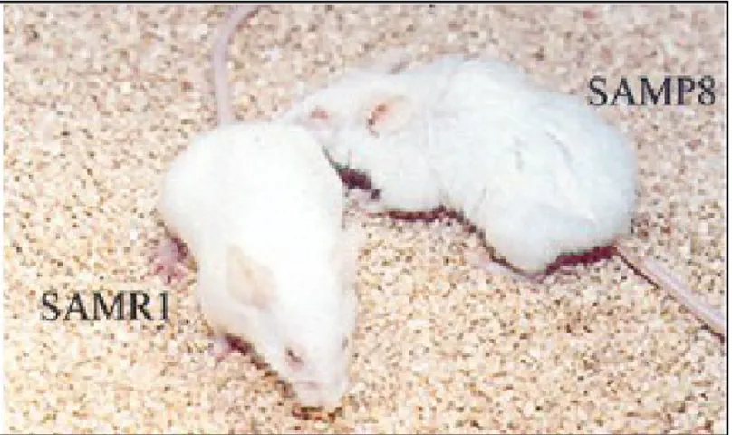
SAMP8 is an accelerated aging model developed by Takeda and his colleagues [129]. SAMP8 strain displays a reduced lifespan of 50% with respect to SAMR1 controls and signs of aging such as ruffled and dull coat, loss of hair, abnormal curvature of the spine and inflammation of the periorbital areas. The phenotypic characteristic are shown in the figure below (Figure 7).
Figure 7. SAMR1 and SAMP8 mice: phenotypic characteristics. The signs of aging typical of SAMP8 mice includes loss of skin glossiness, increased coarseness or hair loss, curvature of the spine and inflammation of periorbital areas. (From Ye et al 2004 [130]).
SAMP8 and SAMR1 mice belong to the SAM family that derives from continuous sister-brother mating of five AKR mice with severe deterioration, and three AKR mice with normal aging [134]. The grade of aging has been assigned phenotypically by evaluating characteristics such as loss of skin glossiness, increased coarseness or hair loss (Figure 7). SAM family includes 9 SAMP (P1-P9) and 3 SAMR (R1-R3) substrains. Among these, SAMP8 mice exhibit signs of liver degeneration. In particular, biochemical markers of hepatic function such as alanine aminotransferase (ALT), aspartate aminotransferase (AST) and alkaline phosphatase (AP) are increased in SAMP8 mice as compared to the relative controls. Notably, AP increase is associated to bile canaliculi damage or cholestasis. SAMP8 mice display hepatic steatosis (macrovesicular fat) and hepatocellular degeneration (e.g. enlarge cell nuclei, swelling cell bodies and necrosis with deposition of cell debris) in the centrilobular zone of hepatic acinus. At this level, immune cell infiltration, fibrosis and cell degeneration has been found. It is not fully
One of the proposed mechanism relies on the increasing titers of MuLV in liver of SAMP8 (50 fold) with respect to controls. MuLV is a retrovirus that following viral RNA retrotranscription in DNA, integrates his genome in the host genome [135], [136]. Once integrated, viral genome is replicated together with the host chromosomal DNA. However, there are no data that link MuLV to liver disease development. Another mechanisms proposed to explain the liver degeneration occurring in SAMP8 is the hypoxia establishment. With this regard, increased levels of lipid peroxide have been evidenced in liver of SAMP8 compared to SAMR1, since early stage of life [137].
3.2.6.2 Twf1+/+ and Twf1
-/-Twf1 knockout (C57BL/6NTac -/-Twf1-/-) are transgenic mice generated through the “knockout-first” strategy (Figure 8). Null allele is generated through allele splicing to LacZ trapping element included in the targeting cassette. This knockout could be easily modified due to the presence of particular sequences in the trapping cassettes. In particular, the Flippase
(Flp) recombinase recognizes the Flp recombinase recognition target (FRT) and it is able to revert the mutation to wild-type upon sequence recognition leaving the LoxP sites in either sides of the gene exon of interest. The crossing with Cre mice results in Cre recombinase-mediated exon excision upon LoxP sequences recognition. The cleavage induces a frameshift mutation resulting in mutant transcript decay.
Figure 8. Illustration of Twf1-/- allele map
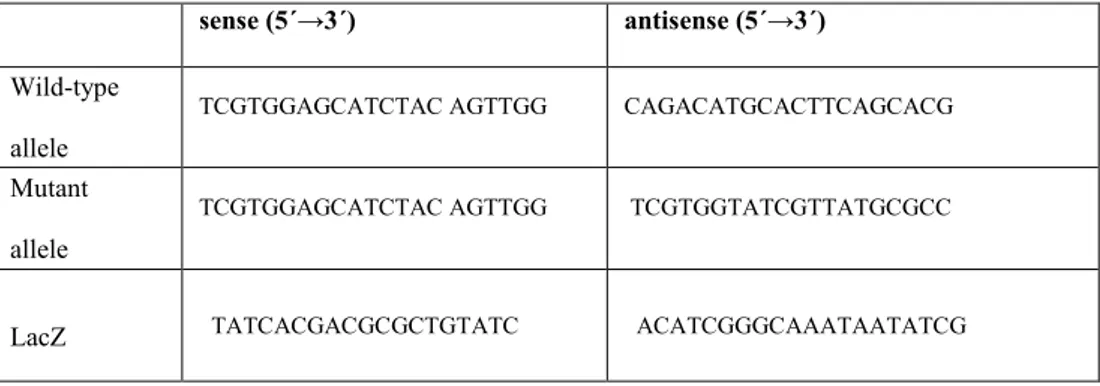
The animals lacking for the expression of Twf1 have been genotyping before starting the treatment. Genomic DNA was extracted from mouse tail with the DirectPCR lysis Reagent Tail (Viagen Biotech Inc, Los Angeles, USA) following manufacturer’s instructions. The protocol for mice genotyping includes separate PCR reactions that detect LacZ, the gene-specific wild type allele, and the mutant specific allele. The set of primers used for the allele discrimination have been listed in the table below (Table 1).
sense (5´→3´) antisense (5´→3´)
Wild-type allele
TCGTGGAGCATCTAC AGTTGG CAGACATGCACTTCAGCACG
Mutant allele
TCGTGGAGCATCTAC AGTTGG TCGTGGTATCGTTATGCGCC
LacZ TATCACGACGCGCTGTATC ACATCGGGCAAATAATATCG
Table 1. Primers set used for mouse genotyping.
The thermal profile for both PCRs are listed below:
3.2.7 RNA extraction, reverse transcription and real-time PCR
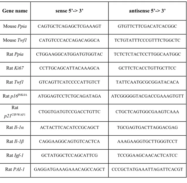
Total RNA was isolated from NRC or liver tissues using the RNeasy Mini Kit (QIAGEN, Hilden, Germany) or alternatively following a manual protocol. A quantity of 1 μg was reverse-transcribed to complementary cDNA with random primers using the High Capacity cDNA Reverse Transcription Kit (Applied Biosystems, Foster City CA, USA) according to the kit’s instruction. Primers for real-time PCR were designed with Oligo software version 6.71 (Molecular Biology Insight, Cascade, CO) using reference mRNA sequences accessed through GenBank. The specificity of primers was confirmed by BLAST analysis. Real-time PCR was performed using a Rotor-Gene 6000 instrument (Corbett Life Science Pty. Ltd., Mortlake, NSW, Australia) through the SYBR Green fluorophore. Relative abundance of target genes was normalized to Peptidylprolyl isomerase B (Cyclophilin B) as internal control. The Gene Expression Macro Genex developed by Bio-Rad (Milan, Italy) was used to calculate relative expression values from real-time PCR data. Oligonucleotide sequences of primers used for real-time PCR are listed in the table above:
![Figure 1. Biliary tree architecture (From de Jong et al. 2018 [8]).](https://thumb-eu.123doks.com/thumbv2/123dokorg/2968439.27106/8.893.201.807.346.689/figure-biliary-tree-architecture-jong-et-al.webp)