2017
translation factor
Flavia Bassani
Supervisor: Prof. Anna La Teana
PhD Program
Biomolecular Sciences: XV Cycle
Università Politecnica delle Marche
Summary
Protein synthesis is a process conserved among the three primary domains of life, in which the genetic information is translated into working proteins.
The translational machinery has been extensively studied in bacteria and eukaryotes, whereas the archaeal one is still poorly characterized, therefore a better understanding of the translation process in Archaea could help to clarify several aspects, which have been conserved during the evolution.
In this regards the structural and functional study of translation factors, with high homology between archaea and eukaryotes, is important to determine not only their role in protein synthesis but also to help understand their role in physiological and pathological cellular processes.
The aim of this thesis is to shed light on the archaeal protein aIF5A, a translation factor highly conserved and essential in eukaryotes and archaea, whose requirement in protein synthesis remains elusive.
The eukaryotic eIF5A and the bacterial ortholog EF-P, post-translationally modified through a distinct pathway, were identified as initiation factors, but subsequent studies highlight their implication in translation elongation.
Recently it was also reported the involvement of the eukaryotic eIF5A in translation termination, however the function of the archaeal homologue is still unknown, as well as its post-translational modification.
The gene appears to be essential and the modification seems to be important, since at least its inhibition in some Archaea causes a rapid and reversible arrest of growth.
Significant studies have been carried out in the Euryarchaeota kingdom, in which it was shown for the first time that aIF5A has RNA degrading activity in vitro and can be modified via an alternative modification pathway.
Information on the archaeal aIF5A in Chrenarchaeota, which appears to be closely related to eukaryotes, is extremely limited and in order to fill this gap we investigate the role of the protein in the model organism Sulfolobus solfataricus (Sso), highlighting unreleased features of this factor.
The first part of the thesis focuses on the subcellular localization of the protein and the identification of its post-translational modification.
The fractionation of Sulfolobus solfataricus whole-cell extract on sucrose density gradients allowed us to determine the behavior and localization of aIF5A.
Whilst the expression of the protein in Sso followed by affinity purification and mass spectrometry analysis enable the detection of the post-translational modification.
The second part of the thesis is aimed at identifying interacting partners (proteins and RNAs) of Sso-aIF5A, providing new hints of the protein involvement in several cellular processes. Taken together our evidences suggest the probable involvement of aIF5A in mRNA stability/turnover and its multifunctional role, which in Sulfolobus can be modulated by the hypusination itself or by the structural conformation of the protein.
Contents
1. Introduction ... 1
1.1. The eukaryotic translation factor eIF5A ... 1
1.1.1. Structural features and hypusination ... 1
1.1.2. The role of the eukaryal eIF5A in translation ... 5
1.1.3. Implications of the eukaryal eIF5A in physiological and pathological cellular processes ... 9
1.2. The bacterial elongation factor EF-P ... 12
1.2.1. Structural features and post-translational modification ... 12
1.2.2. Function of the elongation factor P ... 15
1.3. The archaeal translation factor aIF5A ... 18
1.3.1. Protein synthesis in Archaea ... 18
1.3.2. Structural features of the archaeal factor aIF5A ... 21
1.3.3. Post-translational modification of the archaeal factor aIF5A ... 22
1.4. Aim of the thesis ... 26
2. Materials and Methods ... 27
2.1. Expression of ORF Sso0970 in E.coli and purification of Sso-aIF5A ... 27
2.2. Expression of ORF Sso0967 in E.coli and purification of Sso-DHS ... 29
2.3. Western blot analysis ... 29
2.4. Preparation of Sulfolobus solfataricus cell extract ... 30
2.5. Fractionation of Sulfolobus S30 exctract programmed for translation on linear sucrose gradients ... 31
2.6. Construction of plasmids pMJO5-SsoaIF5A and transformation of Sulfolobus solfataricus PH1-16 cells ... 32
2.7. Synthesis of His-tagged Sso-aIF5A and purification of the recombinant protein in Sulfolobus solfataricus ... 34
2.8. Determination of the Sulfolobus solfataricus aIF5A intact mass by LC-MS ... 35
2.9. Structural analysis of the recombinant Sso-DHS by Static Light Scattering ... 36
2.10. In vitro hypusination assay ... 36
2.11. Identification of Sso-aIF5A protein interacting partners by LC-MS/MS ... 37
2.12. Co-Immunoprecipitation assay ... 38
2.13. aIF2 release by Sso-2509 & Sso-aIF5A ... 39
2.14. Glycerol density gradients ... 40
2.15. Isolation of RNA substrates affinity co-purified with Sso-aIF5A and RNASeq ... 40
2.16. RNA cleavage assay ... 42
2.16.1. Native polyacrylamide gel electrophoresis ... 42
2.16.2. Denaturing urea polyacrylamide gel electrophoresis ... 42
2.17. Zymogram assay ... 44
3. Results ... 45
3.1. Production and purification of Sso-aIF5A in E.coli ... 45
3.2. Subcellular localization of Sso- aIF5A ... 47
3.3. Production of His-tagged Sso- aIF5A proteins in Sulfolobus solfataricus ... 50
3.4. The post-translational modification of Sulfolobus solfataricus aIF5A ... 53
3.5. Production and purification of Sulfolobus solfataricus deoxyhypusine synthase in E.coli ... 55
3.6. The tetrameric structure of Sso-DHS ... 57
3.7. Sso-DHS performs the deoxyhypusine synthesis in vitro ... 58
3.9. Functional interplay between the factor aIF5A and the translation recovery factor (Trf) ... 63 3.10. Structural characterization of the archaeal factor aIF5A ... 65 3.11. The factor aIF5A binds distinct RNA substrates in Sulfolobus solfataricus 69 3.12. Sulfolobus solfataricus aIF5A factor shows ribonucleolytic activity ... 74
4. Discussion ... 80
5. Outlook ... 89
Appendix
91
List of abbreviations
93
References
97
Acknowledgements
108
List of Figures
1. Post-translational modification pathway for the eukaryal eIF5A ... 3
2. Cryo-EM structure of eIF5A bound to the yeast 80S ribosome ... 8
3. Comparison of eukaryal eIF5A and bacterial EF-P structures ... 12
4. β-lysinylation patway of EF-P ... 14
5. Crystal structure of EF-P bound to the ribosome ... 16
6. Model of translation initiation in Archaea ... 20
7. Structural features of the archaeal protein aIF5A ... 22
8. Hypusine and deoxyhypusine modification in the archaeal kingdom ... 23
9. Production and purification of Sso- aIF5A in E.coli ... 46
10. Performance of the polyclonal antibody in the detection of the recombinant Sso- aIF5A ... 47
11. Cellular localization of the native Sulfolobus solfataricus aIF5A ... 48
12. Fractionation of Sulfolobus solfataricus S30 extract and localization of Sso- aIF5A ... 49
13. The synthesis of aIF5A in Sulfolobus solfataricus causes a dramatic slow-down of growth ... 50
14. Production of aIF5A in Sulfolobus solfataricus ... 51
15. SDS-PAGE analysis of mock control and Sso- aIF5A C-terminal His-tagged ... 53
16. The Sulfolobus solfataricus aIF5A factor is hypusinated ... 54
17. Conserved residues in the DHS enzyme sequence involved in the binding to the NAD+ cofactor and to the spermidine substrate ... 56
18. Production and purification of Sso-DHS in E.coli ... 57
19. The recombinant Sso-DHS, purified in E.coli, shows a tetrameric structure in solution ... 58
20. The unmodified Sso- aIF5A is in vitro deoxyhypusinated by the recombinant Sso-DHS
enzyme ... 59
21. Sso- aIF5A N-terminal His-tagged co-purifying proteins ... 61
22. Sso- aIF5A co-immunoprecipitated with Sso-2509 ... 64
23. In presence of Sso- aIF5A, Sso2509 is not able to remove aIF2γ from the 5′-P3-end of the RNA ... 65
24. The recombinant Sso- aIF5A expressed and purified in E.coli is a monomer in solution ... 66
25. The eukaryotic anti-hypusine antibody recognizes the hypusinated archaeal protein ... 67
26. The archaeal aIF5A may forms oligomers or be part of a multi-protein complex ... 68
27. Quality control of RNA samples for the deep-sequencing analysis ... 70
28. Sso- aIF5A, purified from E.coli, shows ribonucleolytic activity ... 75
29. The hypusinated Sso- aIF5A shows ribonucleolytic activity ... 76
30. The native Sso- aIF5A shows ribonucleolytic activity ... 77
31. Ribonucleolytic activity of the native Sso- aIF5A showed by the zymogram assay ... 77
32. Conservation of aIF5A residues involved in the RNA cleavage ... 78
List of Tables
1. Universal translation initiation factors ... 19
2. Effect of the inhibitor GC7 on the archaeal and bacterial growth ... 25
3. PCR program for the amplification of ORF 0970 from Sulfolobus solfataricus P2 genomic DNA ... 27
4. Western blot probing protocols ... 30
5. Analysis of Sso- aIF5A protein interacting partners by LC-MS/MS ... 62
6. ncRNAs co-purifying with Sso-aIF5A ... 70
7. tRNAs co-purifying with Sso-aIF5A ... 72
8. mRNAs co-purifying with Sso-aIF5A and encoding for known products ... 72
1. Introduction
Decoding the evolutionary history, translation is one of the most conserved cellular processes, although each of the three primary domains has elaborated specific variants of some steps; the elongation phase is essentially invariant in all cells, whereas translation initiation, termination and ribosome recycling, have specific features [1].
The translation initiation process is one of the most delicate moment of protein synthesis due to two fundamental events, the recognition of the mRNA start codon and the setting of the correct reading frame.
The selection of the start codon by the ribosome entails the play of accessory proteins called translation initiation factors (IFs).
Due to their crucial role in the translation process, most of them have been functionally characterized, in particular the eukaryal and bacterial ones.
The only ambiguous protein, which might have been improperly included among the initiation factors, is the translation initiation factor IF5A, homologous between Eukarya and Archaea and with an orthologue, EF-P, in Prokaryotes.
1.1. The eukaryotic translation factor eIF5A
1.1.1. Structural features and hypusinationThe eukaryotic translation factor eIF5A is a small protein (17 kDa) highly conserved and essential.
The protein consists of two distinct domains: the N-terminal domain (SH3-like domain) composed of six β-strands, which fold into a partially open β-barrel, and the C-terminal domain, which is formed by 3–5 β-strands and 0–2 α-helices and resembles an OB-fold domain.
Multiple alignment of eIF5A amino acid sequences shows a strong conservation, in particular, in the N-terminal domain.
This is conserved among the three primary domains, and conservation is especially evident in the region surrounding the lysine residue at position 50 (Lys 50) that is the site of a unique post-translational modification: hypusination.
The hypusine residue resides in the long unstructured loop, between strands β3 and β4, which protrudes from the N-terminal domain [2].
The eukaryal eIF5A exists as a dimer, not only in vitro but also in vivo, independently of the presence of the hypusine residue or the ribosome, but dependently on RNA [3]. RNase treatment of the affinity-purified protein eIF5A in yeast, followed by size-exclusion chromatography, leads to the disruption of the dimer in vitro.
The oligomeric state of the protein does not depend on electrostatic interactions and disulfide bridges, in agreement with a previous study [4], which suggested that hypusine is necessary for eIF5A dimerization in vitro; however this discrepancy may be due to different experimental conditions.
This region is positively charged for the presence of the hypusine residue, whereas the overall content of acidic residues is concentrated in the C-terminal domain.
The eukaryotic translation factor eIF5A is the sole protein known that contains the unique polyamine-derived amino acid, hypusine [Nε-(4-amino-2-hydroxybutyl) lysine], discovered in 1971 [5].
Polyamines are synthesized via highly regulated pathways and these small aliphatic molecules are involved in a myriad of cellular processes, as cations they can bind to nucleic acids and promote cellular proliferation and signaling.
This residue was initially called “hypusine” according to the structure that consist of two moieties, hydroxyputrescine and lysine, and its biosynthesis is accomplished by two distinct enzymatic steps (figure 1).
In the first reaction, the enzyme deoxyhypusine synthase (DHS) transfers the 4-amino butyl moiety of spermidine (synthesized in the L-arginine metabolism) to the ε-amino group of one specific lysine residue (Lys50 for the human protein) of the protein precursor to form the deoxyhypusine residue.
The second enzyme deoxyhypusine hydroxylase (DOHH) subsequently hydroxylates this intermediate to form the hypusine residue and mature form of eIF5A [2, 6].
Many insights about the essential protein eIF5A and its modification arise from experimental evidence in Saccharomyces cerevisiae.
The DHS enzyme is essential, since the null mutation in the single copy ydhs gene results in the loss of cell viability in yeast and, upon depletion of deoxyhypusine synthase, cessation of growth is accompanied by a marked enlargement of cells, suggesting a defect in cell cycle progression or in cell division [7].
The enzyme exhibits an absolute specificity toward its protein substrate but also a very narrow specificity toward spermidine and few of its closely related compounds (homospermidine, aminopropyl cadaverine, cis- and trans-unsaturated spermidine, and N8-methyl- and N8-ethyl spermidines) [6].
The deoxyhypusine synthesis occurs in four steps and in the first one the NAD cofactor is required for the dehydrogenation of spermidine to dehydrospermidine. In the second part of the reaction the dehydrospermidine 4-aminobutyl moiety is cleaved and transferred to the ε-amino group of a lysine residue, present in the active site, to form a covalent enzyme-imine intermediate.
This was demonstrated by trapping it into a stable enzyme-substrate adduct after NaBH3CN reduction of the mixture containing the enzyme, [1,8-3H] spermidine, NAD and the deoxyhypusine was the labeled component identified.
Thus is now clear that the acceptor of the amino-butyl group is a lysine residue of the enzyme (Lys329 for the human DHS). Substitutions of this particular residue lead to a totally inactive human enzyme and also S. cerevisiae‘s growth is impaired if the Lys350 of the DHS active site is mutated [8].
In the third step the 4-aminobutyl moiety is transferred to the ε-amino group of a specific lysine of eIF5A, and this imine intermediate is finally reduced to form the deoxyhypusine residue by the enzyme-bound NADH.
An alternative butylamine acceptor of eIF5A lysine is putrescine, which can be converted into homospermidine from spermidine, despite the deoxyhypusine synthesis is the preferred pathway of the DHS reaction.
All the DHS-catalyzed reactions are reversible and this was experimentally validated using a radiolabeled 4-aminobutyl group, which can be transferred to anyone of the three acceptors, eIF5A lysine, putrescine or 1,3-diaminopropane leading to the synthesis of deoxyhypusine, homospermidine or spermidine, respectively [9].
N1-guanyl-1,7-diaminoheptane (GC7) is the most effective inhibitor of deoxhypusine synthase GC7 [10], which leads to anti-proliferative effects, even in human cancer cell lines. The spermidine homolog GC7 inhibits the first step of the hypusination pathway by occluding the DHS binding site for spermidine [11], however, apart from the enzyme inhibition, other effects on cell growth cannot be excluded.
The discovery of this inhibitor was also useful for determining the crystal structure of the human recombinant deoxyhypusine synthase in its ternary complex with the cofactor NAD and the GC7 inhibitor [12, 13].
The human DHS is a tetramer composed of four identical subunits of 40 kDa, two tightly associated dimers and four spermidine-binding sites in each dimer interface.
Deoxyhypusine hydroxylase (DOHH), which completes the modification of eIF5A, through hydroxylation, is also highly conserved among the eukaryotic kingdom.
Although the DHS enzyme has been extensively studied, experimental evidences about DOHH are still limited.
The human DOHH is a α-helix-rich protein (302 amino acids, 32 KDa) characterized by metal-chelating sites in the interior concave structure (diiron core) and belongs to a family of HEAT-repeat-containing proteins [14].
Studies in mammalian cells revealed that the cloned human dohh gene is active and the co-expression of the eIF5A precursor, DHS and DOHH is required to obtain the hypusinated eIF5A [15].The yield of this process is greater than the yield of deoxyhypusinated eIF5A in bacteria co-expressing eIF5A, suggesting that hydroxylation of deoxyhypusine to hypusine blocks this back reaction stabilizing the hypusine.
Moreover the knock-out of dohh in mouse was accompanied by the loss of both the hypusine and deoxyhypusine forms of eIF5A [16].
The crystal structure of DOHH is now available and further studies maybe directed towards the identification of selective inhibitors.
It is relevant to mention that the dohh gene seems to be essential only in higher multicellular eukaryotes, in fact it does not appear to be essential in S. cerevisiae, since the null strain shows only a slow growth phenotype [17].
This may explain also the absence of the dohh gene in Trichomonas vaginalis genome and the peculiar post-translational modification of the eIF5A precursor in this organism. Hypusination, in this protozoan parasite, occurs thanks to the catalytic activity of a single bifunctional enzyme (TvDHS), which performs both the DHS and DOHH reactions [18]. TvDHS is a mixture of DHS and DOHH features, it has a tetrameric structure and contains a HEAT-motif in each monomer, which let this enzyme also capable of hydroxylase activity.
1.1.2. The role of the eukaryal eIF5A in translation
The eukaryotic factor eIF5A was identified for the first time in the late 1970s during the isolation of translation stimulatory factors from fractionated rabbit reticulocyte lysates [19, 20].
IF-M2Bα, renamed eIF-4D and then eIF5A was isolated from ribosomal high-salt washes and this suggest since the beginning an involvement in translation.
The synthesis of methionyl-puromycin (Met-Pmn) is a classical assay used to characterize initiation factors, in which 80S initiation complexes are assembled in vitro and translation is monitored tracing the radioactivity of the radiolabeled methionine, that is transferred from the Met-tRNA in the P site to the puromycin, which mimics the aminoacyl-tRNA in the ribosomal A site.
The eukaryotic factor eIF5A was initially identified as a translation initiator factor that stimulates the Met-Pmn synthesis, facilitating the formation of the first peptide bond and promoting the transition from the initiation to the elongation phase [20].
The Met-Pmn synthesis is a translation initiation step that also reports on elongation activity, anyhow the factor eIF5A was ineffective in any other assay used to characterize translation initiation (e.g. binding of initiator Met-tRNA/mRNA to ribosomes).
Subsequent studies in the yeast Saccharomyces cerevisiae definitively established that eIF5A plays a role in translation, showing effects in total protein synthesis and polysome profiles upon depletion of the gene.
In S. cerevisiae the tif51a and tif51b genes encode for two proteins that share >60% identity with the human eIF5A and their expression is reciprocally regulated by oxygen.
In one study, the tif51a gene, regulated by the galactose promoter, resides in a cassette together with protein-destabilizing elements which rapidly deplete the protein of interest within a single generation; shifting the culture from galactose to glucose, eIF5A depletion causes 4-fold decrease in translation rates [21].
Another approach exploited eIF5A temperature-sensitive mutant and showed that the protein can impairs translation elongation. Once the culture is shortly shifted to the restrictive temperature the inactivation of eIF5A leads to an increase in the average ribosomal transit time and polysome retention [22].
In support of the theory that links eIF5A to translation elongation studies in yeast reported the protein association with ribosomes and polysomes in whole-cell extracts and with the elongation factor eEF2 [23].
Consistent with these findings Gutierrez and coworkers recently reported that the eukaryal protein eIF5A plays a critical role in translation elongation and in particular in translation of polyproline motifs [24]. They developed a set of dual-luciferase reporter constructs in which the 5’ Renilla luciferase and 3’ firefly luciferase open reading frames (ORFs) were separated by ten consecutive codons for each of the twenty amino acids and the activity in the bifunctional Renilla-firefly luciferase fusion protein was measured in wild-type eIF5A and the temperature-sensitive eIF5A-S149P mutant yeast strains.
As mentioned before reduced levels of eIF5A impair the growth at semi permissive temperature (33°C) and causes the retention of polysomes in the absence of cycloheximide, suggesting a general translation elongation defect in the strain.
In this dual-luciferase assay, if eIF5A has an impact in the translation of specific amino acids motifs, it is possible to observe a change of the luciferase activity in the temperature mutant strain, when grown at the semipermissive temperature.
What experimentally came out is that only the expression of the reporter, which contains proline codons, is impaired in eIF5A mutant, so the protein promotes translation of polyproline motifs rescuing stalled ribosomes.
This latter aspect was digged more in details in the same study through the toe-printing analysis of eukaryotic ribosomes translating polyproline sequences: the lack of eIF5A leads to the ribosome stall with a diproline codon bound to the P-site and a single Pro codon in the A site.
The analysis of Saccharomyces cerevisiae proteome reveals that almost 10% of proteins contains at least one tripeptide motif and the expression of some of them was tested by Gutierrez et al. to confirm their data.
Likewise the translation of several yeast polyproline-containing proteins (Ldb17, Eap1, Vrp1) was reduced in vivo in eIF5A mutant strains and the hypusine modification of eIF5A is needful for an efficient polyPro synthesis in vitro. The same authors proposed a probable scenario of eIF5A bound to the 80S ribosomes confirmed by subsequent structural studies [25].
The cryo-electron microscopy reconstruction of eIF5A in the yeast 80S ribosome (figure 2) showed that the protein is located in the ribosomal subunits interface assuming an L-shape tRNA-like structure.
eIF5A binds between the P and the E sites placing the hypusine side chain near the diprolyl-tRNA in the peptidyl transferase center (PTC) of the ribosome, where it facilitates peptide bond formation.
In particular, eIF5A recognizes ribosomes that are stalled, and binds in such a way that the hypusine residue interacts with the A76 of the CCA-end of the P-tRNA; this interaction stabilizes the P-tRNA in the optimal geometry for the peptide bond formation.
A recent study described the structure of eIF5A within a rotated state of the 80S ribosome, proposing a dynamic scenario. The protein can interact with the L1-stalk and with helix H69 of the 25S rRNA, implying that eIF5A affinity for the ribosome may depend on the ribosome conformation and its recruitment or release is coupled to the dynamic motions in the ribosome [26].
The structural evidence that eIF5A binds to the E site of the ribosome stabilizing the peptidyl-tRNA urged the curiosity to investigate other probable involvements of the protein in the ribosomal peptidyl transferase center.
A very recent study uncovered for the first time a role for eIF5A in translation termination and a role in elongation broader than previously reported [27].
Figure 2. Cryo-EM structure of eIF5A bound to the yeast 80S ribosome [25].
To asses this the depletion of eIF5A in yeast was carried out through the fusion of the protein with a mini auxin-inducible degron (mAID) tag which, in the presence of auxin, ubiquitinates the mAID- eIF5A fusion protein for proteasome degradation [28].
This allowed them to avoid the alteration of the transcriptional landscape that occurs in temperature-sensitive mutants, providing a good reproducibility without global changes in gene expression introduced by the temperature shift.
The depletion of eIF5A increases the fraction of ribosomes in polysomes and the ribosome profiling revealed a higher ribosome occupancy in the 5’-end relative to the 3’-end, consistent with a general elongation defect as previously discussed.
More in detail the ribosome occupancy on individual genes showed many strong pauses in eIF5A-depleted cells at positions that do not encode only for polyproline motifs and the computational analysis confirmed that eIF5A stimulates translation elongation in many peptide contexts, certainly not limited to proline stretches.
The involvement of eIF5A in translation termination derived from in vivo profiling data, in which the ribosome occupancy at stop codons and in the 3’ UTR is higher in the depleted strain and from the stimulation of peptidyl-tRNA hydrolysis by eRF1 in vitro.
Taken together these observations suggest that ribosomes interact with eIF5A mainly throughout the elongation and termination phases of translation.
1.1.3. Implications of the eukaryal eIF5A in physiological and pathological cellular processes
The role of eIF5A in the translation process has been widely discussed but, due to its abundance in the cell, the eukaryal protein is also involved in various cellular and RNA-related processes, arousing significant physiological and pathological effects.
Studies on the immunodeficiency virus (HIV) reveal that eIF5A is an RNA binding protein. In particular the protein can facilitate the nucleocytoplasmic transport of viral mRNAs through the RRE (Rev responsive elements) or IRES (iron responsive elements) binding and stimulates their translation initiation events, contributing to human immunodeficiency virus type 1 and human T cell leukemia virus type 1 replication [29].
In this regard, the hypusinated eIF5A is also required for nuclear export and translation of iNos-encoding mRNA, which are crucial steps during the inflammatory damage of islet β cells in the diabetic disease; eIF5A depletion as well as the inhibition of hypusination prevents hyperglycemia in diabetic mouse models [30].
Using SELEX (systematic evolution of ligands by exponential enrichment), it was also possible to show that eIF5A RNA binding is sequence-specific, in fact the SELEX-enriched RNA shares two conserved motifs (UAACCA, AAAUGU) and the binding is hypusine dependent [31]. Later on the same authors combined the affinity co-purification with the differential display, termed SNAAP, and identified in vivo the potential physiological RNA targets of eIF5A in HeLa cells [32].
eIF5A-interacting RNAs founded encode proteins such as ribosomal L35a, plasminogen activation inhibitor mRNA-binding protein, NADH dehydrogenase subunit and ADP-ribose pyrophosphatase and some hypothetical proteins.
However to establish if the interaction of eIF5A with this selected groups of RNA may be related to the regulation of their metabolism further analysis are required.
A first hint comes from the predicted structures of these RNAs that exhibit extensive secondary structures containing structural elements, such as hairpins and internal loops [33]. In particular, the C-terminal domain of eIF5A shares a structural similarity with CspA, a putative RNA chaperone and it may functions as an RNA chaperone during translation of a small class of mRNA with extensive secondary structures.
Even in this case studies on temperature-sensitive yeast mutants revealed important information. At the restrictive temperature, eIF5A mutants displayed mRNA decay defects, like the accumulation of NMD-targeted mRNAs, suggesting a direct role of eIF5A in mRNA degradation [34, 35].
Other evidences from the depleted budding yeast suggested an important role for eIF5A and its unique amino acid residue in the control of the cell cycle and proliferation. High-copy genes, involved in cell-wall integrity and actin cytoskeleton polarization, are suppressed in the yeast temperature-sensitive eIF5A mutant, and a similar defect is observed in yeast cells depleted of spermidine and spermine [36].
Even in mammalian cells, several authors observed that the use of hypusine inhibitors, like GC7, lead to the cell proliferation inhibition.
It is obvious from these findings that there is also a correlation between the eukaryal protein and its possible involvement in cancer.
Most mammalian cells and tissues normally express predominantly the eif5a1 gene and the isoform protein eIF5A1, however only in certain tissues, such as testis and brain another isoform has been identified, eIF5A2, suggesting a tissues specific expression of eif5a2 gene [6].
A high amplification of the eif5a2 gene was reported in human ovarian cancer cells [37] and the eIF5A2-isoform is significantly upregulated in mouse embryonic livers and several cancer cell lines, such as the human hepatocellular carcinoma (HCC) cell lines [38] and colorectal (SW-480) cancer line [39].
Turning again to eIF5A RNA-mediated processes, the protein may promotes cell proliferation facilitating the translation of specific, growth-promoting mRNAs, which support DNA replication and hyperproliferation of tumor cells [40].
The considerable therapeutic interest in the eukaryal eIF5A is now directed towards the identification of selective targets, such as the inhibition of hypusination, since it has been shown already that the inhibitor GC7, together with usual chemotherapeutic agents, causes an additive effect in the treatment of cancer cells [41]. Related to this the inhibition of hypusination or the accumulation of eIF5A precursor led to cell apoptosis and this effect was useful in the treatment of mice multiple myeloma, through a net reduction of eIF5A levels by siRNA nanoparticles [42].
eIF5A silencing by RNAi is also particularly interesting in the treatment of malaria that still remains a great problem of public health. Schizonts, transgenic for the enzymes argonaute and dicer, were transfected with a siRNA construct of eIF5A and the parasitemia in rodents decreased within a couple of days [43].
1.2. The bacterial elongation factor EF-P
1.2.1. Structural features and post-translational modification
Identified in 1975 [44], EF-P, the bacterial ortholog of the eukaryal eIF5A, is a small protein (21 kDa) which consists of three β-barrel domains with an overall shape that resembles the L-shape of the tRNA molecule (figure 3).
One arm of the L contains domain I and II, whereas the other one is formed by domain II and III, which probably originated from a single domain by a duplication event, since they share the same topology [45].
EF-P is an acidic protein (pI = 4.6) and the overall surface is negatively charged, however it has a patch of basic residues in the conserved tip of the N-terminal domain I and in the C-terminal domain III.
Figure 3. Comparison of eukaryal eIF5A and bacterial EF-P structures. (A) Superimposed structure of
Saccharomyces cerevisiae eIF5A and Thermus thermophilus EF-P [2]. (B) Amino acid residues conserved in T. thermophilus EF-P and M. jannaschii aIF5A [45].
The latter one shows a typical OB-fold and is probably favorable for nucleic acid binding, due to the presence of positively charged residues, whereas the negatively charged domain II does not.
Intriguingly both N-terminal and C-terminal domain of the eukaryal eIF5A have the same topology of the N-terminal domain I and domain II of EF-P respectively (figure 3) and, besides their structures are superimposable, these domains share also the same internal flexibility. In addition SAXS analysis of the dimeric yeast eIF5A showed that the molecular envelope has the same topology of EF-P [4].
The alignment of the bacterial EF-P and the eukaryal counterpart eIF5A underlines that around the eukaryal hypusinated Lys50 resides the highest conservation of residues, however neither DHS nor DOHH homologs has been identified in bacteria [6] and in some species the Lys residue in EF-P is replaced by Arg (figure 3).
Nevertheless EF-P undergoes different post-translational modifications that are present in some species, but not in all bacteria.
One of this has been found for the first time in E.coli by mass spectrometry analysis of the native EF-P, that revealed a modified Lys34 which contributes an extra mass of ~144 Da [46].
Subsequent studies in E.coli and Salmonella sp. uncovered the β-lysinylation modification pathway that occurs in three steps and requires three enzymes YjeK, YjeA and YfcM [47-49].
This pathway is predicted to proceed first with the conversion of α-lysine to β-lysine by YjeK, which is a lysine aminomutase. Next the lysyl-tRNA synthetase YjeAtransfers, in an ATP-dependent manner, a β-lysine to the ε-amino group of a specific lysine (Lys34 on E. coli EF-P).
The evidence that the isomerization of the α-lysine is the first event of the β-lysinylation arouse from biochemical analysis in which the β-lysine was a preferred substrate for the lysyl-tRNA synthetase YjeA than was α-lysine [50]. In the last step, recently discovered, YfcM hydroxylates the lysine residue (figure 4).
The β-lysinylation is necessary for EF-P function in species that contain this modification but not much is known about the protein hydroxylation, despite the loss of yfcM in E. coli did not affect bacterial growth or antibiotic sensitivity [51].
However this modification pathway is restricted to only one-third of known bacterial genomes and the substitution of the modified lysine by arginine in several bacteria (e.g. Thermus thermophilus, Pseudomonas aeruginosa, Neisseria meningitidis, Shewanella oneidensis) leads to the recent discovery of other two modification strategies.
The rhamnosyl modification pathway was described and extensively studied first in Pseudomonas aeruginosa and Shewanella oneidensis.
A 2'-deoxy-thymidine-β-L-rhamnose is attached on the conserved arginine of EF-P by a glycosyltransferase (EarP) and this is the only glycosylation on arginine known in bacteria, that is also essential for the function of a translation factor [52].
The study of this alternative modification is clinically relevant since it was shown for Pseudomonas aeruginosa that the depletion of EarP leads to a defect of growth, decrease in motility and a greater susceptibility toward certain antibiotics.
Another modification was identified in the Gram-positive bacterium Bacillus subtilis and consists of a 5-aminopentanol moiety attached to Lys32 of EF-P [53].
Although EF-P and the β-lysinylation have a general housekeeping role in Gram-negative bacteria, the modification in Bacillus subtilis plays a role in the synthesis of proteins required for swarming motility.
1.2.2. Function of the elongation factor P
Despite several parallelisms with the eukaryal factor eIF5A, EF-P is not an essential protein, even if described as essential in E.coli and was initially identified as an elongation factor. It was isolated from fractionating cellular components and at the very beginning was assessed the biochemical activity to stimulate formylmethionyl-puromycin peptide (fMet-Puro) synthesis and the production of some fMet-tRNAi initiator dipeptides [44].
However based on the predominant abundance in the post-ribosomal supernatant and the differential peptide-bond stimulation, which depends on the aminoacyl moiety of the acceptor and not only on the presence of fMet-tRNAi [54], EF-P was proposed to be an elongation factor.
The crystal structure analysis of Thermus thermophilus EF-P bound to the 70S ribosome (figure 5) leads to a better understand of the protein effect in translation [55].
EF-P resides in the interface of 30S and 50S subunits, between the P and the E site.
EF-P domains I and II, which are superimposable to the N-terminal and C-terminal domains of the eukaryal eIF5A, as mentioned before, interact with the D loop and acceptor stem of tRNA respectively in the ribosome PTC; EF-P domain III binds near the anticodon tRNA stem loop, close to the 30S subunit.
EF-P domain II comes also into direct contact with L1 ribosomal protein, which is involved in tRNA translocation and release.
In this reconstituted 70S complex the modified residue (β-lysinylated or rhamnosylated) of the bacterial protein resides near the amino acid attached to the 3’-CCA end of tRNA in the P site.
Looking at the structure is evident that EF-P competes with the tRNA in the E-site but its recruitment during translation, relying only on structural data, is difficult to explain.
Interestingly further studies help to address this question, since it was shown that EF-P has a specialized role in translation elongation.
First of all mutants of EF-P, YjeA and YjeK cause the accumulation of polysomes [51], underlying their involvement in translation elongation and the ribosome profiling analysis in Δefp strains identified regions of pausing motifs where the ribosome stalled and failed to translate [56].
Previous studies solidified the EF-P role in translation elongation showing that the protein enhanced the synthesis of proteins containing consecutive proline codons [57, 58].
Figure 5. Crystal structure of EF-P bound to the ribosome. (A)E- and P-site tRNAs bound to the 70S ribosome. (B) EF-P and P-P-site tRNA–binding in the 70S ribosome. (C) E-site tRNA, P-site tRNA and ribosomal protein L1bound to the 70S ribosome. (D) L1 movement from its location in (C), due to the presence of EF-P together with P-site tRNA in the 70S ribosome[55].
Ude et al. proved that the translation of one of these proteins, CadC, is impaired in strains lacking EF-P or its modifying enzymes, and the mutation of proline codons restored the expression.
Doerfel and coworkers confirmed in vitro the stimulatory effect of EF-P in the synthesis of polyproline containing peptides, enhanced by the β-lysyl-lysine modification.
Taken together all these evidences explain the recruitment of EF-P on the ribosome, which is dependent on the conformation/composition of the peptide in the active site and exit tunnel [59] and the involvement of the bacterial protein in the specialized translation of a certain subset of mRNAs.
Anyhow the number of proteins containing polyP, PPG or other stalling motifs is higher in eukaryotes and this could explain why the eukaryal eIF5A is an essential protein, while the bacterial EF-P is not.
It is now clear that EF-P and its post-translational modification play an essential role in the translation of native proteins containing polyproline or stalling motifs and their deletion leads to pleiotropic effects. These proteins are involved in several cellular processes such as bacterial fitness, cell motility, virulence, susceptibility to hyperosmotic conditions, stress resistance and that is the reason why the study of EF-P and its modification pathway is particularly relevant in the antibacterial therapy field.
Curiously, it was reported very recently that EF-P, beyond the support in the translation elongation of polyproline motifs, has also a role in maintaining coupled transcription and translation, when potential terminators (Rho-dependent terminators or hairpin dependent) are transcribed downstream [60].
To asses this fluorescent reporters containing a polyproline motif upstream of either a Rho-dependent terminator or the intrinsic hairpin one were used and it was detected a significant increase in termination efficiency in Δefp cells.
Further experiments could be directed to the identification of other factors that safeguard coupling between transcription and translation, but also to a better understand of global effects in genes expression due to the EF-P-altered transcription.
1.3. The archaeal translation factor aIF5A
1.3.1. Protein synthesis in Archaea
Among the primary domains of life, Bacteria, Archaea and Eukarya, much has been discovered concerning the fascinating process of translation, the archaeal one dials a medley of bacterial, and eukaryal features.
Starting from the translation initiation the main differences result from the structure of the archaeal mRNA, which can be polycistronic or monocistronic, Shine - Dalgarno (SD) motifs are present only in a minority of cases and in some species transcripts lack a 5’ untranslated region (5’UTR).
These latest ones, named “leaderless” mRNAs, can be considered ancestral forms of mRNAs and are very abundant in some archaeal species such as halophiles (72% in Haloferax volcanii) and extreme thermopiles of the Crenarcheota branch (69% Sulfolobus solfataricus). It is known for the thermophilic archeon Sulfolobus solfataricus that the 30S subunit alone is unable to interact with a leaderless mRNA, which requires the presence of tRNAi, conversely it can contact directly a leadered mRNA endowed with ShineDalgarno (SD) motif in stable binary complex.
This underlines that Archaea such as Sulfolobus can routinely use two distinct mechanisms for translational initiation [61].
The overall size of Archaeal ribosomal subunits is similar to the bacterial one, despite archaeal ribosomes are more protein-rich (68 in total).
Of this set, 34 ribosomal proteins are conserved in all three groups, but the archaeal and eukaryotic homologs are more similar to each other.
Exclusively Archaea and Eukaryotes share the remaining 33 ribosomal proteins and only one is unique to Archaea.
Therefore, the archaeal ribosome can be viewed as a smaller version of the eukaryal particle in terms of protein composition [62].
The selection of the start codon by the ribosome requires the functional involvement of translation initiation factors (IFs).
Archaea and Eukaryotes possess more translation factors in common than with the Bacteria, and primary sequences of these factors are more similar between those of Archaea and Eukaryotes, than the bacterial homologs.
A list of shared and domain specific IFs is presented in table 1 [63].
Starting from the archaeal pre-initiation complex the initiator tRNA, which carries an unformylated methionine, is escorted to the 30S ribosomal P site by the heterotrimeric factor aIF2 [64].
Despite archaeal mRNAs lack the 5’-end 7-methyl-G modification is important to mention that a recent study proved the existence of an archaeal “capping” system. It has been shown that both the complete trimer aIF2 and the isolated γ subunit bind to the 5’ end of mRNAs, protecting them from degradation [65, 66].
Another archaeal initiation factor bound to the small 30S subunit is aIF1 (aSUI1), which facilitates the binding of the initiator tRNA and mRNA to the ribosome [67].
Once the codon–anticodon recognition occurs aIF2–bound GTP is hydrolyzed and aIF2 leaves the ribosome, probably with aIF1 and aIF1A (figure 6).
The last one is known as an homologue of the eukaryal eIF1A, but its function has not been determined yet.
The initiation event ended with the joining of the 50S subunit promoted by the archaeal factor aIF5B, which is responsible also in stabilizing the tRNAi in the P site [68].
Lastly, a ribosome anti-association factor aIF6 has been characterized and is known to bind specifically the 50S ribosomal subunit hindering the formation of the 70S [69].
Its binding and release is currently being investigated as many others translation factors, like aIF5A, whose function remains still elusive.
1.3.2. Structural features of the archaeal factor aIF5A
Structural information of the archaeal protein aIF5A were available since 1992, when the small protein (15kDa) was isolated from the aerobically grown crenarchaeon Sulfolobus acidocaldarius [70].
A strong conservation of residues, surprisingly high in the N-terminal domain around the corresponding eukaryal hypusine site, suggests from the beginning a common ancestry of the archaeal protein and the eukaryotic factor eIF5A.
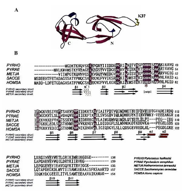
The crystallographic structure of the archaeal protein aIF5A from Pyrococcus horikoshii has been determined [71] and the striking similarity of the euryarchaeal protein with its eukaryotic counterpart is even more evident.
The β-rich structure is composed of two distinct domains, the N-terminal domain is a SH3-like barrel motifs and the C-terminal domain folds in an OB-fold (figure 7A). The N-terminal domain consists of a 310-helix, six stranded anti-parallel highly twisted β-sheets and a long hairpin loop, in which the K37 is supposed to be post-translational modified.
The C-terminal domain comprises two short α-helices and five anti-parallel β-sheets.
A flexible peptide linker connects the two domains, which embrace a hydrophobic core and contacts between them are established by hydrogen bonds.
Regarding the electrostatic potential of the archaeal protein, the N-terminal domain, as well as the interface of the two domains, are positively charged, whereas the C-terminal domain is negatively charged. Based on this the authors speculated that the archaeal aIF5A acts as a bimodular protein, interacting with nucleic acids in the concave surface and in the flexible loop, and with protein partners in the C-terminal domain.
The amino acid sequence alignment of aIF5A from Pyrococcus horikoshii with two euryarchaeal homologs and eukaryotic homologues shows that the overall structure of the protein displays significant similarity and high sequence conservation within the N-domain, while the C-domain is less conserved (figure 7B).
1.3.3. Post-translational modification of the archaeal factor aIF5A
From its discovery, the characterization of the archaeal factor aIF5A was mainly addressed to the identification of the post-translational modification, since a functional assay, like the in vitro initiation complex, cannot be built up.
A
B
Figure 7. Structural features of the archaeal protein aIF5A. (A) Crystal structure of Pyrococcus horikoshii factor aIF5A (PDB ID: 1IZ6). (B) Amino acid sequences alignment (CLUSTAL W) and secondary structure elements (DSSP program) of aIF5A protein from Pyrococcus horikoshii, Pyrobaculum aerophilum, Methanococcus jannaschii, Saccharomyces cerevisiae, Homo sapiens. Identical residues are white and the eukaryotic modified K50 is marked by an asterisk [71].
Almost ten years after the discovery of the eukaryotic translation factor eIF5A, the archaeal homologue was isolated from aerobic crenarchaeota and euryarchaeota archaea [72] and chromatographic methods assessed that the archaeal protein from Sulfolobus acidocaldarius, Halobacterium cutirubrum and Thermoplasma acidophilum is hypusinated. This is phylogenetic relevant to the lysine modification, which could originate in the archaeal kingdom or in the common ancestor of archaea and eukaryotes.
A subsequent study revealed that several species, not only aerobic, in the archaeal kingdom are able to synthesize protein-bound deoxyhypusine and/or hypusine (figure 8) [73].
Deoxyhypusine is mainly present in strictly anaerobic thermophilic archaea (Thermoproteus tenax, Pyrodietium oecultum and Desulfurolobus ambivalens), however the hypusine modification was found also in anaerobic Thermoproteales, suggesting that deoxyhypusine could be an intermediate of the hypusine pathway as in eukaryotes. From this finding the question about an alternative hydroxylation step is obvious, since there is no possibility for anaerobic organisms to perform an oxygenation.
Figure 8. Hypusine and deoxyhypusine modification in the archaeal kingdom. The number of dots and dashes in each box is proportional to deoxyhypusine or hypusine content [73].
Bartig D et al. suggested that a dehydrogenation followed by bond hydratation may occur, likewise the hydroxylation of fatty acid chains during the β-oxidation. Nevertheless, no deoxyhypusine was detected in Sulfolobales and is still an open question if these organisms develop, in aerobic mode, an oxygen way of deoxyhypusine hydroxylation.
Another crucial issue of the hydroxylation step, related to the lack of information about the archaeal protein and its modification pathway, arose when the hypusine residue was identified in Archaea.
To date no DOHH is found in any archaeal genomes or proteomes, whereas DHS homologs has been identified in all sequenced archaeal genomes.
It is worth to elucidate the archaeal post-translational modification, starting from the characterization of the archaeal DHS and the analysis of its substrate.
Indeed this was the approach of a recent study in which it was shown that aIF5A is totally deoxyhypusinylated in the euriarchaeon Haloferax volcanii, through an alternative pathway [74].
The analysis of Hfx. volcanii intracellular polyamines revealed that agmatine is the more abundant in all growth stages, the DHS enzyme is not able to transfer the 4-aminobutyl moiety from spermidine to aIF5A and is not inhibited by the known DHS inhibitor GC7. In addition the essential agmatinase-like gene clusters, in Hfx. Volcanii, with the dhs gene and the agmatinase enzyme may have a role in the modification pathway.
Based on this data the authors proposed a model in which the Hfx. volcanii DHS transfers directly the aminobutyl group from agmatine to aIF5A and the agmatinase enzyme finally hydrolyzes the agmatine moiety of the mature deoxyhypusine protein.
The effect of the inhibitor GC7 was also investigated in members of the genus Sulfolobus and for the first time, several hints on the physiological role of the archaeal hypusinated protein were provided [75].
The growth of four different archaeal species and a bacterial one was analyzed during GC7 treatment, which was effective on all archaea tested but not on the bacterium E. coli (table 2).
The insensitivity of E.coli is consistent with the absence of the dhs gene in bacterial genomes, despite a strong inhibition and a reversible arrest of growth was observed in Sulfolobus acidocaldarius, even at low concentration of GC7.
A previous analysis of the polyamine content in sulfur-dependent archaea [76] revealed that there is a high amount of spermidine, norspermidine and norspermine, however spermidine is the most abundant and this can be one explanation of the strong inhibition by GC7 in Sulfolobus acidocaldarius.
More in detail the inhibitor causes the arrest of growth at the end of the D period (G2), that occurs prior to cell division, but no changes in S.acidocaldarius cell morphology were detectable. The authors suggested that these physiological effects may be due to the presence of unmodified aIF5A, which is unable to synthetize a subset of proteins directly involved in cell cycle progression.
The possibility that the GC7 inhibitor can have targets others than DHS enzyme, causing thus a general effect in the cell, cannot be excluded.
However, these findings paved the way for future studies, which can be directed towards the functional characterization of the protein aIF5A in sulfur-metabolizing archaea, that are closely related to eukaryotes.
Nowadays very little is known about the role of the archaeal aIF5A, even though a comprehensive analysis of initiation factors in Haloferax volcanii, attempted through single gene deletion mutants, showed that aIF5A gene is essential [77].
While the involvement of this factor during the protein synthesis is still unknown, just in Halobacterium sp., a completely novel activity of aIF5A was discovered.
Table 2. Effect of the inhibitor GC7 on the archaeal and bacterial growth. +++, normal growth; ++, slight effect; +, strong effect; -, no measurable growth [75].
Wagner et al. showed for the first time that aIF5A from Halobacterium sp. NRC-1has RNA-degrading activity in vitro, which has not been reported for its eukaryotic homologue [78]. The biochemical characterization revealed that the cleavage is not dependent on the hypusination, but requires charged residues in the N-terminal and C-terminal domain and occurs preferentially between adenine and cytosine, within single stranded regions.
The Halobacterium sp. aIF5A is also able of RNA binding in vitro but, unlike the cleavage, the hypusine residue is needful to stabilize the RNA-protein complex.
In the same study, the authors tested also the ribonucleolytic activity of the eukaryotic eIF5A in vitro and showed for this the hypusine-dependent RNA degrading activity.
Moreover the RNA-cleavage by Halobacterium sp. aIF5A may depends on the oligomeric state of the protein, thus further studies should be aimed to identify its potential interacting partners.
1.4. Aim of the thesis
There is rather limited knowledge on the archaeal factor aIF5A, therefore we aim to characterize this protein in the model organism Sulfolobus solfataricus.
We investigate about the subcellular localization of the protein, focusing, in particular on the interaction with ribosomal particles, in order to clarify whether the archaeal factor aIF5A is involved in the protein synthesis.
We investigated about the post-translational modification of aIF5A in Sulfolobus solfataricus and the structural and functional features of the enzymes that are involved in this pathway. Concomitantly we seek to identify all protein interacting partners and characterize the functional interplay between aIF5A and RNA molecules.
Our goal is to discover whether the function of the archaeal factor aIF5A is confined to the protein synthesis or it is also involved in other physiological processes.
2. Materials and Methods
Materials: all chemicals used were of analytical grade from Sigma-Aldrich and were used as received without any further purification.
2.1. Expression of ORF Sso0970 in E.coli and purification of Sso-aIF5A
The ORF Sso0970 was PCR-amplified from S. solfataricus P2 genomic DNA using a forward primer containing an NcoI site (5′- GCAACCATGGGCATAACGTACACG -3′)
and a reverse primer containing a BamHI site
(5′-GCGCGGATCCCTTAACCCTAACTATT -3′).
100 ng of genomic DNA was used as a template for the PCR (table 3), performed with Phusion High-Fidelity PCR Master Mix (Thermo Fisher Scientific) and 10 µM primers were added to the reaction mixture.
TEMPERATURE TIME CYCLES
Initial denaturation 98 °C 5’ 1X Denaturation 98 °C 30’’ 30X Annealing 50 °C 30’’ Elongation 72 °C 30’’ Final extension 72 °C 10’ 1X
The amplification product, purified with the PCR purification kit (Quiagen), was cleaved with NcoI and BamHI and inserted into the corresponding sites of plasmid pETM11 (a kind gift from Dr. Roberto Spurio, University of Camerino), then the recombinant pETM11-aIF5A expression plasmid was propagate in E.coli DH5.
The recombinant plasmid was extracted using a mini-prep kit (QIAprep Spin Miniprep Kit, Quiagen), sequenced and used to transform Escherichia coli ROSETTA (DE3)/pLysS cells.
Table 3. PCR program for the amplification of ORF 0970 from Sulfolobus solfataricus P2 genomic DNA.
Concentrations of PCR products and plasmids were determined with a NanoDrop 2000c spectrophotometer device.
The cloned aIF5A gene encoded a protein that consists of six histidine residues in the N-terminal position, a peptide linker of ten amino acids and the specific cleavage site (ENLYFQ) for the Tobacco Etch Virus (TEV) protease.
2 l LB medium containing 34 µg/ml chloramphenicol, 25 µg/ml kanamycin and 100 µg/ml ampicillin were inoculated and grown at 37 °C until an OD600 of 0.7, then the culture was induced by adding 0,5 mM IPTG. After 3 hours cells were harvested, pelleted, resuspended in 20 ml of lysis buffer (50 mM Tris-HCl pH 7.4, 150 mM NaCl, 15 mM imidazole, 1mM PMSF, 10 mM β-mercaptoethanol, 10 µg/mL DNase I, 0.1% Triton X-100, 25 µg/mL lysozyme) and incubated for 20 min on ice.
Cells were lysed by sonication and the lysate wasclarified by centrifugation at 25.000 g for 30 min at 4°C.
The supernatant was incubated with 500 µl of pre-equilibrated Ni-NTA Agarose resin (Quiagen) overnight at 4 °C, to allow the binding of the His-tagged Sso- aIF5A.
The lysate and the beads was transferred to Poly-Prep chromatography columns (Bio-Rad, Hercules, CA, USA) and washed with 30 mL buffer (50 mM Tris-HCl pH 7.4, 500 mM NaCl, 40 mM imidazole). The protein aIF5A was eluted in 2 ml elution buffer (50 mM Tris-HCl pH 7.4; 150 mM NaCl; 250 mM imidazole) and the eluate was dialyzed overnight at 4 °C in dialysis buffer (50 mM Tris-HCl pH 7.4, 150 mM KCl, 5% glycerol).
The concentration of aIF5A was determined by the Bradford assay and the purity was assessed by SDS-PAGE, followed by coomassie-blue and silver staining; aliquots of the protein were stored at –80 °C.
An aliquot of the purified protein was incubated with the TEV protease to remove the His-tag. The TEV protease was added in a 1:50 ratio (TEV/protein aIF5A) and they were incubated overnight at room temperature in TEV incubation buffer (50 mM Tris-HCl pH 7.4, 0.5 mM EDTA, 1mM DTT).
The TEV protease contains a His-tag, therefore, to purify the cleaved protein aIF5A, 500 µl of Ni-NTA Agarose beads were added and the reaction was incubated overnight at 4 °C.
The protein of interest, present in the flowthrough, was collected and a small aliquot was analyzed by SDS-PAGE, followed by coomassie-blue staining.
Part of the protein, without His-tag, was used for the production of polyclonal antibodies in rabbit, using the speedy 28-day program, provided by Eurogentec, Belgium.
Different dilutions (1:1000, 1:5000, 1:10000) of anti-Sso aIF5A were tested for the detection of the recombinant Sso- aIF5A, expressed and purified in E.coli, by western blot analysis.
2.2. Expression of ORF Sso0967 in E.coli and purification of Sso-DHS
ORF Sso0967 was amplified by PCR using 100 ng genomic DNA of S. solfataricus P2 using two pairs of oligonucleotides for cloning the gene in two different plasmids:
1- forward 5'GCGGCCATGGTAAATAGAGAGGAC3' (NcoI restriction site) 2- reverse 5'CCGGGATCCTTAGCTTAATAAAGACG-3' (BamHI restriction site) 3- forward 5'AAAAGCATGCGCATAAATAGAGAGGACTTGTTAAAAAACCC3'
(SphI restriction site)
4- reverse 5'AAAAGGATCCGCTTAATAAAGACGCGGCCAAAATAGG3' (BamHI restriction site).
ORF Sso0967 amplified with primers 1 and 2 was cloned in pETM11 and the N-terminal His-tagged DHS was expressed in Escherichia coli ROSETTA (DE3)/ pLysS, using the same protocol described above for Sso- aIF5A.
ORF Sso0967 amplified with primers 3 and 4 was cloned in the plasmid pQE-70 (Quiagen) which adds a C-terminal His-tag to the recombinant protein.
The C-terminal His-tagged DHS protein was purified with the same protocol described above, with the exception of the TEV cleavage, impossible to perform with this expression plasmid.
2.3. Western blot analysis
Proteins were separated by SDS-PAGE using standard protocols [79] and transferred by Semi-dry Western blotting onto a 0,2 µm nitrocellulose membrane (GE Healthcare). Blotting
was performed at 15V for 20 min in transfer buffer (25 mM Tris, 192 mM glycine, 0.1% (w/v) SDS, 20% (v/v) methanol).
Suitable protocols, for each antibody used, are summarized in table 4.
Blocking solution 1h, R.T. Primary antibody, (in the corresponding blocking solution) overnight, 4°C Wash n°1 (3 x 5’) Secondary antibody, (in the
corresponding blocking solution) 1h, R.T. Wash n°2 (4 x 5’) Detection solution TBS + 0,1% Tween 20, 5% milk anti-SsoaIF5A (1:10000) TBS + 0,1% Tween 20 anti-rabbit IgG HRP conjugate (1:20000) TBS + 0,1% Tween 20 Enhanced chemiluminescent reagent
(SuperSignal West Pico PLUS, Thermo Scientific) anti-SsoaIF6 (1:10000) TBS + 0,1% Tween 20, 3% BSA anti-hypusine (1:2000) TBS, 5% milk anti-Sso2509 (1:5000) TBS TBS + 0,05% Tween 20 anti-SsoaIF2γ (1:5000)
The chemiluminescent signal was detected using the Chemidoc detection system (Biorad).
2.4. Preparation of Sulfolobus solfataricus cell extract
Sulfolobus solfataricus P2 was aerobically grown in Brock’s medium [80], supplemented with 0.2% NZamine and 0.2% sucrose, at 75 °C, pH 3.0.
During exponential growth, at an OD600 of 0.8, cells were harvested, pelleted and stored at -80°C. 1,5g (wet weight from 1L culture) of cells was disrupted by grinding [81], with 3g of alumina, using a sterile cold mortar on ice.
1ml of ribosome extraction buffer (20 mM Tris/HCl pH 7.4, 40 mM NH4Cl, 10 mM MgAc2, 1 mM DTT, 2.5 µg/ml DNase I) was gradually added and the cell lysate was shortly centrifuged to remove the alumina and cell debris.
Table 4. Western blot probing protocols. All solutions are in TBS buffer (20 mM Tris HCl pH 7.6, 150 mM NaCl)
Then two centrifugation steps at 26000 g for 30 min at 4°C were performed and the supernatant, crude cell extract S30, was collected.
Part of it was centrifuged at 45000 rpm (Beckman TLA-100.3 rotor) for 2h in order to obtain the S100 supernatant, rich in enzymes and factors.
The crude ribosomes pellet was resuspended in high salt buffer (20 mM Tris/HCl pH 7.4, 500 mM NH4Cl, 10 mM MgAc2, 2 mM DTT) and gently layered on the surface of a 18% sucrose cushion.
Ribosomes were centrifuged at 45000 rpm (Beckman TLA-100.3 rotor) for 3h at 4°C and at the end of this step the supernatant HSW was collected, whereas the salt- washed ribosomal fraction was resuspended in extraction buffer.
The protein concentration in S30, S100, HSW and salt-washed ribosomes fractions was determined by Bradford assay. These fractions were analyzed by western blot, with anti-SsoaIF5A antibody, to determine the sub-cellular localization of the protein.
2.5. Fractionation of Sulfolobus S30 exctract programmed for translation
on linear sucrose gradients
Sulfolobus solfataricus S30 lysates (500µg total proteins) were pre-incubated at 75°C for 10 min and then programmed for protein synthesis.
In vitro translation reaction contained, in a final volume of 50 µl: 10 mM KCl, 20 mM Triethanolamine-HCl (TEA) pH 7, 18 mM MgCl2, 3 mM ATP, 1 mM GTP, 1,5 µg of bulk S. solfataricus tRNA, amino acid mixture 0,1mM (concentration related to each of the 20 essential amino acids), 4 µg of an in vitro transcribed mRNA (Sso2375 mRNA).
The reaction was incubated for 30 min at 75 °C, then 2% formaldehyde was added and after an incubation on ice for 1h. A control sample containing only the S30 extract was incubated with 2% formaldehyde for 1h on ice.
Both samples were layered on a 10%-30% linear sucrose density gradient using 14 ml centrifuge tubes [82].
After a centrifugation step, at 23000 rpm (Beckman SW40Ti rotor) for 17 h at 4°C, 500 μl fractions were collected by continuously monitoring absorbance at 260 nm.
Samples were TCA precipitated (10% of TCA), loaded on 15% SDS-polyacrylamide gels and analyzed by western blot analysis with anti-SsoaIF5A serum.
As a loading control anti-SsoaIF6 was used, in order to detect fractions which correspond to the large 50S subunit [69].
2.6. Construction of plasmids pMJO5-SsoaIF5A and transformation of
Sulfolobus solfataricus PH1-16 cells
The Sso-aIF5A gene (Sso0970) was amplified by PCR (protocol in table 3) from 100 ng of Sulfolobus solfataricus genomic DNA using two pairs of oligonucleotides in order to create two version of the cloned aIF5A gene: an N-terminal and a C-terminal His-tagged.
Sso-aIF5A N-terminal His tagged:
forward primer containing an NcoI site and the 6xHis tag coding sequence (5′-AAAACCATGGAACATCACCATCACCATCACAGCATAACGTACACGACC GTC-3')
reverse primer containing an EagI site (5′-
AAAACGGCCGTTACTTAACCCTAACTATTTTTCTC-3′). Sso-aIF5A C-terminal His tagged:
forward primer containing an NcoI site
(5′-AAAACCATGGACAGCATAACGTACACGACCGTC-3')
reverse primer containing an EagI site and the 6xHis tag coding sequence (5′-AAAACGGCCGTTAGTGATGGTGATGGTGATGCTTAACCCTAACTATTTTT CTCC-3′).
The PCR was performed with Phusion High-Fidelity PCR Master Mix (Thermo Fisher Scientific) supplied with 10 µM primers, using the same protocol shown in table 3.
The productiom of the His-tagged protein aIF5A, in Sulfolobus solfataricus, was achieved using the pMJ05-based vector system [83].
First the PCR products were purified with the PCR purification kit (Quiagen), cleaved with NcoI and EagI and inserted in two different entry vectors, pSVA5 (arabinose inducible promoter) and pSVA11 (tf55α constitutive promoter).
Resulting plasmids pSVA5-SsoaIF5A N-termHis, pSVA5-SsoaIF5A C-termHis, pSVA11-SsoaIF5A N-termHis and pSVA11-pSVA11-SsoaIF5A C-termHis were sequenced and propagated in E.coli TOP10.
Recombinant plasmids were extracted using a mini-prep kit (QIAprep Spin Miniprep Kit, Quiagen), sequenced and digested, together with the shuttle vector pMJ05, with AvrII and EagI. Fragments were purified from agarose gel (QIAquick Gel Extraction Kit, Quiagen), and ligated into the corresponding sites of plasmid pMJ05.
This gave rise to plasmids, pMJ05 (ptf55)-SsoaIF5A N-termHis, pMJ05 (ptf55)-SsoaIF5A C-termHis, pMJ05 (AraP)-SsoaIF5A N-termHis, pMJ05 (AraP)-SsoaIF5A C-termHis used to transform E.coli TOP10.
Since pMJ05 is a huge plasmid (21868bp), which can be easily expelled, the cell recovery, after the heat-shock, was carried out for 2h at 25 °C and plates of transformants were leaved at room temperature for 2 days.
Plasmids were extracted and sequenced with pMJ05 specific primers [84], forward
5'-GGATGCTAAACAACTATTCAAACTG-3' and reverse
5'-GTTGTGTGGAATTGTGAGCGGATAA-3', to assess that each construct is in frame. 300 ng of each plasmid was used for electroporation of Sulfolobus solfataricus PH1-16 (∆pyrEF) [85] and cells were recovered for 3 days in uracil medium and then selected in medium without uracil for other 3 days. Finally cells were plated in glass Petri dishes containing Brock’s media, supplemented with 0,2%w/v sucrose, 0,2% NZamine, Gelrite (0,7% w/v) solidified.
Once grown single colonies were inoculated in liquid medium without uracil, genomic DNA was isolated and the presence of the recombinant plasmid was confirmed using the pMJ05 specific primers previously reported.
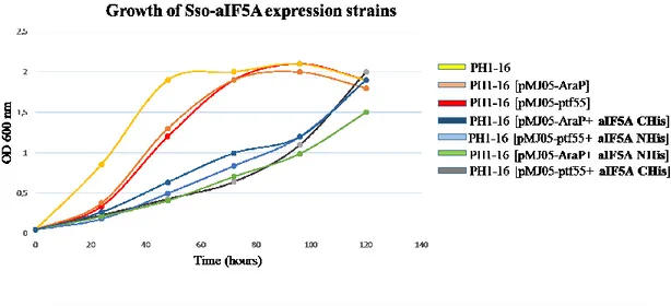
Glycerol stocks of positive clones were made and their growth was monitored for 5 days in order to see if there is an effect due to aIF5A expression.
As controls Sso PH1-16, Sso PH1-16 [pMJ05 (ptf55)-empty plasmid] and Sso PH1-16 [pMJ05 (AraP)-empty plasmid] were grown in parallel.
The two strains in which aIF5A gene is under control of the inducible arabinose promoter, were induced at a certain OD600nm, as described below.
![Figure 1. Post-translational modification pathway for the eukaryal eIF5A [2].](https://thumb-eu.123doks.com/thumbv2/123dokorg/2970187.27234/13.918.271.700.182.483/figure-post-translational-modification-pathway-eukaryal-eif-a.webp)

![Table 1. Universal translation initiation factors [63].](https://thumb-eu.123doks.com/thumbv2/123dokorg/2970187.27234/29.918.223.742.307.575/table-universal-translation-initiation-factors.webp)
![Figure 6. Model of translation initiation in Archaea [63].](https://thumb-eu.123doks.com/thumbv2/123dokorg/2970187.27234/30.918.238.736.181.634/figure-model-translation-initiation-archaea.webp)