SEDE DI CESENA
SECONDA FACOLTA’ DI INGEGNERIA CON SEDE A CESENA
Corso di Laurea Magistrale in Ingegneria Biomedica
TACTILE PERCEPTION – PERCEPTION OF TACTILE
DISTANCE ON A SINGLE SKIN SURFACE CHANGES
WITH STIMULUS ORIENTATION: A NEURONAL
NETWORK MODELLING STUDY
Tesi di Laurea in:
Sistemi Neurali LM
Relatore Presentata da
Prof.ssa Elisa Magosso Luca Monti
Correlatore
Dr. Matthew R. Longo
… To the people who make my London experience much better than an amazing dream …
… We are too concerned with what was and what will be …
There’s a saying:
Yesterday is history, Tomorrow is a mystery
But Today is a gift.
That is why it’s called PRESENT.
Keywords:
• Computational Model • Synaptic Connections • Tactile Perception • Weber’s Illusion • AnisotropyIndex
Introduction
i
1
Touch and Perceive Distance: State of the art
1
1.1 Somatosensory System
3
1.2 Mechanoreceptors and Receptive Fields in the skin
3
1.3 Dorsal Root Ganglion Neuron
6
1.4 Somatic Sensory Cortex
7
1.5 WEBER’S ILLUSION: what is it and why does it exist?
14
2
Computational model
27
2.1
Qualitative Description of the Model
27
2.2
Qualitative Description of the model working
31
2.2.1 Response of lower-level layer area to two points
stimulation
31
2.2.2 Response of lower-level layer area to two points
stimulation
33
2.3.1 Receptive Fields
35
2.3.2 External Input
38
2.3.3 Activity of the lower-level neurons
40
2.3.4 Activity of the higher-level neurons
49
2.4
Border Effects and Periodic Domain
57
3
Simulation Results
61
3.1
1. Case study 1: Mean Distance Value is equal to 3 cm. 67
3.2
2. Case study 2: Mean Distance Value is equal to 4 cm. 73
3.3
3. Comparison between Area1 outputs by considering
different Mean Distance Value
76
3.4
4. Comparison between Area2 outputs by considering
different Mean Distance Value
78
3.5
5. Green considerations
81
3.6
6. Two-point discrimination threshold
85
3.6.1 Transversal two-point discrimination threshold: 1 cm. 86
3.6.2 Longitudinal two-point discrimination threshold: 1.2 cm. 91
3.7
t-Test
102
3.7.1 t-Test: case study 1
109
3.7.2 t-Test: case study 2
110
3.7.3 t-Test: case study 3
111
4
Parameter Sensitivity Analysis
113
4.1
Lateral Synapses within Area2
113
4.1.1 Case 1: Decreasing Lateral Synapses’ dimensions. 113
4.1.2 Case 2: Increasing Lateral Synapses’ dimensions. 117
4.1.3 Case 3: Increasing the inhibitory Gaussian function’s
standard deviation.
121
4.1.4 Case 4: Increasing the excitatory Gaussian function’s
intensity.
123
4.1.5 Case 5: Decreasing the excitatory Gaussian function’s
intensity.
126
4.1.6 Case 6: Increasing the inhibitory Gaussian function’s
intensity.
128
4.1.7 Case 7: No inhibitory effects.
129
4.2
Feed-forward Synapses
132
4.2.1 Case 8: Decreasing Feed-forward synapses’
longitudinal dimension.
132
4.2.2 Case 9: Increasing Feed-forward synapses’
longitudinal dimension.
133
4.2.3 Case 10: Decreasing Feed-forward synapses’
transversal dimension.
136
4.2.5 Case 11: Decreasing Feed-forward synapses’
dimension.
141
4.2.6 Case 12: Increasing Feed-forward synapses’
dimension.
143
4.2.7 Case 13: Increasing the Gaussian function’s
intensity.
145
4.2.8 Case 14: Decreasing the Gaussian function’s
intensity.
147
4.3
Activation Threshold
147
4.3.1 Case 16: Threshold of Activation is equal to 0.7. 148
4.3.2 Case 17: Threshold of Activation is equal to 0.5. 149
4.3.3 Case 18: Threshold of Activation is equal to 0.3. 151
4.3.4 Case 19: Threshold of Activation is equal to 0.1. 154
4.4
Receptive Fields’ shape
155
4.4.1 Increasing the long axis of the RFs.
155
4.5
Considerations
160
Conclusion
161
Introduction
Interaction between the environment and us occurs through any kind of external stimuli (tactile stimuli, visual stimuli, acoustic stimuli, etc.) we receive as well as their elaboration. The brain provides this special ability, which is also a result of our experience. Sometimes integration and elaboration of inputs coming from the environment may produce illusion.
This occurs, for example, in case of tactile perception. In fact, perception of tactile distances changes with body site. The concept that distance on the skin is frequently misperceived, was first discovered over a century ago by Weber. Perceived distance is larger on regions of high tactile sensitivity than over those with lower acuity. The effect is now known as Weber’s illusion. Besides this illusion, another important phenomenon observed in vivo is that perceived distance depends on the stimuli orientation. In other words, perception of tactile distance over a skin surface changes with stimulus orientation.
Recently, Longo and Haggard [Longo & Haggard, J.Exp.Psychol. Hum Percept Perform 37: 720-726, 2011]– in order to investigate how body shape is coded within the brain - compared tactile distances presented in different orientations on the hand. They found that distances between two punctual stimuli applied across the hand are consistently perceived larger than the same distances applied along the hand. This illusion is known as Orientation-Dependent Tactile Illusion and some results in the literature provide evidence that the extent of this illusion also depends on the applied distance. In fact, Green in his paper (Green, Percpept Pshycophys 31, 315-323, 1982) evidenced that the
greater is the applied distance the greater is the orientation-dependent tactile illusion.
Weber’s Illusion and orientation-dependent tactile illusion are generally explained in the literature considering differences in receptor density, differences in dimension and shape of receptive fields (RFs), and cortical magnification effects in the primary somatosensory cortex (SI) (i.e., different extents of somatosensory cortex are allo cated for different body regions)
Anyway, the explanation based only on differences in receptor density, RF’s shape, and cortical extent in SI is unsatisfactory as orientation-dependent tactile illusion is smaller compared to the effect that the previous cited differences would produce. This suggests that tactile information, behind primary somatosensory cortex, receives further processing in higher cortical areas that may operate a sort of “rescaling process” acting in order to reduce the gap of judgment in terms of distance perceived in different orientations, and to preserve tactile size constancy.
The neural mechanisms and neural circuits acting in the brain to produce rescaling are still largely unknown. Aim of the present work is to gain insight into this particular aspect of tactile perception (orientation-dependent tactile illusion) by means of a neural network model.
The main hypothesis included in this work is that tactile information is processed at two different levels, corresponding to two different neural layers. One of them represents a lower-level layer (called Area1) in which a first and distorted tactile representation is created. This layer may mimic the primary somatosensory area, where tactile distance representation can be significantly distorted depending on stimuli orientation and skin surface, because of differences in RFs’ size and shape and cortical magnification. The other one (called Area2) represents a higher-level layer that receives the distorted information from the previous layer and reduces this distortion by rescaling tactile information toward their true size. This layer may correspond to
superior cortical areas (e.g. in the temporal or parietal cortex) that are recruited in distance judgment and implicated in the rescaling process. In the model, neurons within Area1 receive information from the external space (skin) and send information to neurons within Area2 via
feed-forward synapses. Furthermore, neurons with each area
communicate via lateral synapses: lateral synapses provide an important contribution for the elaboration of the external stimuli.
It is worth noticing that the developed model is mainly a conceptual model and it does not aspire to an accurate reproduction of the physiological and anatomical structures. So, I focused on an abstract level of implementation, without specifying an exact correspondence between layers in the model and anatomical brain regions. Anyway, the mechanisms included in the model are biologically plausible.
Hence, the neuronal network could be helpful for a better understanding of the several mechanisms that operate within the brain in order to elaborate tactile inputs. In fact, the model is able to simulate several results both of Longo & Haggard’s paper as well as Green’s paper.
The present Thesis is organized as follows.
Chapter 1 includes a review of the more relevant results of the neurophysiological and psychological literature that have been used for model implementation and validation. Chapter 2 provides an accurate description of the developed neural network: it includes a description of network architecture, contains all the mathematical formulas, and provides explanation of model parameters. Chapter 3 presents all neural network results together with their analysis and interpretation. Finally, in Chapter 4 sensitivity analysis on model parameters has been conducted to test the robustness of the model against variation in some key parameters.
The main part of the dissertation project was developed at the Department of Psychological Sciences of the Birkbeck University of London, under the supervision of Dr. M. Longo. In particular, Dr. Longo helped me in the implementation of the neuronal network, in the
interpretation of model results as well as in the their validation.
This experience also offered me the possibility to improve my knowledge concerning neurosciences. Moreover, this experience in London was the first one abroad for me and it gave me the possibility to improve my English, to make a lot of new friends coming from other country and being more self-confident.
Chapter 1
Touch and Perceived Distance: State of the art
My dissertation project consists in the implementation of a computational model that reproduces a particular aspect of tactile information processing, that is how the brain elaborates touch information coming from a skin surface of the body. In particular, I focused on how the brain could reproduce perceived distance as a function of the orientation of two stimuli applied on a specific skin region (dorsum of the hand).
It is known that the perceived distance between two stimuli applied on a skin surface is judged different from region to region of our body.
The concept that distance on the skin is frequently misperceived, was first discovered over a century ago by Weber. Weber and others have reported that the apparent distance between two pressure stimuli fluctuates with both body site and stimulus orientation. In particular, it is larger on regions of high tactile sensitivity than on those with lower acuity. This effect is known as Weber’s illusion.
This illusion suggests that tactile size perception involves a representation of the perceived size of body parts preserving characteristics of the somatosensory homunculus.
It is well known that somatosensory homunculus shows a representation of how much of somatosensory cortex innervates certain body parts.
Moreover, it seems Weber’s illusion doesn’t exist only on different regions of the body but also considering different orientation over the same skin surface. In fact, Matthew Longo and Patrick Haggard
of the Institute of Cognitive Neuroscience (University College London), made an experiment in which they investigated how the tactile perception on the dorsum of the hand can be affected by the orientation of two stimuli applied over the skin. They found that the perception of the two points distance is larger across the hand (medio-lateral direction) rather than along the hand (proximal-distal direction).
This suggests the existence of orientation anisotropies of both tactile acuity and of tactile receptive fields (RFs) of cortical neurons. So, shape of tactile RFs may partly explain distortions of mental body representations.
In addition, the paper “The perception of distance and location for
dual tactile pressures” written by Barry G. Green of the Princeton
University in New Jersey, reports similar conclusion and provides further information about this effect.
Anyway, differences in tactile perceived distance across different skin regions of the body and/or different orientations on a specific skin area are not as high as expected simply by looking at the sensory homunculus.
This suggests that tactile information, behind primary somatosensory cortex, receives further processing in higher cortical levels that acts in order to reduce the gap of judgment in terms of distance perceived in different body parts or different orientations.
The effect is known as rescaling. However, the neural mechanisms acting in the brain to produce rescaling are still largely unknown. Aim of the present work is to gain insight into this particular aspect of Weber’s Illusion by means of a neural network modelling.
Of course, the model represents an approximation of the behaviour of different cortex levels considering several simplifications.
Before giving an accurate explanation of how the model works, I will present several results of the neurophysiological and psychological literature that have been accounted for in model implementation and model behaviour.
Somatosensory system
The somatosensory system is composed of the receptors and processing centres that produce the sensory modalities such as touch, temperature, proprioception (body position), and nociception (pain). The sensory receptors cover the skin and epithelia, skeletal muscles, bones and joints, internal organs, and the cardiovascular system. While touch (also called somatosensory) is considered one of the five traditional senses, the impression of touch is formed from several modalities. In fact, the term touch is often replaced with somatic
senses to better reflect the variety of mechanisms involved.
The system reacts to diverse stimuli using different receptors: thermoreceptors, nociceptors, mechanoreceptors and chemoreceptors. Transmission of information from the receptors passes via sensory nerves through tracts in the spinal cord and into the brain. Processing primarily occurs in the primary somatosensory area in the parietal lobe of the cerebral cortex.
At its simplest, the system works when activity in a sensory neuron is triggered by a specific stimulus such as heat; this signal eventually
passes to an area in the brain uniquely attributed to that area on the body; this allows the processed stimulus to be felt at the correct location.
Hence, the interaction between our body and the environment involves a complex process that the somatosensory system executes in order to obtain the best output it needs to interact with the environment itself. Therefore, it is important to explain any single step of a tactile stimulus processing.
Mechanoreceptors and Receptive Fields in the skin
Mechanoreceptor in the skin mediate touch, so the higher is the
number of mechanoreceptor in a specific skin region, the higher is the tactile sensitivity. For example, tactile sensitivity is higher on the hairless (glabrous) skin on the palmar surface of the hand rather
then on the dorsum. In general, tactile sensitivity is greatest on the hairless skin on the fingers, the palmar, the sole of the foot and the lips.
Glabrous skin is characterized by regular array of ridges formed by folds of the epidermis. Each ridge contains a dense matrix of mechanoreceptors, which mediate the sense of touch. In fact, any time there is a motion over the skin surface or an indentation on it, they excite. The depth of the indentation depends on the force exerted by the object on the skin as well as on its dimension and shape.
All mechanoreceptors sense these changes in skin contour but differ morphologically in important ways that affect their physiological function.
Thanks to histological and physiological studies it is now clear there are four major types of mechanoreceptors in glabrous skin. Two of these are located in the superficial layers of the skin (Meissner’s corpuscle and Merkel disk receptor) and two are situated in the subcutaneous tissue (Pacinian corpuscle and Ruffini ending). The small superficial receptors sense deformation of the papillary ridges in which they reside. The larger subcutaneous receptors sense deformation of a wider area of skin that extends beyond the overlying ridges [ref. 1].
Moreover, these types of mechanoreceptors have different Receptive Fields (RFs) that play an important role in the processing of any stimulus.
A receptive field is a region of the skin from which a sensory neuron is excited. The size and structure of receptive fields differ for
receptors in the superficial and deep layers of the skin. In fact, receptors in the superficial layers on the skin resolve fine spatial differences because they transmit information from a restricted area of skin. This very fine resolution allows humans to perform fine tactile discrimination of different surfaces.
Conversely, mechanoreceptors in the deep layer of the skin sense more global properties of objects and detect displacement from a
wide area of the skin [ref. 2].
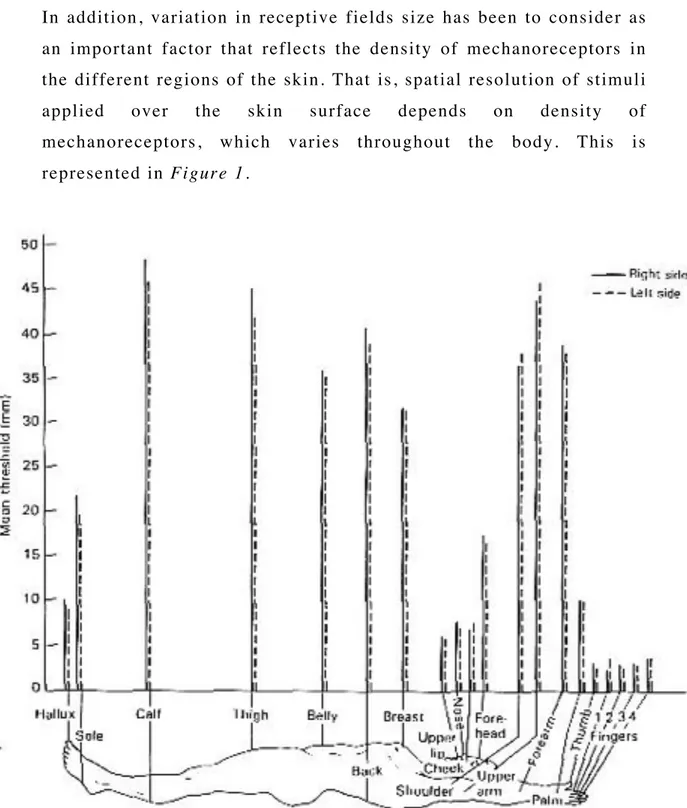
In addition, variation in receptive fields size has been to consider as an important factor that reflects the density of mechanoreceptors in the different regions of the skin. That is, spatial resolution of stimuli
applied over the skin surface depends on density of
mechanoreceptors, which varies throughout the body. This is represented in Figure 1.
Figure 1. Spatial resolution of stimuli varies throughout the body surface.
In other words, the size of the receptive fields in a particular region of the skin establishes the capacity to determinate whether one or more points are stimulated. So, if two points within the same receptive field are stimulated, the sensory neuron innervating
mechanoreceptors within this RF, will elaborated them as single continuous sensation which spans the distance between the points. Instead, if the points are located in the receptive fields of two different nerve fibres, the information about both points of stimulation will be signalled. The minimum distance between two detectable stimuli is called the two-point discrimination threshold. The two-point discrimination threshold varies from body region to body region and it depends on the size of the receptive fields and the innervation density of mechanoreceptors in the superficial layers of the skin.
Anyway, any information gathered by each mechanoreceptor is transmitted by sensory neurons to the spinal cord as well as the brain. These sensory neurons are called Dorsal Root Ganglion
Neuron.
Dorsal Root Ganglion Neuron
As I wrote before, all somatosensory information from the limbs and trunk is conveyed by dorsal root ganglion neurons. In particular, each neuron is well suited to its two principal functions:
• Stimulus transduction.
• Transmission of encoded stimulus information to the central
nervous system.
We can roughly think that this neuron is composed by a central body sitting in a ganglion on the dorsal root of a spinal nerve and an axon
divided into two branches. One of them is linked to
mechanoreceptors in the skin and the other one projects to the central nervous system.
As Dorsal Root Ganglion Neuron transmits encoded stimulus information to the spinal cord or brain system, is usually renamed
primary afferent fibre.
Figure 2. Dorsal Root Ganglion Neuron.
The primary afferent fibre performed two different classes of somatic sensation such as Epicritic sensations and Protopathic sensations. The first class involves fine aspects of touch for example the ability to detect gentle contact of the skin, localize the stimulated position of the skin and resolve spatial details. The second class involves pain and temperature senses. The present work does not consider protopathic sensations.
Somatic Sensory Cortex
Information transmitted to the brain from mechanoreceptors enables us to feel the shape of objects and get other further information about them such as their specific dimensions.
How could we get all this further information by simply touching an object with our fingers for example? In this chapter I will try to reply.
First of all, any information mechanoreceptors on the skin are able to get, is convey to the brain thanks to sensory neurons. So, information should be integrated inside the brain in order to achieve a final output such as dimensions features of objects we have touched. But, how does cerebral cortex integrate and transform sensory information coming from the periphery? How the cortex constructs an image of objects we touch from the fragmented information provided by the
receptors of the skin?
The ability to recognize objects placed on the hand on the basis of touch alone is one of the most important and complex functions of the somatosensory system.
It is known that tactile information about an object is fragmented by peripheral sensors and must be integrated by the brain. In fact, many familiar objects such as an apple, a screwdriver or a set of keys are much larger than the receptive field of any receptor in the hand. These objects stimulate a large population of sensory nerve fibres, each of which scans a small portion of the object. The peripheral sensory apparatus deconstructs the object into tiny segments because Dorsal Root Ganglion Neurons convey information from only a small area of the receptor sheet. When a particular nerve fibre fires an action potential, it signals that its territory has been contacted at intensity sufficient to cause it to fire. By analysing which nerve fibre
has been excited, the brain reconstructs the pattern made by the object.
So, several nerve fibres convey tactile information to the cortex providing many parallel pathways to the brain. It is the job of the central nervous system to construct a coherent image of the object from fragmented information coming from multiple pathways. In particular, a big contribution is given by Somatic Sensory Cortex. It consists of three different major divisions: Primary Somatic Sensory
Cortex (S-I), Secondary Somatic Sensory Cortex (S-II), Posterio Parietal Cortex. S-I is divided into four different areas: Broadmann’s areas 3a, 3b, 1 and 2. These four regions of the cortex
differ functionally. Areas 3b and 1 receive information from the mechanoreceptors in the skin, whereas areas 3a and 2 receive proprioceptive information from receptors in muscles and joints. However, the four areas of the cortex are extensively interconnected, so that both serial and parallel processing is involved in higher-order elaboration of sensory information.
S-II is innervated by neurons from each of four areas of S-I. The projections from S-I are required for the function of the S-II. For
example, when the neural connections from the hand area of S-I are removed, stimuli applied to the skin of the hand do not activate neurons in S-II.
Finally, other important somatosensory areas are located in the Posterior Parietal Cortex (Brodmann’s area 5 and 7). These areas receive input from S-I. Area5 integrates tactile information from mechanoreceptors in the skin with proprioceptive inputs from the underlying muscles and joints. This region also integrates information coming from the two hands. Area 7 receives visual as well as tactile and proprioceptive inputs, allowing integration of visual information.
Since each cortical neuron receives inputs from receptors in a specific skin area, central neurons also have receptive fields. Thus, each cortical neuron is defined by its receptive field as well as by its sensory modality. Any point in the skin is represented in the cortex
by a population of cortical cells connected to the afferent fibres that innervate that point on the skin. When a point on the skin is touched,
the population of cortical neurons connected to the receptors at that location is excited. Stimulation of another point on the skin activates another population of cortical neurons. We perceive contact at a
particular location on the skin because a specific population of neurons in the brain is activated.
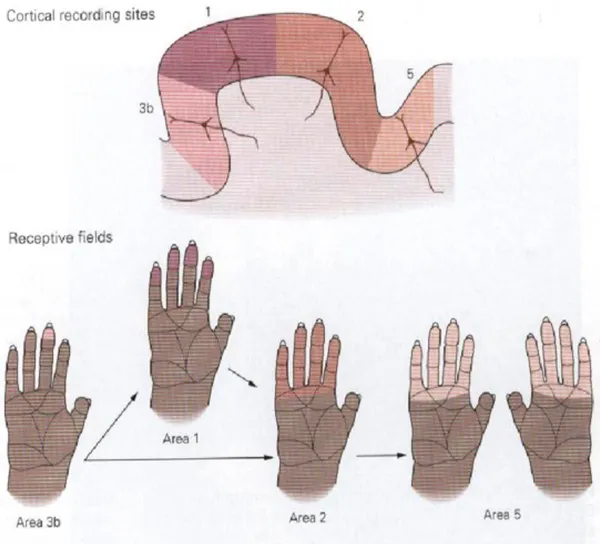
It is important to say that RFs of cortical neurons are much larger than those of dorsal root ganglion neurons. For example, the RFs of sensory neurons innervating a finger cover tiny spots on the skin, while those of the cortical cells receiving these inputs are large areas covering an entire fingertip, or several adjacent fingers, or the palmar surface of the contralateral hand.
Figure 3. The receptive fields of neurons in the Primary Cortex are larger than those of the sensory afferents.
The size and the position of cortical receptive fields on the skin are not fixed permanently but can be modified by experience or by injury to sensory nerves. For example a tennis champion will develop a larger proportion of cortical neurons devoted to sensory input on the arm than a pianist, who needs a larger proportion of cortical neurons devoted to sense inputs coming from each finger.
Although the RFs of cortical neurons cover a large area of the skin, a cortical neuron is nevertheless able to discriminate fine detail because it responds best to excitation in the middle of its receptive field. Thus, a stimulus applied to the tip of the finger strongly
excites some neurons, while others fire weakly or not at all. If a more proximal spot on the finger is touched, many of the same cells are activated but in different proportions. Information provided by the entire population of excited cells localizes a stimulus on the skin. The somatotopic arrangement of somatosensory inputs in the human cortex is called homunculus. This is not more than a visual
representation of the concept of “the body within the brain” that one hand or face exists as much as a series of nerve structures or a “neuron concept” as it does a physical form.
However, the internal representation of our body does not duplicate in a correct way the spatial topography of the skin. In fact, the images of the body in the brain exaggerates certain body regions such as hands, feet, mouth and compress other regions such as trunk, arms and so.
The reason for the bizarre, distorted appearance of the homunculus is that the amount of cerebral tissue or cortex devoted to a given body region is proportional to how richly innervated and sensitive that region is, and not to its size. The homunculus is like an upside-down sensory map of the contralateral side of the body.
It is easy to understand that somatosensory homunculus is represented by huge hands, lips and face in comparison to the rest of the body because there are a lot of sense nerves in these specific regions. Those regions are important sensor of the properties of
Instead, the proximal portions of the limbs and trunk are much less densely innervated; correspondingly, fewer cortical neurons receive inputs from these regions.
An important consequence of the magnification of the hand representation in the cortex is that the size of individual peripheral receptive fields on the hand covers a much smaller area of skin than receptive fields on the arm, which are smaller than receptive fields on the trunk. The figure below gives us an illustration of the sensory homunculus.
Figure 4. Sensory Homunculus is an original model of our body. Each part of the Somatosensory cortex is dedicated to a specific body region.
Another important phenomenon has to be considered in order to explain the spatial resolution within the sensory cortex. In fact, it not depends only on the innervation density over the skin but also on how cortical neurons could communicate each other. As I wrote before, when a particular stimulus is applied on a certain point of the skin, cortical neurons with RFs covering that position will activate, increasing their firing rate (a population of neurons will be activated). However, if many neurons are activated at the same
magnitude, the spot represented by the stimulus could be blurred and the position of it will not be identified in the best way by the brain. In order to find out the best localization, cortical neurons interact to each other not only via excitatory synapses but also with inhibitory ones. That is, if a neuron is activated it will tend to excite proximal neurons nearby and inhibit more distal ones. In other words, stimulation of regions of skin, surrounding the excitatory region of the receptive field of a cortical neuron, may reduce the responsiveness of the neuron to an excitatory stimulus because afferent inputs surrounding the excitatory region are inhibitory.
It is now clear that inhibitory interactions are particularly important for fine tactile discrimination. In fact, if two stimuli applied on a skin surface are very close, the activity of both populations will overlap each other and the distinction between the two peaks might become blurred. However, the inhibition produced by each stimulus also summates in the zone of overlap. As a result of this more effective inhibition, the peaks of activity in the two responding populations become sharpened, thereby separating the two active populations spatially. What I have just explained might become clearer by having a look at the figure below.
Figure 5. Surround inhibition. Inhibitory interneurons provide a better localization of the stimulus, as there is a greater discrimination between the activities of the second-‐ order neurons. Note that the activation of X1 and Z1 is higher than the activation of X2 and Z2.
WEBER’S ILLUSION: what is it and why does it exist?
Although inhibitory synapses between neurons might figure out the discrimination of two stimuli very close to each other, it is not always true that the perceived location of the stimuli and the perceived distance between them correspond to the real physical position and distance of the two stimuli. That is, perceived distance often differ by the physical one. Moreover, perceived distance differs from body region to body region because of sensory acuity changes with skin surface.In other words, supposing to apply two punctual stimuli on the hand of a subject at a certain distance and then to apply the same stimuli on the arm of the same subject and at the same distance, subject will
perceive a larger distance on the hand than on the arm.
How is it possible?
The real distance is the same but it is judged as not as the same. Weber has detected this sort of tactile illusion and that is why it is
called Weber’s Illusion.
In 1834 E. H. Weber described accurately that when compass-points, kept equidistant, are moved with equal pressure over a cutaneous surface of varying sensitivity, the observer experiences a converging or a diverging of the two paths; a converging, when the points pass from an area of greater to one of less sensitivity, and a diverging when they pass from an area of less to one of greater sensitivity. Weber indicated, in some detail, the form of the illusion as found at twelve different regions of the body.
After Weber’s work, other studies have been conducted in order to discover more about Weber’s Illusion as well as if it might appears in different ways.
Interesting results were obtained. In fact, it seems that this sort of tactile illusion is present not only by comparing different body regions but also analysing a specific skin surface by changing the orientation in which stimulation was applied. Further information could be find by taking a look at Matthew R. Longo and Patrick Haggard’s Paper, “Weber’s Illusion and Body shape: Anisotropy of
Tactile Size Perception on the Hand”.
They investigated how body shape is coded within the brain, by comparing tactile distances presented in different orientations on the hand. In particular, the authors applied two punctual stimuli on the hand; in same cases the distance between the two stimuli was oriented along the hand, in other cases the distance between the two stimuli was oriented across the hand. Longo and Haggard found that across (medio-lateral axis) distances are consistently perceived as larger than along (proximo-distal) ones. This is completely true if the experiment is conducted over the dorsum of the hand. A second experiment reveals there is not as such as good discrimination between across and along distances on the palm of the hand. In other words, Weber’s Illusion could change at any skin region of the same body part [ref. 3].
In Longo & Haggard experiment, participants made un-timed
points felt farther apart in the along or across orientation, and responded verbally.
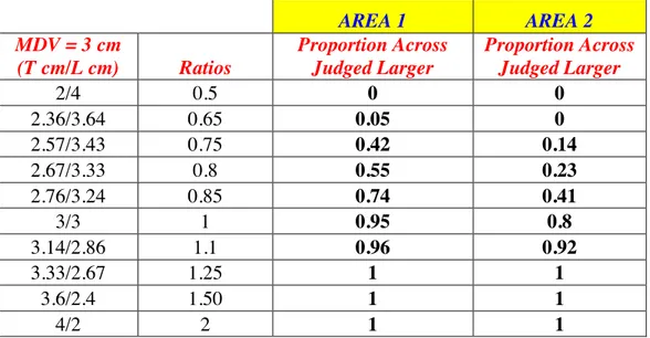
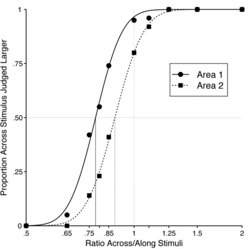
There were five pair of stimuli, according to the size of the transverse and longitudinal orientation (across/along): 2/4 cm, 2/3 cm, 3/3 cm, 3/2 cm, 4/2 cm. Stimuli were applied approximately on the centre of the dorsum of the hand.
Each ratio was applied 14 times for each of twenty participants. So, the total trials number was 70.
The proportion of trials in which the ‘across’ stimulus was judged as larger was analysed as a function of the ratio of the length of the along and across stimuli, plotted logarithmically to produce a symmetrical distribution about the point-of-actual-equality (i.e., ratio equals to 1).
Figure 6 (Figure 3 Normal Case, Longo & Haggard Paper), clearly demonstrates when the ratio is equal to 1, transversal sizes are judged larger than longitudinal sizes. Investigated distances are judged equal to each other if a ratio lower than 1 is provided.
Figure 6. Proportion Across Stimulus Judged Larger than Longitudinal Stimulus. The black vertical line indicates the ratio value that provides an equal Judgment of the applied distances.
Note that Proportion Across Stimulus Judged Larger is 0.5 if a ratio Across/Along equal to 0.729 is provided. This ratio is visualized in the figure thanks to the black vertical line and it is known as Point of Subjective Equality (PSE).
However, I am going to report a better explanation about this experiment in Chapter 3.
So, the perceived distance might be a function of physical distance.
That is, relationship between physical and perceive distance is not a constant.
What I have just maintained is the main subject of some experiments conducted by Barry G. Green of the Princeton University of New Jersey. The paper he wrote, called “The perception of distance and
location for dual tactile pressures” is a clear demonstration.
In each experiment, the perceived distance was investigated by changing orientation of stimuli but also considering different body
parts.
Stimuli spaced from each other at several distances were applied over different skin regions of the subjects.
Each subject responded with a number that reflected the perceived distance between the two stimuli.
Thanks to this judgment a linear relationship between perceived and physical distance has been found [ref. 4].
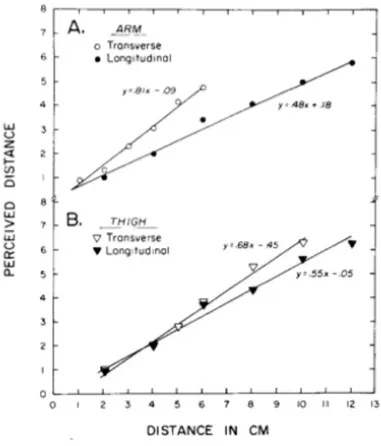
The picture below shows the comparison between two different limbs.
Figure 7. Perceived distance depends on both the body locus and the orientation of the stimulation.
The two limbs appear anisotropic, with, the arm showing greater anisotropy than the thigh.
So, from data reported on Green Paper, I can conclude that perceived tactile distance depends on both the body locus and the orientation of stimulation, and that the size of the orientation effect depends upon the locus. That is, the size of the orientation effect will change if the stimulated skin surface changes.
Now, the key question is: Why perceived distance differ at any body part or by changing the orientation?
As I wrote before, our brain has got an original model of our body. Considering the Primary Sensory Cortex, each part is dedicated to a specific body region. Inputs coming from the hand are elaborated within areas that differ from those areas in which inputs coming from the arm are elaborated. Moreover, each area has different dimensions. Taking a look at the sensory homunculus, it is easy to see that hands are much larger cortical representation than the arms. This is because brain provides larger area for the integration of inputs coming to the hands rather than for those coming from the arms.
Differences in term of magnification have been study by Mriganka Sur, Michael M. Merzenich and Jon H. Kaas of the Department of Psychology and Anatomy of Vanderbit University in Nashville, California.
In the paper “Magnification, Receptive Field Area, and Hypercolumn
Size in Areas 3b and 1 of Somatosensory Cortex in owl Monkeys”
they reported the magnification intensity of several owl Monkeys’ body parts.
An interesting result was found. In fact, the glabrous hand or foot representations occupy nearly 100 times more cortical tissue per unit body-surface area than the trunk or upper arm representations in both
areas 3b and 1 [
ref
. 5].Figure 8 reports magnification magnitude by considering different body part. Magnification is one of several factors that can explain why there are differences in terms of perceived distance from body region to body region.
Figure 8. Difference in the perceived distance from a region to another is attributable to a different cortical magnification.
The cortical magnification is obtained by dividing the cortical representational area devoted to a body region by its skin-surface area.
In formula:
M = Cra/Ss à Cra = M x Ss (1)
where M shorts for cortical Magnification, Cra is the Cortical Representational Area and Ss short for Skin Surface.
As a ratio of square areas, unit square measure is not necessary to describe Magnification.
So, if we consider a square area on the hand equals to 25 cm2
(5 cm x
5 cm) and a square area over the arm equals to 100 cm2
(10 cm x 10 cm) the cortical representational area of each body part will be different. In fact, by looking at the figure above the Magnification
devoted to the hand is 10- 2
whereas Magnification on the arm is equal
to 10- 4
. In particular:
Cra_Hand = 0,01 x 25 cm2 = 0,25 cm2 Cra_Arm = 0,0001 x 100 cm2 = 0,01 cm2 Cra_Hand/Cra_Arm = 0,25 cm2 / 0,01 cm2 = 25
So, the cortical representational area of a smaller skin region on the hand is much bigger than the cortical representational area devoted to a bigger skin surface on the arm. In this example Cra_Hand is 25
times Cra_Arm. Difference in the perceived distance from a region to
another is attributable to a different cortical magnification.
Even if data in Figure 8 are obtained by studying brain of owl Monkeys is legitimate to think that similar data can be obtained by studying human brain. This is because somatosensory cortex of Monkeys is similar to somatosensory cortex of humans.
As stated below, different magnification factors correspond to different amounts of peripheral innervation of the represented body part, with skin surface more densely innervated having higher magnification factor. Different magnification factor correspond to different dimension of neuron RF. In particular, cortical areas with great magnification factor contain cells with smaller RFs (that is, a large number of cells is necessary to represent the surface area); cortical areas having low magnification factor have cells with larger RFs (a small number of neurons is necessary to represent the surface area).
It is known that a population of neurons in the cortex will be activating if a stimulus touches the skin surface over the RF of the population. Therefore if the RF area is large, neuronal population will represent a great skin area. Even if neurons are not activated at the same intensity, two stimuli applied over the same RF could be close enough to be undistinguished from each other by the brain. Conversely, considering smaller RF, the two stimuli could be sitting over two different RF providing the activation of two different
neuronal populations. In this way stimuli can be distinguished because of a better spatial resolution.
As I have written in “Mechanoreceptors and Receptive Fields in the
skin” there are tiny RFs in the skin of the fingertips. RF gets larger
from fingers to the rest of the limb. So spatial resolution operated by the brain gets worst from the hand to the arm.
I think changing dimension of RF from body part to body part and differences in Magnification for each region of our body can explain the “classical” Weber’s Illusion.
Here, I have supposed RFs with circular shape but what happen if RFs have got a shape that differ from the circular one?
The answer to this question can introduce another aspect of the Weber’s Illusion. In fact, by considering a sort of anisotropy in RF’s shape it is clear that tactile illusion should be detected simply by changing the orientation of the stimuli applied on the skin. Try to think at RFs with oval shape. In this way stimuli sitting along the same direction of the long axis of a RF, could fall within the same RF activating a specific neuronal population. So, stimuli could not be discriminated if they are close to each other (as the example gave before in which RF dimension changes). On the other hand, stimuli applied along the same direction of the short axis of the RFs could fall over regions represented by two different RFs. Two different populations will be activated and the two stimuli could be better distinguished. Although distance between stimuli is the same, it is not judged as equal. In other words, perceived distance changes by considering different orientation.
Moreover, a distort representation of the body inside the brain can increase anisotropy in perceived size of tactile objects and perceive distance as well. For example, if the hand is represented as being longer and more slender than it really is, distances oriented proximo-distally, along the body surface, should feel larger than those oriented medio-laterally, across the body surface. Conversely, if the hand is represented as being wider than it actually is, distances oriented across the hand should be perceived as larger than those
oriented along the hand.
Longo and Haggard investigated the perceived size of tactile distances in different orientations on a single skin surface (dorsum and palm of the hand).
Several studies have found RFs representing hairy skin at many levels of the nervous system are generally oval-shaped, with the long axis running proximo-distally. Anyway, it is impossible to estimate exactly how large could be an area representing a RF. There are no studies that find out this information.
Anyway, data obtained by Longo and Haggard show that tactile stimuli running medio-laterally are systematically perceived as larger than stimuli running proximo-distally. This suggests the body model
mediating touch present a dorsum that is stretch along medio-lateral axis. Hand is represented as being wider than it actually is. This would produce the orientation-dependent tactile illusion.
Thus, RF geometry may play a fundamental role in the construction of the implicit body model mediating tactile size and shape perception.
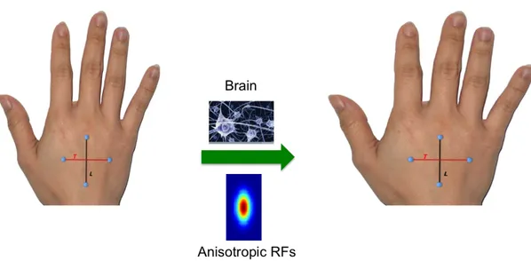
The figure below gives an illustrative explanation of what I have just declared.
Figure 9. Orientation-‐Dependent Tactile illusion. Note that the red line became longer than the black one.
I focused on this relevant aspect in order to implement my model. In addition, the experiment made by Green, shows other interesting results. For example, a physical distance equal to 4 cm, it is not judged as the same by considering different body parts.
In fact:
Physical Distance (cm) à Perceive Distance (points)
4 cm (Arm) à 2 points
4 cm (Hand) à 3 points
Data I have reported are referred to the longitudinal orientation. Difference in terms of Perceived distance is just only one point. It is known that somatosensory area devoted to the hand is much larger than the cortical area devoted to the arm (the ratio between them is closely to 100). So, it seems, there is not a proportional
relationship between Magnification and perceived distance. In fact, a difference higher than one point should be expected by considering what Mriganka Sur et al. have reported.
This result suggests that an integration of inputs elaborated by primary somatosensory cortex, may exist somewhere in the brain in order to reduce the Magnification effects.
I made a similar hypothesis for the orientation-dependent tactile illusion.
Indeed, differences in term of perceived distance between transversal and longitudinal directions are not as significant as expected. In fact, some studies suggest that the long axis of RFs on the hairy skin of the limbs may be more than twice as long as the short axis [Brown et al 1975]. By considering only the shape of the RFs, tactile distance along the transversal orientation should be perceived at least twofold bigger than along the longitudinal orientation. But this is not the case. That is, the illusion is substantially smaller than would be
expected on the basis of sensitivity, cortical magnification or RF geometry. Hence, we can hypothesize that some processes in the
homunculus and RF shape, although the compensations is only partial.
This behaviour of the brain is called rescaling. As the brain is not able to reproduce perfect rescaling, the final result is the Weber’s illusion. Shortly, rescaling decreases Magnification effects.
Unfortunately there are not studies that show how the brain works in order to reproduce rescaling. For that reason, this study tries to clarify this aspect via a neural network modelling study: in particular the model I implemented reproduces the orientation-dependent tactile illusion on the dorsum of the hand both inspiring by Longo and Haggard data and considering simplifying assumptions.
The main hypothesis included in this work is that there are two levels of processing of tactile inputs. The first level (lower level) may correspond to elaboration in the primary somatosensory cortex; we assume that at this lower level, codification of distance between two stimuli is strongly affected by differences in RFs size or shape (for example, at this level the same physical distance may be codified as much larger along the transversal dimension rather than along the longitudinal dimension). The second level of tactile information processing may correspond to higher somatosensory cortices. In this second level, differences in distance codification with orientation are partially rescaled; we assumed that rescaling might emerge as a network property arising from specific patterns of synaptic connections from the lower to the higher levels and inside the higher level.
In the subsequent chapters, the model will be described in details and hypotheses included in the model will be highlighted.
Chapter 2
Computational Model
Qualitative Description of the Model
As I wrote in Chapter 1 the model represents a simplification of how the brain elaborates tactile inputs sensed by skin receptors in order to obtain perceived distance between two stimuli. This is mainly a conceptual model rather than an accurate reproduction of physiological structures. So I focused on an abstract level of implementation, without specifying an exact correspondence between layers in the model and anatomical brain regions. Anyway, the mechanisms included in the model are biologically plausible.
I focused on the dorsum of the hand to study orientation-dependent tactile illusion.
I suppose that two different cortical areas integrate tactile information. One of them represents a lower-level layer (called Area1) in which a first and distorted body model is created. The other one (called Area2) works with the aim to reproduce rescaling. In other words, this is a higher-level layer that reduces the distortion of the lower level by rescaling tactile information toward their true size. Neurons in Area1 receive information from the external space (skin) and send information to neurons in Area2 via feed -forward connections.
The computational model is divided into two main parts:
• First Elaboration Step of the External Stimuli.
28
• Second Elaboration Step of External Stimuli.
Neurons within Area2 and feed-forward connections from neurons in Area1 represent this part.
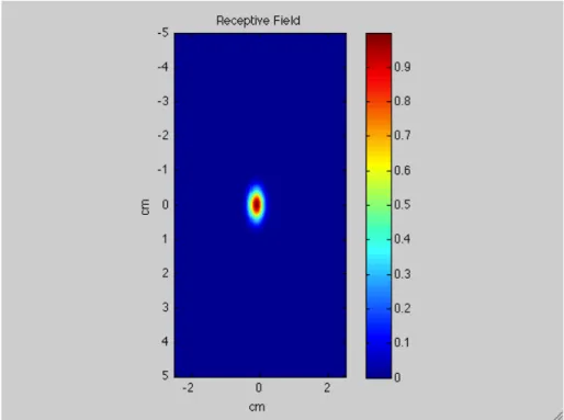
First of all, tactile inputs stimulate a specific skin region of the dorsum of the hand. They have been mimicked with a Gaussian spatial pattern with a tiny deviation standard in order to reproduce stimuli very similar to punctual inputs.
The first layer in the cortex, which consists of 41x26 units, maps a skin surface area of 10 cm (longitudinal dimension) x 5 cm (transversal dimension).
Each neuron in this layer has a Receptive Field covering a specific portion of the skin region. Supposing that a topological organization exists, proximal units in the layer will respond to stimuli coming from proximal positions over the skin.
Taking a look at the matrix (41 units x 26 units) representing the lower-level layer it easy to say that centres of RFs are arranged at two different distances considering transversal (medio-lateral) and longitudinal (proximo-distal) orientation.
In fact, 26 units represent 5 cm along transversal direction whereas 10 cm correspond to 41 units along longitudinal direction. Hence, RFs centres are disposed at a distance of 0.2 cm along the transversal dimension and at a distance of 0.25 cm along the longitudinal dimension.
In formula:
5 cm / 25 units = 0.2 cm/units
(the first unit is centred in -2.5 cm and the last unit in 2.5 cm)
10 cm / 40 units = 0.25 cm/units
(the first unit is centred in -5 cm and the last unit is centred in 5 cm)
According to physiological and behavioural literature (see Chapter 1) there is a greater precision in two points discrimination along
medio-lateral direction rather than proximo-distal direction. To reproduce this aspect, besides considering a different spatial resolution of RFs along the two dimensions (see below), the model considers different dimension and shape of RFs.
In fact, as I wrote in chapter 1, the smaller is the receptive field the better is the discrimination of two tactile stimuli on the skin. Anyway, not only the dimension plays an important role but also the shape of RFs.
According to data finding in literature [ref. 5, ref. 6] the model adopts oval-shape RFs with the long axis along proximo-distal direction. So, considering transversal orientation, RFs are much many and much smaller than on the other orientation. Thanks to these differences, the model reproduces a greater sensory acuity along the transversal dimension. As a consequence, a higher magnification of the dorsum of the hand along this direction is provided.
Like the inputs, RFs are represented by a Gaussian function with standard deviation that differs for the two orientations. In particular, (as justified later) longitudinal standard deviation is two times the transversal one.
Moreover, RFs overlap each other thanks to their dimensions and shape. In this way, a stimulus applied in a certain position on the skin region will active more neurons, specifically all neurons having RF covering that position on the skin. So, a bubble of activation in the first layer occurs.
The first area improves a rough version of the perceived distance. This is why also a second layer has been implemented as involved in tactile distance perception. It receives inputs coming from Area1. As the first layer, a matrix of 41 rows and 26 columns represents Area2.
Feed-forward synapses connect the two layers. So, activation of each unit within the higher-level area, in response to an external stimulus, depends on the pattern of the synaptic connection from the first layer.
30
The figure below shows a schematic view of the model structure
I hypothesize that the second layer may represent higher cortical areas that - starting from a distorted primary representation based on receptor density and cortical magnification - may partially rescale tactile information towards their true size.
The model not only implements feed-forward synapses, but also lateral synapses (not reported in the figure above).
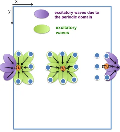
Feed-forward synapses ensure communication from Area1 to Area2. Lateral synapses improve communication between each unit at any layer. In particular, these synapses are arranged according to a Mexican hat disposition, i.e. excitatory synapses among proximal neurons and inhibitory ones among distal neurons. This arrangement of lateral synapses improves two points discrimination.
i = 1
i = 41
x y j = 1 j = 26
First layer: 41x26 units
(correspond ing to 10 cm x 5 cm respectively ).
Uni ts in the first layer send synapt ic connections to each unit in the second layer.
Second layer
Qualitative description of model working
In this section, I will describe how the model works qualitatively. In particular, I will present the pattern of neuron activation that it is expected to obtain in the two layers of the model.
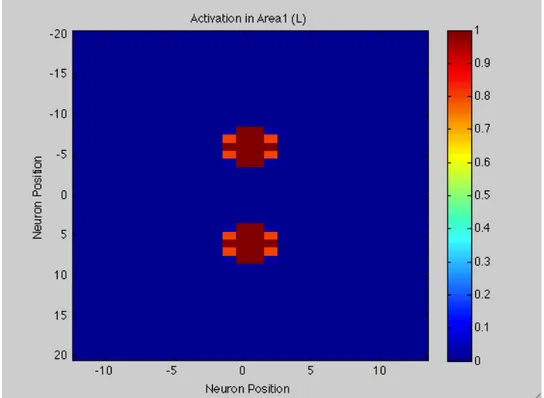
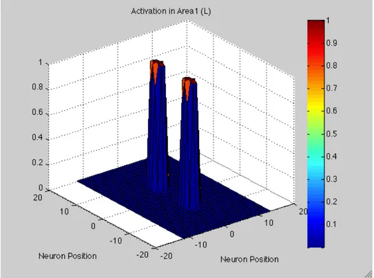
Response of lower-level area to two points stimulation
Suppose that a tactile stimulation consists of two punctual stimuli at about 2.5 cm distance is applied.
For example, assume we stimulate units in the central row of the matrix. Below, a graphical representation is reported.
Transversal Direction:
The red arrows represent the external punctual stimuli applied at two skin positions. Let’s assume that the position of one stimulus corresponds to the RF centre of the neuron of the first layer at position j = 7 (RF centre = j*0.20 cm = 1.4 cm) and the position of the second stimulus corresponds to the RF centre of the neuron of the first layer at position j = 20 (RF centre = j*0.20 cm = 4 cm). The expected activation of the neurons in the row (first layer) is something like this:
i = 21 j = 1 j = 7 j = 20 j = 26 1
7 Position of neurons in the first layer (j) 20 26 0 1 Ne ur on A ct iv at io n
32
In order to solve perceived distance I considered the number of neurons between the two peaks of activation. In this example a perceived distance equal to 13 neurons is reported.
A similar discussion can be made with regard to punctual stimuli applied along the longitudinal orientation.
Longitudinal Direction:
Here, each neuron is design bigger just to remember that along this orientation there is less resolution.
The red arrows represent the external punctual
stimuli applied in two skin positions
corresponding respectively to the RF centre of the neuron of the first layer at position i = 6 (RF centre = i*0.25 cm = 1.5 cm) and to the RF centre of the neuron of the first layer at position i = 16 (RF centre = j*0.25 cm = 4 cm).
The expected activation of the neurons in the row (first layer) is something like this:
Here, the perceived distance (by adopting the same metric as above, that is the number of neurons between the two peaks of activation) is equal to 10.
Now, we can think about the first layer implemented by the model as
1
6 16 41 Position of neurons in the first layer (j)
0 1 Ne ur on A ct iv at io n
that area in the primary somatosensory cortex devoted to the hand. Taking a look at the distance codified by this layer (remember that the applied distance is the same along the two orientations = 2.5 cm), a ratio (Transversal/Longitudinal) higher than one is found.
Transversal = 13; Longitudinal = 10; Ratio = 13/10 = 1.3;
This result may be interpreted in this way: the same distance between two punctual stimuli applied externally is perceived bigger when the stimulation is applied along the transversal orientation rather than along the longitudinal orientation.
Response of higher-level area to two points stimulation
Area2 provides an improvement of the body model obtained within Area1. That is, Area2 acts in order to reduce the discrepancy
between the two distances as reported by Area 1.
So, I assume that the final output of the neuronal network is the activation observed in Area2. This means, activation in Area2 is read out in order to produce the response of our hypothetical subject. We assume that the distances perceived by our hypothetical subject correspond to the number of neurons between the two peaks of activation in Area2.
As I wrote in Chapter 1 it is unknown how the brain rescales tactile inputs coming from lower-level layer. Several hypotheses have been made. A plausible hypothesis is that the brain has learned (by integrating various sensory information such as visual, tactile, proprioceptive information) to rescale tactile information from skin regions having different cortical extents. In this case, the Weber’s Illusion would reflect a failure to perform a complete rescale.
I assume that this brain capability is implemented through the synaptic connections from the first layer to the second layer (and that
34
these synapses would have been learned via training and experience). Area1 sends feed-forward synapses that work in order to preserve perceived distance along transversal direction changing the one along the longitudinal orientation. It means that distance in terms of number of neurons has same value in both lower-level area and higher-level area if a transversal stimulation is considered, whereas it is not the same along longitudinal direction. In particular, perceived distance along transversal direction is preserved while perceived distance along longitudinal direction increases within Area2.
I adopted this solution in order to change perceived distance value along the orientation with less spatial resolution and less sensory acuity. In fact, it is licit to think that nothing should change from Area1 to Area2 (in term of perceived distance) if transversal stimuli are applied because of both better spatial resolution and sensory acuity. Here a better judgment of distance is provided. In other words, it seems ecologically more beneficial to improve the functionality where it is poorer (that is along the orientation showing lower resolution) and to preserve it where it is higher (that is along the orientation showing better resolution).
So, an activation in Area2 due to a longitudinal stimulation have to enlarge the gap between the two peaks, that is, the number of neurons between them must become higher.
Hence, the output, relative to the example made before for Area2 should be something like this:
So, longitudinal perceived distance is equal to 12 neurons in Area2.
1
5 17 41 Position of neurons in the first layer (j)
0 1 Ne ur on A ct iv at io n