UNIVERSITY OF PISA
Research Doctorate School in
BIOLOGICAL AND MOLECULAR SCIENCES
Course in
EXPERIMENTAL AND MOLECULAR ONCOLOGY
XXIII CYCLE (2008-2010)
Research Doctorate Thesis
Molecular targeting and development of a
new radioreceptor therapeutic strategy for
non-iodophil epithelial follicular thyroid
and neuroendocrine-like tumors.
Supervisor Candidate Prof. Giuliano Mariani Dr. Chiara Manfredi
Tutor
“I have no particular talent. I am passionately curious”. Albert Einstein “Somewhere, something incredible is waiting to be known”.
Carl Sagan
To all those that I have been passionate about science.
TABLE OF CONTENTS
ABBREVIATIONS 7
INTRODUCTION 8
1 Somatostatin and it receptors family. 8
1.1 Somatostatin and it actions. 8
1.2 Somatostatin receptors and their expression in normal tissues. 9
1.3 Somatostatin receprors subtypes expression in human cancers. 10
1.4 Incidence and mortality. 14
1.5 Neuroendocrine Tumors aetiology. 15
1.6 Gastroenteropancreatic tumors aetiology. 15
- Stomach 15
- Appendix 16
- Oesophagus 16
- Enteron 17
- Colon and rectus 17
- Pancreas 18
1.7 Medullary thyroid carcinoma. 19
1.8 Papillary and follicular thyroid carcinoma 19
1.9 Lung. 20
1.10 Breast. 21
1.11 Kidney. 22
1.12 Lymphoma. 22
2 Somatostatin and it actions. 23
2.1 Somatostatin and it activations. 23
2.2 Somatostatin functions. 23
2.3 Somatostatin and it analogues. 24
2.3.1 Agonists. 24
2.4 Somatostatin analogues and cancer. 28
2.5 Radiolabeled Somatostatin analogues. 32
3 Strategies to improve pharmacological profile of radiolabeled Somatostatin analogues. 38
3.1 Characteristic of Somatostatin analogs for nuclear medicine. 38
3.2 Modifications of pharmacokinetics/pharmacodinamics of Somatostatin analogs. 40
3.4 Lowering the kidney radioactivity levels 42
3.5 Targeting the nucleus. 43
3.6 Understanding the post-endocytic pathway of SSTRs. 46
AIMS OF THE PROJECT 47
MATERIALS AND METHODS 49
1 Peptides synthesis. 49
2 Determinations of Somatostatin receptor affinity profiles. 50
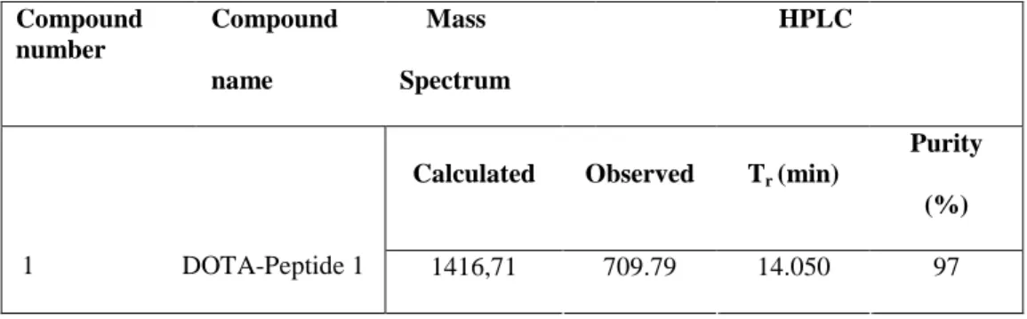
3 Studies performed with 177Lu DOTA-Peptide 1. 51
3.1 Peptide radiolabeling. 51
3.2 Quality control. 51
3.3 Serum stability and identifications of metabolites. 51
3.4 Cell line and culture conditions. 52
3.5 Radioligand internalisations studies. 52
3.6 Cellular retentions studies. 53
3.7 Biodistributions studies in tumor bearing nude mice. 54
4 Studies performed with 111In DOTA-Peptide 1. 54
4.1 Peptide radiolabeling. 54
4.2 Quality control. 55
4.3 Cell line and culture conditions. 55
4.4 Radioligand internalisations studies. 56
4.5 Cellular retentions studies. 56
4.6 Biodistributions studies in balb-c mice. 56
5 Ligand Tracer®. 57
5.1 Cell line and culture conditiotions. 57
5.2 Characterisation of NPA-87 cell line. 57
5.2.1 Western Blot analysis. 57
5.2.2 Immunofluorescence studies. 58
5.3 Ligand Tracer®. 59
5.4 Affinity assays with Ligand Tracer® and pharmacological studies. 60
6 Pharmacological studies. 62
6.1 Immunofluorescence studies: SSTR2 internalisation. 62
6.2 Immunofluorescence studies: investigations intracellular localization of internalized SSTR2. 63
7 Investigation of the activation of the apoptotic pathway. 64
7.1 TUNEL assay. 64
RESULTS 65
1 Peptides synthesis. 65
2 Determinations of Somatostatin receptor affinity profiles. 66
3 Studies performed with 177Lu DOTA-Peptide 1. 67
3.1 Peptide radiolabeling and quality control. 67
3.2 Serum stability and identifications of metabolites. 67
3.3 Radioligand internalisations studies. 67
3.4 Cellular retentions studies. 68
3.5 Biodistributions studies in tumor bearing nude mice. 69
4 Studies performed with 111In DOTA-Peptide 1. 71
4.1 Peptide radiolabeling and quality control. 71
4.2 Radioligand internalisations studies. 71
4.3 Cellular retentions studies. 71
4.4 Biodistributions studies in balb-c mice. 73
5 Ligand Tracer®. 74
5.1 Characterisation of NPA-87 cell line. 74
5.1.1 Western Blot analysis. 74
5.1.2 Immunofluorescence studies. 74
5.2 Affinity assays with Ligand Tracer® and pharmacological studies. 75
6 Pharmacological studies. 77
6.1 Immunofluorescence studies: SSTR2 internalisation. 77
6.2 Immunofluorescence studies: investigations intracellular localization of internalized SSTR2. 78
7 Investigation of the activation of the apoptotic pathway. 79
7.1 TUNEL assay. 79
DISCUSSION 81
CONCLUSIONS 95
AKNOWLEDGEMENTS 97
ABBREVIATIONS
ACTH Adrenocorticotropic Hormone
CCL39 Chinese Hamster lung fibroblast cell line CHO Chinese Hamster Ovary cancer cell line CNS Central nervous system
DCM Dichloromethane DMF Dimethylformamide
DOTA 1,4,7,10-tetraazacyclododecane-1,4,7,10-tetraacetic acid DTPA Diethylentriaminepentaacetic acid
EGF Epidermal growth factor GEP Gastroentropancreatic GF Growth factor
GH Growth hormone
GPCR G-protein coupled receptor HEK Human embryonic kidney (cells) hSSTR Human somatostatin receptor HYNIC 2-hydrazinonicotinic acid IFN-γ Interferon- γ
IGF-1 Insuline-like growth factor IL-1 Interleukine-1
MEN Multiple endocrine neoplasia mRNA Messenger ribonucleic acid MTC Medullary thyroid cancer NET Neuroendocrine tumor
nNOS Neuronal Nitric Oxide Synthase p.i. Post injection
PTP Phoshotyrosine phosphatases RCCC Renal clear cell carcinoma
RT-PCR Reverse Trascrittase Polymerase chain reaction PDGF Platelet-derived growth factor
PET Positron emission tomography PRRT Peptide receptor radionuclide therapy
RP-HPLC Reverse phase-high performance liquid cromatography SPECT Single photon emission computer tomography
SRIF Somatotropin release-inhibiting factor SHP-1 SH2 domain-containing phosphatases SST Somatostatin
SSTR Somatostatin receptor TSH Thyroid-stimulating hormone
TUNEL Terminal deoxynucleotidyl transferase dUTP nick end labeling VEGF Vascular endothelial growth factor
INTRODUCTION
1.
Somatostatin and its receptor family.
1.1 Somatostatin and it actions.
Somatostatin (somatotropin release-inhibiting factor, SRIF or SST) was identified in the hypothalamus as a tetradecapeptide with the goal of inhibiting the release of growth hormone (GH)1. Subsequently it was discovered that the human SST gene encodes a precursor prohormone, that is enzimatically cleaved to yield the two bioactive forms of SST: the first is composed by 14 aminoacids (SST-14), while the second by 28 aminoacids (SST-18). Both SST-14 and SST-28 are found in circulation, secreted not only from hypothalamus but, from gastrointestinal tract, the central and peripheral nervous system and to a lesser extent, other endocrine glands and reproductive organs2. SST functions as a neurotransmitter in the central nervous system (CNS) and as a regulator of endocrine and gastrointestinal functions1,3. SST inhibits hormone release, in particular GH, TSH, insulin and gut hormones, affects gastrointestinal function (inhibits gut exocrine secretion, regulates intestinal absorption, and decreases mucosal proliferation) and serves as neurotransmitter or neuromodulator. SST also blocks the release of growth factors (IGF-1, EGF, PDGF) and cytokines (IL-6, IFN-γ)4,5,6.
These actions are mediated by a family of seven transmembrane domain G-protein coupled receptors composed by five different subtypes of SSTR. These are termed Somatostatin receptors 1-5 (SSTR1–SSTR5) and are encoded on five different chromosome7,8. These receptor subtypes all share common signal pathways of signal transduction involving adenylate cyclase, Ca2+-K+ channels and Na+/H+ exchanger and protein dephosphorylation8 and bring to inhibitory effects on cellular processes such as secretion and cell proliferation7.
1.2
Somatostatin receptors and their expression in normal
tissues.
SSTR1-5 are five different receptors belonging to the same family, encoded by five different genes located on separate chromosomes. SSTR2 can be found in two different isoforms: SSTR2a or SSTR2b, obtained by two forms of alternative splicing, that led to a long variant (SSTR2a) or a short one (SSTR2b). The two isoforms differs only in the length of the cytoplasmic tail. The two variants are present in human too.
Thus, there are six putative SSTRs subtypes composed by 356-391 aminoacid residues. Each receptor is composed by seven α-helix trans-membrane domains typical of G-protein coupled receptors. The individual subtypes display a remarkable degree of structural conservation across species. SSTR1 presents 94-98% sequence identity between human, rat and mouse isoforms; SSTR2 presents 93-96% sequence identity between human rat, mouse, porcine and bovine isoforms. SSTR4 presents 88% of sequence identity between the rat and the human isoforms. SSTR3 and SSTR5 are less conserved and show an homology of 82%-83% between human and rodent isoforms.
The transmembrane domains have a great similarity in sequence, while the carboxy- and amino-terminal sequences are different from each other20. Somatostatin receptors were identify in different tissue using different technologies such as receptor binding studies, RT-PCR and immunohistochemistry and it was determined that SSTRs are expressed throughout the body including the CNS9, particularly in brain and in peripheral organs including GI tract10,11, endocrine and exocrine pancreas12,13,14, kidney15, pituitary, thyroid, adrenals and immune cells16,17,18,19,20,21. SSTR2a is the most expressed SST receptor and particularly, there is a very high expression in pancreatic islet and in peripheral nervous system, but also in adrenals, immune system and in kidney. SSTR4 is expressed in foetal and adult lungs, while a good expression of SSTR3-5 was demonstrated in T-lymphocytes.
1.3
Somatostatin receptor subtypes expression in human
cancers.
Several studies demonstrated that different tumours express or overexpress a particular or several membrane receptor subtypes on their cellular membrane compared to the correspondent healthy organs22. SSTRs were the first receptor family to be identify in tumors. In particular, it is now well described and documented their expression in some malignancies22,23. The majority of human SSTR-positive tumors simultaneously express multiple SSTR subtypes, although there is a considerable variation in SST subtype expression between the different tumor types and among tumors of the same type. The presence of SSTRs in cancers was demonstrated by molecular biology with mRNA level using in situ hybridazation24,25, RNase protection assays methodologies and RT-PCR26,33, or by immunohistochemistry
adopting specific antibody against synthetic peptide sequences of the SSTR1-527. It is by the combination of these different technique that more accurate results are obtained. SSTR2 is the most expressed receptor subtype in the majority of tumors28.
There are some malignancies that express SSTRs. The most common SSTRs expressing tumors are: neuroendocrine tumors (NET)29 and gastroenteropancreatic tumors (GEP)30,31. Characteristic of these cancer types is the interface between the endocrine (hormonal) system and the nervous system. NETs derive from diffused neuroendocrine system and, this is the consequence because, NETs can spread to different parts of the body. NETs are a heterogeneous group of neoplasm, originating from neural crest, that can be classified in two different sub-groups: the first and most common, is composed by well-differentiated and slowly-growing tumors, the second composed by rare pathologies, is made by poorly differentiated and malignant tumors with an aggressive behaviour. NET tumors includes: pituitary tumors32,33, pheochromocytoma34, meningioma35, glioma36, head and neck tumours37, paragangliomas38, medullary thyroid carcinomas (MTCs)39, small cell lung cancer40.
Endocrine tumors of the gastroenteropancreatic (GEP) axis (involving the gastrointestinal system, stomach, and pancreas) are rare, generally slow-growing tumors that occur in the pancreas and the gastrointestinal tract, which includes the stomach, small and large intestine41.
They have a common embryologic origin, indicated by the term “protodifferentiated stem cell,” and it is now believed to derive from the endoderm and capable of giving rise to a variety of tumors.
GEP tumors include carcinoid tumors and pancreatic endocrine tumors (also called pancreatic islet cell tumors). GEP tumors includes: carcinoid tumors, islet cells carcinomas42 of the pancreas such as gastrinomas, glucoganomas,
GRFomas, vipomas, insulinomas, non functioning islet cells carcinomas, midgut and gastrointestinal tumors.
Moreover, there are two hereditary pathologies that have as consequent the establishment of different kinds of expressing SSTRs NET. These are: Multiple Endocrine Neoplasia, type 1 (MEN-1) and Multiple Endocrine Neoplasia type IIa-b (MEN-2a/b).
MEN-1 is a hereditary syndrome and is characterized by the presence of parathyroid glands, pancreatic islet cells, and pituitary gland43,44,45 tumors. These tumor insurance is associated with the loss of a tumor suppressor gene on chromosome 11q1346,47. Moreover, it seems that allelic loss might be responsible for sporadic parathyroid and pituitary tumors as well as stomach, pancreas, and intestine48,49,50 NETs.
The second is Multiple Endocrine Neoplasia type 2a (MEN-2a) syndrome is characterized by the occurrence of the following tumors: pheochromocytomas, medullary thyroid carcinomas (MTC) and parathyroid hyperplasia.
Multiple endocrine neoplasia type IIb (MEN-2b), has stigmata of cutaneous and mucosal neuromas and is not associated with parathyroid hyperplasia. MEN-2a, MEN-2b and familial MTCs are associated with RET protooncogene51,52 mutations, a conventional dominant oncogene located on 10q11.2 chromosome. Although mutations in this region have been associated with sporadic MTC, the role of this gene in sporadic GEP tumors is not known.
Finally, the presence of SSTRs have been demonstrated both in vitro and in vivo in some other not endocrines malignancies such as adenocarcinomas of the breast53, kidney53,53, prostate54, de-differentiated papillary and follicular thyroid cancer55, ovary, lymphomas56.
De-differetiated thyroid cancer includes papillary thyroid cancer (PTC). This pathology is usually curable by the combination of surgery, radioiodine ablation, and thyroid-stimulating hormone suppressive therapy, but recurrence occurs in 20–40% of patients57. During tumor progression, cellular dedifferentiation occurs in up to 5% of cases and it is usually accompanied by more aggressive growth, metastatic spread, and loss of iodide uptake ability, making the tumor resistant to the traditional therapeutic modalities and radioiodine. Along with the loss of radioiodine concentrating ability, de-differentiated thyroid cells develop some features typical of other tumors such as NETs. Beside the occurrence of transition from follicular epithelial thyroid cancer to medullary thyroid cancer, the expression of SSTRs on thyroid cells has recently been documented, demonstrating that these tumors highly express SSTR-1, SSTR-3, SSTR-558.
In conclusion, from the current scientific literature, it can be asserted that:
• SSTR2 is the most frequently expressed SSTR subtype in a majority of cancers;
• There is a high heterogeneity in the expression of individual SSTR within and between different tumors;
• SSTR1-2-3-5 are often found in GEP tumors, MTCs and in ephitelial ovarian cancers;
• SSTR3 is overexpressed in thymomas and inactive pituitary adenomas;
• Human cervical and endometrial cancers has a great expression of SSTR1-2-3;
• SSTR2-3-5 were found in human lung cancers;
• non-iodophil follicular epithelial thyroid cancer expresses SSTR-1, SSTR-3, SSTR-5.
The predominant expression of SSTR2 in so different kinds of tumor threw the basis of the successful clinical application of radiolabelled or not SST analogs (octapeptide) [see section 2.3].
1.4 Incidence and mortality.
NET has a low incidence on population (about 5 cases on 100,000 inhabitants that is 0.5% of the total of malignancies) but, in the last 30 years, incidence is growing up of about five times, while survival is the same. Both men and women can be affected by NETs, but an higher frequency is recorded in middle age (40-45 years old) and old (70-75 years old) male59. Children are attacked too.
In Italy, every year 1,200 new NET diagnosis are registered. Diagnosis is often difficult, because typical symptoms are low and sometimes difficult to understand. These include: diffuse rash, abdominal cramps accompanied by diarrhoea.
GEP tumors are the most frequent type of SSTRs expressing tumors, corresponding to the 70% of the totality, while brunchopulmonary carcinoids represent more than 25%. Regarding entero-pancreatic district, enteron carcinoids represent 29% of the patients, while ductal and gastric carcinoids are less common. Finally, appendicular and pancreatic carcinoids60 are quite rare.
1.5 Neuroendocrine tumors aetiology.………
NETs aetiology still remains unknown. Carcinoids incidence is mainly considered sporadic.
Hereditary proneness is observed in MEN-1 syndromes. Hemminki59 reports that less than 1% of Swedish has a carcinoid history in their family.
However, it seems that some ethnic groups are mainly predisposeded to be affected by neuroendocrine tumors. This suggests that hormonal and ethnic factors have an influx on the neuroendocrine tumors growth.
1.6
Gastroenteropancreatic tumors aetiology.
Around 80% of GEP NETs express somatostatin receptors (SSTRs) on their cellular membrane. Tumours expressing SSTRs often contain one or more receptor subtypes. In addition, recent studies have shown that such receptors are preferably expressed in well-differentiated forms, that some advanced tumours can loose a particular receptor subtype while keeping others61.
Stomach
Stomach NETs correspond to 5% of all GEP tumors62, but a more accurate data were obtained after endoscopy technique using.
The most diffused stomach NETs are carcinoids that can be divided into three groups, where prognosis represents the main difference between these groups.
These are:
- type 1, where the pathology is associated with chronic atrophic
(60-80 years) are prevalently attacked. It is a benign tumor. Only in rare cases show metastasis;
- type 2, it is associated with MEN-1: men and women are attacked with the same incidence;
- type 3, it is a sporadic tumor: it has a predilection for male. 70% of
the cases are associated with metastasis and a strong therapeutic approach is required. It has a poor prognosis.
Appendix.
Appendix is a frequent site for carcinoids insurgence, but Modlin62 demonstrated a progressive decrease of appendicular NETs in last years. Appendicular carcinoids rapresent 60-70% of all appendicular tumors, but only 0.3-0.5% of all the appendicectomy63.
Only in rare cases, appenicular NETs symptoms are shown and a precise diagnosis come out with histological examinations after appendicectomy. Appendicular tumor onset age is between the second/third life decade. Another incidence spike was observed between 70-74 years.
Appendicular carcinoids incidence is 2.1 higher in women compared to men64.
Mayo Clinic case-report reported that only 4% of appendicular carcinoids has metastasis insurgence. This is the reason because survive rate is about 5 years in 95% of patients.
Oesophaghagus.
Primitive oesophaghagus NET are extremely rare (0.05% of all GEP tumors and less than 1% of the neoplasia of this organs)60. 60-70 year old men are
the most attacked. ………. Oesophaghagus tumors are divided in typical carcinoids, atypical carcinoids and neuroendocrine carcinoids with small or big cells65. The last one is very rare. Atypical carcinoids and small cells oesophaghagus NET have poor prognosis.
Enteron
Enteron NETs are more frequent. Black people, particularly male, are predominantly predisposed in this tumor growth.
Duodenum and proximal jejunum carcinoids are associated with Zollinger-Ellison syndrome, a pathology that demonstrated about 10 years of survival in 84% of the cases.
Other histotypes are: SST secreting tumors neurofibromatosis 1 associated or not, poorly differentiated carcinoids and ganglytic paragangliomas.
Distalis and ileal jejunum carcinoids are associated with carcinoid syndrome66.
At diagnosis, about 60% of patients shows distant metastasis. Five years is the average of survival prognosis in 60% of patients.
Colon and rectus.
If colon NETs are extremely rare, rectus NETs represent about 20% of all GEP tumors65. In western counties, colon NETs are diagnosed during the seventh decade of life, while rectal carcinoids during the sixth63.
Poorly differentiated colon tumors are typical and male are mostly attacked, while rectus tumors usually present differentiated tumoral cells. It is rare that colon and rectus syndromes could be associated to carcinoids.
Pancreas.
Pancreas NETs incidence is about 10/1.000.000 population/year. Women are mainly attacked, particularly during the third/fourth decade of life. Pancreas NET are 2% of all the pancreas neoplasia67.
Pancreas NETs are divided into two groups: functional and not-functional, depending on whether a clinical syndrome resulting from the autonomously released hormone is present (gastrinoma, insulinoma, glaucagoma, VIPoma, somatostinoma, GRFHoma, ACTHoma). Not-functional tumors frequently release hormones and peptides (chromogranin A, pancreatic polypeptide, neurotensin, enolase) that do not cause distinct clinical syndromes. 90% of not-functional pancreatic NETs are malignant.
Not-functional pancreas NET corresponds to 15% of all pancreas tumors. Clinical showing is due to the tumoral mass effect on nearer organs. Cellular proliferation is usually slow but, at diagnosis, more than 50% of patients presents liver and lymph nodes metastatic diffusion.
Insulinomas are the most frequent functional pancreas NETs (about 70% of all pancreas NETs). 40-60 years old female shows an high insulinoma incidence. Diagnosis occurs early, because patients show an incorrect glucidic metabolism. 10% of insulinomas are included in MEN-1 panel. 20% of pancreas NETs are gastrinomas (incidence 4-10 case/1,000,000 persons/year). Maximal incidence are registered during the fourth decade of life. Symptoms are poor. 25-50% of the patients is affected by MEN-1. VIPomas corresponds to 2-8% of pancreatic NETs, 70% of patients are women.
Glucoganomas represent 5% of all pancreatic NETs and 8% of all functional tumors. Even in this last case females are mostly attacked, incidence spike is obseved at 40 years.
Pancreatic cancers demonstrated an heterogeneous expression profile of SSTR subtype68. Somatostatin receptors have been extensively mapped in different pancreatic tumours by means of autoradiography, reverse-transcription polymerase chain reaction, in situ hybridization and immunohistochemistry; SSTR-1, -2, -3 and -5 are usually expressed in pancreatic NETs. Pancreatic insulinomas had heterogeneous SSTRs expression while 100% of somatostatinomas expressed SSTR 5 and 100% gastrinomas and glucagonomas expressed SSTR 269.
1.7 Medullary thyroid carcinoma.
Medullary thyroid carcinoma (MTC) arises from thyroid’s parafollicular C cells. MTCs correspond to 5% of all thyroid cancers. It may occur as sporadic, as a component of the MEN-2b syndrome, or as familiar medullary thyroid cancer syndrome. These hereditary syndromes are autosomal dominant disorder and are caused by mutation of the ret proto-oncogene. MTC patients typically present thyroid nodule, cervical adenopathy, distant disease, high level of circulating calcitonin and consequent flushing, pruritus and diarrhea. Other elements and typical features of MEN-2a/-2b are observed.
MTC demonstrated a coexpressionof more than one SSTR. The presence of different SSTR subtypes mRNA were observed by RT-PCR with the exception of SSTR4 mRNA70.
1.8
Follicular and Papillary Thyroid Cancer.
Thyroid cancer incidence increased 2.6-fold from 1973 to 2006. This change can be attributed primarily to an increase in papillary and follicular thyroid
carcinoma, which increased 3.2-fold (P<0.0001)71. Follicular and papillary thyroid cancers are generally characterised by excellent clinical outcome after conventional treatment (thyroidectomy and 131I treatment). But during tumor progression, cellular dedifferentiation occurs in up to 5% of cases and is usually accompanied by more aggressive growth, metastatic spread, and loss of iodide uptake ability, making the tumor resistant to the traditional therapeutic modalities and radioiodine. Conventional chemotherapy and radiotherapy have a modest, if any, effect on advanced dedifferentiated thyroid cancer, which is responsible for a large number of deaths attributed to thyroid cancer. Various recent studies have described the visualization of metastases from follicular cell-derived thyroid carcinomas by means of somatostatin receptor scintigraphy although, in contrast to medullary thyroid carcinoma, these tumors are not of neuroendocrine origin. These findings are in agreement with the in vitro demonstration of specific somatostatin binding receptors in thyroid carcinoma cells. In the majority of these tumors, the expression of SSTR-1 seems to predominate, but a high positivity was observed in SSTR-3, and SSTR-5 expression too72.
1.9 Lung.
Lung cancer is the most common cause of cancer death both in men and women throughout the word. The two main lung cancer type are: small cell lung cancer and non-small cell lung cancer and diagnosis occurs by microscopy. The American Cancer Society estimates that 219,440 new cases of lung cancer were diagnosed in the U.S. and 159,390 deaths due to lung cancer occur in 2009. In U.S., incidence on population is one out of every 14 men (almost 70% of people diagnosed with lung cancer are over 65 years of age, while less than 3% of lung cancers occur in people under 45 years of age). The management of pulmonary neuroendocrine tumours is poorly
standardised and data about somatostatin receptors (SSTR) expression or therapeutic guidelines for somatostatin analogue administration are still debated. From a study it was demonstrated that SSTR2a was strikingly overexpressed in metastatic typical carcinoids as compared with atypical carcinoids and clinically benign typical carcinoids73.
1.10 Breast.
Breast cancer is the most common cancer disease among women74. Each year 210,000 new cases are diagnosed in U.S., of which 50,000 are ductal carcinoma in situ and 41,000 dies. Incidence continues to increase slowly, but mortality started to decrease from 1990s, thanks to mammographic screening, improvement of surgical techniques, radiation therapy and systemic adjuvant therapy, but about 1,000,000 new cases are diagnosed worldwide each year and death incidence dramatically changes in each nation. 5% of breast cancer are hereditary, however, the majority of breast cancers75 are sporadic and are critically influenced by hormonal exposure to ovarian steroids76. Normal breast development, breast carcinogenesis as well as growth and progression of breast cancer depends on several hormones produced by the ovary (estrogens, progesteron), pituitary (GH, prolactin), and endocrine pancreas (insulin), as well as growth factors (epidermal growth factor, transforming growth factor, insulin-like growth factor) synthesized locally by normal or cancerous epithelial cells and by stromal cells. SSTR1-5 expression were determined in primary ductal breast tumors through semi-quantitative RT-PCR and immunocytochemistry. All five SSTR subtypes are variably expressed at the mRNA level in breast tumors
are localized to both tumor cells and the surrounding peritumoral regions as detected by immunocytochemistry77.
1.11
Kidney.
Renal cell carcinoma (RCC) corresponds of about 2% of all cancer worldwide, and thanks to the increasing use of imaging techniques diagnosis occurs easily and it incidence is increasing till 1.5-5.5%78. RCC has a major lethality if compared to the other urinary tract tumors. Metastatic disease occurs in 30-40%79 of RCC patients, and metastatic disease occurs 2 years after partial or total nephrectomy80, while not treated metastatic RCC patients have a 5 years survival of 0-18%81. It was demonstrated by immunohistochemistry that not pathological kidney express SSTRs, SSTR-1 and 2a in glomus, while SSTR1-2-2a-2b-4-5 in tubule16.
1.12
Lymphoma.
Lymphoma is a cancer in the lymphatic cells of the immune system and presents as a solid tumor of lymphoid cells. These malignant cells often originate in lymph nodes, presenting as an enlargement of the node. Lymphomas are closely related to lymphoid leukemias, which also originate in lymphocytes but typically involve only circulating blood and the bone marrow (where blood cells are generated in a process termed haematopoesis) and do not usually form static tumors. There are many types of lymphomas, and in turn, lymphomas are a part of the broad group of diseases called hematological neoplasms. Lymphomas represent 5.3% of all cancers in the United States and 55.6% of all blood cancers. Prognosis depends to the lymphoma type and stage. In particular, SSTRs are expressed in non-Hodgkin's lymphoma82, lymphomas of the mucosa-associated lymphoid
tissue (MALT)-type arise in the stomach, but extragastric locations are also frequently encountered83.
2
Somatostatin and it actions.
2.1
Somatostatin and it activation.
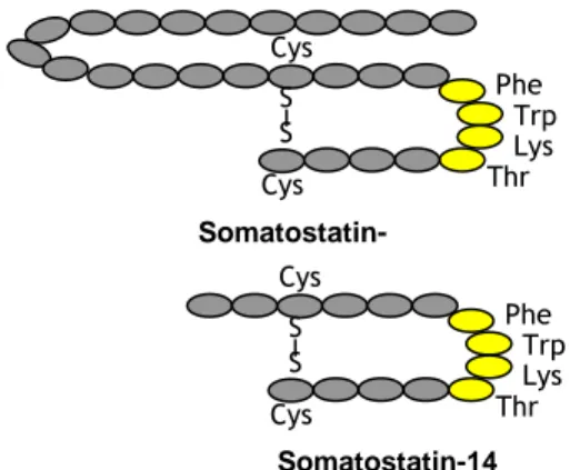
SST is encoded by a single gene and it mature form is obtained by two subsequent proteolitic cleavages that bring from the primitive pre-prosomatostatin to the pre-prosomatostatin that is newly modified in the two final and mature products: 14 amino acids SST (SST-14) and 28 amino acids SST (SST-28)28 (Figure 1). Pharmacological studies reveals that SST-14 and SST-28 binds to SSTR1-5 receptors with high affinity, but SST-28 has a 10 times higher affinity for SSTR-5 respect to SST-14.
SST functions were discovered in 1973 by Guillemin and Gerich84. It was isolated in the hypothalamus and researchers initially hypothesized that the only own function was to inhibit growth hormone release by pituitary gland. Subsequently, SST was discovered in most brain regions and in peripheral organs, but SST is typically contained in neurons or endocrine-like cells, such as central and peripheral nervous systems, in the endocrine pancreas and the gut, and in small number in the thyroid, adrenals, submandibular glands, kidney, prostate and placenta, where it executes different functions7.
2.2
Somatostatin functions.
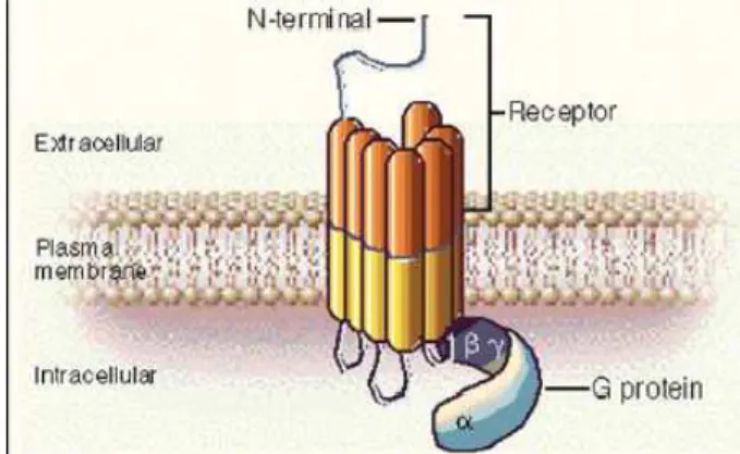
SST action is executed when it binds to it specific receptor. There are five SSTRs subtypes, composed by seven transmembrane domains and coupled by G-protein (Figure 2).
Figure 1: SST-28 and SST-14. The two forms have the same aminoacidic
sequence, with the length is the only exceptions. The yellow dots represent the pharmacophore.
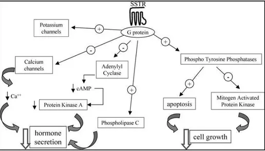
When the SST-SSTR binding occurs, the second messenger systems is activated. This system includes: inhibition of Adenylyl Cyclase, inhibition of calcium channels and activation of phospholipase-c. All these processes bring to the inhibition of the hormone secretion. Moreover, phosphotyrosine phosphatases (PTPs) are activated by SST. PTPs can activate two different pathways: the first shows as consequence cytostatic effects by the inhibition of the Mitogen Activated Protein Kinase, the second leds to the apoptosis activation, having as last event cell death85,7 (Figure 3).
Because SST is involved in the inhibition of hormone secretion and brings to cytostatic or cytotoxic effects and because SSTRs are over-expressed in different types of tumors, SST and it activated pathways are the most studied processes for cancer treatment and diagnosis.
2.3
Somatostatin and its analogues.
2.3.1
Agonists.
When a SST-analogue with an agonist behaviour binds to it specific SSTR subtype, G-protein are activated by phosphorilation through protein kinases
Somatostatin-14 Thr S S Cys Cys Lys Trp Phe Thr S S Cys Cys Lys Trp Phe Somatostatin-28
A/C and G-protein coupled receptors (GPCR) kinases and the mechanism that derives is the internalisation of the SSTR-agonist complex.
Figure 2: G-protein coupled receptor94.
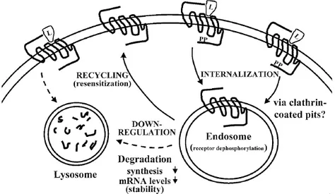
Internalisation occurs with the formation of clathrin-coated pits (involving β-arrestins). The internalised complex is then channel to endosomes, where dephosphorilation occurs. Finally, the receptors are recycled and go back to the membrane as functional receptors28 (Figure 4). GPCR down-regulation results from lysozomal degradation of intracellular receptors, this led to a decrease of mRNA and protein synthesis.
Due to its actions, SST started to be studied in particular for the therapy of SSTR-expressing tumors. Native SST has a very short half life in human serum (about 3 minutes), because it is rapidly attacked and degraded by endogen reducing agent as glutathione oxidase, thioredoxin reductase or basic and nucleophilic agents, thus preventing it application to the clinic. In order to get over this difficulty, SST-analogs with different affinity to each SSTR subtypes and with different behaviour were synthesized.
Figure 3: SST mechanism of actions92.
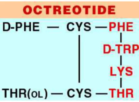
The first and well known SST-analogue is Octreotide, also named SMS 201-995 or Sandostatine®. It is a cyclic octapeptide that maintain the SST pharmacophore together with a protection against degradation, that are represented by the presence of a D-Phe residue at N-terminus together with an alcoholic residue (Thr-ol) at C-terminus. Metabolic stability was given by the presence of a disulphide bridge between Cys2-Cys7 (Figure 5). Octreotide has an half life of 117 minutes in human serum, a great affinity for SSTR-2 and a moderate affinity for SSTR-3/5 (Table 1).
Later, new peptides with different affinity profiles and different behaviour were synthesised, but Octreotide is still considered the milestone in the SST-analogues history. For example, other SST-analogues that are now in clinical trials are Lanreotide and Vapreotide but, even in these cases the main limiting factor remains the affinity to each SSTR subtypes. Because SST has a large spectra of action and in particular for it antisecretory action, the potential of SST analogues in cancer treatment is very high, especially for
those tumors such as pituitary tumors, gastrointestinal tract, acromegaly, prevention and treatment of pancreatic surgery complications.
Figure 4: Rapresentation of intracellular routing of GPCRs after agonist
activation (L= ligand, PP= phosphate group)28.
Even if these drugs are very promising, an important problem to underline is the typical behaviour of SSTRs. In fact, this receptors have the particularity to create homo- and heterodimers with the receptors of other system (dopamininergic and opioid receptors), becoming very difficult to predict the therapeutic potential of SST-analogues.
Compound hsstr1 hsstr2 hsstr3 hsstr4 hsstr5 SS-14 1.1 1.3 1.6 0.53 0.9 SS-28 2.2 4.1 6.1 1.1 0.07 Octreotide > 1000 2.1 4.4 > 1000 5.6 Lanreotide > 1000 1.8 43 66 0.62 Vapreotide > 1000 5.4 31 45 0.7
Table 1. Binding affinities (Ki, nM) to hSSTR of clinically used
Figure 5: Octreotide. Pharmacophore corresponds to the red aminoacids.
2.4
Somatostatin analogues and cancer.
Due to it antisecretory activity, the therapeutic potential of SST in cancer treatment is multiple.
Different neoplasms express a particular SSTR or co-express more than one SSTR subtypes. This was abundantly studied during the last twenty years. For example, pituitary adenomas express SSTR-2 and -5, receptors that are important in inhibiting the excessive GH secretion in acromegaly. Since GH stimulates production of IGF-I, that has been implicated in promoting cancer87, SST-analogues may retard additional cancers by decreasing systemic GH/IGF levels88.
Another important mechanism of anti-neoplastic action of these drugs appears to be the inhibition of neoangiogenesis.
Angiogenesis is a fundamental process in the context of tumourgrowth, and one of the main factors involved in the appearanceof new tumour vessels is vascular endothelial growth factor (VEGF). SST-analogues inhibit the production and secretion of many angiogenic factors89. It has been demonstrated thatoctreotide induced inhibition of angiogenesis by a process that is G-protein, calcium- and cAMP dependent and is protein
protein-kinase C (PKC) and PTP independent90. SSTRs expression has been demonstrated in peritumoral vessels in different tumourtypes, and it appears to be unrelated to the receptor expressionin the tumour cells24. SSTR2 is expressed when the endothelial cells begin growing91. In fact, SSTR2 gene is expressedon proliferating angiogenic vessels.These data were confirmed by immunohistochemistry and in vivo scintigraphy. This is the reason, because SSTR2 may be a specific target for anti-angiogenic therapy with SSTR2-binding SST conjugated to radioisotopes (section 2.5)or cytotoxic agents. Preclinical studies on the potentiality of SST analogues in inhibiting angiogenesis were performed in different experimental models, including the chicken chorioallantoic membrane model, the human umbilical vein endothelial cell proliferation model and the human placental vein angiogenesis model.
Recent studies have focused on SSTRs signalling and its effects on cell growth, because it is recognized that SST has activity as endogenous antiproliferative agent in many different experimental tumor models both in vivo and in vitro92. However, these effects, that are highly significant in preclinical study, become much more questionable when the data are translated to clinical trials, so that the research is still moving further and a significant progress is doing in understanding the mechanism by witch SSTR activation may lead to cytostatic or apoptotic effects. In particular, it is now accepted that, the main transduction system involved in the antiproliferative activity of SST is represented by the activation of a subset of phosphotyrosine phosphotases92,92.
Therefore, the effects of SST on tumor cell growth may take place at different levels93: directly blocking the cell cycle progression through the binding to SSTRs expressed on cell and the activation of PTPs, and
indirectly through the modulation of tumor growth mediated by the inhibition of the production of GF, or via an antiangiogenic effect.
Figure 6: Somatostatin receptors characteristics94.
Frequently, the activation of SSTRs causes cytostatic effects, with cell’s block in the G1 phase. The role of SST as an endogenous regulator of cell cycle is a recognised activity and, using different in vitro and in vivo experimental models, all the five SSTR subtypes were reported to induce arrest of cell proliferation94 or induce apoptosis88 too.
Different SSTRs (SSTR1, SSTR2, SSTR4and SSTR5) have been implicated in vitro in the G1–G0cell cycle blockade, while the apoptotic effect of SST being mediatedthrough SSTR3 and less throughSSTR2.
SSTR-3 was shown to increase wild type p53 through a dephosphorylation-dependent conformational change and to induce Bax, but not p21, in apoptosis caused by octreotide treatment of Chinese Hamster Ovary cancer (CHO) cell line stably transfected with human-SSTRs and MCF-7 human breast adenocarcinoma cells95, where transient G2/M blockadeandapoptosis were demonstrated96. In these cells, octreotide had cytotoxic effects leading toapoptosis, with a rapid time-dependent induction of wild-typep53.
In human pancreatic adenocarcinoma, it was demonstrate that, during the tumoral progression, the cells lose the ability to express SSTR2 but, reintroducing this receptor into the pancreatic cancer cells by stable expression, leadsto a constitutive activation of theSSTR2 gene and evokes a negative feedback loop, inhibiting cellproliferation. This may suggest that SSTR2 gene transfer might be considered as a possible novel therapy for pancreatic cancer 97 (Figure 6; Table 2).
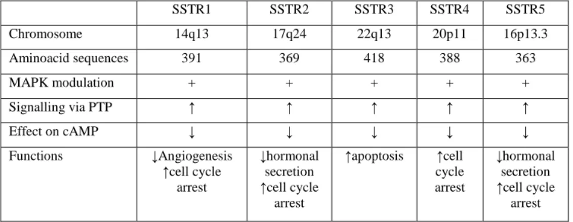
SSTR1 SSTR2 SSTR3 SSTR4 SSTR5 Chromosome 14q13 17q24 22q13 20p11 16p13.3 Aminoacid sequences 391 369 418 388 363 MAPK modulation + + + + + Signalling via PTP ↑ ↑ ↑ ↑ ↑ Effect on cAMP ↓ ↓ ↓ ↓ ↓ Functions ↓Angiogenesis ↑cell cycle arrest ↓hormonal secretion ↑cell cycle arrest ↑apoptosis ↑cell cycle arrest ↓hormonal secretion ↑cell cycle arrest
Table 2: Somatostatin receptors characteristics (chromosomal localisation of
the genes encoding the five SSTR subtypes; aminoacids structure; G-protein coupling and activation; effect on cAMP; signalling via tyrosine phosphatases and receptor-specific functions)98.
Even if, the cytotoxic and cytostatic effects of SST analogues were abundantly studied in vitro, currently, no consistent results were proved in patients to inhibit cell proliferation or metastasis in NETs. In fact, treatment with SST-analogues has produced variable results in clinical practice especially when used as single agent. Poor results were seen with rapidly progressive tumors, with high proliferation capacity despite the presence of SSTRs99. Conversely, well-differentiated tumors such as mid-gut carcinoids respond well, with stabilization of tumor growth over many years.
What is well established is that SST can decrease in tumor growth from indirect effects, through suppression of synthesis and secretion of GFs and
some hormones (insulin, prolactin, IGF-1, EGF, TGF-α, gastrin, cholecystokinin and GH). An important example is constituted by IGF-1, that plays as modulator of many neoplasms, because SST analogues suppress the GH-IGF-1 axis by both central and peripheral mechanisms.
Experimental studies of combinations of octreotide with antimitotic drugs resulted in slightly additive actions100,101.
Furthermore, somatostatin analogs have also been used as carriers to deliver cytotoxic agents to cancer cells.
2.5
Radiolabeled Somatostatin-analogs.
The presence of SSTRs in different neoplasm can be exploited, not only in long term therapy with not-cytotoxic SST-analogs, but also in tumor diagnosis and therapy with radioactive analogues. The use radiolabeled SST analogues derives from the necessity to obtain consistent inhibition of cell proliferation or metastasis in NETs, exploiting cytotoxic effects due to radiation.
Thus, basic knowledge of SSTR subtype profiles in different neoplasm, the affinity profile, binding/internalization of SST analogues-carried radionuclides features are critical for the evaluation of the potential usefulness of receptor-mediated radiotherapy. The uptake of radiolabeled SST-analogues depends on the number of SSTR on the cell membrane, on the internalization rate and on the of the recycling time and mass of the radiolabeled peptide.
In nuclear medicine, two are the techniques for diagnosis using radionuclides: γ-scintigraphy and positron emission tomography (PET). The main differences between the two technologies consisting in the different emission spectra of the radionuclides exploited. In fact, it is necessary to
adopt radiopharmaceuticals containing a radionuclides that emits γ-radiation with an energy between 100-250 KeV in γ-scintigraphy and the required instrumentations are γ camera and SPECT camera. PET camera requires pharmaceuticals radiolabeled with β+ emitting radionuclides (positron emission) and a PET or PET-CT camera.
The first radiolabeled SST-analogue was [123I-Tyr3]-octreotide, a radiopharmaceutical with γ-emission102. The radiopeptide was used for in vivo localisation of tumors, but although the pharmacological profile was optimal with a very high SSTR2 affinity (IC50= 2.0±0.7 nM) and a very high internalisation rate in tumoral cells, the radiopharmaceutical was turned out be useful as a diagnostic tool. This was due to the lipophilic feature of [123 I-Tyr3]-octreotide, that bring to an hepatobiliary exctretion, with the consequence that abdomen imaging results with low sensitivity.
Subsequently, a first chelator was united to octreotide: DTPA (Diethylentriaminepentaacetic acid). This chelator allows the radiolabelling with 111-Indium, giving a hydrophilic feature to the radiopeptide and a renal excretion. 111In-DTPA-Octreotide gave a better biodistribution profile, even if affinity for SSTR2 was reduced (IC50= 22±3.6 nM). 111 In-DTPA-Octreotide was the first commercialized radiopeptide for diagnostic imaging in nuclear medicine (Octreoscan®, 111In-pentereotide, Millinckrodt Med., St. Louis, MO, USA).
Subsequently, new strategies were developed and research was moved to find new solutions to allow the radiolabelling of SST-analogues with other radionucides, both for γ-scintigraphy and PET.
Particularly, DTPA was substituted by HYNIC ( 2-hydrazinonicotinic acid) to be possible radiolabeling with 99mTc. This radiometal has a very low cost production and a short half life (6 h). At moment, there are two different successful 99mTc-radiolabeled SST-analogue: these radioconjugates
demonstrated good results if compared to Octreoscan®. These are 99mTc-N4 -Tyr3-Octreotide and 99mTc-HYNIC-Tyr3-Octreotide (HYNIC-TOC). Tyr3 -Octreotide is synthesised in a more recent times compared to octreotide and presents the octreotide skeleton, but of the third aminoacid is a Tyrosine instead of a Phenilalanine (Phe is replaced by a Tyr).
SST-analogues were radiolabeled with 18F too. In this case, inconveniences are a rapid washout of the radiopeptide, an high liver uptake and a consequent not clear imaging of the abdominal area103. Another radiometal with β+ emission is 64Cu, that is also used for SST-analogues radiolabelling and from preclinical data, particularly biodistribution profile on animal model seems favourable104.
The introduction of the macrocyclic chelator DOTA (1,4,10-tetraazacyclododecane-1,4,7,10-tetraacetic acid) gave advances in this technology. The obtained metal complex is kinetically and thermodynamically stable. This radiometal is encapsulated and embedded inside the macrocyclic cage which protects it from the attack of competing ligands present in the human tissues and the body fluids. DOTA chelator makes possible the radiolabelling with different radiometals, such as: 111In, 90/86
Y, 177Lu and 66/67/68Ga. 68Ga-Tyr3-octreotide (68Ga-DOTATOC) is a very successful radiopeptide for tumor imaging with PET scan, in fact the radiopharmaceutical offers a very high quality imaging and a very high tumor-to-background ratio105,104.
Consequence of peptide receptor mediated scintigraphy was peptide receptor mediated radionuclide therapy (PRRT). To have a successful PRRT, four requirements are necessary. These are:
• The number of receptors on tumoral cells have to be high in number.
• The radionuclides have to emit radiations with an high linear energy transfer (LET), in order to destroy the tumor tissue.
• The whole metal-chelator-peptide complex must have suitable pharmacokinetics.
There are three types of therapeutic radionuclides: α-emitters, β-emitters and Auger-electron emitters, each one with different range of energy deposition and LET properties (Figure 7). Radionuclides with β emission are the most used in the current clinical practise for therapeutic applications. The advantage for β–emitters is that it is not required to target all the tumoral cells for their killing (crossfire effect, low LET). There are different type of β-emitters radionuclides and are classified considering their energy of emission. So, there are β-emitters radionuclides with a low range (mean range <200µm, as 177Lu), with a medium range (mean range between 200µm and less than 1 mm, as 67Cu, 153Sm) and high range (mean range >1 mm, as 90
Y). Moreover, for those cases where γ-emissions are present too, biodistribution profile is reproducible, for example 177Lu has a γ-emissions (160-202 KeV), while Y-90 is a pure β—emitter.
α-emitters particles emits with an high LET over a path length of 3-4 cells diameters.
Auger-electron emitters have an electron energy between 10 KeV and several eV. This type of radionuclides have a very short effect range (several nanometers) and high toxicity, giving them ideal for small cluster metastatic cells therapy.
The first widely used radiolabelled SST-analogues for PRRT was 90 Y-DOTA-Tyr3-octreotide (90Y-DOTATOC)106,107. Studies have demonstrated that 90Y-DOTA-Tyr3-octreotide is successful for the therapy of metastatic
NETs, in particular in pancreatic tumors and carcinoids, but some evidences are described for other NETs too108.
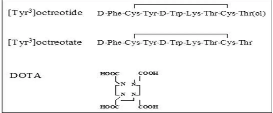
Another radiopharmaceutical used for PRRT is DOTA-Tyr3-Thr8-octreotide (DOTATATE), which differs from DOTA-Tyr3-octreotide by a aminoacidic modification in the aminoacid number 8 (the alcoholic molecule was take off: Thr (ol) became Thr) (Figure 8). The peptide retains high affinity for human SSTR2 as demonstrated by YIII-DOTATATE binding studies (7-fold higher compared to YIII-DOTATOC) (Table 3).
Despite this higher affinity for SSTR2, biodistribution profiles of 111 In-DOTATOC and 111In-DOTATATE are very similar. Further, preclinical studies on nude mice bearing SSTR2-xenograft tumor demonstrated that 177
Lu- has performed better compared to 90Y in small or medium tumors. This behaviour is related to Lutetium-177 maximum tissue range of 2 mm compared to 11 mm of Yttrium-90. Additionally, 177Lu emits γ-radiation, giving the possibility to obtain the biodistribution profile after the PRRT.
Compound hSSTR1 hSSTR2 hSSTR 3 hSSTR4 hSSTR5 SS-28 5.2±0.3 2.7±0.3 7.7±0.9 5.6±0.4 4.0±0.3 InIII-DTPA-octreotide > 10,000 22±3.6 182±13 >1,000 237±52 YIII-DOTA-OC >10,000 20±2 27±8 >10,000 57±22 YIII-DOTA-TOC >10,000 11±1.7 389±135 >10,000 114±29 YIII-DOTA-TATE >10,000 1.6±0.4 >1,000 523±239 187±50 YIII-DOTA-Lanreotide >10,000 23±5 290±105 >10,000 16±3.4 YIII-DOTA-Vapreotide >10,000 12±2 102±25 778±225 20±2.3
Table 3:Affinity profiles for human SSTR1-5 of a series of Somatostatin
analogues (values are expressed as IC50 ± SEM, in nM)109.
Summarizing, it is important to underline that when a chelator is added to the peptide, some changing in affinities profile are obtained. For example, it can be present a loss of affinity for a particular subtype receptor, especially for
SSTR5, but also for SSTR3 and SSTR2. Not only, changing in affinity profiles are obtained when a metal is present and a particular metal instead of another.
Although the in vivo metabolism, excretion pathway and retention times of a molecule are important parameters for its evaluation as a new tracer for diagnosis or therapy, there is no doubt that the in vitro characterization of the receptor binding affinity of such a molecule is crucial information, particularly nowadays when several studies on the receptor expression pattern on tumors are available.
Figure 7: Penetration of Particulate and Electromagnetic Radiation.
Therefore, the conclusion from the informations presented here is that the efficacy of the currently used radiolabeled somatostatin analogues derives mainly from their moderate to high affinity for SSTR2, the receptor with the widest distribution among the SSTR family.
Epidermis Dermis Subcutaneous tissue
Alpha particles
Betaparticles
Gamma rays
Having complex structures, in which every component influences the biological efficacy in vivo, it is still a challenge to find the best metal-chelator-SST analogue structure with not only suitable pharmacological properties, but also with optimal pharmacokinetics and pharmacodynamics.
Figure 8: Structures of the somatostatin analogues Tyr3-octreotide and Tyr3 -octreotate and of the chelator DOTA110.
3
Strategies to improve pharmacological profile of
radiolabeled Somatostatin analogues.
3.1
Characteristics of Somatostatin analogues for Nuclear
Medicine.
Summarizing, the factors limiting therapeutic efficacy of SST analogues are their selectivity for a particular SSTR, requiring a precise determination of receptor subtypes expression in tumor tissue before therapy and their partial answer in therapeutic protocols used in clinic until now, consisting in their antisecretory activity. On the other hand, native SST with great affinity for all five SST receptors, are not practise in clinical, because SST has a short
plasma half-life (1–3 minutes in humans), giving necessary continues infusion on treated patients.
Mainly for this reasons and for the unsuccessful results in therapeutic protocols in the last years oncologic research was devoted to the development and synthesis of new pharmaceuticals which, once radiolabeled and injected in vivo, can be used in for imaging and therapy of tumours. For this purpose, labelled molecules must possess high selectivity towards tumour cells or tissues to reach efficient targeting activity.
At moment, in nuclear medicine the current SST analogues used both for diagnosis and therapy shows good affinity for SSTR2 and a moderate affinity for SSTR3-5 (Table 4).
SST-14 served as model for all the analogues synthesized over the years and all the radiolabeled somatostatin based compounds used in clinical applications are derivates of octreotide. Octreotide have a critical position between two aminoacids, that is maintained from the native molecule and, if it is replaced, it could give modification in the biological properties of the molecule. This sequence corresponds to the Lys5- D-Thr6 where a β-turn is present. The main modifications present in the SST analogues in clinical practise are: a changing in position 3 (Phe >Tyr) for vapreotide, lanreotide and TOC and a substitution of the sixth aminoacid (Thr>Val) for lanreotide and vapreotide.
Other modifications could are present at C-terminus and at N-terminus of the peptide and of course each of these modifications induce changing in the affinity profiles (Table 4).
Even if some important results are obtained by PRRT, new SST analogues with high affinity for a particular SSTR subtype as SSTR3 and SSTR5 or a pan-SST analogue (analogues that possess high affinity for all the subtype
receptors) are required, so that PRRT with maximal therapeutic effects can be performed on patients. Compound hSSTR 1 hSSTR 2 hSSTR 3 hSSTR 4 hSSTR 5 SST-28 5.2±0.3 2.7±0.3 7.7±0.9 5.6±0.4 4.0±0.3 InIII-DTPA-OC > 10,000 22±3.6 182±13 > 1,000 237±52 YIII-DOTA-OC >10,000 20±2 27±8 >10,000 57±22 YIII-DOTA-TOC >10,000 11±1.7 389±135 >10,000 114±29 YIII-DOTA-TATE >10,000 1.6±0.4 >1,000 523±239 187±50 YIII-DOTA-Lanreotide >10,000 23±5 290±105 >10,000 16±3.4 YIII-DOTA-Vapreotide >10,000 12±2 102±25 778±225 20±2.3
Table 4: Affinity profiles for human SSTR1-5 of a series of somatostatin
analogues (values are expressed as IC50 ± SEM, in nM)109.
3.2 Modification of pharmacokinetics/pharmacodynamics of
radiolabeled Somatostatin analogues.
Affinity is a crucial parameter for a new peptide development, in fact this parameter corresponds to the tendency of a drug to bind to a binding site including specific receptor. Affinity can be evaluated at equilibrium by the affinity or association constant (sometimes given the symbol K), which is the reciprocal of the dissociation constant (KD), or it can be measured in pD2 units, that corresponds to the negative logarithm of the concentration of the agonist that produce half of the maximal response.
So, the molar concentration of a drug inducing 50% of the maximal response is considered the affinity between a drug to it proper receptor (IC50) and it is expressed as the negative logarithm of the concentration of the agonist that produce half of the maximal response.
Affinity studies are the first step of a very long study, if a new drug is the target of the research.
If affinity profile is favourable, the next step will be performed. In nuclear medicine, it is usual that the new peptide is linked to a chelator molecule that allows the radiolabeling with different radiometal so, it is important to perform affinity assay even after the chelator linking, because this molecule can induce some modifications in binding assaysresults. After this passage, radiolabeling and stability tests are to be performed. Another important step consisting in the measurement of human serum stability and in vitro assays on an adapt cell line. This last step is important to establish an eventually biological effect of the new radiopeptide. After this phase, experiments will be performed on animal models. Objective of this phase is to establish the biodistribution profile of the radiopharmaceutical, if the radiopharmaceutical goes to it target, the washout, if there are any secondary effects (kidney burning, for example).
After all these evaluations, a clinical phase on patient could be started. Of course, it could be that a very promising radiopharmaceutical from preclinical data gives unlucky results on men, infect, in vivo studies on animals can not predict the drug behaviour in humans. This is the case of 123
I-[3-iodo-Tyr3]-octreotide where, despite the very spectacular first results, the radiopharmaceutical was abandoned, because of the high hepatobiliary excretion, with the consequence that abdomen imaging results with low sensitivity.
There are a lot of different SST analogues, each one with a different affinity and different behaviour and, for researchers, the purpose still remains to design new pharmaceuticals with a more elective action, a better affinity for a particular SSTR subtype and a prolonged action replacing the others in the current trade.
3.3
Modulation in hydrophilicity/lipophilicity.
Clearance refers to the volume of blood or plasma cleared of a drug in a unit time. It is defined as the product of the volume of distribution rate constant and only unbound proteins are available for clearance. Physiologic excretion pathway of the drugs are mainly through liver or kidney, it depend by the characteristic of the pharmaceutical.
For radiopharmaceuticals, the excretion pathway is of crucial importance for both the diagnostic performances (high tumor/background ratios, thus signal intensity in diagnostic by PET or SPECT) and low toxicity profile in therapy. Regarding SST analogues, particularly the DOTA chelated analogues, the hydrophilic characteristic of these peptides determine a predominant renal clearance making the kidney the dose limiting toxicity organ for PRRT. For this reason, efforts are directed to increase lypophilicity of the compounds. Of course, these modifications have not to influence the affinity profile. The current radiolabeled SST analogues, 111In/90Y-DOTA-octreotide and octreotide derivates demonstrated an increased metabolic clearance with a gallbladder accumulation. The same was observed in 68Ga-DOTA-octreotide derivates, where the abdominal uptake did not demonstrate any refinement. This problem can be crossed adding an hydrophilic spacer, hoping that pharmacological performance will be maintained.
3.4
Lowering the kidney uptake .
The treatment of patients with SSTR positive tumours with peptide receptor radionuclide therapy (PRRT) has with no doubt beneficial effects. One of the
limits of this therapy is represented by kidney burning. In fact, the radiolabelled peptides are rapidly cleared via the glomeruli in the kidneys into the urine, but a low percentage is reabsorbed and retained in the cortical proximal tubules. After glomerular filtration a fraction of the administered peptides is internalized via endocytic receptors; megalin is responsible to this process. Transfer of the radiopeptides to the lysosomes is followed by degradation of the peptide, after which aminoacid chelate conjugates are trapped in the lysosomes of the tubular cells, delivering a high radiation dose to the renal cortex during PRRT. In PRRT, the maximum tolerated dose in patients is not exactly known, but dosimetric studies applying the principle of the biological equivalent dose (correcting for the effect of dose fractionation) suggest that a dose of about 37 Gy is the threshold for development of kidney toxicity. This threshold is lower when risk factors for development of renal damage exist: age over 60 years, hypertension, diabetes mellitus and previous chemotherapy, but the upper limit for kidney safety is fixed to 27 Gy111.
In Erasmus Medical Centre in Rotterdam, co-infusion of lysine and arginine (Lys/Arg) has become a standard procedure during PRRT with 177Lu- or 90 Y-labelled somatostatin analogues, reducing the renal retention of the radiopeptides by approximately 35%112.
3.5
Targeting the cell nucleus.
To have a successful PRRT, there are some parameters that a clinician have to consider. Of course the presence of an high density of SSTRs on tumoral cells is the first one, but it is not the unique. In fact it has to consider if the radiopeptide has a very high affinity for the expressed SSTR subtype and if the SSTR are functional. After these considerations, others important
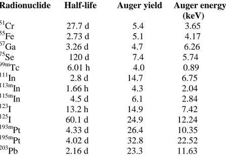
reflections have to be done: is the receptor-peptide internalised into the tumoral cells? And in case of affirmative answer: how long is the radiopharmaceutical retention time in cells? Retention time is a very crucial parameter, because if it is very long, the radiopharmaceutical have a maximal therapeutic effect. In PRRT, the radiometals mainly used are 90Y and 177Lu, these shows respectively an intermediate and high energy, making fruitful a therapy on medium or large tumor diameter, where the cytotoxic effect is due to the cross-fire effect. The problem is that β-emitters do not show any effectiveness on small metastasis cell cluster, but the problem can be easily surpassed using radiometals with different characteristics. The ideal radioemitters are Auger-electron emitters, because of their high toxicity and short effect range (several nanometers).
There are some radionuclides that emits Auger electrons (Table 5), some of them are currently used in the routine clinical practise. Auger electron emitters, inducing DNA damage by indirect mechanism, led to cell death. Moreover, a prolonged retention of the radiopeptide in the target cells will bring to an unquestionable success of PRRT.
Auger-electron emitter antibodies conjugates demonstrated positive results for the therapy of different pathologies, in particular for B-cell lymphomas113 and some preclinicals studies were also started for SST analogues and the results seem encouraging.
Finally, it is important to point the attention to those SST analogues that demonstrated an antagonist behaviour, where their importance in PRRT seems to be controversial.
The rationale is that agonists, after high-affinity binding to the receptor, usually trigger internalization of the ligand–receptor complex114. The internalization process is the basis for an efficient accumulation of the radioligand in a cell over time and it has been considered a crucial step in the