Università degli Studi di Pisa
FACOLTA’ DI MEDICINA E CHIRURGIA
DOTTORATO DI RICERCA IN CHIRURGIA,
BIOTECNOLOGIE E IMMUNOLOGIA DEI TRAPIANTI
Tesi di Dottorato di Ricerca
(Settore Scientifico Disciplinare MED 18)
Applications of Robotic Surgery
in Organ Transplantations -
Applicazioni della Chirurgia
Robotica nei Trapianti d’Organo
Relatore:
Chiar.mo Prof. Ugo Boggi
Presidente:
Chiar.mo Prof. Franco Mosca
Candidato:
Dott. Fabio Vistoli
Index
Abstract
pag.
3
Introduction
pag.
4
Aim of the study
pag.
8
Materials e methods
pag.
9
Results
pag.
25
Discussion
pag.
32
Conclusion
pag.
45
Tables
pag.
46
Figures
pag.
50
References
pag.
55
Abstract
Introduction: Surgical complications are a major disincentive to transplantation despite the undisputed benefits of restored organ function. Robot-assisted surgery is the new technological advance of the recent years. The da Vinci surgical system, a computer assisted electromechanical device, provides the unique opportunity to test whether laparoscopy can reduce the morbidity in the setting of transplantation. We evaluate the feasibility and safety of this new surgical technique in living kidney donation, kidney transplantation and pancreas transplantation.
Materials and methods: Robot assisted living donor nephrectomy was performed on 2 subjects. The immediate post-operative courses for these donors, and their respective recipients, were compared with those of 20 laparoscopic living donor nephrectomies, performed in the same period. Moreover robot assisted kidney transplantation was performed on 2 living kidney recipients and robot assisted laparoscopic pancreas transplantation was performed in further 3 recipients, performing a pancreas after kidney transplant, a simultaneous pancreas kidney transplantation, and a pancreas transplant alone. The grafting procedures were carried out through an 11 mm optic port, two 8 mm operative ports, and a 7 cm incision (midline for pancreas and suprapubic for kidney). The latter was used to introduce the grafts, handle vascular crossclamping, and create pancreas exocrine drainage into the jejunum or uretero-vescical anastomosis in case of kidney transplant.
Results: No significant differences between the two donor groups with respect to age, gender, body mass index or renal vasculature were found. The average operative times and the warm ischaemia times were similar. There was no conversion to open surgery in both groups. The estimated blood loss was slight. Following nephrectomy, no complication occurred. The average duration of hospitalization was similar. The estimated creatinine clearance rate was equivalent for all donors, at 5 days and 1 month after nephrectomy. All kidneys started functioning immediately after the transplantation. The mean recipient estimated creatinine clearance was similar. Two kidneys, one from a 56-year-old mother to her 37-year-old daughter and one from a 49-year-old sister to her 48-49-year-old brother were transplanted laparoscopically using the DaVinci surgical system. Vascular anastomoses were carried out through a total of three additional ports. Surgery lasted 154 and 148 min, including 51 and 39 min of warm ischemia of the graft, respectively. Urine production started immediately after graft reperfusion. Renal function remains optimal at the longest follow-up of 10 and 3 months. The two solitary pancreas transplants lasted 3 and 5 hrs, respectively, the simultaenous pancreas kidney transplantation lasted 8 hrs. Mean warm ischemia time of the pancreas graft was 34 minutes. All pancreata functioned immediately, making their recipients insulin-independent. The kidney graft, revascularized after 35 minutes of warm ischemia, also functioned immediately and fully. No patient had complications during or after surgery; mean hospital stay was 21 ±5 days. After a mean follow-up period of 3.7 months, all recipients are alive with optimal graft function.
Conclusion: Robotic assisted living donor nephrectomies were associated with no morbidity among donors, in which both the operative and warm ischaemia times were no longer duration, moreover had no observable adverse effects upon short-term graft function. On the other hand the daVinci surgical system allows the performance of kidney transplantation under optimal operative conditions. Further experience is needed, but it is likely that solid organ transplantation will not remain immune to robotics. We have also shown the feasibility of laparoscopic robot-assisted solitary pancreas and simultaneous pancreas and kidney transplantation. If the safety and feasibility of this procedure can be confirmed in larger series, laparoscopic robot assisted pancreas transplantation could become a new option for diabetics needing beta-cell replacement.
Introduction
Live kidney donation is an important alternative for patients with end-stage renal disease. Renal transplantation from living donors confers several advantages as compared with dialysis and transplantation from deceased donors, including improved longer-term patient survival, better quality of life, immediate functioning of the transplant, better transplant survival, and the possibility of transplanting pre-emptively (1–9).
To date, the health of live kidney donors at longterm follow-up is good, and the procedure is considered to be safe (2). Currently, attention to donor wellbeing has become a priority, and therefore the surgical technique must be optimized continually. The surgical practice has evolved from the open lumbotomy, through mini-incision muscle-splitting open (mini-incision open donor nephrectomy; MIDN), to minimally invasive laparoscopic techniques. There are different minimally invasive techniques, including standard laparoscopic, hand-assisted laparoscopic, hand-assisted retroperitoneoscopic, pure retroperitoneoscopic, and robotassisted live donor nephrectomy. At present, these minimally invasive techniques are being subjected to clinical trials focusing on surgical outcome, quality of life, costs, long-term follow-up, and morbidity of donor, recipient, and graft.
Other issues that surgeons encounter with live kidney donation are related to the type of kidney to select, the factors to be reckoned while dealing with obese donors, and the strategies to be adapted while approaching donors with multiple arteries and veins. Many centers still restrict donor nephrectomy to relatively younger, normal weight donors, categorized as American Society of Anaesthesiologists group I. They tend to choose the left kidney, with simple
renovascular anatomy. Nowadays, donors with isolated abnormalities, i.e. hypertension or obesity, can also be accepted for live kidney donation, as longterm renal function and health is good.
Open nephrectomy is the accepted standard procedure for live donor kidney removal (10), but attempts are now being made to duplicate the outcomes of this traditional open donation method with less invasive surgical techniques. Laparoscopic nephrectomy has recently gained popularity as it provides the potential advantages of decreased post-operative pain, shorter hospital stay and faster recuperation (11, 12). However, the surgical techniques required for this procedure are demanding, extremely difficult to master (13) and, consequently, have been adopted by only a few centres. Modifications such as hand-assisted techniques, and more recently, robotic assistance, have been suggested to improve surgical outcomes.
The robotic system provides steady imagery with threedimensional visualization and additional degrees of freedom that mimic human wrist motions, and eliminate both exaggerated hand motions and fine tremors (14, 15). To our knowledge, no reports are currently available regarding laparoscopic donor nephrectomy performed completely with the assistance of a robot, especially without the conjunction of a hand-assisted procedure (16), and there have been no observations made on either donor safety or the quality of the recovered organ with this approach. For robotic assisted living donor nephrectomy to become a viable option for procuring kidneys for renal transplantation, it is essential that the donor suffers no additional morbidity and that the prognosis for recipients should be at least equivalent to the ‘gold standard’ of open nephrectomy. In this regard, open live-donor nephrectomy sihas proven to be very safe for donors, with reported mortality rates of between 0.03–0.06%, and
the transplanted kidney is usually of excellent quality following this procedure (10).
The technique for kidney transplantation (KT) has evolved little since 1950s (17). Rosales et al. recently reported on a patient undergoing successful laparoscopic KT (18). Although this case report shows that a kidney can be transplanted laparoscopically, it does not demonstrate that this operation can be reliably duplicated by the average transplant surgeon. Laparoscopy is indeed used infrequently in operations requiring multiple vascular anastomosis because of loss of hand–eye coordination, use of long instruments amplifying natural surgeon’s tremor and carrying a fulcrum effect, and poor ergonomy causing surgeon’s fatigue (19). The daVinci™ SiHD surgical system (dVss)
(Intuitive Surgical, Sunnyvale, CA, USA) is a computer-assisted electromechanical device acting as a remote telepresence manipulator controlled by a surgeon (20). The dVss provides the operating surgeon with 3D high-definition view including 10 to 15·magnification, fully restoring hand–eye coordination; it employs wristed instruments, with seven degrees of freedom, and it tracks surgeon’s movements 1,300 times/s, providing for tremor filtration and scaled motion. Furthermore, the surgeon simultaneously drives the binocular endoscope, achieving steady view, and toggles between three operative arms (21). These features translate into significant operative advantage, especially when the operative field is deep and narrow, and when fine dissection and microsuturing are required (21). The dVss is currently used in urology, for radical prostatectomy, pyeloplasty, and ureteral reimplantation (20), as well as in vascular surgery for coronary artery by-pass (22), repair of renal artery aneurysm (19), and repair of abdominal aorta (23). Thus, it would seem that the dVss could facilitate the implementation of laparoscopy in KT.
Vascularized pancreas transplantation is the only treatment that routinely and consistently restores endogenous, servo-regulated, insulin secretion making beta-cell-penic diabetic patients euglycemic (24). The main penalties for insulin independence are operative risk (24, 25) and need for chronic immunosuppression(26, 27).
Despite recent improvements, pancreas transplantation continues to have the highest rate of surgical complications among all kinds of solid organ transplantation (25). The intrinsic fragility of diabetic recipients further compounds operative risk (28). A reduction of post-transplant morbidity would be very much welcome and could possibly make pancreas transplantation a more appealing treatment option for selected diabetic patients.
As compared with conventional operations, laparoscopy is associated with reduced pain, earlier recovery, quicker return to daily life activities, lower incidence of wound complications, and better cosmetic result (29, 30). Until recently, however, laparoscopy was not deemed suitable for organ transplantation. Experience in several abdominal (31, 32) and thoracic operations(33) shows that robot-assistance greatly enhances surgeon’s power in endoscopic operations, especially when fine dissection and microsuturing are required (34). Based on these backgrounds, few groups, including our own, have successfully performed laparoscopic, robot-assisted, renal transplantation (35, 36).
Aim of the Study
Robot-assisted surgery is the new technological advance of the recent years. The applicability and safety of this new surgical technique to the settings of transplantation procedures is not still assessed. In this study It is evaluated the application of the robot-assisted laparoscopic approach to living kidney donation, kidney transplantation and pancreas transplant settings.
Robot-assisted living donor nephrectomy was planned and performed. Data regarding perioperative morbidity and mortality of the donors and early outcomes of the recipients were analyzed. Results were then compared with similar data obtained from most recent laparoscopic living donor nephrectomies, where the same surgical team had performed both procedures of harvesting and transplantations.
Robot-assisted laparoscopic approach was applied to what we believe to be the first two European cases of robotic kidney transplantation, presenting the technique we have employed, it is evaluated the safety and feasibility and it is discussed the pros and cons of the use of this new technology in kidney transplantation.
Morover, robot-assisted laparoscopic approach was applied to the world first three whole pancreas transplants performed laparoscopically with the assistance of the dVss, evaluating feasibility and safety of the procedure.
Materials and methods
ROBOTIC ASSISTED LIVING DONOR NEPHRECTOMY
Kidney donors and recipients
In Pisa live donor nephrectomy for living kidney transplantation started in 1972. From 1972 to March 2000 live kidney donor nephrectomy was exclusively performed by open surgery and resulted in 58 living kidney transplantations. Since April 2000, live kidney donors have been presented with every possible surgical option at Pisa transplantation center facility and have consistently chosen the laparoscopic technique. Thus, between April 2000 and October 2008, 110 laparoscopic living donor nephrectomies and 34 open living donor nephrectomies were performed at Pisa transplant centre.
In November 2008 the robot-assisted laparoscopic donor nephrectomy option was introduced at Pisa transplant centre and in the next 18 months (November 2008-May 2010) applied it to 2 donors. The clinical course of these latter individuals, and of the corresponding recipient patients, was compared with 20 laparoscopic live-donor nephrectomies which had been performed in the same period.
Pre-operative donor evaluation
Patient evaluation for robot assisted living donor nephrectomy was similar to the evaluation method used for laparoscopic donor operations. Potential candidates for donor nephrectomy underwent a standard pre-operative evaluation by our transplant division. The presence of two functional kidneys and the assessment of vascular anatomy were determined by multi-slice spiral computed angio-tomography. Standard arteriography only if either the computed tomographic
angiography results were equivocal or if renal artery dissecative or occlusive disease was suspected.
Surgical technique
All the live donor nephrectomy procedures have been performed with the patient positioned in the dorsal decubitus position, ipsilateral side lifted up and table rotated 45 degrees axially in order to bring the patient in a lateral kidney position. General anaesthesia is routinely used.
Laparoscopic nephrectomies were done in different way according to the side of the removed kidney, 1 surgeon and 2 surgical assistants were involved in the procedures. In case of left kidney laparoscopic living donor nephrectomy it was used a pure laparoscopic approach with 3 trocars (1 optical and 2 operatives) for mobilization of the kidney, of the ureter and for dissection of the vessels. Then the kidney graft was removed after been loaded on a Endocatch bag (Ethicon s.p.a., Pomezia, Italy) through a Pfannestiel incision. In case of right kidney laparoscopic living donor nephrectomy an hand assisted laparoscopic approach was applied using 2 trocars (1 for the optical device and 1 operative) combined with a 7 cm midline upper umbilical incision in which a Gelport TM
laparoscopic system (Applied Medical, Rancho Santa Margarita, CA, USA) was placed for the introduction of left hand of the surgeon. This approach was used for: kidney mobilization, vessels dissection and kidney graft extraction. In both cases the peritoneal cavity was insufflated with carbon dioxide to a pressure of 12 mmHg and the urine output was maintained throughout the surgery by administering intravenous fluids. At the critical point of ligature and section of the vessels, an additional surgeon was available to facilitate the removal and rapid flushing of the kidney. The organ was extracted alternatively through the
Pfannenstiel incision or the service incision for the hand, using an entrapment sac, placed on ice and flushed with cold heparinazed Ringer Lactate solution Robotic assisted live donor nephrectomy procedures are performed completely robotically, using the dVss. The surgeon is seated at a remote console, once the robotic arms are docked to the trocars. One surgical assistant is stationed at the operating table to perform suction-irrigation, assist with instrument exchanges, introduce and remove suture material and apply sutures to the renal vessels. The procedure then follows a transperitoneal approach. The peritoneal cavity is insufflated with carbon dioxide to a pressure of 12 mmHg and the urine output is maintained throughout the surgery by administering intravenous fluids. At the critical point of ligature of the vessels, an additional surgeon is available to facilitate the removal of the kidney. The organ is extracted through a Pfannenstiel incision, using an entrapment sac, placed on ice and flushed with cold heparinazed Ringer Lactate solution.
The principle of leaving a healthier and better-functioning kidney with the donor was also adopted in every case and the left kidney was used preferentially for technical reasons. In cases involving two or more renal arteries, vascular reconstruction was performed before implantation to the recipient vessels. All the donors received calcium heparin for thromboprophylaxis for 2 weeks after their nephrectomy, irrespective the surgical technique applied.
Recipient evaluation and transplantation
Patients were selected for transplantation based on established evaluation criteria. Organ recipients underwent surgery in an adjacent operating theatre. In all cases, the transplantation was performed in a standard fashion. Ureteral implantations were performed according to the method described by Gregoire
and Lich, and double J ureteral stents were routinely used. The patients received standard regimens of immunosuppressive agents, which included tri-therapy based on an inhibitor of calcineurin (tacrolimus) with mycophenolate mofetil and prednisone. Antibody induction was also used alternatively with basiliximab or thymoglobulin. A Doppler ultrasound was systematically performed during the first 48 h and once a week during the first month post-transplantation.
Patient parameters
The charts of each of the 20 donors and the 20 corresponding recipients incorporated into this study were prospectively analyzed. The donor parameters that were assessed included surgical data, operative times, warm ischaemia times, blood loss, intra-operative complications, post-operative complications, length of hospital stay and renal function. Creatinine clearances were estimated utilizing the Cockroft– Gault formula and changes of clearances on the first five post-operative days and on the first month were calculated. Intra-operative blood loss was estimated by the decrease in haemoglobin levels at 24 h following nephrectomy. Donors in both groups were discharged home when they were free of post-operative complications and spontaneous pains. For the recipients, data about the necessity for dialysis during the first week after transplantation as well as serum creatinine levels on days 1, 2, 3, 4 and 5 after transplantation were collected. Early evolution of renal function was investigated by the measurement of the creatinine reduction ratio (CRR2) from post-transplantation day 1 to day 2.
The formula to define CCR2 was:
where Cr1 and Cr2 are serum creatinine values on posttransplantation day 1 and day 2, respectively. Any immediate post-operative complications were also noted.
Statistical analysis
Data are presented as the mean±SD. Statistically significant differences between the laparoscopic living donor and robotic assisted living donor groups were analysed by utilizing the chi-square or Student’s t-test for parametric data and the Mann–Whitney rank sum test for non-parametric data, with a P-value of <0.05 considered significant.
ROBOTIC ASSISTED KIDNEY TRANSPLANTATION
Recipients
Recipient 1 she was a 37-year-old Caucasian woman on dialysis since 32 months because of lupus nephritis. She was 164 cm tall and weighed 59 kg. Her surgical history included hysterectomy, performed through a Pfannenstiel incision. On July 3, 2010 she received a left kidney from her mother, a 56-year-old woman. The graft had no vascular or urologic variations and was procured laparoscopically. It was perfused with cold Celsior solution and was transplanted after 58 min of cold storage.
Recipient 2 he was a 48-year-old Caucasian man on dialysis since 24 months because of glomerulonephritis. He was 182 cm tall and weighed 73 kg. His surgical history was negative. On February 15, 2011 he received a left kidney from her sister, a 49-year-old woman. The graft had no vascular or urologic variations and was procured laparoscopically. It was perfused with cold Celsior solution and was transplanted after 38 min of cold storage.
Surgical technique
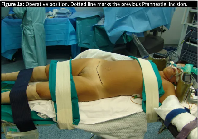
The patient was positioned supine, with the right flank slightly elevated, and was secured to the operating table using wide bandings (Figure 1a). The table was then tilted 25° to the left, further elevating the right flank, and 15° in Trendelenburg’s position. A 7-cm suprapubic incision was made along the previous Pfannenstiel incision where a hand access device was inserted (Lap DiscTM; Ethicon spa, Pomezia, Italy).
Figure 1a: Operative position. Dotted line marks the previous Pfannestiel incision.
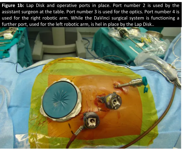
Through a 12-mm port, placed within the lap disk, pneumoperitoneum was created at a pressure of 12 mmHg. Under laparoscopic view, an 11 mm port, to be used for the endoscope, was placed slightly to the left of the mid-line and some centimeters below the navel, and an 8 mm robotic port was placed along the right pararectal line some 5 cm below the costal margin. A final port (12 mm), to be used by the assistant surgeon at the table, was placed along the left
pararectal line halfway between the Pfannenstiel incision and the camera port (Figure 1b).
Figure 1b: Lap Disk and operative ports in place. Port number 2 is used by the
assistant surgeon at the table. Port number 3 is used for the optics. Port number 4 is used for the right robotic arm. While the DaVinci surgical system is functioning a further port, used for the left robotic arm, is hel in place by the Lap Disk..
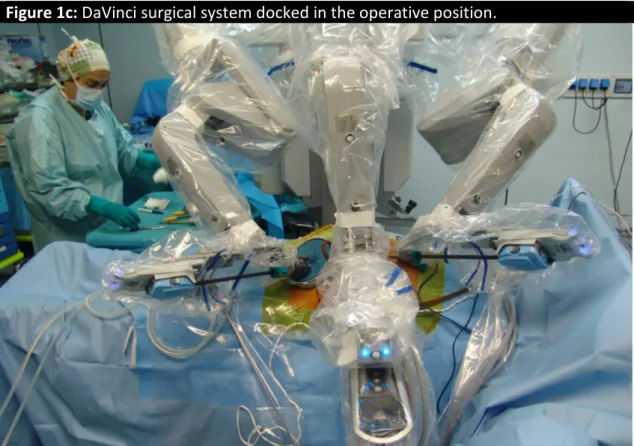
The dVss, placed to the patient’s right side, was docked into position (Figure 1c) and a 0° endoscope was advanced through the 11 mm port. Two operating arms were used. The distal robotic arm operated through a port placed within the suprapubic lap disk. The operation began by mobilizing the cecum until the common iliac vessels were exposed (Figure 2a). Lymphatics were individually ligated and cut. Dissection was carried out using either bipolar Maryland forceps or micro bipolar forceps on the left robotic arm, and monopolar curved scissors on the right robotic arm (Figure 3). Iliac vessels were then crossclamped using laparoscopic bulldogs and the kidney was pushed into the abdomen through the Pfannenstiel incision and dragged over the right psoas muscle using a Cadiere
Figure 1c: DaVinci surgical system docked in the operative position.
Figure 1c: DaVinci surgical system docked in the operative position.
Figure 2: (a) Common iliac vessels exposed; (b) Venotomy being made using Potts
scissors; (c) Venous anastomosis being made using black diamond micro forceps and De Backey forceps; (d) Arterial anastomosis being made using black diamond forceps and De Backey forceps; (e) Venous anastomosis after graft reperfusion; (f) Arterial anastomosis after graft reperfusion.
forceps. The left robotic arm was re-docked and armed with DeBackey forceps and the right-one with Potts scissors. After creating a venotomy (Figure 2b), the
renal vein was anastomosed end-to-side to the common iliac vein using two half running sutures of 6–0 expanded polytetrafluoroethylene using black diamond micro forceps on the right robotic arm and DeBackey forceps on the left-one (Figure 2c). The same steps were followed to create an end-to-side arterial anastomosis between the renal artery and the common iliac artery (Figure 2d).
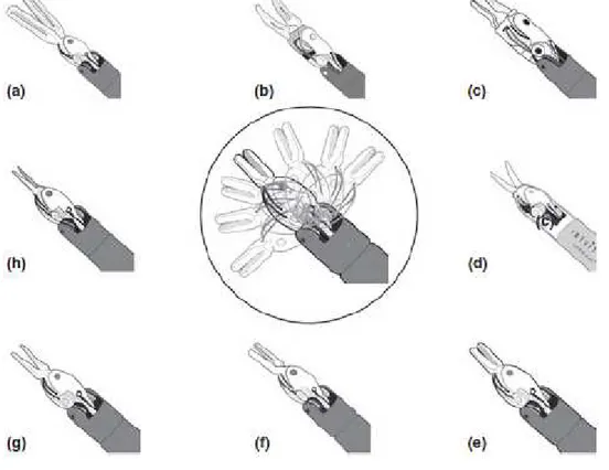
Figure 3: Drawing depicting the full set of robotic instruments used for kidney
transplantation. The central drawing, within the circle, shows the range of motion of wristed robotic instruments. (a) Cadiere forceps; (b) fenestrated Maryland bipolar forceps; (c) micro bipolar forceps (d) monopolar curved scissors; (e) large needle driver; (f) black diamond micro forceps; (g) De Backey forceps; (h) Potts scissors..
ROBOTIC ASSISTED PANCREAS TRANSPLANTATION
Setting.
Pancreas transplantation was the last step of this feasibility and safety study regarding the application of the robotic assisted surgery to transplantation procedures. It was developed in the context of a multidisciplinary team having 39 years of experience in transplantation of abdominal organs. Surgery and
anesthesia teams, in particular, have extensive experience in whole pancreas transplantation and advanced laparoscopic procedures, including laparoscopic robot-assisted auto- and allo-transplantation of the kidney (36). All modern laparoscopic technologies are available at our Institution and the laparoscopic robot-assisted transplant, using the last generation of dVss was proposed to the candidate recipients, who were informed of the innovative nature of the procedure and gave a written consent.
Donor selection, graft procurement and back-table preparation.
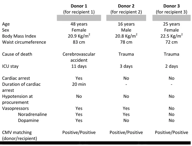
A summary of donors’ characteristics is provided in Table 1. Deceased donors were selected according to standard criteria, as previously described (37). On the contrary, departing from our institutional policy of quick en-bloc procurement of abdominal organs (38), the grafts planned for robotic transplantation were fully dissected before aortic crossclamping. This decision was based on the willingness to ensure hemostasis in the donor, thus reducing the possibility of graft bleeding after reperfusion, which might be difficult to control laparoscopically. Vascular pedicles of the pancreas graft were dissected out, but they were not ligated or divided until completion of visceral perfusion. Grafts were perfused through an aortic cannula with University of Wisconsin solution (60 ml/kg) by gravity flush from a height of 80 centimetres. Direct portal perfusion of the liver was avoided.
At the back table, a donor Y iliac arterial graft was joined to graft arterial pedicles, in a standard fashion (38). No further graft preparation was needed. The left kidney, used for the simultaneous pancreas-kidney transplantation, was not further checked at the back table since it had been procured as in a live donor operation.
Table 1: Summary of donor characteristics. Donor 1 (for recipient 1) Donor 2 (for recipient 2) Donor 3 (for recipient 3)
Age 48 years 16 years 25 years
Sex Female Male Female
Body Mass Index 20.9 Kg/m2 20.8 Kg/m2 22.5 Kg/m2
Waist circumeference 83 cm 78 cm 72 cm
Cause of death Cerebrovascular
accident
Trauma Trauma
ICU stay 11 days 3 days 2 days
Cardiac arrest Yes No No
Duration of cardiac arrest 20 min - - Hypotension at procurement No No No
Vasopressors Yes Yes No
Noradrenaline Yes Yes No
Dopamine Yes No No
CMV matching (donor/recipient)
Positive/Positive Positive/Positive Positive/Positive
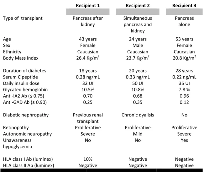
Recipients
A summary of recipient characteristics is provided in Table 2. All patients had a long lasting history of type 1 diabetes.
Recipient 1 (pancreas after kidney), who had previously received a renal transplantation from a deceased donor into her left iliac fossa, had an estimated creatinine clearance of 55 ml/min under a triple maintenance immunosuppression regimen including tacrolimus, mycophenolate mofetil and steroids. She had also undergone hysterectomy, through a Pfannienstel incision, because of uterine fibromata.
Recipient 2 (simultaneous pancreas-kidney) had chronic hepatitis B (HBV-DNA 26.600 IU/ml), without HDV superinfection.
Table 2: Summary of recipient characteristics.
Recipient 1 Recipient 2 Recipient 3
Type of transplant Pancreas after
kidney Simultaneous pancreas and kidney Pancreas alone
Age 43 years 24 years 53 years
Sex Female Male Female
Ethnicity Caucasian Caucasian Caucasian
Body Mass Index 26.4 Kg/m2 23.7 Kg/m2 20.8 Kg/m2
Duration of diabetes 18 years 20 years 28 years
Serum C peptide 0.28 ng/mL 0.33 ng/mL 0.22 ng/mL
Daily insulin dose 32 UI 50 UI 35 UI
Glycated hemoglobin 10.5% 10.8% 7.8 %
Anti-IA2 Ab (≤ 0.75) 0.70 0.68 0.96
Anti-GAD Ab (≤ 0.90) 0.25 0.35 0.12
Diabetic nephropathy Previous renal
transplant
Chronic dyalisis No
Retinopathy Proliferative Proliferative Proliferative
Autonomic neuropathy Severe Mild Severe
Unawareness hypoglycemia
No No Yes
HLA class I Ab (luminex) 10% Negative Negative
HLA class II Ab (luminex) Negative Negative Negative
Recipient 3 (pancreas transplant alone) had no further associated morbidities in addition to the ones presented in Table 2.
Immunosuppression and perioperative care were carried out in all recipients according to our standard protocol, as described previously (39).
Robotic transplants.
The operations followed a precisely established step-by-step protocol.
The operating table was equipped with a heating blanket and CO2 heater.
Anesthesia was induced using fentanyl (0.2 mg), sodium thiopental (3mg/kg) and atracurium besilate (0.2 mg/kg), and it was maintained using sevofluorane
in a 50% air oxygen low flow (2L/min) respiratory mixture delivered by a volumetric ventilator. Atracurium besilate was used in a continuous infusion (0.01 mg/kg/min) to achieve the necessary neuromuscular blockade. Intraoperative hemodynamic monitoring included ECG, mean arterial pressure, and central venous pressure. Respiratory monitoring included end tidal CO2 and
pulse O2 levels.
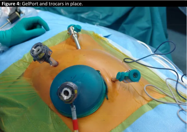
Patients were positioned supine, with the right flank slightly elevated, and were secured to the operating table using wide bandings. The table was then tilted 25 degrees to the left, further elevating the right flank. A 7 centimetres midline incision was made just above the navel and a hand access GelPortTM device
was inserted.
Through a 11 mm port, placed within the GelPortTM, pneumoperitoneum was
created at a pressure of 12 mmHg. Under laparoscopic view an 11 mm port, to be used for the endoscope, was placed slightly to the left of the mid-line some centimeters below the navel. Two 8 mm robotic ports were placed along the right pararectal line some 5 centimetres below the costal margin and 3 centrimetres above the pubis, respectively(Figure 4).
The dVss, placed to the patient’s right side, was docked into position and a 30° endoscope was advanced through the 11 mm optic port (Figure 5).
Figure 4: GelPort and trocars in place.
The operation began by mobilizing the ascending colon until the common iliac artery and the proximal segment of the inferior vena cava were exposed. Lymphatics were individually ligated and cut. Dissection was carried out using either bipolar Maryland forceps or micro bipolar forceps on the left robotic arm, and monopolar curved scissors on the right robotic arm. Following intravenous injection of 5000 units of sodium heparin, the iliac artery was crossclamped and the inferior vena cava was partially occluded using bulldog clamps, manually applied through the GelPortTM. The graft was hence introduced through the
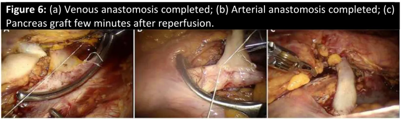
hand port access and placed over the psoas muscle. After excising a longitudinal segment of vena cava, donor portal vein was anastomosed end-to-side to recipient inferior vena cava using two half running sutures of 7-0 expanded polytetrafluoroethylene (Figure 6a). Next, the arterial anastomosis was created, using 6-0 expanded polytetrafluoroethylene, between donor Y graft and recipient common iliac artery (Figure 6b). The posterior wall of vascular anastomoses was sutured from within the lumen (40).
After graft revascularization, (Figure 6c) the pneumoperioneum was interrupted and exocrine drainage was handled, working through the hand port access, by means of Roux-en-Y duodeno-jejunostomy as previously described (39). At the end, the operative field was inspected and two close suction drains were placed along the graft.
In the simultaneous pancreas-kidney transplant, the kidney was transplanted in the ipsilateral iliac fossa (41), according to the technique previously described for robotic kidney transplantation (36). The uretero-vesical anastomosis (Gregoir-Lich extravesical anastomosis) was performed manually, after converting the suprapubic robotic port access into a mini-incision approximately 3 centimetres long.
Figure 6: (a) Venous anastomosis completed; (b) Arterial anastomosis completed; (c)
Results
ROBOTIC ASSISTED LIVING DONOR NEPHRECTOMY
Patient demographics
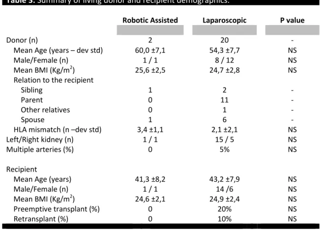
Table 3 shows the pre-operative characteristics of donors and corresponding recipients for the two types of nephrectomy under review. There were no significant differences in age, gender or body mass index between the robotic assisted and laparoscopic groups. There was no significant dialysis duration difference in the recipients who did not receive a pre-emptive transplant (22.2 ± 24.6 months in the robotic assisted group and 36.6 ±72.5 months in the laparoscopic group, P=NS).
Table 3: Summary of living donor and recipient demographics.
Robotic Assisted Laparoscopic P value
Donor (n) 2 20 -
Mean Age (years – dev std) 60,0 ±7,1 54,3 ±7,7 NS
Male/Female (n) 1 / 1 8 / 12 NS
Mean BMI (Kg/m2) 25,6 ±2,5 24,7 ±2,8 NS
Relation to the recipient
Sibling 1 2 -
Parent 0 11 -
Other relatives 0 1 -
Spouse 1 6 -
HLA mismatch (n –dev std) 3,4 ±1,1 2,1 ±2,1 NS
Left/Right kidney (n) 1 / 1 15 / 5 NS
Multiple arteries (%) 0 5% NS
Recipient
Mean Age (years) 41,3 ±8,2 43,2 ±7,9 NS
Male/Female (n) 1 / 1 14 /6 NS
Mean BMI (Kg/m2) 24,6 ±2,1 24,9 ±2,4 NS
Preemptive transplant (%) 0 20% NS
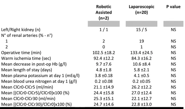
Intra-operative variables among donors (Table 4)
The left kidney was procured from all donors with only one exception. The indication of the single right nephrectomy performed in the robotic assisted live donor group was an anomaly in the right ureter. The vascular complexity between the two groups was similar and the median number of ipsilateral veins is 1 in both cases. Mean operative times and warm ischaemia times, however, were not significantly longer in the robotic assisted live donor group.
Table 4: Intraoperative and postoperative recovery data from live donors.
Robotic Assisted (n=2) Laparoscopic (n=20) P value Left/Right kidney (n) 1 / 1 15 / 5 NS N° of renal arteries (% - n°) 1 2 19 NS 2 0 1 NS
Operative time (min) 102.5 ±18.2 133.4 ±24.5 NS
Warm ischemia time (sec) 92.4 ±12.2 84.3 ±16.2 NS
Mean decrease in post-op Hb (g/l) 9.7 ±7.6 10.6 ±8.4 NS
Mean length of stay (days) 4.8 ±1.8 5.8 ±2.1 NS
Mean plasma potassium at day 1 (mEq/l) 3.8 ±0.18 4.1 ±0.5 NS
Mean blood urea nitrogen at day 1 (g/l) 0.2 ±0.08 0.2 ±0.05 NS
Mean ClCr0-ClCr5 (ml/min) 21.1 ±14.9 26.2 ±12.2 NS
Mean [(ClCr0-ClCr5)/ClCr0]x100 (%) 24.4 ±15.8 27.0 ±12.4 NS
Mean ClCr0-ClCr30 (ml/min) 25.3 ±15.1 22.1 ±12.7 NS
Mean [(ClCr0-ClCr30)/ClCr0]x100 (%) 24.7 ±14.6 22.8 ±13.0 NS
ClCr0, ClCr5 and ClCr30 = creatinine clearance at day 0, 5 and 30 after nephrectomy
Intra-operative complications in donors
No complications were noted in both group donors and intraoperative conversion from laparoscopic to open surgical nephrectomy has not been necessary in any cases. Blood transfusions were also not required and the mean decreases in post-operative haemoglobin levels were minimal in both groups. Symptomatic pneumothorax, and bowel injury were not experienced in this series.
Post-operative courses for live donors
There were no fatalities in either group of donors following nephrectomy and any morbidity was recorded in both groups. There were no cases of re-exploration, incisional hernia, wound infection and pneumonia among patients of either group. Oral intake was resumed within the first 24 h in both the robotic assisted and laparoscopic donors. The duration of hospitalization was similar in both groups. The mean pre-operative estimated creatinine clearance was 92.16±40.67 ml/min in the robotic assisted live donor group and 89.46±25 ml/min in the laparoscopic group (P=NS). Furthermore, the mean decrease in creatinine clearance levels, compared with the pre-operative values, was the same for all donors: 24.4%, 5 days after nephrectomy and 24.71% 1 month later for the robotic assisted live donor nephrectomy patients and 26.97% at 5 days and 22.82% after 1 month for laparoscopic donors. The laparoscopic group reached a peak in estimated creatinine clearance (74.7±27.4 ml/min) on the seventh post-operative day, while the robotic assisted nephrectomized patients achieved maximal estimated creatinine clearance (69.8±15.2 ml/min) (P=NS) on the fourth post-operative day (P=NS).
Post-operative courses for transplant recipients (Table 5)
Cold ischemia times were not significantly longer among the robotic assisted live donor recipients,. Each of the transplanted kidneys functioned correctly following surgery and none of the recipients required posttransplant dialysis. According to the CCR2 values, graft function improved more rapidly in the first 2 days after transplantation in robotic assisted live donor group, however, the mean estimated creatinine clearances at day 5 showed no differences between
robotic assisted live donor and laparoscopic live donor recipients. In addition, no thrombotic complications were observed, even in the case of the right donor kidney.
Table 5: Renal allograft outcomes.
Robotic Assisted (n=2) Laparoscopic (n=20) P value
Cold ischemia time (min) 49.6 ±18.2 52.4 ±16.2 NS
Ureteral complications 0 0 -
Vascular thrombosis 0 0 -
Pyelonephritis 0 0 -
Sepsis related allograft dysfunction 0 0 -
Delayed graft function 0 0 -
CRR2 (%) = (Cr1 –Cr2) x 100/Cr1 42.6 ±15.1 32.6 ±13.9 0.01
Day 5 creatinine clearance (ml/min) 62.2 ±17.6 58.2 ±26.7 NS
Cr1 and Cr2, serum creatinine values on post-transplantation day 1 and day 2
ROBOTIC ASSISTED KIDNEY TRANSPLANTATION
After removal of laparoscopic bulldogs, kidney revascularization was prompt and homogeneous. No bleeding was noted, no additional stitches were placed, and urine production started immediately.
Warm ischemia time was 51 and 38 min respectively. The uretero-vesical anastomosis was fashioned through the suprapubic incision using standard technique (Gregoir-Lich extravesical anastomosis). Before closure of the Pfannenstiel incision, the graft was covered by cecum and pelvic peritoneum thus making it a retroperitoneal graft. Total operative time was 154 and 138 min.
Postoperative course
Postoperative course was uneventful and the kidney functioned immediately. Serum creatinine reached 1.4 and 1.5 mg/dl (normal value 0.5–0.9) on
postoperative day 10, respectively. The day after the transplant, the patients were mobilized and started on oral intake. Pain was described as minimal, and no analgesic was required beyond 48 h after surgery. The patients were discharged on postoperative day 10. At the longest follow-up of 6 and 2 months, they have not been readmitted and renal function remains optimal (serum creatinine 1.4 and 1.6 mg/dl, respectively).
ROBOTIC ASSISTED PANCREAS TRANSPLANTATION
Cold ischemia time was 8 hrs and 52 min for the first pancreas graft, 5 hrs and 35 min for the second, and 7 hrs and 35 min for the third. Cold ischemia time for the kidney graft was 8 hrs and 25 min.
The pancreas after kidney transplant took overall 3 hours. The simultaneous pancreas-kidney transplantation took 8 hours. The pancreas transplant alone took 5 hours. In all recipients pancreas transplantation was carried out with ease.
Warm ischemia, measured from the moment in which each graft was inserted into the abdomen to the time of reperfusion, was 28 minutes for the first pancreas, 30 minutes for the second pancreas, 35 minutes for the kidney, and 33 minutes for the third pancreas. All grafts were reperfused immediately and homogeneously. No bleeding requiring additional suture was noted from vascular anastomosis. Hemorrhage requiring intervention occurred in the third pancreas graft at the level of mesenteric root and at the back of the pancreatic head. Bleeding was easily controlled by selective suture ligation using 4-0 and 5-0 polypropylene. Overall, blood loss was negligible in the first recipient, 200 ml in the second, and 300 ml in the third.
Each recipient became euglycemic soon after graft reperfusion. Figure 7 summarizes the course of serum concentration of pancreatic enzymes checked daily during the post-transplant course.
Figure 7: Course of pancreatic enzymes in the first post-transplant week.
0
1
2
3
4
5
6
7
0
50
100
150
200
250
300
350
400
450
500
Mean Serum Amylase
POD
IU
0
1
2
3
4
5
6
7
0
100
200
300
400
500
600
700
800
Mean Serum Lipase
POD
Kidney transplantation also progressed smoothly. Urine production started immediately.
The post-operative course of all patients was uneventful. Morphine was administered during the first post-transplant day, and no analgesic was required 48 h after surgery. Recipients were out of bed on the first post-transplant day, and were able to stand and walk alone by post-transplant day two. Nasogastric tube was removed on the first post-transplant day in all patients. Recipient 2 and 3 were able to tolerate a full diet by post-transplant day 4. Recipient 1, instead, had a slower recovery of gastrointestinal function, presumably because of severe background autonomic neuropathy. She was able to tolerate a full diet only on post-transplant day 10. Recipients were discharged from the hospital on day 33, 23, and 21 respectively.
At the longest follow-up of 8, 6, and 3 months, respectively, all recipients are doing well and are fully insulin-independent. Renal function is also normal in each of them.
Discussion
ROBOTIC ASSISTED LIVING DONOR NEPHRECTOMY
The results of this study address the technical feasibility and the safety of robotic assisted live donor nephrectomies, as an alternative to the commonly used laparoscopic procedures, in the hands of experienced surgeons. It is noteworthy that the donors at our facility were not randomized to either form of surgery as all chose the robot-assisted technique. That being the case, comparisons between patient demographic data show striking similarities between the robotica assisted and laparoscopic groups. There were no fatalities resulting from either procedure and postoperative complications were minor among the two donor groups. Potential complications associated with open nephrectomy, such as pneumothorax or long-term wound problems (1), were not observed following the use of the laparoscopic or the robotic assisted approach, which both use short incisions and minimize cosmetic defects. The postoperative hospital stays were longer than those reported for the United States. This point can be explained by the health system in Italy. Nevertheless, as previously reported (2,3,7), it was observed that patients treated by conventional surgery are hospitalized for longer periods than those treated using a laparoscopic approach. Significantly, It was found that hospitalization periods were reduced by 50% in robotic assisted live donor group. This may prove to be financially advantageous in the future and lead to a lower risk of nosocomial infections in such patients. Laparoscopic approaches in surgical procedures, whilst offering many benefits, are associated with potentially life-threatening complications that are usually not seen with the traditional ‘open’ approach. Specifically, laparoscopy may induce unique complications related to the creation of pneumoperitoneum, patient positioning, the longer duration of
the operation and surgical instrumentation (42, 43). In our patients, however, each step of the robotic assisted live donor nephrectomy procedure was successfully performed, without the need to revert to an open procedure. In addition, none of robotic assisted live donor patients experienced bleeding, which is the most threatening complication that has been described for laparoscopic procedures (4, 43–45). Furthermore, none of the donors in our robotic assisted live donor initial experience suffered an overt bowel injury, which is another intra-operative complication reported for laparoscopic surgery (44). The increased operative times for robotic assisted nephrectomies also did not lead to adverse events in the donors, such as rhabdomyolysis (46), or cardiovascular and pulmonary complications associated with prolonged pneumoperitoneum (43). Elevated intra-abdominal pressures may cause venous compression and reduce femoral vein flow velocity, and may be because intra-abdominal pressure was always kept near 12 mmHg, none of our patients did develop a deep vein thrombosis following laparoscopic surgery. The production of pneumoperitoneum can also decrease renal blood flow and clinical and experimental studies have previously reported a transient and self-limiting oliguria after laparoscopy in patients with normal renal function (47). This phenomenon was not observed, however, in the donors undergoing robotic assisted or laparoscopic live donor nephrectomy at Pisa transplant centre, and we found that serum creatinine levels increased in a comparable manner in both the robotic assisted and laparoscopic groups. Amongst our recipient patients, there was no evidence to show that a robotic assisted live donor nephrectomy adversely affects allograft function. For laparoscopic donor kidney transplants, there are controversial data in the literature concerning early graft function, in which some investigators have found no significant differences
when comparing laparoscopic and open kidney grafts (44). In contrast to this evidence, a survey of US transplant centres revealed significantly slower early post-transplant graft function in laparoscopic donor kidney graft recipients, with no differences in serum creatinine levels at later time points (48), and similar results have been reported by others (49, 50). At Pisa transplant centre, however, initial graft survival and function rates, after robot-assisted laparoscopic procurement, compare very favourably with our laparoscopic control subjects. None of our patients required dialysis within the first week of transplantation and we observed no incidences of delayed graft function, assessed by CRR2 measurements. CRR2 values (51) were used as they enable clinicians to consider the factors that influence early graft functions, such as components of donation, preservation variables and recipients variables, rather than immune responses, which usually impact upon the transplant recipient at a later date. In addition, CRR2 has been proven to correlate well with graft function during the first year (52). This is an important consideration, as laparoscopic approaches entail increased intra-abdominal pressure and also a traumatic removal of the organ through a Pfannenstiel incision, also if it does not necessitate longer mean warm renal ischaemia times than robotic assisted live donor nephrectomies (84.3 ±16.2 seconds vs 92.4 ±12.2 seconds in this series).
We consider it to be unlikely, however, that warm ischaemia times will drop much below 2 min using current techniques. In their experience with robotic assisted live donor nephrectomies, Horgan et al. (16) reported short warm renal ischemia times, ranging between 70 and 95 s.. The excessive manipulation and prolonged extraction of the kidney, during robot-assisted laparoscopic surgery, could lead to ischaemia-reperfusion injury and hamper organ function recovery.
In an experimental renal transplantation study, Yilmaz et al. (53) reported that prolonged ischaemia times induce intimal proliferation, vascular obliteration, glomerular sclerosis and increase the mesangial matrix. The eventual sclerotic destruction of glomeruli cannot yet be evaluated. Moreover, several laparoscopic urologists have observed a higher incidence of ureteral necrosis, or late ureteral stricture requiring operative repair, in comparison to laparoscopic live-donor nephrectomy recipients and open nephrectomy recipients (54). These lesions, which are thought to be due to impaired vascularity of the ureter, have been attributed to suboptimal mobilization and visualization of donor kidneys during the laparoscopic approach. In robotic assisted live donor group, however, no ureteral complications of vascular origin were observed in the recipients. It seems clear that, in addition to thorough preoperative imaging for evidence of aberrant vessels and a careful patient selection protocol, the training and experience of the surgeon, in addition to proper perioperative management of fluid and electrolyte balance, have greatly influenced the low rate of postoperative complications that we observed. Increased experience of laparoscopic procedures and also of transplantation surgery is well known to decrease the incidence of such complications (43). It must be noted that robotic assistance has been recognized to improve laparoscopic training and skill acquisition, and the low levels of morbidity observed robotic assisted live donor nephrectomy group is partly attributable to this robotic assistance. The three-dimensional vision of the robotic assistance also enhances the ability of the surgeon to perform delicate endoscopic manoeuvres, such as dissection or precise laparoscopic suturing. Surgeon fatigue and tremor levels during robotic suturing are also reduced, when compared with conventional laparoscopic intracorporeal methods. Dissection of
the ureter is also facilitated, avoiding excessive stripping. Finally, robot-assisted donor nephrectomy may minimize intra-operative complications by allowing surgeons to dissect rapidly and efficiently and to control problematic bleeding and lymphatic leaks more easily and efficiently (6).
ROBOTIC ASSISTED KIDNEY TRANSPLANTATION
Surgical robotics is a refinement of classic laparoscopy. The only current available system, the dVss, is not a classical robot, in the narrower sense of the word, but rather an electromechanical surgical actuator faithfully translating movements of surgeon’s hands into wristed instrument actions (20). As such, the dVss should enhance surgeon’s ability to accomplish complex laparoscopic operations requiring fine dissection and microsuturing. On the other hand, the greatest limitations of the dVss are high cost and lack of haptic feed-back. Other drawbacks are risk of technical failure, loss of direct contact between surgeon and patient, and poor adaptability to multiquadrant surgery (20). The high cost of the dVss is a significant problem that has probably limited the diffusion of this new technology. However, like other computer-driven technologies, costs are expected to drop over time, especially when the patent of ‘‘remote center-of-motion robot for surgery’’ (US patent number: 5397323; Issue date: March 14, 1995) will expire (on October 30, 2012) and competitors of Intuitive Surgical will have a chance to propose alternative systems. Lack of haptic feed-back is a further main drawback of current dVss. Theoretically, it could lead to an increased risk of inadvertent tissue injury but, to date, robotically performed operations have not been associated with higher clinical complication rates than their standard laparoscopic or open counterparts (20). On the other hand, reduction in suture strength is known to occur following robotic needle driver
manipulation (55, 56). While research on haptic sensors is ongoing (57-59), improved visual clues seem to act as a substitute for haptic feedback (59, 60). No device or technology is impervious to malfunction. The dVss is no exception to this rule. Current systems, however, are designed to minimize the deleterious effects of such failures on patients thanks to system redundancy features (20). The dVss can incur into recoverable and nonrecoverable faults. Only in the latter instance, the robotic procedure has to be aborted and/or there may be a real hazard on patient safety. In a series of 725 radical prostatectomies, the mean rate of recoverable and nonrecoverable faults per procedure was 0.21 and 0.05, respectively. Interestingly, all nonrecoverable faults occurred before the beginning of the operation resulting in rescheduling of surgery (61). Loss of direct contact between surgeon and patient requires adaptation and improved coordination with the assistant surgeon who, instead, maintains a direct contact with the patient. This process requires a learning curve. Paradoxically, this limitation of current dVss may also have positive implications. Lack of direct interaction between surgeon and patient could reduce the risk of disease transmission, especially in kidney transplant recipients in whom there is a high prevalence of hepatitis infection. Overall, it would seem that the dVss could be used for kidney transplantation under well-controlled, investigational conditions. The first use of the dVss for kidney transplantation was reported by Hoznek et al. in 2002. Iliac vessels, however, were dissected through a standard oblique incision and the dVss was used only to complete the anastomoses (62). The first fully laparoscopic kidney transplantation using a dVss was reported by Giulianotti et al. (Chicago, IL, USA), early past year (63), although the first world case was performed by Geffner at the Saint Barnabas Medical Center (New Jersey, USA) in January 2009 (unpublished data). As of June 25, 2010, a total
of 25 robotic kidney transplantations had been performed in the USA, eight at the University of Illinois and 16 at Saint Barnabas Medical Center (Communication at 5th International Conference: ‘‘Living donor abdominal organ transplantation: state of the art.’’ June 25–26, 2010; Florence, Italy); to our knowledge, the cases described in this study are the first performed in Europe. The technique that it was applied differs substantially from that used in Chicago (63) and New Jersey.
At the University of Illinois, Giulianotti et al. decided to adopt a hand-assisted technique making the incision in the periumbelical area and placing the graft intraperitoneally (63). Regarding the site of incision, a periumbelical incision is known to carry a higher risk of incisional hernia as compared with the bikini type incision it was adopted in these initial cases. Furthermore, a suprapubic incision allows direct performance of uretero-vescical anastomosis. Although this anastomosis can easily be constructed using the dVss, it requires repositioning of the robot (63) and prolongs the period during which the freshly revascularized graft is exposed to the detrimental effects of pneumoperitoneum (64). Hand assistance, easier through a periumbelical incision, could facilitate some operative steps, such as handling the graft during performance of vascular anastomoses, could improve exposure especially in obese recipient, and could be useful in case of sudden hemorrhage. However, with all the limitations of comparisons made between single case descriptions, warm ischemia period in the cases of the study was identical to the one reported by the Chicago group. Further experience will clarify which incision is more suitable. Perhaps, the periumbelical incision will eventually be preferred in obese patients and the suprapubic incision be reserved to thinner recipients. Moreover Giulianotti et al. decided to place their kidney graft intraperitoneally. Although grafts placed in
this location are known to work efficiently, this option is not routinely adopted in conventional kidney transplantation. Intraperitoneal renal graft placement may actually be associated with unique complications, such as paratransplant hernia (65) and renal pedicle torsion (66).
The technique used at the Saint Barnabas Medical Center has not been published yet, but we have learned of it directly from Dr. Geffner at the 5th International Conference: ‘‘Living donor abdominal organ transplantation: state of the art.’’ (June 25–26, 2010; Florence, Italy). Dr. Geffner places the kidney graft extraperitoneally, through a small incision made along the line that would be followed in case of conventional kidney transplantation. A working space is hence created, using the same technique employed in retroperitoneoscopic nephrectomy, and the anastomoses are performed robotically. At the end, the graft lies in the classic retroperitoneal location.
The technique that it was adopted in the cases of the study, which might be identified as ‘‘hybrid’’, employs a transperitoneal approach, but eventually leaves the graft in the retroperitoneum.
In our view, working transperitoneally avoids the traditional disadvantages of retroperitoneoscopy, such as limited working space, ease collapse during suction, and blurred vision, while maintaining the advantage of eventual graft placement in a retroperitoneal pocket. The most prominent advantage of Geffner’s incision is that in case of conversion to open surgery, there would be no additional incision. Of course, the periumbelical incision used by Giulianotti et al. (63) should be extended significantly to gain full access to iliac vessels. Prolonging our small transverse suprapubic incision, toward the iliac fossa where the kidney is being transplanted, would result in a ‘‘hockey stick’’ incision, probably only a bit larger than the one performed under standard conditions.
Minimally invasive kidney transplantation might require more time to complete vascular anastomoses thus prolonging second warm ischemia time and possibly resulting in higher incidence of delayed graft function (67). It is indeed known that kidney temperature increases according to a logarithmic curve and at a speed of 0.48 °C/min. Kidney temperature at th e time of revascularization depends on anastomotic time and is inversely proportional to kidney weight (68). A prerevascularization graft temperature ≤ 15 °C is associated with reduced incidence of acute tubular necrosis (67). Topical graft cooling may slow the rate of graft rewarming (67), but is impractical to use during laparoscopic kidney transplantation, as cold irrigation would blur the vision of the vessels to be anastomosed and would require concurrent suction, decreasing the level of pneumoperitoneum. The use of a cooling pocket (69, 70) might be advantageous. However, the ideal laparoscopic cooling pocket should be friendly to use. To our knowledge, none of the described laparoscopic devices (71, 72) has been tested enough as to prove its efficacy and ease of use. On the other hand, the yet limited experience with kidney transplantations through minimal skin incision (73-75), sharing with laparoscopic kidney transplants the issue of graft rewarming, do not demonstrate a detrimental effect on kidney function. The decision to avoid additional renal graft cooling during robotic transplantation was based on all these considerations. The consequences of progressive graft rewarming occurring during minimally invasive kidney transplantation cannot be defined at the moment. We anticipate that this issue will be debated extensively and will provide new impetus to research.
ROBOTIC ASSISTED PANCREAS TRANSPLANTATION
The high incidence of surgical complications is among the major disincentives to pancreas transplantation, despite the potentially unlimited pool of candidate recipients (76). Islet transplantation, the only possible alternative to pancreas transplantation, was indeed conceived and pursued to achieve beta-cell replacement with lower morbidity, but it has not achieved the expected results yet (24).
In the last two decades, laparoscopy was probably the greatest innovation in abdominal surgery. The advantages of this approach were so evident that it quickly became the standard for many operations. Refinements in surgical techniques and advancement in equipments currently permit complex operations to be safely performed laparoscopically, especially when there is little need for multiple intracorporeal reconstructions (77, 78). In laparoscopy, fine sutures are demanding because of :
1) loss of hand-eye coordination;
2) use of long instruments with only four degrees of freedom, amplifying natural surgeon’s tremor and carrying a fulcrum effect;
3) poor ergonomy leading to surgeon’s fatigue (35).
The dVss is a computer-assisted electromechanical device allowing a remote surgeon to manipulate tissues, handling electromechanical devices and throwing microsutures through laparoscopic ports. As compared with conventional laparoscopy, the dVss offers 3D high-definition view, including 10x to 15x magnification and restoring hand-eye coordination, employs wristed instruments with seven degrees of freedom, and tracks surgeon’s movements 1,300 times per second, providing for tremor filtration and scaled motion. Surgeon power is further enhanced by driving the binocular endoscope,
providing steady view, and by toggling among three operative arms. These technology addenda significantly improve surgeon ability to operate within deep and narrow spaces, especially when fine dissection and microsuturing are required.
The already cited greatest limitations of current dVss are high cost and lack of haptic feed-back. Other drawbacks are risk of technical failure, loss of direct contact between surgeon and patient, and poor adaptability to multiquadrant surgery (32). Lack of haptic feed-back is the reason why in this study was used expanded polytetrafluoroethylene instead of polypropylene for vascular anastomosis. There is indeed a good amount of experimental evidence that repetitive needle driver manipulations weaken suture materials. The maximal failure force of monofilaments is reduced by 35% as compared with 3% for braided sutures (79). In particular, expanded polytetrafluoroethylene shows no loss in strength after repetitive robotic manipulations while polypropylene is weakened after three robotic manipulations at the same point (80).
We have recently shown that kidney transplantation is feasible laparoscopically under robotic assistance (36). A similar experience was reported from the United States (35). Based on this background we speculated that a pancreas graft could be transplanted laparoscopically as well. Indeed, pancreas transplantation, alike kidney grafting, requires only one arterial and one venous anastomosis. In the technique that we elected to employ, which is a modification of the one that we have previously described (39), the pancreas lies on the right retroperitoneal space over the psoas muscle and behind the right colon. This position is similar to the one in which we had placed the grafted kidney during laparoscopic robot-assisted renal transplantation (36).
Beyond technical facilities, dVss provides a wide set of advantages over laparoscopic surgery:
1) The ability of dVss to work within narrow spaces makes it possible to safely construct vascular anastomosis with minimal vessels exposure. This could be advantageous in patients with limited vascular access and could contribute to reduce incidence and severity of perigraft fluid collections.
2) Reduced tissue handling results in reduced activation of coagulation systems (81). This is likely the reason why robotic surgery has been associated with a reduced rate of peripheral vein thrombosis than conventional laparoscopic surgery (82, 83). Graft thrombosis is the leading cause of morbidity and early graft failure after pancreas transplantation. A reduced activation of systemic coagulation is hence expected to also reduce incidence of graft thrombosis and lead to better graft survival. This hypothesis requires anyway confirmation in larger case series.
3) Laparoscopic surgery has been associated with a reduced proinflammatory response (84) and with a reduced immune suppression (85). Since the length of surgical incisions is clearly reduced in robotic versus conventional laparoscopic surgery, the rate of immune suppression, and consequent wound infections, might be further reduced. From the point of view of anticipated benefits to the patients, perhaps no other recipient population could expect greater advantage from minimally-invasive transplantation than diabetics, whose post-transplant course is typically plagued by multiple surgical complications.