patient specific systems for
computer assisted robotic
surgery simulation, planning,
and navigation
faculty of medicine department of oncology, transplantation, and advanced technologies in medicine
november 2011
botic Surgery Simulation, Planning, and Navigation , Philosophiæ
Dedicated to my friend Diego,
and to my grandparents:
The evolving scenario of surgery: starting from modern surgery, to
the birth of medical imaging and the introduction of minimally
inva-sive techniques, has seen in these last years the advent of surgical
robotics. These systems, making possible to get through the
difficul-ties of endoscopic surgery, allow an improved surgical performance
and a better quality of the intervention.
Information technology contributed to this evolution since the
be-ginning of the digital revolution: providing innovative medical
imag-ing devices and computer assisted surgical systems. Afterwards, the
progresses in computer graphics brought innovative visualization
modalities for medical datasets, and later the birth virtual reality
has paved the way for virtual surgery.
Although many surgical simulators already exist, there are no
pa-tient specific solutions.
This thesis presents the development of patient specific software
systems for preoperative planning, simulation and intraoperative
as-sistance, designed for robotic surgery: in particular for bimanual
robots that are becoming the future of single port interventions.
The first software application is a virtual reality simulator for this
kind of surgical robots. The system has been designed to validate
the initial port placement and the operative workspace for the
poten-tial application of this surgical device. Given a bimanual robot with
its own geometry and kinematics, and a patient specific 3D virtual
anatomy, the surgical simulator allows the surgeon to choose the
custom format.
The robot capabilities to accomplish specific tasks can be virtually
tested using the deformable models: interacting directly with the
target virtual organs, trying to avoid unwanted collisions with the
surrounding anatomy not involved in the intervention.
Moreover, the surgical simulator has been enhanced with
algo-rithms and data structures to integrate biomechanical parameters
into virtual deformable models (based on mass-spring-damper
net-work ) of target solid organs, in order to properly reproduce the
physical behaviour of the patient anatomy during the interactions.
The main biomechanical parameters (Young’s modulus and density)
have been integrated, allowing the automatic tuning of some model
network elements, such as: the node mass and the spring stiffness.
The spring damping coefficient has been modeled using the Rayleigh
approach. Furthermore, the developed method automatically detect
the external layer, allowing the usage of both the surface and
inter-nal Young’s moduli, in order to model the main parts of dense organs:
the stroma and the parenchyma. Finally the model can be manually
tuned to represent lesion with specific biomechanical properties.
Additionally, some software modules of the simulator have been
properly extended to be integrated in a patient specific computer
guidance system for intraoperative navigation and assistance in
ro-botic single port interventions. This application provides guidance
functionalities working in three different modalities: passive as a
and active as a tutor preventing unwanted collision during the
inter-vention.
The simulation system has beed tested by five surgeons: simulating
the robot access port placemen, and evaluating the robot movability
and workspace inside the patient abdomen. The tested
functionali-ties, rated by expert surgeons, have shown good quality and
perfor-mance of the simulation. Moreover, the integration of biomechanical
parameters into deformable models has beed tested with various
ma-terial samples. The results have shown a good visual realism
ensur-ing the performance required by an interactive simulation. Finally,
the intraoperative navigator has been tested performing a
cholecys-tectomy on a synthetic patient mannequin, in order to evaluate: the
intraoperative navigation accuracy, the network communications
la-tency and the overall usability of the system.
The tests performed demonstrated the effectiveness and the
us-ability of the software systems developed: encouraging the
introduc-tion of the proposed soluintroduc-tion in the clinical practice, and the
imple-mentation of further improvements.
Surgical robotics will be enhanced by an advanced integration of
medical images into software systems: allowing the detailed
plan-ning of surgical interventions by means of virtual surgery simulation
based on patient specific biomechanical parameters. Furthermore,
the advanced functionalities offered by these systems, enable
surgi-cal robots to improve the intraoperative surgisurgi-cal assistance:
benefit-ting of the knowledge of the virtual patient anatomy.
«Techniques for Computer Assisted Surgery» - Turini, G. and
Pie-troni, N. and Ganovelli, F. and Scopigno, R. - Eurographics Italian
Chapter Conference 2007 - 14-16 February 2007, Trento, Italy; [239].
«New Techniques for Computer-Based Simulation in Surgical
Train-ing» - Turini, G. and Pietroni, N. and Megali, G. and Pietrabissa,
A. - International Conference on the Management of Healthcare &
Medical Technology 2007 - 3-5 October 2007, Pisa, Italy; [238].
«EndoCAS Navigator Platform: a Common Platform for Computer
and Robotic Assistance in Minimally Invasive Surgery» - Megali, G.
and Ferrari, V. and Freschi, C. and Morabito, B. and Cavallo, F. and
Turini, G. and Troia, E. and Cappelli, C. and Pietrabissa, A. and
Tonet, O. and Cuschieri, A. and Dario, P. and Mosca, F. - The
International Journal of Medical Robotics and Computer Assisted Surgery
-vol. 4, p. 242-251, 2008; [135].
«GHOST: Piattaforma Basata su Realtà Virtuale per il Telementoring
in Laparoscopia» - Pietrabissa, A. and Ferrari, V. and Moglia, A. and
Turini, G. and Troia, E. and Mosca, F. - MIMOS MMVR 2009 - 4-5
October 2009, Lecce, Italy; [182].
A. and Turini, G. and Ferrari, V. and Ferrari, M. and Mosca, F.
-MIMOS MMVR 2010 - 14 December 2010, Pisa, Italy; [147].
«Integration of Biomechanical Parameters in Deformable Models for
Interactive Virtual Surgery» - Turini, G. and Sala, A. and Ferrari, V.
and Ferrari, M. and Mosca, F. - MIMOS MMVR 2010 - 14 December
2010, Pisa, Italy; [240].
«Patient Specific Surgical Simulator for the Evaluation of the
Mov-ability of Bimanual Robotic Arms» - Moglia, A. and Turini, G. and
Ferrari, V. and Ferrari, M. and Mosca, F. - Studies in Health
Technol-ogy and Informatics - vol. 163, p. 379-385, 2011; [148].
«New Techniques for Computer-Based Simulation in Surgical
Train-ing» - Turini, G. and Pietroni, N. and Megali, G. and Ganovelli, F.
and Pietrabissa, A. and Mosca, F. - International Journal of
Biomedi-cal Engineering and Technology - vol. 5 (4), p. 303-316, 2011; [241].
«Integration of Biomechanical Params in Tetrahedral Mass-Spring
Models for Virtual Surgery Simulation» - Sala, A. and Turini, G. and
Ferrari, M. and Mosca, F. and Ferrari, V. - Proceedings of the 33rd
An-nual International Conference of the IEEE Engineering in Medicine
and Biology Society (EMBC 2011) - p. 4550-4554 - 30 August - 3
September 2011, Boston, US; [194].
«Patient Specific Virtual Simulator for the Pre-operative Planning
of Robotic Single Incision Laparoscopic Surgery» - Moglia, A. and
Turini, G. and Ferrari, V. and Ferrari, M. and Mosca, F. - MIMOS
MMVR 2011 - 3 November 2011, Bologna, Italy; [149].
«Computer Guidance Software for Navigation and Assistance for
Sin-gle Incision Bimanual Robotic Surgery» - Carbone, M. and Turini, G.
and Petroni, G. and Niccolini, M. and Menciassi, A. and Ferrari, M.
«Computer Guidance System for Single Incision Bimanual Robotic
Surgery» - Carbone, M. and Turini, G. and Ferrari, V. and Petroni,
G. and Niccolini, M. and Menciassi, A. and Ferrari, M. and Mosca, F.
- Computer Aided Surgery - 2011 - Submitted; [36].
Richard Phillips Feynman,
Nobel Prize in Physics in 1965,
on his blackboard at time of death in 1988.
A C K N O W L E D G E M E N T S
It goes without saying that this Philosophiæ Doctor thesis would not
have been possible without the support of a legion of professors,
col-leagues, and friends. Too many to be all cited. But there are certainly
a few that need particular mention here.
I would first like to thank Prof. Franco Mosca for his essential
support to the research activity. I am also particularly grateful to
Prof. Mauro Ferrari for his medical guidance and to Vincenzo Ferrari
for the constant supervision of my scientific research.
I would also thank Prof. Andrea Pietrabissa and Prof. Sir Alfred
Cuschieri not only for their competence and encouragements, but
especially for the passion they always transmit.
A special mention to Fabio Ganovelli, my very first scientific tutor.
His competence and generosity make him a role-model for every
re-searcher. Without his support and encouragements this thesis would
not have been completed. He is a friend.
I wish to thank all the present and past members of the EndoCAS
Research Center: Alessandro, Andrea, Bruno, Carla, Cinzia, Cristina,
Elena, Filippo, Francesca, Gianni, Giuseppe, Liuba, Marina, Orazio,
Paolo, Sara, Silvia, Simona, and Stefano. It has been a pleasure to
work with all of you.
in particular: Andrea, Giuseppe, and Nico.
Finally, I would like to thank my family in Italy and in the US, and my
friends: Caterina, Emanuele, Federico, Luisa, and Michele.
I am also grateful to Diego for his enduring and precious
friend-ship, and my old online pal Giorgio for the myriad of challenges we
faced together.
1 introduction 1
1.1 Introduction to Modern Surgery . . . 1
1.1.1 Medical Imaging and Image Guided Surgery . . 3
1.2 Minimally Invasive Surgery . . . 3
1.2.1 Brief History . . . 4
1.2.2 The Concept . . . 4
1.2.3 Endoscopic Equipment . . . 6
1.2.4 Fields of Application . . . 8
1.2.5 Innovative Minimally Invasive Techniques . . . 9
1.3 Computer Assisted Surgery . . . 11
1.3.1 Essentials . . . 12
1.3.2 Applications . . . 14
1.3.3 State of the Art . . . 15
1.4 Robotic Surgery . . . 16
1.4.1 Commercial Robotic Surgical Systems . . . 17
1.4.2 Telesurgery . . . 21
1.5 Surgical Planning and Assistance . . . 24
1.5.1 Preoperative Planning Systems . . . 25
1.5.2 Intraoperative Assistance Systems . . . 25
1.6 Virtual Surgery . . . 27
1.6.1 Introduction to Interactive Simulation . . . 28
1.6.2 Commercial Surgical Simulators . . . 30
1.7 Objective and Contribution . . . 38
1.7.1 Objective of the Work . . . 39
1.7.2 Contributions of the Thesis . . . 39
2.2.2 Visualization Functionalities . . . 47
2.3 Patient Specific Virtual Anatomy . . . 48
2.3.1 Generating 3D Virtual Organs . . . 48
2.3.2 Modeling Deformable Virtual Organs . . . 50
2.4 Virtual Bimanual Surgical Robot . . . 51
2.5 Algorithms and Data Structures . . . 52
2.5.1 Software Application Technical Description . . 53
2.5.2 Physical Models for Deformable Organs . . . . 54
2.5.3 Interactions with the Virtual Anatomy . . . 55
2.5.4 Interactive Dynamic Simulation . . . 57
2.5.5 Hardware Interface . . . 58
2.6 Results and Conclusions . . . 60
2.6.1 Preliminary Results . . . 60
2.6.2 Final Considerations . . . 61
2.6.3 Acknowledgment . . . 63
3 biomechanical modeling for virtual surgery 64 3.1 Intro to Biomechanical Modeling . . . 65
3.2 Biomechanical Params Integration . . . 67
3.2.1 Nodes Mass Parametrization . . . 68
3.2.2 Springs Stiffness Parametrization . . . 71
3.2.3 Dampers Coefficients Parametrization . . . 75
3.3 Software Development . . . 76
3.4 Preliminary Tests and Results . . . 77
3.4.1 Testing Global Deformations . . . 77
3.5 Final Considerations . . . 80
3.5.1 Acknowledgment . . . 81
4 intraoperative guidance for robotic surgery 83 4.1 Intro to Surgical Navigation . . . 84
4.2 Guidance System Overview . . . 87
4.2.1 Hardware Components . . . 87
4.2.2 Navigation Functionalities . . . 89
4.3 Navigator Technical Description . . . 91
4.3.1 Modeling the Virtual Scene . . . 92
4.3.2 Patient Registration . . . 92
4.3.3 Software Implementation . . . 93
4.4 Testing, Evaluation and Results . . . 94
4.4.1 Test Session Setup . . . 95
4.4.2 Navigation System Evaluation . . . 96
4.4.3 Test Session Results . . . 97
4.5 Final Considerations . . . 97
4.5.1 Acknowledgment . . . 99
5 results and conclusions 100 5.1 Summary . . . 100
5.2 Results . . . 101
5.3 Conclusions . . . 102
Figure 4 AESOP and ZEUS Surgical Robots . . . 16
Figure 5 Robotic Surgery System . . . 17
Figure 6 ROBODOC and Neuromate Surgical Robots . . 19
Figure 7 Cyberknife and RIO and SpineAssist Robots . . 21
Figure 8 First Telesurgery Interventions . . . 24
Figure 9 Commercial Surgical Simulators . . . 32
Figure 10 Commercial Surgical Simulators . . . 34
Figure 11 Commercial Robotic Surgical Simulators . . . . 36
Figure 12 Surgical Simulator Overview and Screenshot . 46 Figure 13 Surgical Simulator Viewing Modalities . . . 48
Figure 14 Surgical Simulator Anatomy Generation . . . . 49
Figure 15 Surgical Simulator Bimanual Robot . . . 51
Figure 16 Surgical Simulator Virtual Bimanual Robot . . . 52
Figure 17 Skeleton Based Deformable Model . . . 54
Figure 18 Surgical Simulator System Diagram . . . 55
Figure 19 Surgical Simulator Hardware Interface . . . 58
Figure 20 3D and Physical Models . . . 67
Figure 21 Spring Network Structure and Topologies . . . 75
Figure 22 Biomechanical Modeling Simulator Overview . 77 Figure 23 Testing Global Deformations . . . 79
Figure 24 Testing Local Deformations . . . 80
Figure 25 Surgical Navigator Screenshot . . . 87
Figure 27 Surgical Navigator Assistive Modality . . . 90
Figure 28 Surgical Navigator Passive Modality . . . 91
Figure 29 Surgical Navigator System Diagram . . . 95
Figure 30 Surgical Navigator Test Session . . . 98
L I S T O F T A B L E S
Table 1 Surgical Simulator Evaluation . . . 59Table 2 Comparing Mass-Lumping Strategies . . . 72
Table 3 Surgical Navigator Evaluation . . . 97
L I S T O F A C R O N Y M S
API Application Programming Interface
ARAKNES Array of Robots Augmenting the KiNematics of
Endo-luminal Surgery
ARFI Acoustic Radiation Force Impulse
CT X-Ray Computed Tomography
DAQ Data AcQuisition system
DICOM Digital Imaging and COmmunications in Medicine
DOF Degrees Of Freedom
EG ElastoGraphy
ENT Ear, Nose and Throat surgery
FDA Food and Drug Administration
FEM Finite Element Method
FPS Frames Per Second
GPU Graphics Processing Unit
GUI Graphical User Interface
IGS Image Guided Surgery
IR Infra-Red
LAN Local Area Network
LOD Levels Of Detail
LOS Line Of Sight
MICS Minimally Invasive Cardiac Surgery
MIS Minimally Invasive Surgery
MRE Magnetic Resonance Elastography
MRI Magnetic Resonance Imaging
MSDm Mass-Spring-Damper Model
NOTES Natural Orifice Transluminal Endoscopic Surgery
RAM Random-Access Memory
SAES Single Access Endoscopic Surgery
SILS Single Incision Laparoscopic Surgery
SPRINT Single Port lapaRoscopy bImaNual roboT
SVD Singular Value Decomposition
TURP Trans-Urethral Resection of the Prostate
UDP User Datagram Protocol
The future of surgery is not about blood and guts;
the future of surgery is about bits and bytes.
Richard M. Satava,
pioneer of surgical robotics and virtual surgery.
Innovative technologies impact all fields of healthcare, and surgery
in particular. This change affects not only diagnostic processes and
therapies, but also education and training of the medical staff.
This chapter describes the evolving scenario of surgery. Initially
traditional surgery and medical imaging are introduced, highlighting
the revolution occurred with the birth of Minimally Invasive Surgery
(MIS) techniques. Therefore, the limitations of conventional surgery are presented: motivating the design concepts of Computer
Assis-ted Surgery (CAS) and robotic surgery, describing advantages and problems of these innovative approaches.
At last, the scope and the contributions of this thesis are exposed.
1.1
introduction to modern surgery
By the late Middle Ages the ancient idea that the hand was sub- Surgeons from the Middle Ages to the Modern Era.
servient to the head, the senses to the intellect, had been
incorpo-rated into the organisation of medicine. Physicians regarded
(a)Thomas Eakins, The Gross Clinic, 1875.
(b)Thomas Eakins, The Agnew Clinic, 1889.
Figure 1:Two paintings by Thomas Eakins (Figure1aand Figure1b) docu-menting the history of medicine: honoring the emergence of sur-gery and showing a typical surgical theater in the late XIX century [202,203].
selves as the reasoning head of this body and surgeons as the hands
[112].
Slowly, after the XVII century, surgeons gained accurate
knowl-edge in science and technology: raising their status to that of the
physician. Science, technological innovation, natural history, and
ex-periment made surgery what it was, allowing surgeons to be
gentle-men and do what was once one of the most ungentlemanly of things:
use their hands on other people’s bodies [112].
Later, in the XIX century, surgeons began to present themselves
as the embodiment of heroism and manliness (see Figure1) [112].
During the XX century, surgery progressed farther and faster than Surgery in the XX century.
in all preceding ages.
Despite the operating theatre and surgical instruments still
re-main almost the same of the past (see Figure1), and the anesthesia progressed slowly, the scope of surgery increased leading to
special-ization. Before long it became clear that, to achieve progress in
certain areas, surgeons had to concentrate their attention on that
ical engineering and the exploitation of new materials [33].
1.1.1 medical imaging and image guided surgery
The progresses in medical imaging techniques induced great
im-provements on surgery. Medical imaging has beed accepted in
es-tablishing diagnosis and guiding therapies. Moreover, even if
medi-cal imaging was always used in the therapeutic process, the digital
revolution improved image-guided therapies with high quality digital
data manipulated by computer systems.
Although interventional radiology has the potential to push further
developments in medical imaging, Image Guided Surgery (IGS) can Interventional radiology and IGS.
play an important role in the advancement of intraoperative imaging
[144,207,249,261].
Reaching the new millennium, modern surgery experienced a
dra-matic change: the revolution leaded by minimally invasive surgery.
1.2
minimally invasive surgery
During the last years of the XX century, Minimally Invasive Surgery
(MIS) has influenced the techniques used in every specialty of sur-gery: leading to the replacement of conventional procedures with
reevalu-ate conventional approaches with regard to perioperative morbidity
[64].
The advent of MIS and the availability of new image-based tech-nologies leaded to a significant change in surgery. These, both
ad-dressing the improvement of healthcare quality, have been combined
to overcome the problems introduced by novel surgical approaches,
toward a revolution in surgical performance.
1.2.1 brief history
The very first endoscopic attempt to explore the inner workings of
the human body was cystoscopy performed by Pierre Salomon
Sé-galas d’Etchépare in 1826, using an innovative lichtleiter (light
con-ductor) introduced by Philipp Bozzini in 1804. Thereafter, a
mile-stone was reached in 1881 in Vienna with a gastroscopy carried
out by Jan Mikulicz-Radecki. Fifty years later, in 1932 a novel
semi-flexible gastroscope was introduced to the market, and then, in 1938
Janos Veress designed a spring-loaded needle (named after him) to
insufflate gas into the abdominal cavity (i.e. to create the
pneu-moperitoneum). The further development of endoscopy to video
en-doscopy resulted in manifold novel endoscopic interventions, as: the
first laparoscopic appendectomy performed by Kurt Semm in 1981,
and the first laparoscopic cholecystectomy (the flagship of minimaly
invasive surgery) carried out by Erich Mühe in 1985 [248].
1.2.2 the concept
AMISprocedure is any surgical procedure that is less invasive than open surgery used for the same purpose. This kind of intervention
typically involves use of laparoscopic devices and remote-control
imaging instead of exploratory surgery [263].
Medical dictionaries describe Minimally Invasive Surgery (MIS), also called endoscopic surgery or keyhole surgery, as follows:
minimally invasive surgery is a surgical procedure done in MIS definition.
a manner that causes little or no trauma or injury to the patient,
such as through a cannula using lasers, endoscopes, or laparoscopes;
compared with other procedures, those in this category involve less
bleeding, smaller amounts of anesthesia, less pain, and minimal
scar-ring [54,144,157].
The surgeon performs the procedure using several long and thin
in-struments through natural body openings or small artificial incisions,
viewing the surgical field on a monitor displaying the video output
from a endoscopic camera (see Figure3a on page 11).
In comparison to traditional open surgery, this novel technique offers MIS pros and cons.
several benefits for the patient, as: less operative trauma, reduced
hospitalization time, and small wounds. Moreover, even ifMIS pro-cedures are expensive, the reduced operative trauma and
hospital-ization time allow to cut costs: enabling at least to compensate the
increased expense for the surgical procedure. In fact, in many cases,
it is also cost effective for the healthcare system [187,195]. Finally, an important supplementary benefit is that the whole surgical equipe
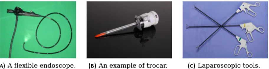
can watch the intervention on monitors and be ready to assist the
(a)A flexible endoscope. (b)An example of trocar. (c)Laparoscopic tools. Figure 2:An example of endoscopic instrumentation: a flexible endoscope
used to examine the interior of a hollow organ or cavity of the body (Figure 2a), an example of trocar used to introduce ports in the abdomen (Figure2b), and some laparoscopic instruments (Figure2c) [24,54,65,80,144,157,207,264].
However, MIS procedures present some drawbacks for the geon too. In fact, in traditional open surgery the guidance of
sur-gical instruments is carried out under direct visual control of the
surgeon, and the intervention can be executed with one dominating
hand performing the critical phases. On the contrary, in MIS true ambidextrous skill is required and the handling of the instruments is
carried out viewing the surgical field using an endoscopic camera. In
particular, this means to lose the natural 3D vision replaced by a
mag-nified but restricted bidimensional vision, requiring good hand-eye
coordination skill. Furthermore, the handling of endoscopic surgical
instruments is difficult due to the constraints imposed by the
inser-tion points, and to the reduced workspace. Finally, unlike open
sur-gery, the surgeon has not tactile perception and only limited haptic
feedback while performingMIS interventions. All these difficulties usually result in a prolonged learning curve and in longer operative
time in respect to an open surgery approach [64,201].
1.2.3 endoscopic equipment
Endoscopic surgery requires special medical equipment designed to
or part (as the bladder or esophagus) for diagnostic or therapeutic
purposes that typically has one or more channels to enable passage
of instruments (as forceps or scissors) (see Figure2a on the previous page) [70].
the trocars are medical instruments with a sharply pointed end,
often three-sided, that is used inside a hollow cylinder (cannula) to
introduce this into blood vessels or body cavities. Trocars are also
used to introduce ports in the abdomen, such as during laparoscopic
surgery. Sometimes the combined trocar and introduced cannula are
referred to as a trocar. The trocar is often passed inside a cannula,
and functions as a portal for the subsequent placement of other
de-vices, such as a chest drain, or an intravenous cannula (see Figure2b on the preceding page) [80,144,157,249].
the endoscopic instruments are long and thin surgical tools
ergonomically designed to facilitate grasping, mobilization,
dissec-tion, and transection of tissue (see Figure2c on the previous page).
Clinical research, as the size of the instruments is reduced, has
ex-panded the indications for minimally invasive approaches to take
ad-vantage of the shorter hospitalization and of the reduced surgical
1.2.4 fields of application
The MIS concept has been applied in several surgical specialties, as: laparoscopy, arthroscopy, endovascular surgery, and cardiac
sur-gery.
laparoscopic surgery is a modern surgical technique in which
operations in the abdomen, or in pelvic cavities, are performed
us-ing small incisions with the aid of an endoscopic camera. At present,
cholecystectomy is the most common laparoscopic procedure
per-formed (see Figure3a on page 11) [264].
arthroscopic surgery is a common orthopedic procedure that
is used to diagnose and treat problems in joints. Technically
speak-ing, arthroscopic surgery could potentially be performed on any joint,
such as: knee, shoulder, hip, ankle, elbow or wrist. Knee and
shoul-der arthroscopy are by far the most common arthroscopic
proce-dures performed, including: repairing cartilage and meniscus
prob-lems in the knee, and repairing rotator cuff tears in the shoulder
[54,65,80,144,157,207,249].
endovascular surgery is a form of minimally invasive surgery
designed for the treatment of vascular diseases from the inside of
blood vessels.
Endovascular techniques were originally designed for diagnostic
purposes. Basic techniques involve the introduction of a catheter
percutaneously (or rather through the skin), into a large blood
ves-sel. The catheter is injected with a radio-opaque dye that can be seen
on live X-ray or fluoroscopy. As the dye courses through the blood
vessels, characteristic images are seen by experienced viewers and
vas-composed of fine wire materials such as: titanium, stainless steel,
and nickel titanium (or nitinol NiTi). These that can be inserted
through a thin catheter and expanded into a predetermined shape
once they are guided into place [80,101,204].
minimally invasive cardiac surgery (mics) (also called
min-imally invasive heart surgery or the McGinn technique) is an
ap-proach to heart surgery that does not involve cutting open the chest
and dividing the breast bone. Using a MICS approach, the sur-geon can access the heart through a small incision between the ribs.
Heart repair is performed using special elongated instruments,
view-ing scopes, and video assistance. This results in a dramatical
reduc-tion of the surgical trauma, shortening the patient recovery process
[207].
1.2.5 innovative minimally invasive techniques
The continuous development of new concepts pushed the frontiers
ofMISto innovative ultra-minimally invasive techniques, as: Single Access Endoscopic Surgery (SAES) and Natural Orifice Transluminal Endoscopic Surgery (NOTES).
single access endoscopic surgery (saes) is a recently
devel-oped minimally invasive surgical technique, also known as Single
In-cision Laparoscopic Surgery (SILS), in which the surgeon operates almost exclusively through a single entry point, typically the
umbili-cus of the patient but also other abdominal sites.
SAES interventions are accomplished through a single incision, SAES pros and cons.
minimizing the scarring and incisional pain associated with the
mul-tiple points of entry used during traditional endoscopic surgery.
TheSAEStechnique has been used to perform many types of sur-gery, including: adjustable gastric banding, appendectomy,
cholecys-tectomy, colectomy, hernia repair, hysterectomy, sleeve gastrectomy,
nephrectomy, and sacrocolpopexy [110,271].
natural orifice translumenal endoscopic surgery (or
trans-luminal surgery) is an extension of endotrans-luminal procedures,
allow-ing the surgeon to perform the intervention by way of a lumen.
This approach has been referred to as Natural Orifice
Translumi-nal Endoscopic Surgery (NOTES), and consists in an experimental surgical technique performed endoscopically by initially passing the
endoscope through a natural orifice (e.g. mouth, urethra, or anus),
then transluminally into areas that would not otherwise be
accessi-ble endoscopically (e.g. abdomen or pelvis).
Theoretical advantages ofNOTESover the laparoscopic approach NOTES pros and cons.
include less invasiveness, elimination of any external incisions or
scars (scarless surgery), and a reduction in postoperative
abdomi-nal wall pain, wound infection, hernia formation and adhesions [18, 77,266].
During the initial years, endoscopic surgery was limited by several From MIS to CAS.
factors such as: bidimensional restricted vision, the difficulties in
handling the surgical instruments, the lack of tactile perception, and
the limited working area are factors which add to the technical
com-plexity of the minimally invasive surgical approaches. Nevertheless,
the number of operations grew, surgeons became skilled over the
limitation imposed byMISand along the years this gap was almost recovered [118].
(a)Physicians perform laparoscopic sur-gery.
(b)An example of Computer Assisted Surgery.
Figure 3:An example of Minimally Invasive Surgery procedure (Figure3a)
and Computer Assisted Surgery system (Figure3b) [139,264].
MIS has been enhanced by specialized tools for decades, as well as by Computer Assisted Surgery (CAS) systems. CAShas been par-ticularly effective to improve the performance of minimally invasive
interventions, and has been a lead in factor for the development of
robotic surgery [64,257].
1.3
computer assisted surgery
Even modern imaging techniques do not offer anything more than
the possibility of preoperative “map briefing”. Computer Assisted
Surgery (CAS) introduce innovative instruments into the operation theater [2].
computer assisted surgery (cas) is the concept and set of CAS definition.
methods that use computer technology to enhance the surgical
pro-cedures including: surgical training, preoperative planning,
intraop-erative navigation and assistance [257].
Two major advances can be achieved byCAS. On the one hand, be- CAS benefits.
fore starting the intervention, the surgeon can navigate through the
impres-sion of the region and its neighborhood. This enhanced visualization
of the operative field can also increase the accuracy of the
preop-erative diagnosis and surgical planning. Furthermore, the planned
approach can be assessed using a virtual surgery simulator in order
to optimize the strategy and decrease the surgical morbidity. On
the other hand, during the surgery, the computer guidance helps the
surgeon in navigating through the patient anatomy: avoiding time
consuming procedures, decreasing the risk of surgical errors, and
increasing the safety and quality of the intervention [2].
1.3.1 essentials
Despite the name,CASincludes all steps from patient data acquisi-tion to final surgery, and even post-operative control. The general
principles of aCASsystem can be summarized in: the generation of a virtual image of the patient, followed by the medical diagnosis. The
same virtual model can be used for the preoperative planning and/or
simulation of a surgical intervention, as well as for the intraoperative
surgical navigation.
generating the virtual patient
The Computer Assisted Surgery requires the development of an
ac-curate model of the patient. This virtual representation can be
gener-ated using several medical imaging techniques as: X-ray Computed
Tomography (CT), Magnetic Resonance Imaging (MRI), and 3D ultra-sound imaging.
Initially, the target anatomical region is scanned and the output
dataset is stored into a computer system. During this phase, it is
also possible to apply different medical imaging methods, combining
The final result is a 3D representation (volumetric or geometric) of
the anatomical region to be operated, including pathological tissues
and structures.
medical diagnosis and preoperative planning
The patient medical dataset generated can be visualized using
spe-cific software suites, allowing the surgeon to examine the 3D target
anatomy from different views. Inspecting the anatomy using a
vir-tual 3D visualization improves the accuracy of the medical diagnosis,
and enhances the assessment of the surgical case.
Additionally, before the surgical procedure takes place, the
inter-vention can be planned using the virtual patient. In this way the
surgeon is able to evaluate various strategies in order to detect
po-tential difficulties that may arise during the surgery.
surgical simulation and intraoperative navigation
The 3D model of the patient is not only useful to plan the intervention,
but it allows also the virtual simulation of the procedure. Thanks to
surgical simulators, designed to reproduce specific surgical setup
(e.g. using virtual laparoscopic devices), the surgeon can perform
the operation in a virtual environment training and validating the
planned strategy.
Eventually, during the surgical intervention, intraoperative
naviga-tion systems assist the surgeon. These softwares, using the medical
dataset, provide a 3D visualization of the virtual anatomy aligned
im-proved view of the surgical field, avoiding loss of orientation or
un-wanted collisions.
1.3.2 applications
CAS systems have been used in several interventions of different
surgical specialties: from neurosurgery to orthopedic procedures.
computer assisted neurosurgery started to use
telemanipula-tors in 1980, allowing a greater development in brain microsurgery
and increasing the accuracy and precision of the intervention. This
long experience using telemanipulators has favoured the
develop-ment of innovativeCASsystem for minimally invasive brain surgery, furthermore reducing the risk of post-surgical morbidity by
acciden-tally damaging adjacent centers.
computer assisted oral and maxillofacial surgery uses a
surgical approach, based on aCASsystem, called the bone segment navigation. This is applied to: orthognathic surgery, temporo
man-dibular joint surgery, and the reconstruction of the mid-face and
or-bit [126].
computer assisted orthopedic surgery (caos) (togheter with
the usage of robots) is widespread in orthopedics, especially in
rou-tine interventions, like total hip replacement. CAOS is especially useful in pre-operative planning and intraoperative guidance (e.g. to
find the correct anatomical position of displaced bone fragments in
fractures, allowing a good fixation by osteosynthesis) [73].
computer assisted general surgery utilizesCAS systems al-lowing important progresses in general surgery towards MIS
ap-cal robots contributed in laparoscopic approaches for pyeloplasty or
nephrectomy or prostatic interventions [72,156].
1.3.3 state of the art
In 1991, VPL Research Inc. created a computerized 3D model of the
optic nerve that can be studied from any point of view.
Few years later, in 1994 at the Brigham and Women’s Hospital
in Boston, the radiologists Ron Kinkinis and Ferenc A. Jolesz and
the neurosurgeon Peter M. Black began performing neurosurgeries
superimposing a 3D representation of the brain, generated and
up-dated using images fromMRIscans, on a live video frame [158,201, 232].
In these last decades, several universities and research laboratories
have developedCASsystems, addressing different surgical interven-tions. Unfortunately, at the moment, many of these systems are
ex-perimental devices.
Only few CASplatforms have been approved for clinical use and are commercially available, some examples are : the Medtronic®
StealthStation®, the Stryker®NavSuite®, and the Brainlab®
Vector-Vision®[29,133,229,257].
Most of theCASsystems use an optical infrared (IR) tracking tem, but there are also solutions using electromagnetic tracking
(a)The AESOP®System. (b)The ZEUS®Robotic Surgical System. Figure 4:Two examples of the first commercial surgical robots: the AESOP®
(Figure 4a) and the ZEUS® (Figure 4b), both by the Computer
Motion®Inc. [43,44,274].
Technological innovations in the field of robotics and computer
sci-ence will drive the future of minimally invasive and computer
assis-ted surgery toward robotic surgery or cybersurgery [118,207].
1.4
robotic surgery
The initial concept of robotic-assisted surgery involved mainly
re-mote presence or telesurgery: that means enable the surgeon to
operate at a remote site. It was thought that the transposition of
surgical and technical expertise had the potential to expand surgical
applications. Although simple surgical procedures have been
per-formed remotely, there are some difficulties for an extensive clinical
use due to high costs, transmission latency and medical and legal
issues.
Recently, robot assisted surgery has emerged as a popular method, The concept.
often combined withCASsystems, to enhanceMISinterventions. Ac-tually, the unaided human hand cannot reliably position a surgical
Figure 5:A photo of the da Vinci®S Surgical System by Intuitive Surgical
Inc. [91,179].
instrument to within less than a hundred microns of its target.
Fur-thermore, muscle fatigue in the hand rapidly creates a tremor, at a
frequency of8Hzto14Hz. Surgical robots allow finer surgical ges-ture: providing enhanced dexterity and motion filtering and scaling.
Moreover, novel surgical robotic systems often incorporate sensors
and miniaturized tactile devices to convey to the surgeon the tactile
feedback [201].
Even if surgical robots offer many advantages, the number of robot
assisted interventions is increasing slowly, mainly due to the high
investment required and running costs of the devices [118].
1.4.1 commercial robotic surgical systems
The first known surgical procedure using a robot to improve the
qual-ity of the intervention, was performed in 1985, when an Unimation
PUMA®560 was used to place a needle for a brain biopsy usingCT guidance [108,159,269].
Three years later, in 1988, the PROBOT by Imperial College
In 1992, the ROBODOC®was presented to mill out precise fittings
in the femur for hip replacement (see Figure6a on the next page) [50,52,257,269].
The first robot for endoscopy appeared one year later and was the
Computer Motion®AESOP®, which received the US Food and Drug
Administration (FDA) approval in 1993. It was a voice-controlled robotic endoscopic positioning system, providing a steady video
dur-ingMISprocedures (see Figure4a on page 16) [7,43,104,257,269, 274].
A second generation of this technology, as the Computer Motion®
ZEUS™ Robotic Surgical System, approved by FDAin 1994, made endoscopic procedures even more accessible. This device had three
robotic arms remotely controlled by the surgeon: the first was an
AESOP®, whereas the other two act like extensions of the surgeonís
own arms, mimicking his movements and also allowing for more
pre-cise executions of the surgical gesture (see Figure 4b on page 16) [44,257,269,274].
In 1997, the Intuitive Surgical® da Vinci® Surgical System (see
Figure5 on the preceding page) received theFDAapproval to assist the surgeon. Few years later, in 2000 it received also theFDA ap-proval to perform stand-alone surgical interventions as remote
ma-nipulator. The complete platform consists in a robotic surgical
sys-tem controlled by a surgeon from a console, and designed to improve
conventional laparoscopic procedures. The console, typically in the
same room as the patient, controls four interactive robotic arms:
three arms are for surgical instruments like scalpels, scissors etc.,
whereas the fourth arm is for an endoscopic stereo camera. The
in-terface at the console provides: stereo 3D vision of the surgical field,
and two foot pedals together with two hand controllers to maneuver
the robot arms. The da Vinci®Surgical System provides motion
(a)The ROBODOC®Surgical System.
(b)The Neuromate®stereotactic robot.
Figure 6:Two examples of commercial surgical robots for orthopedics and
neurosurgery: the ROBODOC®(Figure6a) by Curexo Technology
Corporation, and the Neuromate® (Figure 6b) by the Renishaw
PLC [23,50,52,188].
precise micro-movements of the endoscopic instruments operating
through small incisions in the patient body [91,179,257,262,269].
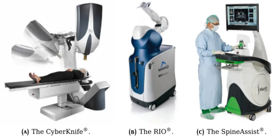
Innovative surgical robots have been developed also for orthopedics,
neurosurgery, and radiosurgery. Some examples are: the already
mentioned ROBODOC®Surgical System, the Neuromate®
stereotac-tic robot, the CyberKnife® Robotic Radiosurgery System, the RIO®
Robotic Arm Interactive Orthopedic System, and the SpineAssist®
Robot combined with the Renaissance™ Guidance System (see
Fig-ure6and7 on page 21) [1,50,122,127,128,188].
The complete ROBODOC®Surgical System for orthopedics
devel-oped by Curexo Technology Corporation, combines the technologies
of ORTHODOC®and ROBODOC®Surgical Assistant (see Figure6a). The simulator enables the virtual pre-surgical 3D planning to
opti-mize the prosthetic selection and alignment. It also improves the
subsequent execution of the intervention using the high-precision
robotic capabilities in preparing the bone to achieve optimal fit of
the prosthetic implant [50,52,257].
The Neuromate® stereotactic robot marketed by Renishaw®PLC,
provides a platform solution for several functional neurosurgical
in-terventions (see Figure6b). The system has been used in different procedures, as: electrode implantation procedures for deep brain
stimulation, stereotactic electroencephalography, stereotactic
appli-cations in neuroendoscopy, radiosurgery, biopsy, and transcranial
magnetic stimulation [23,188,257].
In 2001 appeared the CyberKnife® Robotic Radiosurgery System
by Accuray Inc., consisting in a robot that incorporates a linear
accel-erator (see Figure 7a on the next page). The system was designed to treat tumors throughout the body non-invasively using a
radio-surgery approach, or rather delivering beams of high dose radiation
to tumors with extreme accuracy. The CyberKnife®is able to follow
soft tissue deformations due to the patient breathing. This robot
of-fers a pain-free, non-surgical option for patients who have inoperable
or surgically complex tumors [1,3,257].
Few years later, MAKO®Surgical Corp. announced the release of
the RIO®Robotic Arm Interactive Orthopedic System (see Figure7b on the following page). This system includes tactile feedback and high definition patient specific visualization, allowing the
preopera-tive planning for orthopedic interventions. Furthermore, integrated
software applications enable a virtual simulation to optimize implant
positioning and placement in order to restore biomechanical
align-ment and joint motion [69,109,122,190,231].
In 2011, Mazor Robotics Ltd. presented the SpineAssist® Robot
for spine surgery. Together with the Renaissance™ Guidance
Sys-tem, the surgical robot allows accurate interventions also reducing
the exposure to radiations (see Figure 7c on the next page). The platform enables a variety of spine procedures: from open surgery
to minimally invasive techniques, and also percutaneous posterior
thoracolumbar approaches. Moreover, it allows the treatment of
scoliosis and other complex spinal deformities, and also to perform
(a)The CyberKnife®.
(b)The RIO®.
(c)The SpineAssist®.
Figure 7:Three examples of commercial surgical robots for radiosurgery and orthopedics and spine surgery: the CyberKnife® Robotic
Ra-diosurgery System by Accuray Inc. (Figure 7a), the RIO®
Ro-botic Arm Interactive Orthopedic System by MAKO® Surgical
Corp. (Figure7b), and the SpineAssist®Robot together with the Renaissance™Guidance System both by Mazor Robotics Ltd. (Fig-ure7c) [1,122,127,128].
1.4.2 telesurgery
Telesurgery systems were initially designed to implement the remote
surgery concept, but the initial difficulties in long distance
telecom-munications pushed the development toward the exploitation of
sur-gical robotics in order to enhance the dexterity of the surgeon. In
fact, the first commercialized systems allowed the surgeon to
con-trol the surgical robot using a console located near the patient, in
the same operating room. However, later the advancements in
com-puters, electronic systems, and communication technologies made
feasible the implementation of the remote surgery. The computer
system converts the motion of the surgeon into electronic signals
that can be transmitted to distant sites [201,275].
Today telesurgery mainly means remote surgery, and this form of
telepresence can be defined as:
telesurgery is the surgery performed at a distance from the Telesurgery definition.
technol-ogy and, when distances from the surgeon to the patient are great,
robust formats for data transmission [207,249].
In telesurgery, the surgeon operates using a control console located
in the same room as the patient of in a remote location. The
con-cept is to provide the surgeon enough sensory input to simulate the
feeling of directly performing surgery on the patient [201].
Telesurgery combines: surgical robotics, cutting edge
communica-tion technology, and informacommunica-tion management. Even if robotic
sur-gery is fairly well established, most of these systems are controlled
by surgeons at the location of the surgery. Remote surgery takes
ad-vantage of advanced telecommunications to make the physical
dis-tance between the surgeon and the patient immaterial. These
sys-tems allow the remote surgical expertise by specialized surgeons to
be available worldwide [64,268].
applications
In 1997, the surgeons Jacques Himpens and Guy-Bernard Cadière First telesurgeries.
performed the first human gallbladder operation via telesurgery, at
the Saint Blasius Hospital in Brussels. One year later, in 1998, the
heart surgeons Alain Carpentier and Didier Loulmet performed the
first closed-chest human telesurgery heart operation, at Broussais
Hospital in Paris [201].
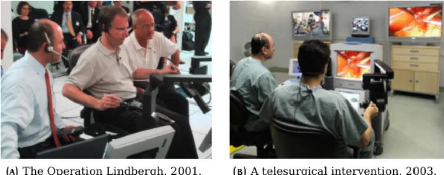
One of the first long-distance telesurgery interventions was the Long-distance telesurgery.
Operation Lindbergh, performed on 7 september 2001 across the
Atlantic Ocean. Dr. Jacques Marescaux in New York (US) carried
out a cholecystectomy on a 68-year-old female patient in Strasbourg
(France). The entire surgical procedure was conducted using
tele-communications via dedicated fiberoptic link to ensure guaranteed
connectivity and minimal lag: the established round-trip latency
dur-ing the surgery was135ms(see Figure8a on page 24) [92,125,265, 268].
with regular telecommunications, no connection issues were raised
during the remote surgical procedures (see Figure 8b on the next page) [92,268].
Since then, remote surgery has been conducted many times in
numerous locations, and the rapid development of technology has
allowed remote surgery rooms to become highly specialized [268].
Commenting on the Operation Lindbergh, Dr. Jacques Marescaux
said: “I believe that this demonstration of the feasibility of a
com-pletely safe remote surgery ushers in the third revolution in surgery,
after the arrival of MIS and the introduction of CAS... These pro-posed the concept of distance between the surgeon and the patient.
Therefore, we can imagine that this distance could potentially be up
to several thousand kilometers.” [265].
Unfortunately, at present time, all the telesurgical systems under Limitations.
development use either a direct cable connection or a wireless link
over a very short distance. This is mainly due to the
communica-tion latency required by these systems. The lag time is a limitacommunica-tion
for telesurgery, since the surgeon is not able to compensate for the
delays [201].
Researches on astronaut training explain how delays affect the
dexterity in remotely manoeuvring a servomechanism (e.g. the
ro-botic arm of the NASA Space Shuttle). Delays of less than25msare impercetible. If the delay is of about50ms, the operator perceives that something is wrong but is able to automatically compensate for
com-(a)The Operation Lindbergh, 2001. (b)A telesurgical intervention, 2003. Figure 8:Two photos of the first telesurgeries: Dr. Marescaux and Dr.
Gag-ner in New York (US) performing the Operation Lindbergh on 7 september 2001 (Figure8a), and Dr. Anvari in Hamilton (Canada) performing a laparoscopic telesurgery on 28 february 2003 (Fig-ure8b) [92,265,268].
pensate only if properly trained. Reaching a latency of 200ms is nearly impossible for anyone to manage the delay, and additionally
telesurgical systems become unstable, tending to crash [201]. Therefore, telesurgery is quite popular in theory, though
telesurgi-cal techniques have not been widely adopted due to several factors.
Its acceptance requires many issues to be resolved, as: the
establish-ment of clinical protocols, the developestablish-ment of specific training, the
design of global compatible equipment, and the need of a backup
surgical team to recover in case of a lack in telecommunications or
a malfunction in the robotic system. Nevertheless, the first
experi-ences in the early years of the XXI century demonstrated that the
technology exists to enable the delivery of expert care worldwide
[268].
1.5
surgical planning and assistance
3D visualization of medical datasets enables the surgeons the
com-plete understanding of the patient anatomy. Image processing,
modali-For many procedures in different surgical specialties, specific
soft-wares have been developed to improve the presurgical planning or
to simulate treatments on a virtual patient model. In orthopedic
sur-gery, planning systems assist the surgeon in the determination of
the optimal placement for the prosthesis. In endovascular surgery,
preoperative planning softwares enable the surgeon to perform
mea-surements on the target vessel in order to design the stent graft prior
to surgical intervention. In neurosurgery, path-planning systems
as-sist the surgeon to find the optimal path for the surgical instrument
or the radiation beam [59,99,192,206].
Preoperative planning systems, allowing the determination of the
optimal surgical strategy for a specific intervention, not only ease
surgical training encouraging rehearsal, but also support
intraoper-ative image guidance during the execution of the planned procedure
[200].
1.5.2 intraoperative assistance systems
The developments in registration and real-time sensing enabled the
design of innovative intraoperative assistance systems to aid the
sur-geon in following the preoperative planned strategy during the
inter-vention [40,111,166].
A proper simulation of the surgical procedure results in a
well-designed and geometrically correct preoperative plan, which is the
All the data, preoperative and intraoperative, have to be properly
integrated in the same frame of reference, performing an
image-to-patient registration. After this initial alignment, tracking the image-to-patient
motion and using real-time imaging, the registration can be updated.
These allow the usage of robotic devices and the integration of
real-time tracking of surgical instruments [12,120,252].
Surgical robotic systems are able to enhance the surgical
per-formance not only improving the accuracy of the surgical gesture,
but also performing routine tasks or simply assisting the surgeon
[31,68,83,99,123,233,250].
Additionally, the vision of the surgeon can be augmented
perform-ing the fusion of preoperative and intraoperative data from several
medical imaging sources, such as: video,CT,MRI, ultrasound, fluo-roscopy, microscope and endoscope [15,31,42,58,62,71,212].
Surgical robots, 3D visualization, real-time sensing and
multimo-dal medical imaging provide new capabilities to enhance surgical
performance [114,155,253,279].
Finally, tele-presence and tele-surgery systems can be developed
integrating both CAS and robotic technologies, as in the Intuitive Surgical® da Vinci® Surgical System (see Figure 5 on page 17) or the Computer Motion® ZEUS™ Robotic Surgical System (see
Fig-ure4b on page 16) [44,91,199].
As already described, surgery in the information age has advanced
mainly through the integration of different technologies.
Advancements in computer graphics, especially with the birth of
virtual reality concept, provide immersive virtual environments
en-abling surgeons to easily manipulate 3D anatomies even with haptic
feedback. Stereo imaging devices, advanced computer systems and
innovative surgical robots allow the development of a unique
some of the most simulated procedures. During a virtual surgery
the surgeon uses a computer display visualizing the surgical field
in 3D. The surgical instruments are connected to specific devices to
perform motion-tracking and tactile feedback. The user can carry
out the surgical procedure upon the virtual patient anatomy by
ma-nipulating the surgical tools, displayed in the virtual scenario using
specific virtual 3D replicas.
Preliminary studies suggest that an hour of training on a surgical Advantages.
simulator is worth three hours practicing on an animal or human
cadaver. Virtual surgery allows surgeons to practice interventions
multiple times without requiring cadavers or animals. Hence, an
an-cillary benefit of these virtual systems could be to reduce the number
of animals required for the medical education of surgeons.
Addition-ally, the simulation software can track the surgical gesture during
the whole virtual procedure, in order to analitically evaluate the
sur-geon dexterity or the learning curve of a trainee. Moreover, these
systems allow the simulation of rare pathological cases enabling
sur-geons to extensively train difficult interventions or complications.
Fi-nally, virtual training not only may help trainees to enhance their
skills before the first real surgery, but can also be used to
periodi-cally assess the dexterity of expert surgeons [201].
The first surgical intervention following a virtual surgery practice, The first preoperative virtual surgery.
was performed on 17 august 2009 by Dr. David Clarke in Halifax
(Canada) performing the removal of a brain tumour 24 hours after
Moreover, these software simulators have applications not only in
virtual surgery, but they have also great potential for use in medical
education [201].
Despite the several advancements in medicine and surgery,
medi-cal education is almost the same of thirty years ago and most medimedi-cal
schools still use a rote learning approach [201].
Virtual enviroments can provide the intuitive learning framework
students need. In fact, in 1994 the National Library of Medicine
released the first version of the Visible Human Project®, two digital
image datasets of a human male and a human female. Once the
project is complete, students will be able to gain a comprehensive
understanding of the anatomy and the interrelations between organs
[164,201].
Technically speaking, virtual surgery software is based on an
inter-active dynamical simulation, carried out by a physics engine
manag-ing the physical behaviour of all the entities in the virtual scenario
[259,267].
1.6.1 introduction to interactive simulation
Interactive simulation is one of the main topics of Computer
Graph-ics (CG). Usually simulators are based on real-time physics engines providing models to represent and simulate natural phenomena
us-ing computer systems.
a physics engine is computer software that provides an approx- Phisics engine definition.
imate simulation of certain physical systems, such as rigid body
dy-namics (including collision detection), soft body dydy-namics, and fluid
ing lower accuracy, but ensure a high update rate as needed by video
games or interactive simulators [267].
real-time physics engine
The main task of a real-time physics engine is to solve the forward
dynamics problem. This means to compute the motion of the system
knowing all the forces and constraints acting on that system.
The main factors describing a physics engine are: the simulation
paradigm, the numerical integrator, and the collision detection and
response. These determine the overall performance of the engine,
but also provide quite different results even if simulating the exact
same system [25,55,255,256,259,267].
A brief description of these essential factors is reported below:
the simulator paradigm determines which aspects can be
ac-curately simulated, affecting the precision in resolving constraints.
There are three major paradigms for the physical simulation of solids:
penalty based, constraint based, and impulse based methods; but
also hybrid solutions are possible combining different techniques
[16,25,55,146,267].
the numerical integrator determines the numerical accuracy
of the simulation [17,25,55].
the collision detection and response detects the
(or deformation) following a collision, also contributing to the
effi-ciency and accuracy of collisions in the simulation [25,76,98,255, 256,267].
the object representation contributes to the efficiency and
accuracy of collisions in the simulation [25,76,186].
the material properties determines which physical models the
simulation can approximate (e.g. the Coloumb friction) [25,97].
the constraint implementation determines which constraints
are supported and how accurately they can be simulated [25,55].
The main limitation of a physics engine is the numerical precision of Limitations.
the positions and forces. Low accuracy may affect results and the
simulation can lead to an unexpected behaviour. Higher precision
reduces the errors on positions and forces, but at the cost of greater
computing power [267].
1.6.2 commercial surgical simulators
The very first virtual surgical simulator was developed in 1991 by
Lanier and Satava. The system was designed for abdominal surgery
enabling the trainees to navigate the virtual anatomy in order to fully
understand the physical interrelations between organs. Finally, the
simulator incorporated various laparoscopic tools to practice several
endoscopic surgical techniques [201].
In 1994, MusculoGraphics Inc. developed a limb trauma
simula-tor recreating tissue properties, bleeding, and wounding. It even
en-laparoscopic surgery simulators
mentice mist is a minimally invasive surgical trainer developed
by Mentice Inc., designed for training and assessment of endoscopic
surgical skills. MIST™ includes a frame holding a pair of standard
laparoscopic instruments connected to a laptop displaying the
move-ment of the virtual surgical instrumove-ments in the 3D environmove-ment.
Trainees are guided through a series of exercises of progressive
complexity; but the system also allows the customization of the
train-ing programs, and provides data on the performance scores for
com-parative analyses and objective assessment [137].
simbionix lap mentor is a laparoscopic surgical simulator
de-veloped by Simbionix™USA Corporation, providing a complete
train-ing solution for: laparoscopy, gynecology, urology and general
sur-gery (see Figure9b on the following page). The system includes a library of modules for basic and advanced procedure training.
The system enables a robust simulation and has an advanced
er-gonomic design, it also allows a tactile experience of tissue
resis-tance feedback via the laparoscopic haptic interface [219].
surgical science lapsim is a surgical skills training system
de-signed for: basic skills, cholecystectomy, gynecology, suturing and
anastomosis, and appendectomy. The simulator is adaptable and
ver-satile, allowing the trainee to learn and practice several exercises:
Further-(a)Mentice VIST™System. (b)Simbionix™LAP Mentor™. Figure 9:Two examples of commercial surgical simulators: the Mentice
VIST™ System for endovascular training (see Figure9a) and the
Simbionix™ LAP Mentor™ for laparoscopic surgical simulation
(see Figure9b) [138,219].
more, LapSim®can be extended with additional add-on modules for
specific surgical intervention simulation [230].
simsurgery sep is a training and educational platform for
la-paroscopic surgery. The system combines surgical simulation and
multimedia content to practice skills, knowledge and judgment. The
SEP offers the possibility to customize a structured training program
for surgical trainees, including performance evaluation
functionali-ties. The complete package comprehends modules for the virtual
practice of: basik skills, cholecystectomy, and ectopic pregnancy
[225].
endovascular simulators
mentice vist is an endovascular simulator developed by Mentice
Inc., which enables procedural training for clinicians and medical
professionals (see Figure9a). The high fidelity simulation provides optimal environment for proficiency based training, and enhances
the clinical training by using real devices and equipment.
Further-more, VIST™supplies tactile feedback and realistic cath lab
choosing appropriate instruments. The platform incorporates also a
haptic interface providing realistic visual and tactile feedback while
using guidewires, balloons, stents and other interventional devices
[216].
immersion laerdal virtual i.v. simulator is a learning system
for training intravenous catheterization. The simulator integrates
real-time 3D visualization with a force feedback device to provide an
immersive experience [86].
bronchoscopy and gastrointestinal simulators
simbionix gi mentor is an endoscopic medical simulator for the
training of gastrointestinal upper and lower endoscopic procedures
developed by Simbionix™USA Corporation. The system offers a
com-prehensive library of modules including several basic and advanced
tasks and virtual patient cases. The simulator includes a colonoscope
and a duodenoscope customized to provide realistic visual and
tac-tile feedback [218].
simbionix bronch mentor is an additional module for the
endo-scopic medical simulator Simbionix™ GI Mentor™, providing a
com-prehensive solution for the flexible bronchoscopy training. The
learn-ing platform combines basic and advanced tasks to provide an
opti-mal training environment, supporting: posterior and lateral
work-ing positions, oral or nasal scope introduction, various classification
More-(a)The Immersion Laparoscopy VR™. (b)The Simbionix™VirtaMed HystSim™. Figure 10:Two examples of commercial surgical simulators: the Immersion
Laparoscopy VR™, one of the first laparoscopic simulators (see Figure10a); and the Simbionix™ VirtaMed HystSim™for the vir-tual hysteroscopy (see Figure10b) [223].
over, the system instrumentation includes a physical syringe to
simu-late a realistic fluids delivery and BAL performance; whereas a
physi-cal master tool simulates a wide variety of bronchoscopic tools, such
as: biopsy forceps, cytology brush, aspirating needle, balloon,
elec-trocautery probes and more [217].
immersion cae endoscopyvr surgical simulator is a surgical
simulation platform designed for virtual training in both
bronchos-copy and gastrointestinal procedures. The Endosbronchos-copyVR allows
sur-geons to practice basic and advanced skills, offering: haptic
feed-back, physiological simulation, metrics reports, vital signs and drug
administration [85].
other surgical simulators
simbionix uro mentor is a medical simulator providing a
sim-ulation platform for training in endourology interventions as:
cys-toscopy or ureteroscopy. The system enables the simulation of
ei-ther rigid and flexible cystoscopes and ureteroscopes, allowing also
the practice of several tasks as: stone extraction, stone lithotripsy,
in-which operates on the Simbionix™URO Mentor™ platform properly
extended. The system includes a mannequin representing the virtual
patient equipped with different interchangeable layers of epidermis
and underlying tissue combined with simulated ribs [221].
simbionix virtamed hystsim is a training system developed by
Simbionix™USA Corp. together with VirtaMed AG, designed to
sim-ulate diagnostic and therapeutic hysteroscopy (see Figure 10b on the preceding page). The system provides realistic training for both experienced and novice surgeons, and includes a custom
resecto-scope to easily acquire experience with real hysteroscopic
instru-ments [223].
simbionix virtamed turpsim is a comprehensive educational
so-lution developed by Simbionix™USA Corporation together with
Vir-taMed AG, enabling realistic training in the transurethral resection
of the prostate (TURP). The system includes a computer provided with two monitors, and an adapted original resectoscope to improve
the realism of the simulation experience [224].
simbionix pelvic mentor is an advanced simulator designed to
train gynecologists and urogynecologists for pelvic floor
reconstruc-tion and transvaginal mesh procedures. The system allows trainees
to acquire knowledge of pelvic anatomy and to enhance their skills
in pelvic reconstructive surgery. The PELVIC Mentor™ includes a
![Figure 1: Two paintings by Thomas Eakins (Figure 1a and Figure 1b) docu- docu-menting the history of medicine: honoring the emergence of sur-gery and showing a typical surgical theater in the late XIX century [202, 203].](https://thumb-eu.123doks.com/thumbv2/123dokorg/7552481.109532/22.892.239.680.134.348/paintings-history-medicine-honoring-emergence-surgical-theater-century.webp)
![Figure 3: An example of Minimally Invasive Surgery procedure (Figure 3a) and Computer Assisted Surgery system (Figure 3b) [139, 264].](https://thumb-eu.123doks.com/thumbv2/123dokorg/7552481.109532/31.892.240.679.134.329/figure-minimally-invasive-surgery-procedure-computer-assisted-surgery.webp)

![Figure 5: A photo of the da Vinci ® S Surgical System by Intuitive Surgical Inc. [91, 179].](https://thumb-eu.123doks.com/thumbv2/123dokorg/7552481.109532/37.892.228.686.118.394/figure-photo-da-vinci-s-surgical-intuitive-surgical.webp)
![Figure 6: Two examples of commercial surgical robots for orthopedics and neurosurgery: the ROBODOC ® (Figure 6a) by Curexo Technology Corporation, and the Neuromate ® (Figure 6b) by the Renishaw PLC [23, 50, 52, 188].](https://thumb-eu.123doks.com/thumbv2/123dokorg/7552481.109532/39.892.241.679.139.294/commercial-surgical-orthopedics-neurosurgery-technology-corporation-neuromate-renishaw.webp)
![Figure 9: Two examples of commercial surgical simulators: the Mentice VIST ™ System for endovascular training (see Figure 9a) and the Simbionix ™ LAP Mentor ™ for laparoscopic surgical simulation (see Figure 9b) [138, 219].](https://thumb-eu.123doks.com/thumbv2/123dokorg/7552481.109532/52.892.247.676.142.339/examples-commercial-simulators-endovascular-simbionix-laparoscopic-surgical-simulation.webp)
![Figure 10: Two examples of commercial surgical simulators: the Immersion Laparoscopy VR ™ , one of the first laparoscopic simulators (see Figure 10a); and the Simbionix ™ VirtaMed HystSim ™ for the vir-tual hysteroscopy (see Figure 10b) [223].](https://thumb-eu.123doks.com/thumbv2/123dokorg/7552481.109532/54.892.248.680.134.301/commercial-simulators-immersion-laparoscopy-laparoscopic-simulators-simbionix-hysteroscopy.webp)
