SAPIENZA Università di Roma
Facoltà di Scienze Matematiche Fisiche e Naturali DOTTORATO DI RICERCA IN BIOLOGIA CELLULARE E
DELLO SVILUPPO
30° Ciclo (A.A. 2016/2017)
Effects of acute and sub-chronic glucocorticoid treatments on hippocampal neurons of wild type and dystrophin-deficient
DMDmdx
mice: an in vitro and in vivo study
Dottorando:
Francesca Cosmi
Docente guida
Prof.ssa Maria Egle De Stefano
Coordinatore
“I do not know what I may appear to the world, but to myself I seem to have been only like a boy playing on the seashore, and diverting myself in now and then finding a smoother pebble or a prettier shell than ordinary, whilst the great ocean of truth lay all undiscovered before me.”
Sir Isaac Newton
INDEX
Abstract 11
Introduction 13
1. The Duchenne Muscular Dystrophy and the dystrophin protein
13 2. Dystrophin expression and localization in the nervous
system
16 3. Therapeutic approaches for DMD treatment 21 4. The glucocorticoid-mediated stress signalling 22 4.1 Glucocorticoids and their receptors 22 4.2 The glucocorticoid receptor genomic response 25 4.3 The glucocorticoid receptor non-genomic signalling 26
5. The stress response 29
6. Glucocorticoid effects on adult hippocampal
neurogenesis 34
7. Glucocorticoids and DMD 37
Aim and Scope 39
Materials and Methods 41
Animals 41
Primary hippocampal neuron cell cultures 41 In vitro glucocorticoid administration 42 In vivo glucocorticoid administration 42
Acute treatment 42
Sub-chronic treatment 43
Plasma corticosterone measurement 43
Real-Time PCR 43
RNA extraction and retro-transcription 43
Quantitative RT-PCR 44
Western Immunoblot 45
Primary antibodies 45
Preparation of cell lysates and tissue extracts 45
Electrophoresis 46
Protein revelation 46
Immunofluorescence in vitro 47
Cell counting and statistical analysis 49 Peroxidase anti-peroxidase immunohistochemistry 50
Primary antibodies 50
Immunostaining 50
Statistical Analysis 51
Results 53
In vitro treatments 53
GR protein levels in the hippocampus of E18 mdx mice are significantly lower compared to wild type
53 GR and pGR protein levels after acute corticosterone
treatment
55 GR mRNA levels are differently modulated in wild type
and mdx mouse hippocampal neurons after acute treatment with high CORT concentrations
57 Acute administration of high corticosterone
concentration promotes neuronal death
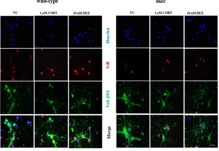
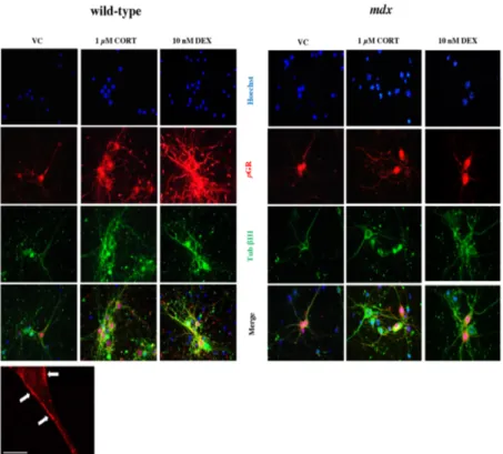
58 Immunofluorescence analysis confirms CORT effects on
hippocampal neurons disclosed by biochemical analyses
60
In vivo treatments 63
Ninety minutes after acute corticosterone treatment, GR protein levels are differently modulated in wild type and mdx mouse hippocampal neurons
64
Modulation of hippocampal GR and GILZ mRNA after 90 min from acute corticosterone treatment are different between wild type and mdx mice
65 Immunohystochemical analysis of pGR and
GR localization
66 Analysis of the levels of proteins involved in the rapid
non-genomic signalling mediated by glucocorticoids 90 min after acute corticosterone treatment
69 Six hours after acute corticosterone treatment, GR
protein levels are still differently modulated in wild type and mdx mouse hippocampal neurons
72 Modulation of hippocampal GR and GILZ mRNA after 6
h from acute corticosterone treatment are different between wild type and mdx mice
Analysis of the levels of proteins involved in the rapid non-genomic signalling mediated by glucocorticoids 6 h after acute corticosterone treatment
75
In vivo sub-chronic treatment with dexamethasone 78 GR protein levels are de-regulated in mdx mouse
hippocampi compared to wild type after sub-chronic dexamethasone treatment
79 Modulation of hippocampal GR and GILZ mRNA after
sub-chronic DEX treatment are different between wild type and mdx mice
81 Analysis of the levels of proteins involved in the rapid
non-genomic signalling mediated by glucocorticoids after sub-chronic dexamethasone treatment
82
Quantitative analysis of the neural precursor cell proliferation in the hippocampal dentate gyrus
84
Quantitative analysis of apoptotic cells in the
hippocampal dentate gyrus 88
Discussion 91
The response of cultured hippocampal neurons to acute corticosterone administration is different between wild type and mdx mice
92 In vivo, wild type and mdx mouse hippocampal neurons
respond differently to acute corticosterone treatment
93 In vivo, wild type and mdx mouse hippocampal neurons
respond differently to dexamethasone sub-chronic treatment
98 Proliferation of neural precursor cells in wild type and
mdx mouse hippocampus is differently affected by dexamethasone sub-chronic treatment
100
Conclusion 103
Glossary
Ach: acetylcholine
ACTH: adrenocorticotrophic hormone
ADHD: attention-deficit/hyperactivity disorder BDNF: brain-derived neurotrophic factor BLA: basolateral amygdala
BSA: bovine serum albumin
cAMP: cyclic adenosine monophosphate Cav1: caveolin 1
Cav2: caveolin 2
CNS: central nervous system CORT: corticosterone
CRF: corticotrophin-releasing factor CRH: corticotrophin releasing hormone DBD: DNA-binding domain
DEX: dexamethasone DG: dentate gyrus
DGC: dystrophin glycoprotein complex DMD: Duchenne Muscular Dystrophy DMSO: dimethyl sulfoxide
E18: embryonic day 18 ECM: extracellular matrix EdU: 5-ethynyl-2'-deoxyuridine GC: glucocorticoids
GILZ: glucocorticoid-induced leucine zipper GJIC: gap junction intercellular communication GPCRs: G-protein coupled receptors
GR: glucocorticoid receptor
GRE: glucocorticoid response element HDAC: histone deacetylase
HPA: hypothalamus-pituitary-adrenal axis HSP: heat shock proteins
IQ: intelligence quotient LBD: ligand-binding domain LTP: long-term potentiation
MAPK: mitogen-activated protein kinase mGR: membrane-associated GR
MR: mineralcorticoid receptor MT1X: metallothionein 1X
nAChRs: nicotinic acetylcholine receptor
NF-kB: nuclear factor kappa-light-chain-enhancer of activated B cells
NGF: nerve growth factor
nGRE: negative glucocorticoid response element NLS: nuclear localization signals
NOS: nitric oxide synthase
NPSC: neural pluripotent stem cell NTD: N-terminal domain
OD: optical density
PI3K: phosphoinositide 3-kinase PKA: protein kinase A
PNS: peripheral nervous system PS: population spike
PVN: paraventricular nucleus SCG: superior cervical ganglion ANS: autonomous nervous system CNS: central nervous system SGZ: subgranular zone STD: short-term depression STP: short-term potentiation 14 DIV: 14 days in vitro
Abstract
Duchenne muscular dystrophy (DMD) is a lethal X-linked disease characterized by progressive muscular wasting due to lack of full-length dystrophin (Dp427), a cytoskeletal protein expressed in muscle and selected brain regions (i.e. hippocampus). Dp427 binds to a large multi-proteic complex (Dystrophin Glycoprotein Complex, DGC), endowed with structural and functional properties, as the modulation of several intracellular signaling pathways. The presence of the dystrophin-DGC in areas involved in cognitive functions suggests that lack of Dp427 may be responsible for the neurological disturbances described in DMD patients. These could be further aggravated by the glucocorticoid (GC) therapeutic treatments of the muscular inflammation in DMD patients. As the hippocampus is one major GC target, in this study I analyzed whether in vitro (acute) and in vivo (acute and sub-chronic) treatments with either corticosterone (CORT) or dexamethasone (DEX) affected the already compromised hippocampal neuron physiology. Under any conditions we analyzed several parameters of the neuronal response to GCs: a) protein levels of the glucocorticoid receptor (GR) and of its phosphorylated (active) form pGR; b) mRNA levels of GR and GILZ; c) changes in the intensity of GR and pGR immunohistochemistry; d) protein levels of GR intracellular signaling effectors (i.e. caveolin 1, ERK 1/2); e) proliferation of hippocampal neural progenitor cells (NPC) (in vivo sub-chronic treatment only). In both in vitro and in vivo studies,
mdx mouse hippocampal neurons respond differently than wild
type to GC treatments. The general picture emerging is that they could be more sensitive to GCs and, therefore, more predisposed to be damaged. In fact, even acute GC administrations elicit a response similar to the more damaging
chronic administration: i.e. reduction in GR levels, increase in the ration pGR/GR, possible reduction in GR gene expression, all aspects that are connotative of a chronic stress response. During high level of stress, which correspond to high and prolonged levels of secreted GCs, several physiological responses are altered, including those typical of hippocampal activity: i.e. synaptic plasticity, cognitive functions. These are accompanied by a reversal of the GC effects on hippocampal neurons: from the promotion of neuronal activity, and hence of its inhibitory control over the HPA axis, to its reduction, with consequent depression of HPA axis activity and increase in GC secretion. These are the basis for psychopathologies, as post-traumatic disorders. Therefore, the already compromised activity of the hippocampus in dystrophic subjects could be further damaged even by mild doses of GC, amplifying the risks for serious neural hilliness. Another crushing data is that sub-chronic treatments with DEX induce an increase in the proliferation of NPC in adult hippocampus, in contrast to what occurs in the wild type. This de-regulation of precursor cell cycle, responsible for of glia and neuronal self-renewal in adult brains, could further compromised hippocampal physiology. In conclusion, in the hope that new therapies could extend the life span of the young DMD patients, it is important to go deeper in the comprehension of how hippocampus and other brain areas affected by DMD, respond to anti-inflammatory (GCs) treatments.
Introduction
1. The Duchenne Muscular Dystrophy and the dystrophin protein
The Duchenne Muscular Dystrophy (DMD) is the most common form of muscular dystrophy and the second most common genetically inherited disease, affecting approximately 1 in 3500 live male births (1). The disease is characterized by a progressive and devastating muscular degeneration, which primarily affects skeletal muscles, and subsequently cardiac and respiration muscles, causing premature death of DMD patients within their 20s (2). The disease is caused by the lack of a large cytoskeletal protein of 427 KDa called dystrophin (Dp427) (3).
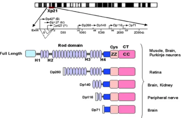
The dystrophin gene, localized on the short arm of the X chromosome, is one of the largest human genes so far described, comprising almost 0.1% of the genome, (4) and consisting of 79 exons (5) encoding a primary transcript of 2400 kilo-bases. Due to its large size, this gene has a high mutation probability, so that nearly one third of DMD cases are non-familial (6). The dystrophin gene is quite complex, hosting at least eight independent and tissue-specific promoters. The full-length dystrophin isoform (Dp427), for instance, is transcribed by three independently regulated promoters, labelled as B (brain), M (striated muscle), or P (Purkinje cell), the respective letters reflecting the major sites of expression (7). Apart from the Dp427, five additional isoforms exist, produced by activation of different promoters along the gene or by alternative mRNA splicing. These shorter isoforms are named according to their molecular weights: Dp260 (predominantly in the retina), Dp140 (central nervous system and kidneys), Dp116 (peripheral nervous system), Dp71 (most tissues, but not muscles), and Dp40 (brain) (8, 9, 10 ).
Fig 1: Schematic diagram showing the position of all promoters within the dystrophin gene and the molecular structure of the dystrophin isoforms. Tissue distribution of dystrophin and its isoforms are also indicated (11)
Muscle dystrophin is localized at the cytoplasmic face of the sarcolemma membrane and consists of an N-terminal actin-binding domain, a central large rod-like domain composed of spectrin-like repeats, and a cysteine rich C-terminus (Fig. 1) that connects to a multiproteic complex called the dystrophin-associated glycoprotein complex (DGC) (Fig. 2).
Fig 2: The dystrophin-associated glicoprotein complex in skeletal muscles (12)
Central protein of the DGC is dystroglycan (DG), composed by the transmembrane b-DG and the extracellular a-DG, sarcoglycans (a, b, g and d), sarcospan, syntrophins (a1, b1, b2, g1, g2), and dystrobrevin (13). In this complex, while the b-DG binds to dystrophin, which in turn links to the cortical actin filaments, the a-DG binds to extracellular matrix (ECM) proteins, as laminin. Therefore, the DGC provides a link between cytoskeleton and ECM (14), which is thought to protect the muscle plasma membrane (sarcolemma) from mechanical damages. Sarcoglycan is the second transmembrane component of the DGC, connected to b-DG and dystrobrevin. On the cytoplasmic side, instead, dystrophin directly interacts with syntrophins and dystrobrevin, which recruit other scaffolding proteins onto which signalling proteins (i.e. nitric oxide synthase) and ion channels are anchored to the plasma membrane (15). In this way, the DGC provides both a physical and functional connection between the internal and external environment of muscle cells.
In DMD, the absence of Dp427 results in the destabilization of the DGC, which not only hinders muscle integrity, but also induce secondary changes reflecting an impairment in intracellular signalling, as could be the reduction in nitric oxide synthesis and, hence, protein nitrosylation (16).
2. Dystrophin expression and localization in the nervous system
Selected neuronal populations (within hippocampus, cortex, cerebellum, autonomic ganglia) and glial cells (i.e. oligodendrocytes, Schwann cells) of both central and peripheral nervous systems also express Dp427, some of its isoforms and proteins composing the DGC (17). In brain, the Dp427 is primarily located in the hippocampus, prefrontal cortex, amygdala and cerebellum (18, 19). Here, similarly to skeletal muscles, it associates to the DGC, although several brain DGC variants exist because of the various dystrophin isoforms, alternatively spliced variants, and the presence of other DGC components, such as b-dystrobrevin, e-sarcoglycan, and the g-syntrophins (Fig. 3), which are not expressed in muscle. Moreover, the Dp427-DGC localizes not only in domains along the plasma membrane, but also at post-synaptic specializations, in which the complex does not bind to laminin but to the pre-synaptic protein neurexin (Fig. 3) (15).
Fig 3: Neuronal DGC localized within post-synaptic specializations. When Dp427-DGC are localized in the post-synaptic domains, dystrophin binds to β-dystrobrevin and the dimer β-DG/a-DG, which in turn associates with the pre-synaptic protein neurexin-α. This contributes to synaptic stabilization. In addition, part of the DGC is also α1- and γ1-syntrophins, absent in muscles. These proteins, by binding Dp427 and dystrobrevin, are involved in the stabilization of ion channels within the membranes, among which voltage-gated Na+ channels. In addition, syntrophin also bind and stabilize at the DGC the neuronal nitric oxide synthase. The all complex is also implicated in the stabilization of post-synaptic receptors, as GABAA receptors. (Abbreviations: DGC dystrophin-associated glycoprotein complex; nNOS neuronal nitric oxide synthase; SAST syntrophin- associated serine/threonine kinase; ABD actin-binding domain; EFI/EFII EF hand domains; ZZ zinc finger domain; H1/H2 helical domains) (15)
Due to these differences, the DGC in the nervous system is often referred to as a DGC-like (15, 20). This complex associates not only with the Dp427, but also with all its shorter informs. Localization of full-length and short dystrophin
isoforms is cell-specific: for example, Dp427 is mainly neuronal, although has also been described in oligodendrocytes; Dp260 and Dp140 are highly represented in the retina, especially in neurons of the inner nuclear layers (21); Dp140 has also been found in perivascular astrocytes, along the Dp71, the most ubiquitous dystrophin isoform (22, 16, 8, 23); Dp116 is mainly expressed by Schwann cells of peripheral nerves.
Full-length dystrophin is mainly localized in specific neuronal populations, as hippocampal neurons (all regions), cortical pyramidal cells, and cerebellar Purkinje cells (24). Major localization in adult individuals (both human and animal models) is at the postsynaptic sites, which has suggested a role in the maintenance of synaptic structure and function (16). The Dp427 has been found to co-localize with the g-aminobutyric acid type A (GABAA) receptors possibly through their linkage
to syntrophins and gephyrin. This would prevent receptor diffusion (25), playing a critical role in their clustering, stabilization and synaptic signal transmission (26, 27, 1, 28, 29, 30).
In both autopsy brains of DMD patients and mdx mice (elective animal model of DMD), absence of Dp427 does not induce gross anatomical alterations, but a number of diversified cellular and sub-cellular abnormalities, among which: 50% decrease in cortical neuron number and neural shrinkage (31), abnormal dendritic development (32), altered post-synaptic density organization and pre-synaptic ultrastructure in hippocampus, mainly in the CA1 region (33, 34, 35, 17). In addition, in mdx mice, loss of Dp427 has been associated with a reduction in 40–70% of GABAA receptor clusters in CA1 and
CA3 hippocampal neurons (containing the alpha-2 subunit), cerebellar Purkinje cells (containing the alpha-1 subunit), amygdala and cerebral cortex (containing both alpha-1 and
alpha-2 subunits) ( 36, 26, 37). GABAA alpha-1 and/or alpha-2
receptor subunit gene expression was also decreased in these brain regions. In brain, dystrophin deficiency has also been associated to a reduction in the response to nicotine in a passive avoidance memory task (engaging hippocampal activity) (38). This suggested a role of Dp427 in the stabilization of nicotinic acetylcholine receptor (nAChR) subtypes, similarly to GABAA
and glutamatergic receptors. Whether all these alterations are the morpho-functional basis of cognitive dysfunctions has been a matter of discussion for long time, others failed to demonstrate this correlation (39, 17).
Because brain dystrophin is more abundant in the hippocampus compared to other sub-cortical areas (19), selective behavioural deficits involving hippocampal function were predicted to occur in the mdx. Indeed, several studies showed that dystrophin deficiency in mdx mice is associated with impaired memory retention at long delays, in certain procedural learning and spatial alternation tasks (40, 41), suggesting a role for Dp427 in the consolidation of certain forms of long-term memories. Absence of Dp427 also causes unbalanced calcium homeostasis (42), with consequent alterations in hippocampal long-term potentiation (LTP) (43), a form of plasticity widely believed to be critical for memory formation. Intriguingly, dystrophin deficiency in CA1 hippocampal neurons appeared to facilitate induction of short-term potentiation (STP) and depression (STD) of the glutamatergic transmission (41, 29), with no apparent alteration in the maintenance of the LTP phase (41). Recently, brain dystrophin was found in association with a sub-population of GABAA receptors at inhibitory synapses (36, 44) and the abnormal enhancement of hippocampal STP and STD was shown to be occluded by a GABAA-receptor antagonist (29), suggesting that decreased inhibitory tone may be one possible
mechanism of the altered plasticity in dystrophin-deficient neurons. At the functional level, this induces alterations in other mechanisms of plasticity, such as the LTP of the population spike (PS), with important consequences on the output of the neuronal circuitry.
Duchenne De Boulogne was the first describing cognitive limitations in boys with DMD (45). General intelligence among boys with DMD is one standard deviation below the normal population mean IQ and mental retardation has been reported in approximately one third (34.8%) of patients (46). Moreover, boys with DMD can be affected by attention-deficit/hyperactivity disorders (ADHD) (1.7 %), autism spectrum disorders (3.1 %) and reading problems (20 % moderate, 20 % severe) (47). Mutations in the dystrophin gene can even cause intellectual disability in the absence of muscular dystrophy (48). However, since these cognitive deficits do not seem to depend on the location of the gene mutations, a clear genotype-phenotype correlation for cognitive impairment in DMD has yet been established (17, 20). As the gene encodes several dystrophin isoforms, the number of affected gene products and/or cell-type specific isoforms may correlate with the occurrence and severity of cognitive impairment (49, 20).
DMD also associates to peripheral nervous system (PNS) alterations. Autonomic dysfunctions have been reported in DMD patients (50), and a number of morphological and functional alterations have been reported by studies conducted in our laboratory on the sympathetic neurons of the superior cervical ganglion (SCG) of mdx mice. Specifically, loss of Dp427 induces an early (since post-natal day 0, P0) and persistent reduction in the SCG noradrenergic innervation of iris and heart, compared to wild type mice, which associates
with a significant loss in muscle-innervating ganglionic neurons between P5 and P10 (51). Moreover, all mdx mouse ganglionic neurons, regardless of the type of target they innervate, showed reduced defasciculation and terminal branching (51). De-regulated protein levels for components of the nerve growth factor (NGF) signalling system (i.e., NGF receptors TrkA and p75 NTR) (52), unbalanced proNGF/mature NGF ratio (52), and reduction in the NGF-mediated intracellular signalling cascade were also described (53). Moreover, a reduction in intra-ganglionic a3, b2/ b4 nAChR stabilization (54) and activity (55), as well a different modulation in the expression of genes encoding proteins involved in neuron survival and differentiation (56) were also reported.
3. Therapeutic approaches for DMD treatment
Although DMD was discovered more than twenty years ago, there is currently no cure to resolve the disease. In fact, the most widespread pharmacological therapy focuses on the use of a glucocorticoid-based treatment, aimed to reducing muscle inflammation and the activity of the immune system, to alleviate the dystrophic phenotype and increase motor activity in DMD affected patients (57). The action of these drugs can improve muscle strength, ambulation and cardiac activity. However, this therapy is associated with multiple side effects, such as weight gain, CNS disorders, gastrointestinal and metabolic disorders (58). To date, new therapies based on genetic techniques, able to restore the products of the dmd gene, are being tested. In fact, by acting on the dystrophin pre-mRNA, it can induce the exclusion of exon containing the mutation and generate a truncated and partially functional protein. However, the presence of a semi-functional dystrophin improves the patient's motor skills, reducing the severity of
symptoms and lengthening life expectancy, but there were no significant improvements in cognitive function following treatment with drugs, mainly because the administration is intramuscular and the drug has a local action (59).
One of the main objectives in this area is to find a non-invasive treatment, effective both on the muscular and the nervous phenotype.
4. The glucocorticoid-mediated stress signalling
4.1 Glucocorticoids and their receptors
Glucocorticoids (GC) are steroid hormones and the end product of following the activation of the hypothalamus-pituitary-adrenal (HPA) axis, which regulates stress responses. GC, like all steroid hormones, are synthesized from cholesterol, which undergoes multiple transformations in a multi-enzymatic process called steroidogenesis. GCs are neurosteroids, a large group of steroid hormones, which includes allopregnanolone and allotetrahydrodeoxycorticosterone (THDOC), testosterone-derived androgens (androstanediol) and estradiol. Neurosteroidogenesis occurs in the brain regions such as cortex, hippocampus, and amygdala. The type of steroid hormone synthesized in a particular endocrine gland is determined by the combination of the enzymes it expresses. The main enzymes for the synthesis of glucocorticoids are: 17-hydroxylase, 3β-hydroxysteroid dehydrogenase, 21-hydroxylase and 11β-21-hydroxylase (60).
Two types of receptors mediate the GC effects: the mineralcorticoid receptor (MR) and the glucocorticoid receptor (GR), both expressed in several brain regions. Among these areas there is the hippocampus, in which the great abundance of GC receptors makes it an important relay for both stress appraisal and adaptative processes. Through MR, GCs influence the brain’s appraisal of novel information and memory retrieval, and thereby influence behavioural coping responses. As GC concentrations increase in response to stressors, GR are activated to promote stress adaptation, reallocation of energy resources in preparation for future events and recovery of the system (61). One important target of GCs is the BDNF-signalling, which crucially contributes to the modulation of axonal guidance, synaptic plasticity and neurite outgrowth (62). MR, GR and the BDNF receptor, TrkB, are co-expressed in hippocampal neurons, supporting this region as the primary site of immediate interactions between the GC- and BDNF-signalling pathways (63). Ultradian fluctuations of GCs (Fig. 4) drive GR activation and reactivation, whereas MR occupancy is more constant and promotes excitability (64). This balance has implications for the genomic and non-genomic activity of adrenal steroids within target cells.
The GR belongs to the nuclear receptor superfamily, which includes receptors for steroid hormones (e.g. glucocorticoids, estrogens, androgens and mineralocorticoids) as well as receptors for other hydrophobic molecules, such as
Fig. 4: Schematic ultradian fluctuation of GCs in healthy individuals
prostaglandins, fatty acids and thyroid hormones. Nuclear receptors share a similar structural organization and mode of action, via transcriptional activation (transactivation) or repression of transcription (trans-repression). The GR consists of three main domains; the N-terminal domain (NTD), the DNA-binding domain (DBD) and the ligand-binding domain (LBD), with a hinge region between the LBD and DBD. This modular structure is broadly conserved across the nuclear receptor superfamily. The DBD is the most conserved region across the nuclear hormone receptor superfamily. It contains two “zinc finger” structural motifs, the N-terminal zinc finger binds specifically to the DNA response element (glucocorticoid response element, GRE), and the second is thought to be involved in protein-protein interactions, such as when the receptor forms a dimer. Once a dimer of GR binds GRE, it can recruit co-regulators and chromatin-remodelling complexes that modulate gene transcription rates by affecting the activity of RNA polymerase II (65). The LBD is localized at the C-terminal end of the protein. The structure of the binding pocket consists of 12 α-helices and four β-sheets that form a hydrophobic pocket to which the steroid hormone preferentially binds. This conformational structure is dependent on the co-association of the chaperone protein hsp90, which maintains the receptor in the open position in order to accept ligand binding (66). Once a GC binds the LBD, a conformational change leads to the dissociation of chaperones and exposure of the nuclear localization signals. Post-translational modifications of the NTD occur via
phosphorylation and sumoylation. Human GR phosphorylation occurs at serine residues 203 and 211 in response to hormone binding, and is thought to influence receptor localization within the cell, with phosphorylation of Ser203 causing the receptor to be cytoplasmic, and phosphorylation of Ser211 promoting
translocation into the nucleus (67). GR phosphorylation changes its interaction with cofactors and influence target gene expression (68).
4.2 The glucocorticoid receptor genomic response
Steroid hormones have a typical arrangement of four cycloalkane rings, and are derived from enzymatic processing of cholesterol. Their lipophilic nature allows them to enter the cell by diffusion through the plasma membrane, in order to bind the cytoplasmic GR, which is held in the open conformation by heterocomplex of chaperones, such as immunophilins and heat shock proteins (hsp). As said, this binding induces a receptor conformational change, dissociation of chaperone proteins, receptor phosphorylation and exposure of nuclear localization signals (NLS). This signal is recognized by importins, which are proteins for the import into the nucleus, and translocated into this compartment (69). Here, GRs dimerize and bind, via the zinc finger motifs to the GRE, which are conserved signals upstream to target genes. This binding can either enhance or repress gene transcription, depending on the GRE present and the availability of other transcription factors. If GRE is a promoter of transcription, bound GRs recruit co-activators and other transcription factors, leading to increased chromatin remodelling, recruitment of further co-factor, and eventual recruitment of RNA polymerase 2 (RNA Pol II). In case of a “negative” GRE (nGRE), GR binding causes repression of target gene transcription, possibly by blocking the promoter site, or other transcription factor binding sites. Rogatsky and collaborators (70) identified some genes directly regulated by GC binding to GRE. These include glucocorticoid-induced leucine zipper (GILZ), IGFBP1, and metallothionein 1X (MT1X). In particular, GILZ is involved in cell proliferation, epithelial sodium channel activity (71), and
plays key roles in GC signal-modulation, control of protein trafficking and signalling, modulation of T-lymphocyte and of other immune cell activation, IL-2 production and apoptosis (72, 73). GILZ interacts, inhibiting, with NF-kB, AP-1, Raf-1 and Ras, which are negative regulators of GC signalling, as they normally inhibit GC target gene expression, as trans-repression of pro-inflammatory genes (i.e., cytokines and their receptors, nitric oxide synthase, COX-2). Treatments with dexamethasone (DEX), a potent GR synthetic agonist, up-regulate GILZ expression, which mimics some of the GC effects, adding further immunosuppressive effects (74).
4.3 The glucocorticoid receptor non-genomic signalling
GC genomic response is necessarily a rather long process. Gene transcription and expression, along with post-translational processing of proteins, may take several hours, with the minimum time for an effect to be measurable of at least 15 minutes. However, GC also elicit a number of fast responses (within a few seconds to minutes), which are not sensitive to inhibition of both transcription and protein synthesis, and that can be observed also in cells that don’t have a nucleus, such as platelets. These non-genomic signalling can be investigated by using GR inhibitors, such as RU-486, which inhibit the classical ligand-binding activation of cytosolic GR, or by stimulation with DEX conjugated to BSA (DEX:BSA). BSA complexing gives rise to a compound unable to cross the plasma membrane and approach intracellular receptors, hence triggering a signalling cascade, which must be via a membrane-associated receptor (75, 76, 77) (Fig. 5).
Several mechanisms have been proposed for the non-genomic effects of GC signalling. These include effects mediated by a membrane-associated GR (mGR), via GC ligand-binding to a different receptor to the GR, such as glutamate receptors, or
acting via G-protein coupled receptors, or having a direct effect on the membrane (75, 78). Effects of GC signalling to the cytosolic GR have also been proposed to elicit non-genomic effects via proteins released when the receptor complex is disrupted upon ligand binding. The effects observed have included activation of second-messenger systems, changes in ion flow, and activation of kinase pathways (79). Xiao and collaborators (80) demonstrated that GC rapidly and non-genomically activate ERK1/2, JNK, and p38 mitogen-activated protein kinases (MAPK) in GR-deficient neurons, but had no detectable effects on their neuronal viability. CORT rapidly activates both the cAMP-PKA pathways and the ERK1/2 pathway in neurons, important in the hippocampal response to stress (81, 82). Evidence for the involvement of the PI3K pathway has not yet been studied in the brain.
Over the last decade, highly ordered plasma membrane micro-domains with particular lipid and protein composition have been identified. There is evidence that these domains, termed “lipid rafts,” orchestrate some control over intracellular signalling pathways and mediate cross talk between membrane-associated receptors (83). Current evidence suggests that the estrogen receptor is associated with a particular sub-set of lipid rafts termed “caveolae” (84). Caveolin-1, the major protein component of caveolae, has been implicated as a structural scaffold for the oligomerization and organization of cytoplasmic signal complexes (85). Interaction with, and modulation by, caveolin-1 has been shown in many signal transduction pathways, including those regulated by mGR. Caveolae can act as signalling organizers, and several works established a role for Cav1 in a rapid GC signalling pathway that triggers MAPK activation in embryonic mouse neural progenitor cells (NPC) cultures. In this case, one of the consequences of Cav1-dependent activation of MAPK by GCs
is an inhibition of intercellular communication between NPSCs coupled by gap junction (86). Studies with other steroid hormone receptors (i.e., androgen and estrogen receptor) have revealed mechanisms of cross talk between the genomic and the rapid GR responses (87).
MAPKs are a superfamily of serine-threonine kinases composed by the well-characterized extracellular signal-regulated kinases (ERK1 and ERK2 MAPK isoforms of 44- and 42-kDa, respectively), the c-Jun-NH2-terminal protein kinase/stress-activated protein kinase (JNK/SAPK) and the p38 MAPK (88). They are activated by diverse stimuli ranging from cytokines, growth factors, neurotransmitters, hormones, as well as under conditions of cellular stress and cell adherence. They play a key role in the regulation of cell growth, differentiation, cytoskeletal function, and gene expression (89, 90). As demonstrated in PC12 cells, GCs might act via its membrane receptor and activate ERK1/2 MAPK. ERK1/2 MAPK sustained activation results in its nuclear translocation (91, 92), which is then capable of regulating gene transcription itself (93, 94).
Current evidence suggests the estrogen receptor interact with caveolin-1 (84, 95). In addition, association of the GR with membrane lipid rafts has also been proposed (96), as confirmed by immunoprecipitation studies showing co-precipitation of GR and caveolin-1 (97). Moreover, as a control, loss of caveolin expression prevents GC inhibition of cell proliferation, an effect mediated by blocking the cells in G1/S transition (97). Recent data support integration of different signalling cascades to determine integrated cell responses (98).
Fig 5: GR signalling pathways. Glucocorticoid-activated GR regulates gene expression by three main routes: (A) binding directly to DNA, (B) tethering itself to other DNA-bound transcription factors, (C) binding directly to DNA and interacting with neighbouring DNA-bound transcription factors. GR can also signal using a non-genomic pathway, by using membrane-bound GR and activating various intracellular signalling kinases. (77) 5. The stress response
Stress represents a condition that implies a modification of homeostasis, which may occur through a number of different events regulating emotion, behaviour, cognition as well as physical health. Indeed, stress represents the main environmental components for the susceptibility to mental illness, although it is known that the response to stress is
modulated by the genetic signature, as well as by a number of other factors, including earlier exposure to adverse life events that may have ‘primed’ the brain toward enhanced susceptibility. The majority of studies have reported that exposures to stress or elevated levels of corticosteroids impair performance on memory tasks dependent on the hippocampus (99, 100). Memory impairments have also been reported in transgenic mice with elevated corticosterone (CORT) production, due to central overexpression of corticotrophin-releasing factor (CRF). Recent findings of stress altering the firing properties of place cells in the hippocampus (101), which are thought to support spatial navigation and memory, are consistent with the stress effects on spatial memory tasks. The discovery of a relationship between stress and hippocampal LTP is significant, because it offers both a testable synaptic mechanism that may explain stress effects on memory and a “neurophysiological marker” to compare behavioural results from studies that use different stress paradigms. For example, a chronic restraint stress causes reduction in hippocampal volume from the pre-stress size (102), which determines reduction in the number of dendritic spines and branches of pyramidal neurons in the CA3 (103) and suppression of the production of new granule neurons in the dentate gyrus (DG) (104). The abundant presence of the GR in rodent and human hippocampus makes this brain structure, together with the hypothalamic paraventricular nucleus (PVN), very sensitive to the action of GCs and key to the regulation of the stress response (61, 105).
GRs are central in the regulation of stress response and other situation where GC levels are elevated (106, 61, 107), since basal levels of CORT will not occupy all the receptors, leaving some vacant for signalling the response to stress. Prolonged exposures to CORT have also been shown to cause
31
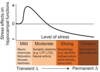
morphological and molecular changes, reduced neurogenesis and impaired synaptic plasticity in the hippocampus, physiological outcomes which are thought to precipitate hippocampus-dependent memory impairments and anxiety- and depression-like behaviours (108) (Fig. 6). These long-term effects of chronic CORT elevation on the hippocampus have been hypothesized to occur via epigenetic mechanisms (i.e., DNA methylation and histone modification) in the HPA axis (109).
Fig. 6: Biological effects of stress on the hippocampus. As the severity (intensity, duration) of stress increases, alterations in neurochemicals, synaptic plasticity, neural activity, cytoarchitecture, and neurogenesis occur in the hippocampus that can influence subsequent cognitive functions, such as learning and memory, and contribute to psychopathologies. + and – represent an increase and decrease in hippocampal functioning, respectively. (110)
In response to stressful or threatening situations, corticosteroids are released by the HPA axis (Fig 7). Neuronal signalling to the PVN of the hypothalamus in response to proposed that when an organism internalizes that its action has no bearing on the aversive outcome (stressor), this learn-ing produces changes in the cognitive, emotional, and motiva-tional systems that impede subsequent learning. The learned helplessness phenomenon, where stress-associated learning nega-tively influences subsequent learning, in essence, is the negative counterpart to the concept of meta-learning in humans, which Maudsley (1979) described as “the process by which learners be-come aware of and increasingly in control of habits of perception, inquiry, learning, and growth that they have internalized.” In re-cent decades, much research has shown that stressful experiences can alter hippocampal mnemonic functioning in animals and humans.
The vast majority of studies have reported that exposures to stress or elevated levels of CORT impair performance on memory tasks dependent on the hippocampus (McEwen and Sapolsky 1995; Kim and Diamond 2002). In human studies, individuals di-agnosed with PTSD and depression are impaired in various verbal recall tests (Bremner et al. 2000). The evidence for the direct role of CORT is based on findings of memory deficits in patients with Cushing’s syndrome (with chronic hypercortisolemia; Starkman et al. 1992), and in healthy subjects administered with CORT (Newcomer et al. 1994). Similarly, rodent studies have shown that exposures to stress and injections of high doses of CORT pro-duce deficits in spatial memory tasks that involve the hippocam-pus (de Quervain et al. 1998). Memory impairments have also been reported in transgenic mice with elevated CORT due to cen-tral overexpression of corticotropin-releasing factor (CRF; Heinrichs et al. 1996). Recent findings of stress altering the firing properties of place cells in the hippocampus (e.g., Kim et al. 2007), which are thought to support spatial navigation and memory (O’Keefe and Dostrovsky 1971), are consistent with the stress ef-fects on spatial memory tasks.
However, stress does not produce global memory deficits as it has been shown to enhance cerebellum-dependent eyeblink con-ditioning (Beylin and Shors 2003) and amygdala- and hippocam-pus-dependent contextual fear conditioning, which is thought to be due to prior stress augmenting glucocorticoid release during training (Cordero et al. 2003). Interestingly, the same stress that impairs hippocampal memory tasks seems to enhance the relative use of competing nonhippocampal (e.g., the caudate-dependent stimulus-response) memory tasks in rats and humans (Kim et al. 2001; Schwabe and Wolf 2012). At present, it is un-known whether these differing effects on memory are indirect, i.e., the result of stress decreasing the hippocampus’ ability to
in-teract (e.g., compete) with other brain–memory systems, or direct facilitatory effects on nonhippocampal memory systems.
Stress and hippocampal plasticity
Long-term potentiation (LTP), resulting from a brief high-frequency stimulation of afferent fibers initially demonstrated in the hippocampus, has characteristics desirable of a synaptic mod-el of memory, such as rapid induction, longevity, stimulation threshold requirement (cooperativity), strengthening by repeti-tion, input specificity, and associativity (see Bliss and Collingridge 1993). The first evidence linking LTP and stress was reported by Thompson, Levine and colleagues who found impairments in the Schaffer collateral/commissural-CA1 LTP in hippocampal slic-es prepared from rats that experienced 30 intermittent tailshocks during 30 min of restraint (Foy et al. 1987). Importantly, CA1 LTP was relatively normal in hippocampal slices from rats that re-ceived the same amount of tailshocks but were able to terminate them voluntarily (Shors et al. 1989), indicating that, similar to learned helplessness, the LTP impairment is largely due to the psy-chological, rather than physical, qualities of stress.
Stress-associated LTP impairments have also been demon-strated in the dentate gyrus (e.g., Shors and Dryver 1994) and CA3 regions of the hippocampus (e.g., Pavlides et al. 2002). Other studies have found time-dependent, biphasic effects of stress on LTP—an initial enhancing effect followed by a longer-lasting, suppressing effect on LTP (Akirav and Richter-Levin 1999). Stress has also been reported to enhance LTP induced by u-burst stimulation but impair LTP induced by high-frequency stimulation. These findings indicate that differences in stress par-adigms, in vitro versus in vivo recordings, and stimulation pat-terns produce a more complex picture of stress effects on LTP.
The discovery of a relationship between stress and hippocam-pal LTP is significant because it offers both a testable synaptic mechanism that may explain stress effects on memory and a
Figure 1. Biological effects of stress on the hippocampus. As the severity (intensity, duration) of stress increases, alterations in neurochem-icals, synaptic plasticity, neural activity, cytoarchitecture, and neuro-genesis occur in the hippocampus that can influence subsequent cognitive functions, such as learning and memory, and contribute to psychopathologies.+ and 2 represent an increase and decrease in hip-pocampal functioning, respectively. Adapted from Kim and Yoon (1998).
Figure 2. The hypothalamic-pituitary-adrenal (HPA) axis. A major neu-roendocrine system associated with various bodily and behavioral activities. None of these compounds or structures responds uniquely to stress. (PVN) paraventricular nucleus of hypothalamus, (CRF) corticotro-pin-releasing factor, (AVP) arginine-vasopressin, (ACTH) adrenocortico-tropic hormone, (GCs) glucocorticoids, (CAs) catecholamines, (Enk) enkephalins.
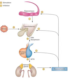
perceived danger causes the release of corticotrophin releasing hormone (CRH) and vasopressin, which stimulates the release of adrenocorticotrophic hormone (ACTH) from the pituitary gland into the blood stream. This is carried to the adrenal glands, above the kidneys, where it stimulates production of corticosteroids, such as mineralocorticoids and GC, from cholesterol by the adrenal cortex (61).
HPA axis activity is organized in a circadian rhythm with high levels in the morning, enabling individuals to cope with energy demands ahead of them. It is important to note that rapid HPA axis activation by acute stress and the subsequent turn-off of the HPA axis by the negative feedback response of CORT is healthy, as it helps an individual to cope with the stressor (111) (Fig. 7).
paraventricular nucleus of the hypothalamus to release CRH and vasopressin, which induce the release of ACTH from the anterior pituitary gland. ACTH stimulates the synthesis and release of glucocorticoids from the adrenal cortex. As a result of the deleterious effects of long-term exposure to glucocorticoids, a strict glucocorticoid-feedback mechanism, acting at the pituitary, hypothalamic and hippocampal levels, is fundamental to modulate the activity of the HPA axis. Particularly, the activation of hippocampal glucocorticoid-receptor-expressing neurons exerts a potent inhibition of the HPA axis. Modified from Vitale et al., 2013
In order to respond quickly to dangerous stimuli, the body must produce an adaptive behavioural response such as increased locomotion and risk assessment. A physiological response is also produced by the sympathetic action of the autonomic nervous system to produce the so-called “fight or flight” response via acetylcholine (Ach), epinephrine and norepinephrine signalling.
Corticosteroid signalling in the brain modulates the stress response, with mineralocorticoids affecting appraisal of the situation, and GC acting on consolidation of information (112). The hippocampal formation is a powerful upstream modulator of the HPA axis stress response and consequent emotional states (114). Imbalances in circulating levels of GCs can lead to defects in hippocampal functioning, and vice versa (106). The production of GC has an inhibitory effect on the HPA axis, inhibiting the release of CRH and ACTH, to decrease the release of further corticosteroids, this occurs in a delayed genomic manner, but effects have been found to occur within 5-15 minutes. The neurons of the PVN of the hypothalamus have a high number of GRs, which would account for the genomic inhibitory effect on the HPA axis. The production of ACTH at the pituitary is also inhibited in a fast, as well as delayed manner, and although the mechanism is still unclear (113), GR-dependent and GR-independent signalling may be involved (114).
6. Glucocorticoid effects on adult hippocampal neurogenesis The hippocampal formation is a powerful upstream modulator of the HPA axis stress response and consequent emotional states (115). Imbalances in circulating levels of GCs and stress context can lead to defects in hippocampal functioning and vice
versa (106); among these is adult neurogenesis, which occurs at
the DG level (116, 117). On the other hand, correct hippocampal neurogenesis is important in the hormonal regulation of stress response, as it gives rise to a small subset of neurons that are critical for a powerful negative control of the hippocampus over the HPA axis. Defects in hippocampus structure and function may be manifested by HPA-axis deregulation (118), which is associated with many forms of affective disorders (119). Conversely, enhancing neurogenesis can improve mood-related behaviour and restore central control over stress response systems (120).
The hippocampus is divided into three main sub- regions, named CA1, CA3, and DG. Neuronal cells derived from these three sub-regions are connected by the so-called tri-synaptic pathways. It is generally assumed, but not proven, that information processing by this pathway is crucially involved in learning and memory formation. Neurogenesis occurs only in the DG and there is no evidence that other hippocampal regions generate new neurons (121). Adult hippocampal neurogenesis refers to neural stem/progenitor cells (NSPCs) present in the sub-granular zone (SGZ) of the hippocampal DG, where they continue to produce new neurons during adulthood in many species, including human (122, 123). New neurons have been implicated in various hippocampus-related functions and disorders, such as spatial learning and memory, pattern separation, epilepsy, anxiety, depression and dementia (124, 125, 126, 127, 128). Recent reports further indicate that new
neurons also play an important role in HPA axis feedback regulation after stress: mice lacking new hippocampal neurons have a slower recovery of CORT levels to baseline after stress. Other studies also indicated that hippocampal new born neurons play an important role in stress regulation, as increased adult hippocampal neurogenesis exerts antidepressant effects, improves behaviour and regulation of stress response (118, 120, 129, 130, 131).
Both acute and chronic stress can suppress one or more phases of adult hippocampal neurogenesis (132, 133, 117). For example, predator stress rapidly raises GC levels, which cause significant reductions in hippocampal NSPC proliferation, (134, 135). While many, if not all, of these effects are generally attributed to increased GC levels, a simple interpretation of the effects of corticosteroids on the regulation of adult hippocampal neurogenesis is difficult to provide and it is important to realize that many other variables can influence both hippocampal neurogenesis and the way GCs regulate it (136, 117). Therefore, the reactions of the endocrine system to stress, and the results of that activation on neuronal function, are modulated by other events and may vary in complex ways. The sum of this pattern determines how a given stressor alters adult hippocampal neurogenesis in a given individual at any one time. Multiple stages of the neurogenic process are affected, including proliferation, as subsequent neuronal differentiation, connections to output pathways (e.g. CA3) and dendritic growth. Stress not only reduces NSPC proliferation and adult hippocampal neurogenesis, it may also control subsequent NSPC fate specification and differentiation through the action of the GR, which has important consequences for hippocampal network connectivity, function and behaviour (137). Direct effects of GCs on NSPCs have been demonstrated in the absence of known stressors, showing that the GR plays a
central role in mediating the direct effect of CORT on hippocampal NSPCs, as selective reduction in GR expression in new born cells results in altered neurogenesis (137). Clearly, the effects of stress and stress hormones on adult neurogenesis are complex. Firstly, neuronal stem cells in the SGZ of the DG are located in a specialized microenvironment, the so-called neurogenic niche, consisting of numerous different cell types, including astrocytes, ependymal cells, blood vessels, interneurons, oligodendrocytes, and myeloid cells, i.e., microglia cells and dendritic cells. All this cell types may modulate adult neurogenesis. For example, depending on the type of microglia and on the challenge, activated microglia cells release cytokines that may have detrimental or beneficial effects on adult neurogenesis (138). Also, these cell types express GRs indicating that stress-induced GC elevation targets these cells, which may modulate the rate of neurogenesis. Secondly, the nature of the stressor is an important factor.
NSPC express both GR and cell-specific mechanisms regulating its activity at the level of intracellular trafficking, suggesting an important biological function for the GR in these cells (139, 140). Consistent with a role in NSPC differentiation, knocking down GR expression in the new born cells results in increased NSPC differentiation (137), demonstrating that direct effects of CORT on these cells via the GR exist as well.
An important question that emerges is what type of environmental factors regulating adult neurogenesis also affect GR levels in neuronal progenitor cells. Interestingly, early life events, such as maternal separation and parental care, are known to reduce GR levels at adult age by epigenetic programming of the GR promoter (141, 142, 143, 144) and associate with impaired adult neurogenesis (145, 146). Aging is associated with lower rates of hippocampal neurogenesis (147), impaired negative feedback of CORT on the HPA axis, and
reduced levels of the GR (148, 149). Chronic stress is another factor negatively affecting both GR levels and adult neurogenesis attenuating multiple excitatory and inhibitory signalling cascades through the GR (150, 151, 152).
GCs inhibit cell differentiation and synaptic development in the various brain regions, resulting in alterations in neural fine structure and functions (153, 154, 155). Prenatal stress diminishes neurogenesis in the DG of juvenile rhesus monkeys (156) and, throughout postnatal life, GCs exert suppressive effects on cell proliferation in the DG. Moreover, persistently high CORT levels decrease cell proliferation in the adult rat DG (157). DEX has been shown to produce cell loss in the pyramidal layers and DG (158) and elicit various effects on neural cells. It has also been reported to cause an arrest of HT-22 cells (mouse hippocampal neuronal cell line) in the G1 phase of the cell cycle (159)and inhibit platelet-derived growth factor-induced differentiation of HiB5 cells (hippocampal stem cell line) (160). The sensitivity of DG to glucocorticoids is of particular concern because DG is also an important brain center involved in the synaptic plasticity. The two aspects of neurogenesis, which includes proliferation and differentiation, could be independently affected by GC activation (161).
7. Glucocorticoids and DMD
CNS alterations associated to DMD are reported in patients and animal models. However, differently from muscles, nervous system defects are mainly established prenatally and difficult to identify, as functional alterations become apparent during growth. Different anatomical alterations induce diversified neurological disorders with different degree of severity. Among these, children and adolescent with DMD are at great risk of developing depression and anxiety disorders (162).
neurological aspects, but new pharmacological treatments are succeeding in extending patients life span. Despite different innovative therapeutic approaches are in the stage of clinical trial, the treatment based on corticosteroid administration, which reduces skeletal muscle recurrent inflammation, continues to be the main adopted therapy (163). In healthy individuals, increase in GC synthesis and release follows the coordinated activation of the HPA and autonomic system. The ubiquitous expression of GR and the multitude of functions of GCs confer this system an essential role in the response to stress and restoration of homeostasis. Precise regulation of the HPA axis activity is very important for the organism; indeed, chronic exposure to GCs end up in various adverse side effects, such as osteoporosis, diabetes, hypertension, neurodegeneration (164) and inhibition of neurogenesis (111, 165, 161). On the other hand, a deficient HPA axis is observed in a wide range of autoimmune and inflammatory disease. GCs reach every organ by way of the circulation and cross the blood brain barrier, which allows the coordination of brain and body functions. Several experimental evidences underline the relevance of corticosteroids as structural modulators in the limbic areas. Both acute (single) and chronic (repeated) stress stimuli induce prominent changes in neuronal activity and synaptic functions, in hippocampus and medial prefrontal cortex, which rely on neuronal remodelling (i.e. dendrite shortening and pruning) (166). A great number of genes are also inhibited directly by GCs, through the formation of a repressor complex containing histone deacetylases (HDACs). Alteration, at any level, in GC mode of action is observed also in aging and in neurodegenerative diseases (167).
Aim and Scope
Goal of this research project is to explore the effects that increased levels of GCs exert in brain regions particularly susceptible to stressful stimuli, such as hippocampus, in DMD. The animal model used has been the mdx mouse, a genetic model of the disease in which a spontaneous point mutation in the exon 23 induces an anticipated stop codon in the dystrophin gene, resulting in absence of full-length dystrophin synthesis. Numerous experimental evidences are shedding a light on the role that Dp427 plays in different brain regions. Among these are hippocampus and cerebellum, which are field of demonstrated brain physiological failures and neurological disorders in both DMD patients and animal models. These areas are in part overlapping with those recognized as major targets of GCs, which are released at high levels in response to both emotional and physical stressors. This makes imperative to better investigate whether the mode of action of GCs in the brain of wild type and dystrophic mice is similar, or whether important differences related to dysfunction of the DGC complex, determined by lack of Dp427, can be revealed. Since DMD patients are subjected to repeated and prolonged application of corticosteroids, directed at lowering recurrent muscular inflammatory events, it is of some importance identifying factors, which could aggravate the already compromised neurological conditions of young DMD patients.
The first part of the project, conducted on wild type and
mdx mouse hippocampal neurons in vitro, has been centered to
uncover the response of these neurons, isolated from their physiological context and from other influence, to acute administration of different concentrations of CORT.
The second part of the project was performed by using an in vivo approach, and investigates the effect of acute and sub-chronic treatment with GCs on adult mdx and wild type
mice. CORT and DEX were used, respectively. In all experimental conditions, GR phosphorylation, expression, synthesis and cellular localization has been used as a marker of the response to GCs by hippocampus. This choice was based on the characteristic physiological regulation of GR levels by circulating GCs: GR modulation not only represents one of the first responses to GC, but it is also highly modulated by levels of circulating hormones and time of exposure.
The final part of the research compares the effects of a sub-chronic exposure to DEX on the proliferation rate of NPCS localized in the sub-granular zone of adult wild type and mdx mice hippocampus. This quantitative study was performed by labelling dividing cells with green fluorescent EdU, injected intra-peritoneum alone or in combination with DEX. The dramatic impact on hippocampal physiology by elevated and/or persistent levels of GC, and the negative regulation they have on both NPCS proliferation and subsequent neurogenesis in healthy brains has been well cleared by several studies.
The results obtained by this research highlights important aspects of the response of dystrophic mouse hippocampus, which could suggest future lines of research on the impact that GC-based therapeutic treatments have on the already compromised brain areas of DMD patients.
Materials and Methods
Animals
Wild type and genetically dystrophic mdx/mdx C57/Bl/10 mice (The Jackson Laboratory, Bar Harbor, Maine, USA) were used. All procedures were conducted in accordance with guidelines established by National Istitute of Health and in accordance with the Code of Ethics of the EU directive 2010/63/EU. The experimental protocols were approved by The Ethical Committee of Animal Research of the Italian Ministry of Public Health.
Mice were housed in cages (max five per cage) and were maintained on a 12 h light–dark cycle with free access to food and water.
Primary hippocampal neuron cell cultures
Hippocampal neurons were cultured using reagents from Sigma-Aldrich (Milan, Italy), unless otherwise indicated. Briefly, hippocampi were dissected from embryonic day 18 (E18) mice embryos in Ca2+
- and Mg2+
-free HBSS, containing 3 mM HEPES (GIBCO, Milan, Italy) and antibiotics (100 unit/ml penicillin and 0.1 mg/ml streptomycin). Hippocampi were washed 5 times in HBSS and then incubated, for 15 min at 37 °C, with 0.5 % trypsin and 100 µg/ml DNAse in Ca2+
- and Mg2+
-free HBSS, transferred in fresh HBSS and washed 5 times. Dissociated cells were then obtained by mechanical dissociation, counted and plated on 35 mm Petri dishes (for Western blot, 1 x 10⁶
cells per Petri dish) or on 12 mm glass coverslip (for immunofluorescence, 1 x 10⁵
per coverslip), previously coated with 100 µg/mL poly-L-lisine (4 h at 37 °C) and 3 µg/mL laminin (overnight at 37 °C). Cells were grown in Neurobasal medium (GIBCO, Milan, Italy) containing antibiotics, B27 serum-free supplement (Invitrogen, Milan,
Italy), 0.5 mM glutamine and 23 µM glutamate, for 14 days (14 DIV), in humidified atmosphere, at 37 °C with 5 % CO2 In vitro glucocorticoid administration
After 14 DIV, both wild type and mdx mouse hippocampal neurons were incubated for 1 h at 37 °C with either 1 µM or 10 µM corticosterone (CORT) (Sigma-Aldrich), diluted in dimethyl sulfoxide (DMSO, Sigma-Aldrich) and glutamate free medium. Control cultures wee incubated with vehicle alone. Some of the cultures were, instead, incubated, for 1 h at 37 °C, with 10 nM dexamethasone (DEX) a more powerful GR activator, and used for immunocytochemical experiments (see the “Immunofluorescence in vitro” section)
In vivo glucocorticoid administration
Acute treatment. Six-seven week-old male wild type and mdx
mice were randomly subdivided in three experimental groups: the CORT group (n=9/genotype), which received a single intra-peritoneal injection of 40 mg/kg of CORT, diluted in saline and containing 15 % of hydroxypropyl-h-cyclodextrin (HBC; Sigma Chemical), 0.1 % dimethyl sulfoxide (DMSO) and 0.1 % Tween-20; the vehicle group (VC) group (n=9/genotype), which was injected with vehicle alone; the control (CTRL) group (n=9/genotype), which was left untreated. Injections were delivered between 9:30 and 11:30 h. Mice were killed by decapitation after either 90 min or 6 h from injection, following Isoflurane (Merial, Milan, Italy) anaesthesia. In the case of mRNA and protein level evaluation, the hippocampi were rapidly dissected out and stored at –80 °C until use. Three mice/genotype of the CTRL and 90 min CORT groups were used for immunohistochemistry. In this case, mice were perfused as described in the appropriate section (see the “Immunohistochemistry” section).
Sub-chronic treatment: Six-seven weeks-old male wild type
and mdx mice were randomly subdivided in three experimental groups: the DEX group (n=8 /genotype), which received a daily intra-peritoneal injection of 100 µg/kg dexamethasone (DEX) dissolved in saline as described by Kim et al. (161), for 9 consecutive days; the VC group (n=8 /genotype), which was injected with vehicle alone; the CTRL group (n=4 /genotype), which was left untreated. Injections were always delivered between 9:30 and 11:30 h. Twenty four hours after the last DEX injection, mice were sacrificed by decapitation, as described for the acute treatment, hippocampi were rapidly dissected out and stored at -80 °C until use.
Plasma corticosterone measurement
Animals were euthanized by rapid decapitation in a benchtop fume hood. Trunk blood was collected into tubes, and samples were centrifuged at 3000 rpm for 20 min at 4 °C; sera were collected and stored at -80 °C until assayed. Plasma corticosterone was measured by specific radioimmunoassay. A single experimenter was responsible in acquiring all blood samples. CORT was measured in 5 µl plasma sample using a commercial radioimmunoassay (RIA) kit (ImmunChemTM 125I Corticosterone RIA, MP Biomedicals, Orangeburg, NY) with 200 µl trace and 200 µl antibody as described by De Filippis et al. (168).The sensitivity of the assay was 7.7 ng/ml. All samples to be statistically compared were run in the same assay to avoid inter-assay variability.
Real-Time PCR
RNA extraction and retro-transcription
Total RNA was extracted from both cultured neurons (n=10) and brain tissue (n=6/9) by using Directzol RNA MiniPrep Kit