Contents lists available atScienceDirect
Redox Biology
journal homepage:www.elsevier.com/locate/redox
Review article
Endothelial cells, endoplasmic reticulum stress and oxysterols
F. Luchetti
a,⁎, R. Crinelli
a, E. Cesarini
a, B. Canonico
a, L. Guidi
a, C. Zerbinati
b, G. Di Sario
a,
L. Zamai
a, M. Magnani
a, S. Papa
a, L. Iuliano
baDepartments of Biomolecular Sciences, University of Urbino Carlo Bo, Urbino, Italy
bDepartment of Medico-Surgical Sciences and Biotechnologies Vascular Biology, Atherothrombosis & Mass Spectrometry, Sapienza University of Rome, Latina, Italy
A R T I C L E I N F O
Keywords: Endothelial cell Oxysterols
Endoplasmic reticulum stress Unfolded protein response Autophagy
A B S T R A C T
Oxysterols are bioactive lipids that act as regulators of lipid metabolism, inflammation, cell viability and are involved in several diseases, including atherosclerosis. Mounting evidence linked the atherosclerosis to en-dothelium dysfunction; in fact, the enen-dothelium regulates the vascular system with roles in processes such as hemostasis, cell cholesterol, hormone trafficking, signal transduction and inflammation. Several papers shed light the ability of oxysterols to induce apoptosis in different cell lines including endothelial cells. Apoptotic endothelial cell and endothelial denudation may constitute a critical step in the transition to plaque erosion and vessel thrombosis, so preventing the endothelial damaged has garnered considerable attention as a novel means of treating atherosclerosis. Endoplasmic reticulum (ER) is the site where the proteins are synthetized and folded and is necessary for most cellular activity; perturbations of ER homeostasis leads to a condition known as en-doplasmic reticulum stress. This condition evokes the unfolded protein response (UPR) an adaptive pathway that aims to restore ER homeostasis. Mounting evidence suggests that chronic activation of UPR leads to cell dys-function and death and recently has been implicated in pathogenesis of endothelial dysdys-function. Autophagy is an essential catabolic mechanism that delivers misfolded proteins and damaged organelles to the lysosome for degradation, maintaining basal levels of autophagic activity it is critical for cell survival. Several evidence suggests that persistent ER stress often results in stimulation of autophagic activities, likely as a compensatory mechanism to relieve ER stress and consequently cell death. In this review, we summarize evidence for the effect of oxysterols on endothelial cells, especially focusing on oxysterols-mediated induction of endoplasmic reticulum stress.
1. Introduction
The endothelial cell (EC) lining of vessels walls is a critical reg-ulatory component of cardiovascular homeostasis that can be seen as an organ supporting important functions. On one hand, it influences vas-cular tone and inhibits coagulation and platelet activation allowing the blood to flow in the vessel conduits. Among others, endothelial cells produce nitric oxide and prostacyclin that modulate function of cells both in the intravascular compartment, i.e platelets, and in the vessel wall, i.e smooth muscle cells. On the other hand, the endothelial lining is the gate to the interstitial space for nutrients and cells of the immune system. Alteration in the EC barrier function has important implications in precipitating the loss of vascular homeostasis. The term endothelial dysfunction is used to refer to diverse changes in endothelial cell me-tabolism that are a prerequisite to triggering the mechanisms of athero-thrombosis, which is the underlying cause of the myocardial infarction,
stroke, unstable angina, and sudden cardiac death[1]. EC dysfunction occurs in conditions of high blood cholesterol and glucose levels, in insulin resistance, and in conditions of up-regulated oxidative stress.
It is well established that atherosclerotic lesions develop in a non-random fashion typically around areas where blood vessels branch or curve. Physical computational models have determined that these sus-ceptible regions have low time-average shear stress, a high oscillatory shear index, and a steep temporal and spatial gradient in shear stress. In contrast, unbranched arteries that are exposed to uniform laminar shear stress largely do not develop lesions[2].
Fluid dynamics in atherosclerosis-prone areas translates into im-paired barrier function and higher rates of turnover and senescence compared with cells present in atherosclerosis-resistant regions.
Among the potential drivers of EC dysfunction, recent evidence highlighted the role of endoplasmic reticulum stress (ERS). Atherosclerotic plaques express markers of chronic ERS [3] that
http://dx.doi.org/10.1016/j.redox.2017.07.014
Received 21 June 2017; Received in revised form 21 July 2017; Accepted 28 July 2017
⁎Corresponding author.
E-mail address:[email protected](F. Luchetti).
Abbreviations: EC, endothelial cell; ER, endoplasmic reticulum; ERAD, ER-associated degradation; ERGIC, ER-Golgi intermediate compartment; ERS, endoplasmic reticulum stress; LDL, low-density lipoprotein; ROS, reactive oxygen species; TLR, Toll like receptor; Ub, ubiquitin; UPR, unfolded protein response; UPS, ubiquitin proteasome system
Available online 29 July 2017
2213-2317/ © 2017 The Authors. Published by Elsevier B.V. This is an open access article under the CC BY-NC-ND license (http://creativecommons.org/licenses/BY-NC-ND/4.0/).
MARK
precipitate apoptosis and eventually promote athero-thrombosis me-chanisms. It is plausible that ERS may promote atherosclerosis by in-itially causing cell dysfunction and later inducing endothelial apoptosis [4].
The early detectable changes of atherosclerosis focus on endothelial dysfunction that favors the transfer and modification of circulating li-poproteins into the sub-endothelial space. In particular, free radicals and the oxidative modification of low-density lipoprotein (LDL) are mediators for inflammatory cells recruitment and foam cell formation. Oxidized-LDLs (Ox-LDL) induce different biological effects in cultured vascular cells depending on several factors, including the nature of the oxidized lipids [5]. LDLs are the main carriers of cholesterol in the circulation and are very prone to oxidative insult. Oxidation of the lipid components transported by LDL includes cholesterol that is transformed into a number of oxysterols, biologically active products that are emerging candidates in several disease settings[6].
Given the central role of endoplasmic reticulum (ER) pathway in cellular metabolism and function, and the bioactivity of oxysterols in the context of atherosclerosis, this review focuses on the potential links between of oxysterols on the ER pathway.
2. Endoplasmic reticulum physiology
The ER plays a central role in lipid and protein and synthesis, and governs several signaling pathways by controlling Ca2+movements. ER is composed by a series of continuous membranes organized into sub-domains that include the rough-, smooth- and transitional-ER, and the nuclear envelope. The rough ER, which is mainly laminar, is associated with polyribosomes for protein synthesis and Ca2+ signaling. The smooth ER is primarily composed of tubular structures providing the site of lipid biosynthesis, has a main role in Ca2+signaling, and is re-ferred as the chief point of contact with other organelles[7].
The ER is the main cellular biosynthesis compartment of a variety of lipids - including phospholipids, cholesterol and ceramides – subse-quently transported to others organelles and cellular membranes via vesicles of the secretory pathway[8]. The ER is involved in monitoring lipid membrane composition, helping to activate appropriate responses to preserve lipid homeostasis. Newly synthesized proteins are trans-ferred into the ER to sustain specific modification, including folding, glycosylation and disulfide bond formation[9]. The ER quality control system prevents protein aggregation by promoting correct folding or selective degradation of the improperly folded polypeptide[10]. The varied ER functions are tightly connected. For example, an accumula-tion of misfolded proteins can alter Ca2+homeostasis and, vice versa, a change in the luminal content of Ca2+has a major effect on the process of protein synthesis[11]. Up to 30% of all proteins in the eukaryotic cells are targeted to the secretory pathway. Proteins destined for se-cretion are translocated across or inserted into the ER membrane, whereupon they fold and assemble to their native state before being transported to the Golgi apparatus. Proteins that fail to fold correctly are translocated back across the ER membrane to the cytosol where they become substrates for a cytosolic degradation machinery, the proteasome, in a process known as ER-associated degradation (ERAD) [12,13].
The ER is capable of both signal reception and signal transmission. The input signals include Ca2+, inositol 1,4,5-triphosphate, sphingo-sine-1-phosphate, reactive oxygen species (ROS), and sterols. In re-sponse to these input signals, the ER generates a variety of output signals such as Ca2+transients, activators of store-operated channels, stress signals, arachidonic acid metabolites, and various transcription factors (NF-kB, CHOP, ATF6, and SREBPs). Structural differences in the organelle morphology correlate with differences in ER function[7]. As mentioned above the ER serves as a nexus for the folding and ma-turation of proteins that transit to secretory pathways. Protein folding is both regulated and sensed by ER resident chaperones, such as GRP78/ BiP and GRP94 [14–17]. GRP78 was first discovery as a 78,000 Da
protein whose synthesis was enhanced in cultured cells grown in medium deprived of glucose. Subsequently, GRP78 was determined to be an ER resident protein whose synthesis could be stimulated by a variety of environmental and physiological stress conditions able to perturb ER function and homeostasis. GRP78, commonly referred to as BiP, the immunoglobulin heavy chain-binding protein, is a well estab-lished marker of ER stress (ERS)[18]. GRP94, like GRP78, participates in protein folding, interacts with other components of the ER protein folding machinery, governs calcium storage, and assists the targeting of misfolded proteins to ERAD[19,20]. Compared to GRP78, the client proteins of GRP94 are more selective and have critical roles in im-munity, growth signaling, and cell adhesion.
3. ER stress
In the last decade, several studies showed that lipid oxidation pro-ducts, including oxysterols, contribute to trigger ERS and eventually play a key role in the progression of atherosclerosis[4,21,22]. In par-ticular, activation of different ERS markers has been demonstrated to occur in endothelial cells exposed to oxysterols and ox-LDL. Proteins are particularly vulnerable to oxidative stress. It has been estimated that under oxidative stress 69% of proteins are oxidized compared to 31% that accounts for lipids and DNA[23]. Free radicals-derived protein modification can result in either gain- or loss-of-function due to the protein misfolding or unfolding[24–26]. Given that proteins are the machinery that virtually performs all major cellular functions, it is not surprising that cells invest a significant energetic effort to retain a fully functional proteome. Cells placed under proteotoxic stress activate specific and integrated cellular pathways involved in maintaining the integrity of the proteome. This is referred to as the proteostasis network that is composed of several protein quality control machineries, which under conditions of proteotoxic stress aims at rescuing or degrading unfolded, misfolded or non-native polypeptides. Thefirst function is usually accomplished by molecular chaperones, while the ubiquitin proteolytic system is mainly responsible for protein degradation.
Protein misfolding in the ER is sensed by the unfolded protein re-sponse (UPR), a signaling network able to re-establish homeostasis of the protein folding function. UPR induces the synthesis of folding cat-alysis to increase folding activity and attenuates the global translation to reduce folding load. On the other hand, ERAD eliminates misfolded proteins from the ER. Removal of abnormal protein products by ERAD occurs after their selective retro-translocation into the cytosol where they are degraded by the ubiquitin proteasome system (UPS)[12,13].
The UPS, which is highly coordinated with autophagy to maintain cellular homeostasis[27], specifically targets proteins for proteasomal degradation by adding the small polypeptide ubiquitin (Ub) at various lysine residues. The UPS pathway ensures high levels of specificity in labeling proteins for degradation through three classes of enzymes - E1 Ub-activating, E2 Ub-conjugating, and E3 Ub-ligase. ER substrates are ubiquitinated on the cytosolic side of the ER by ER-transmembrane E3 Ub-ligases. In the context of ERAD, Ub tagging serves for proteasome recognition, and as a signal for the retro-translocation and regulation of the ERAD machinery components. In addition, ubiquitinated proteins can be selectively degraded by autophagy. Suppression of the UPS pathway by siRNA is offset by an increase in autophagy. However, in-hibition of autophagy results in inhibited degradation of UPS substrate. 4. UPR in the regulation of ER stress
In higher eukaryotes ER is constantly monitored by at least three ER trans-membrane sensors, each initiating a set of distinct but intersecting signaling pathways oriented in maintaining ER homeostasis. The trans-membrane sensors acting as transducers of ER stress signaling include the serine/threonine kinase and endoribonuclease IRE1; PERK serine/ threonine kinase (also referred as PEK); and the basic leucine-zipper transcription factor ATF6. Thefirst pathway is the IRE1 pathway, which
regulates the transcriptional induction of genes encoding ERAD com-ponents. IRE1 is a type I trans-membrane protein residing on the ER membrane[28–31], whose cytosolic portion contains kinase and RNase domains [32–34]. IRE1 is an inactive monomer in normal growth conditions, but becomes an active oligomer and forms clusters on the ER membrane in response to ER stress[35,36]. IRE1 oligomers auto-phosphorylate each other to activate the RNase domain. IRE1 cleaves the pre-mRNA of XBP1 at two levels, and an unidentified RNA-ligase binds the two exons of the XBP1 mRNA, resulting in splicing of XBP1 mRNA and the excision of a small intron[37–39]. Because the length of the intron is 26 nt, splicing of XBP1 mRNA by IRE1 causes a frame shift. Thus, the pre-mRNA and mature mRNA of XBP1 encode different pro-teins, pXBP1(U) and pXBP1(S), respectively. pXBP1(S) is an active transcription factor and contains both the DNA-binding domain and the transcriptional activation domain. pXBP1(S) forms a heterodimer with pATF6(N) and binds to the enhancer element called the unfolded pro-tein response element, resulting in the transcriptional activation of ERAD genes such as HRD1, EDEM and Derlins[40,41]. pXBP1(S) is a very unstable protein that is degraded by the proteasome, unless bound to UBC9[42]. The second pathway is based on PERK, a sensor molecule residing on the ER membrane[43,44]. The molecular structure of PERK is similar to that of IRE1, but the cytosolic domain of the PERK contains only the kinase domain. In the absence of ER stress, PERK is an inactive monomer, whereas PERK becomes an active oligomer upon ER stress, like IRE1. Activated PERK phosphorylates theα subunit of eukaryotic transcriptional initiation factor (eIF2α), resulting in the inactivation of eIF2α and translational attenuation, which prevents further accumu-lation of unfolded proteins in the ER. ATF6, consisting of the closely related ATF6α and ATF6β in mammals, is constitutively synthesized as a type II trans-membrane protein in the ER, designed as pATF6α/β. Upon ERS, pATF6α/β relocates from ER to the Golgi apparatus to be cleaved by site-1 and site-2 proteases. The resulting cytoplasmic frag-ment liberated from the membrane - designated pATF6α/β (N) - enters the nucleus to activate transcription of its target genes[45,46], such as GRP78/BiP, CHOP and XBP1. In this manner ATF6 indirectly regulates autophagy and apoptosis via XPB-1 and CHOP[47].
5. ER stress and autophagy
Oxidative stress and ERS are relevant factors in the mechanisms that induce autophagy. Oxidative stress also triggers ERS that in turn in-duces autophagy. A certain degree of autophagy brings to removal of ubiquitinated unfolded/misfolded proteins and as a consequence re-duces ER stress. However, excessive activation of autophagy after per-severing ER stress can aggravate cell injury eventually leading to apoptosis and cell self-digestion. Autophagy is a tightly regulated in-tracellular bulk degradation/recycling system that has fundamental roles in cellular homeostasis. Autophagy is a major catabolic process that delivers proteins, cytoplasmic components, and organelles to ly-sosomes for degradation and recycling. A well-orchestrated program including over 30 AuTophaGy-related (ATG) genes controls autophagy, which can be activated by nutrient starvation and subsequent inhibition of mechanistic target of rapamycin (mTOR) signaling[48], or by the UPR in response to accumulation of aggregated misfolded proteins [49]. A long-standing question in the autophagyfield is the origin of the autophagosome membrane. Apparently, autophagy has a direct bidir-ectional connection with the ER membrane. The double-layer lipid membrane of autophagosomes most likely originates from two main sources, the ER and mitochondria[50,51]. A peculiar form of ER stress-induced autophagy is referred as reticulophagy, which is essential to counterbalance ER expansion during UPR[52,53]. Recently, Ge and co-workers [54] suggested the ER-Golgi intermediate compartment (ERGIC) as the most efficient membrane substrate for LC3 lipidation. The authors demonstrated that the ERGIC plays an essential role in triggering LC3 lipidation and autophagosome biogenesis by recruiting the key early autophagic factor ATG14. The ERGIC is a recycling
compartment characterized by a tubule-vesicular structure, which is located between ER and cis-Golgi compartment, subjected to a constant flux of membrane traffic. A subset of the ERGIC may became diverted to specialized events such as the formation of phagophore membranes in starved cells. Considering the location of the ERGIC, which is adjacent to the ER, a burst initiation of autophagosome biogenesis could quickly mobilize the ER for further membrane acquisition and expansion. Cel-lular stress induced by different stimuli can activate several processes aimed at either restoring cellular homeostasis or committing cell death. ERS response, which includes UPR and autophagy, represents a me-chanism used by the cell in the attempt of restoring the cellular homeostasis. It is important to underline that the degree of initial insult, often regarded as the degree of ERS and UPR activation, can determine the balance between pro- and anti-survival signals, in which autophagy may serve to either promote or attenuate ERS and UPR signals. In the canonical ERAD system, ubiquitinated unfolded/misfolded proteins are degraded by the UPS and the autophagic pathway is viewed as a sec-ondary response to ERS. However, recent evidence suggests that au-tophagy can be engaged both in the canonical and non-canonical ERAD system. B’Chir et al.[55]showed that the eIF2α/ATF4 pathway drives an autophagy gene transcriptional program in response to amino acid starvation or ERS. The eIF2α kinases GCN2 PERK, the transcription factors ATF4, and CHOP are also required to increase the transcription of a set of genes implicated in the formation, elongation, and function of autophagosome. Furthermore, Haberzettl et al. [56]showed that IRE1 is also implicated in the activation of the autophagy. Increased IRE1 activity and IRE1-dependent inflammation was observed in in-testinal epithelial cells of ATG-knockout mice. This observation implies that deregulated autophagy may also trigger IRE1 activation and con-comitant activation of the sXBP arm of the UPR, suggesting a possible feedback mechanism in the control of UPR signaling.
6. Oxysterols: metabolism and signaling
LDL are the main carriers of cholesterol in the circulation and are very prone to oxidative insults. Oxidation of the lipid components transported by LDL includes cholesterol that is transformed into a number of biologically active oxysterols[6]. A mounting body of evi-dence highlights the role of oxysterols in health and disease.
Diverse oxysterols are cytotoxic, induce apoptosis to cultured cells [57], and are implicated in inflammatory diseases, atherosclerosis, neurodegenerative diseases and cancer[22,58–60]. Pioneristic work by Lizard et al. demonstrated that oxysterols are able to activate apoptotic signaling pathways in endothelial cells[61,62], thus contributing to the development of atherosclerosis. Notably, among investigated oxy-sterols, 7beta-hydroxycholesterol and 7-ketocholesterol (7KC) were the most potent inducers of apoptosis[63].
In addition, oxysterols are emerging candidates for the control of cell function by acting as signaling molecules at cellular level[64]. For example, 7KC through its ability to activate PKC (Protein Kinase C) is able to block the release of NO from vascular endothelial cells thus affecting the relaxation of arterial wall[65,66]. Moreover, several lines of evidence suggest that oxysterols act as stressors that can lead to prolonged activation of the UPR[4,5]. 7KC, in analogy to that observed with LDL, has been shown to induce ER stress in human EC and mediate apoptosis[67]. Several authors showed that changes in the antioxidant status contribute to the induction of redox-sensitive transcription fac-tors, which in turn trigger autophagy[68,69]. Lizard and co-workers [70]reported that 7KC induces a mixed type of cell death on 158N cells, associated to ROS overproduction, apoptosis and autophagy, suggesting the new designation of“oxiapoptophagy”. Yuan et al.[71] reported that the induction of autophagy, in a human monocytic cell line, is associated to a significant reduction in 7KC- mediated cell death. On the other hand, Muller[72]reported that ox-LDL induce the un-folded protein response (UPR) and trigger ERS and autophagy in HMEC-1 cells. The authors suggested that autophagy could act as
anti-atherogenic mechanism by favouring the degradation of cellular com-ponents after ox-LDL insult. In agreement, Yuan et al.[71]reported that autophagy minimizes cellular lipid accumulation induced by oxysterols, preventing the formation of atherosclerotic plaques.
Oxysterols induce the activation of several transcription factors that appear to be redox modulated and are in connection with the ER. Among them the most important and extensively characterized are the LXRs. Activation of LXRs by oxysterols induces the expression of genes involved in cellular cholesterol trafficking, including Niemann Pick type C1 and 2 proteins, and cholesterol efflux, including ABCA1, ABCG1 and apolipoprotein E [73]. Moreover, LXRs maintain choles-terol homeostasis by suppressing LDL uptake through transcriptional induction of Idol (inducible degrader of the LDL receptor), an E3 ubi-quitin ligase that triggers ubiubi-quitination of the LDL receptor. In addi-tion, LXR is reportedly to regulate ER stress by dynamically modulating the incorporation of polyunsaturated fatty acids into phospholipids through the induction of Lpcat3[74].
On the other hand, LXRs regulate fatty-acid-related genes, such as FAS (fatty acid synthase), SCD (stearoyl-CoA desaturase), FADS (fatty acid desatu-rase), ELOVL5 (elongation of long-chain fatty acids), and SREBP1c (sterol regulatory element-binding protein 1c). SREBPs con-trol cholesterol synthesis by a regulated transport from the ER to the Golgi that is inhibited by oxysterols[75,76].
By modulating LXRs activity, oxysterols can negatively affect the signaling of Toll like receptors (TLRs). Joseph et al.[77]demonstrated that activation of LXRs by oxysterols in macrophages inhibits NF-κB signaling and the consequent expression of TLR-inducible inflammatory genes such as inducible nitric oxide synthase (iNOS), interleukin-1β, and monocyte chemoattractant prote1 in response to bacterial in-fection or LPS stimulation. On the other hand, Gargiulo et al.[78] re-cently reported an enhanced NF-κB nuclear translocation promoted by the activation of TLRs in response to oxysterol treatment of promono-cytic cells. In analogy, NF-kB activation in response to oxysterols has been shown in bovine aortic endothelial cells by Umetani et al.[79]. In this paper the pro-inflammatory effect exhibited by oxysterols was at-tributed, at least in part, to the strong activation and nuclear translo-cation of NF-κB promoted by the ERK/c-Jun N-terminal kinase (JNK) pathway. Involvement of NF-kB in transducing proinflammatory signals was also indirectly demonstrated in HUVEC exposed to a mixture of oxysterols[80]. Thus, oxysterols appear to have proinflammatory ac-tivity in resting cells, while they function as immunosuppressants upon cell activation by inflammatory agents such as LPS.
According to recent reports [79,81] the pro-inflammatory effect exhibited by oxysterols is based, at least in part, on the strong activation and nuclear translocation of NF-κB, through the ERK/c-Jun N-terminal kinase pathway.
Side-chain oxysterols are efficient ligands and/or activators of SREBPs, which are synthesized and located on the ER membrane in an inactive form. Nuclear translocation requires that the active N-terminal region of the bHLH to be cleaved. The SREBP cleavage-activating pro-tein (SCAP) and Insigs function as cholesterol and oxysterol sensors, respectively. When the cellular cholesterol levels are depleted, SCAP binds to and escorts SREBP in COPII vesicles to the Golgi apparatus, where site 1 and site 2 proteases cleave the SREBPs[82,83]. Upon re-storation of cellular cholesterol, Insig, a key regulator of ER membrane proteins, traps and retains the SREBP–SCAP complex in the ER to in-hibit SREBP cleavage in the Golgi. Interestingly, 25-hydroxycholesterol was found to potently inhibit proteolytic processing of SREBPs, pre-sumably by binding to Insig proteins. Therefore, oxysterols appear to negatively regulate cholesterol cellular content by promoting efflux, by limiting uptake through LXRs, and by inhibiting de novo synthesis through SREBPs. In this regard, Shentu et al. [84]have shown that exposure to ox-LDL in vitro, and in dyslipidemia in vivo, significantly increases the stiffness of aortic endothelial cells, which in turn is as-sociated with an increase in endothelial contractility, enhanced angio-genic potential, and sensitivity to shear stress. The authors
hypothesized that ox-LDL induces endothelial dysfunction by inserting oxysterols into the plasma membrane, resulting in the disruption of cholesterol-rich membrane domains and causing endothelial stiffening [84]. However, it cannot be excluded that the effects may be mediated by the ability of oxysterols to affect cholesterol metabolism, although total content of membrane-associated cholesterol resulted apparently unaffected.
Another, possible target of oxysterols-mediated cell signaling is nuclear factor-erythroid 2 p45-related factor 2 (Nrf2). Nrf2 is a master transcriptional activator of protective genes. It activates transcription in response to electrophiles and reactive oxygen species. Under normal conditions, Nrf2 is constantly ubiquitinated by Keap1 (Kelch-like ECH-associated protein 1)/Cul 3 (cullin RING E3 ligase complex) and de-graded by the proteasome. Exposure to oxidative stress leads to Keap1 inactivation and Nrf2 stabilization. Nrf2 migrates into the nucleus where it binds to the ARE (Antioxidant Response Element) sequences in the promoter region of genes encoding key components of the glu-tathione-based and thioredoxin-based anti-oxidant systems [85]. Al-though Nrf2 activation in response to oxysterol-induced ROS produc-tion has been reported in promonocitic cells[86], elevated expression of Nrf2 has been observed in HUVECs only after exposure to ox-LDL, but a direct evidence of oxysterol involvement in activating Nrf2 in endothelial cells is still missing[87]. Nrf2 is thought to protect cells against oxidative stress, and has displays anti-inflammatory and an-giogenic functions in endothelial cells[88].
Oxysterols are also recognized by the OSBP and OSBP-related pro-tein (ORP) family, which have been postulated as intracellular lipid sensors or transporters, and carry targeting determinants for ER[89]. Finally, a very rapid increase in cytoplasmic Ca2+is observed in re-sponse to oxysterols in a time frame that precedes the increases in levels of reactive oxygen species, or changes in gene expression[90]. 7. ER stress and oxysterols
In the last two decades, the physiology and pathophysiology of the ER has become a very active area of research due to the evidence that malfunction of the ER stress response caused by aging, genetic muta-tions, or environmental factors can result in various diseases[91]. The involvement of ER stress in atherosclerosis initiation and progression has been well established[92,93]. In addition, pro-atherogenic effects of ERS have been reported almost in every cell types present in ather-osclerotic lesions and in endothelial cells. To this regard Zeng et al.[94] have demonstrated how the transient activation of XBP1 splicing is related to EC proliferation, while sustained activation induced EC apoptosis, cell loss from vessel walls, and atherosclerotic lesion devel-opment.
Both physical factors (disturbed bloodflow) and pro-atherogenic molecules such as ox-LDL have been proposed to contribute to ER stress in endothelial cells[95–98]. In human microvascular endothelial cells activation of the ER stress sensors IRE1α, PERK and ATF6 and sub-sequent activation of eIF2α, XBP1, CHOP and the chaperons Grp78, Grp94 and ORP150 have been found associated to ox-LDL treatment [67,72]. Significantly, ox-LDL also triggered the activation of autop-hagic processes [72]. The evidence that ER stress takes part in the apoptotic effect of ox-LDL through the Ire1α/c-Jun N-terminal kinase pathway was demonstrated by the protective effect exerted by specific small interfering RNAs and c-Jun N-terminal kinase inhibitors [67]. Similarly, induction of the unfolded protein response was observed in bovine aortic endothelial cells (BAECs) exposed to ox-LDL, as demon-strated by phosphorylation of PERK and increased expression of GRP78 [99], and in HUVEC [21]. In the latter case ox-LDL induced cell apoptosis through activation of the ER stress sensors IRE1 and PERK, and nuclear translocation of ATF6, in agreement with the results ob-tained by Muller et al.[72]and Tao et al.[100]. Consistently, down-stream pathways resulted activated as evidenced by the occurrence of eIF2α phosphorylation, increased expression of JNK, XBP1 and
chaperone GRP78, and up-regulation of the proapoptotic proteins CHOP and Bcl-2[21].
Although the link between ox-LDL and ER stress has been well es-tablished by several reports, investigation of the activity/contribution of the different oxidized components is still in its emphasis. As far as oxysterols are concerned, several reports showed their ability to induce sustained ER stress in different cell types and in different pathological contexts. However, different oxysterols may elicit different cellular ef-fects in a dose and cell-specific manner, highlighting the need of further investigation. To the best of our knowledge, only in one case, a com-parative analysis of the effects of ox-LDL and 7KC in endothelial cells has been conducted [67]. The authors concluded that 7KC, used at concentrations relevant to those generated during LDL oxidation, mi-micked the effect of ox-LDL by triggering the phosphorylation of Ire1α and eIF2α.
The molecular mechanism(s) used by oxysterols to induce ER stress
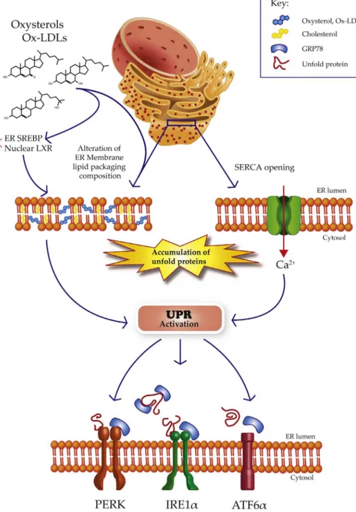
are largely unknown. By modulating cholesterol/lipid metabolism and trafficking or by directly being inserted in ER membranes, oxysterols could alter lipid packing/composition, which is expected to be detri-mental to endothelial function (Fig. 1). Interestingly, perturbation of the lipid composition could impair the folding of ER proteins, thereby activating the UPR[101]. On the other hand, oxysterols may activate the UPR by directly causing accumulation of unfolded proteins. Dong et al., showed that ox-LDL oxidize sarcoplasmic/endoplasmic reticulum Ca2+ATPase (SERCA) causing its partial inactivation[102]. It is well known that inhibitors of SERCA, such as thapsigargin induce a decrease in ER calcium levels. When calcium levels are lowered in the ER, the calcium-dependent ER chaperones, lose their activity, leading to the accumulation of unfolded proteins[103].
Fig. 1. Schematic representation of oxysterols-induced ER stress on endothelial cells. At least three major signaling are involved in the UPR acti-vation. Oxysterols may alter the lipid membrane composition and oxidize SERCA channels. Oxysterols are activators of SREBPs which are synthetized and located on the ER membrane and can modulate the activity of LXR. This last pathway can differently affects the ER status.
8. Concluding remarks
ER plays a central role in cell metabolism as it governs protein and lipid synthesis, and controls Ca2+movements. In response to oxidative insult ER activate a series of responses aimed at preserving cell function or drive signals towards apoptosis and autophagy. ER stress has been implicated in EC dysfunction and atherothrombosis. Ox-LDL, which are considered the initial insult that promotes vascular changes leading to atherogenesis, and their oxidized lipids, are involved in ER stress. Oxysterols, which are found in ox-LDL and in almost all biological fluids, have been implicated in several pathophysiological processes and are potential candidates in the mechanisms of ER stress. However, detailed studies focusing on relevant physiological concentrations and comparing the dose-dependent effect of the diverse oxysterols in in-ducing ER stress is still lacking and deserve further investigation. References
[1] A.J. Lusis, Atherosclerosis, Nature 407 (2000) 233–241.
[2] I. Tabas, G. Garcia-Cardena, G.K. Owens, Recent insights into the cellular biology of atherosclerosis, J. Cell Biol. 209 (2015) 13–22.
[3] M. Civelek, E. Manduchi, R.J. Riley, C.J. Stoeckert Jr., P.F. Davies, Chronic doplasmic reticulum stress activates unfolded protein response in arterial en-dothelium in regions of susceptibility to atherosclerosis, Circ. Res. 105 (2009) 453–461.
[4] I. Tabas, The role of endoplasmic reticulum stress in the progression of athero-sclerosis, Circ. Res. 107 (2010) 839–850.
[5] A. Negre-Salvayre, N. Auge, C. Camare, T. Bacchetti, G. Ferretti, R. Salvayre, Dual signaling evoked by oxidized LDLs in vascular cells, Free Radic. Biol. Med. 106 (2017) 118–133.
[6] C. Zerbinati, L. Iuliano, Cholesterol and related sterols autoxidation, Free Radic. Biol. Med. (2017).
[7] S.H. Park, C. Blackstone, Further assembly required: construction and dynamics of the endoplasmic reticulum network, EMBO Rep. 11 (2010) 515–521.
[8] G. van Meer, D.R. Voelker, G.W. Feigenson, Membrane lipids: where they are and how they behave, Nat. Rev. Mol. Cell Biol. 9 (2008) 112–124.
[9] S.W. Fewell, K.J. Travers, J.S. Weissman, J.L. Brodsky, The action of molecular chaperones in the early secretory pathway, Annu. Rev. Genet. 35 (2001) 149–191. [10] L. Ellgaard, A. Helenius, Quality control in the endoplasmic reticulum, Nat. Rev.
Mol. Cell Biol. 4 (2003) 181–191.
[11] M.J. Berridge, The endoplasmic reticulum: a multifunctional signaling organelle, Cell Calcium 32 (2002) 235–249.
[12] B. Meusser, C. Hirsch, E. Jarosch, T. Sommer, ERAD: the long road to destruction, Nat. Cell Biol. 7 (2005) 766–772.
[13] K. Araki, K. Nagata, Protein folding and quality control in the ER, Cold Spring Harb. Perspect. Biol. 3 (2011) a007526.
[14] A.J. Dorner, L.C. Wasley, R.J. Kaufman, Overexpression of GRP78 mitigates stress induction of glucose regulated proteins and blocks secretion of selective proteins in Chinese hamster ovary cells, EMBO J. 11 (1992) 1563–1571.
[15] W.W. Li, Y. Hsiung, Y. Zhou, B. Roy, A.S. Lee, Induction of the mammalian GRP78/BiP gene by Ca2+ depletion and formation of aberrant proteins: activa-tion of the conserved stress-inducible grp core promoter element by the human nuclear factor YY1, Mol. Cell. Biol. 17 (1997) 54–60.
[16] A. Bertolotti, Y. Zhang, L.M. Hendershot, H.P. Harding, D. Ron, Dynamic inter-action of BiP and ER stress transducers in the unfolded-protein response, Nat. Cell Biol. 2 (2000) 326–332.
[17] J. Melnick, S. Aviel, Y. Argon, The endoplasmic reticulum stress protein GRP94, in addition to BiP, associates with unassembled immunoglobulin chains, J. Biol. Chem. 267 (1992) 21303–21306.
[18] A.S. Lee, The glucose-regulated proteins: stress induction and clinical applications, Trends Biochem. Sci. 26 (2001) 504–510.
[19] D. Eletto, D. Dersh, Y. Argon, GRP94 in ER quality control and stress responses, Semin. Cell Dev. Biol. 21 (2010) 479–485.
[20] M. Marzec, D. Eletto, Y. Argon, GRP94: an HSP90-like protein specialized for protein folding and quality control in the endoplasmic reticulum, Biochim. Biophys. Acta 1823 (2012) 774–787.
[21] D. Hong, Y.P. Bai, H.C. Gao, X. Wang, L.F. Li, G.G. Zhang, et al., Ox-LDL induces endothelial cell apoptosis via the LOX-1-dependent endoplasmic reticulum stress pathway, Atherosclerosis 235 (2014) 310–317.
[22] N. Miyoshi, L. Iuliano, S. Tomono, H. Ohshima, Implications of cholesterol auto-xidation products in the pathogenesis of inflammatory diseases, Biochem. Biophys. Res. Commun. 446 (2014) 702–708.
[23] A. Corcoran, T.G. Cotter, Redox regulation of protein kinases, FEBS J. 280 (2013) 1944–1965.
[24] V.I. Perez, R. Buffenstein, V. Masamsetti, S. Leonard, A.B. Salmon, J. Mele, et al., Protein stability and resistance to oxidative stress are determinants of longevity in the longest-living rodent, the naked mole-rat, Proc. Natl. Acad. Sci. USA 106 (2009) 3059–3064.
[25] I.P. Trougakos, F. Sesti, E. Tsakiri, V.G. Gorgoulis, Non-enzymatic post-transla-tional protein modifications and proteostasis network deregulation in
carcinogenesis, J. Proteom. 92 (2013) 274–298.
[26] O. Nedic, S.I. Rattan, T. Grune, I.P. Trougakos, Molecular effects of advanced glycation end products on cell signalling pathways, ageing and pathophysiology, Free Radic. Res. 47 (Suppl. 1) (2013) 28–38.
[27] A.N. Hale, D.J. Ledbetter, T.R. Gawriluk, E.B. Rucker 3rd, Autophagy: regulation and role in development, Autophagy 9 (2013) 951–972.
[28] K. Mori, W. Ma, M.J. Gething, J. Sambrook, A transmembrane protein with a cdc2+/CDC28-related kinase activity is required for signaling from the ER to the nucleus, Cell 74 (1993) 743–756.
[29] J.S. Cox, C.E. Shamu, P. Walter, Transcriptional induction of genes encoding en-doplasmic reticulum resident proteins requires a transmembrane protein kinase, Cell 73 (1993) 1197–1206.
[30] X.Z. Wang, H.P. Harding, Y. Zhang, E.M. Jolicoeur, M. Kuroda, D. Ron, Cloning of mammalian Ire1 reveals diversity in the ER stress responses, EMBO J. 17 (1998) 5708–5717.
[31] T. Iwawaki, A. Hosoda, T. Okuda, Y. Kamigori, C. Nomura-Furuwatari, Y. Kimata, et al., Translational control by the ER transmembrane kinase/ribonuclease IRE1 under ER stress, Nat. Cell Biol. 3 (2001) 158–164.
[32] J.S. Cox, P. Walter, A novel mechanism for regulating activity of a transcription factor that controls the unfolded protein response, Cell 87 (1996) 391–404. [33] C. Sidrauski, J.S. Cox, P. Walter, tRNA ligase is required for regulated mRNA
splicing in the unfolded protein response, Cell 87 (1996) 405–413. [34] C. Sidrauski, P. Walter, The transmembrane kinase Ire1p is a site-specific
en-donuclease that initiates mRNA splicing in the unfolded protein response, Cell 90 (1997) 1031–1039.
[35] Y. Kimata, Y. Ishiwata-Kimata, T. Ito, A. Hirata, T. Suzuki, D. Oikawa, et al., Two regulatory steps of ER-stress sensor Ire1 involving its cluster formation and in-teraction with unfolded proteins, J. Cell Biol. 179 (2007) 75–86.
[36] A.V. Korennykh, P.F. Egea, A.A. Korostelev, J. Finer-Moore, C. Zhang, K.M. Shokat, et al., The unfolded protein response signals through high-order assembly of Ire1, Nature 457 (2009) 687–693.
[37] M. Calfon, H. Zeng, F. Urano, J.H. Till, S.R. Hubbard, H.P. Harding, et al., IRE1 couples endoplasmic reticulum load to secretory capacity by processing the XBP-1 mRNA, Nature 415 (2002) 92–96.
[38] X. Shen, R.E. Ellis, K. Lee, C.Y. Liu, K. Yang, A. Solomon, et al., Complementary signaling pathways regulate the unfolded protein response and are required for C. elegans development, Cell 107 (2001) 893–903.
[39] H. Yoshida, T. Matsui, A. Yamamoto, T. Okada, K. Mori, XBP1 mRNA is induced by ATF6 and spliced by IRE1 in response to ER stress to produce a highly active transcription factor, Cell 107 (2001) 881–891.
[40] H. Yoshida, T. Matsui, N. Hosokawa, R.J. Kaufman, K. Nagata, K. Mori, A time-dependent phase shift in the mammalian unfolded protein response, Dev. Cell 4 (2003) 265–271.
[41] A.H. Lee, N.N. Iwakoshi, L.H. Glimcher, XBP-1 regulates a subset of endoplasmic reticulum resident chaperone genes in the unfolded protein response, Mol. Cell. Biol. 23 (2003) 7448–7459.
[42] A. Uemura, M. Taniguchi, Y. Matsuo, M. Oku, S. Wakabayashi, H. Yoshida, UBC9 regulates the stability of XBP1, a key transcription factor controlling the ER stress response, Cell Struct. Funct. 38 (2013) 67–79.
[43] H.P. Harding, Y. Zhang, D. Ron, Protein translation and folding are coupled by an endoplasmic-reticulum-resident kinase, Nature 397 (1999) 271–274.
[44] H.P. Harding, Y. Zhang, A. Bertolotti, H. Zeng, D. Ron, Perk is essential for translational regulation and cell survival during the unfolded protein response, Mol. Cell 5 (2000) 897–904.
[45] H. Yoshida, T. Okada, K. Haze, H. Yanagi, T. Yura, M. Negishi, et al., ATF6 acti-vated by proteolysis binds in the presence of NF-Y (CBF) directly to the cis-acting element responsible for the mammalian unfolded protein response, Mol. Cell. Biol. 20 (2000) 6755–6767.
[46] H. Yoshida, T. Okada, K. Haze, H. Yanagi, T. Yura, M. Negishi, et al., Endoplasmic reticulum stress-induced formation of transcription factor complex ERSF including NF-Y (CBF) and activating transcription factors 6alpha and 6beta that activates the mammalian unfolded protein response, Mol. Cell. Biol. 21 (2001) 1239–1248. [47] S. Song, J. Tan, Y. Miao, M. Li, Q. Zhang, Crosstalk of autophagy and apoptosis:
involvement of the dual role of autophagy under ER stress, J. Cell. Physiol. (2017). [48] N. Mizushima, M. Komatsu, Autophagy: renovation of cells and tissues, Cell 147
(2011) 728–741.
[49] S. Deegan, S. Saveljeva, A.M. Gorman, A. Samali, Stress-induced self-cannibalism: on the regulation of autophagy by endoplasmic reticulum stress, Cell. Mol. Life Sci.: CMLS 70 (2013) 2425–2441.
[50] D.W. Hailey, A.S. Rambold, P. Satpute-Krishnan, K. Mitra, R. Sougrat, P.K. Kim, et al., Mitochondria supply membranes for autophagosome biogenesis during starvation, Cell 141 (2010) 656–667.
[51] O.M. de Brito, L. Scorrano, Mitofusin 2 tethers endoplasmic reticulum to mi-tochondria, Nature 456 (2008) 605–610.
[52] T. Yorimitsu, U. Nair, Z. Yang, D.J. Klionsky, Endoplasmic reticulum stress triggers autophagy, J. Biol. Chem. 281 (2006) 30299–30304.
[53] S. Bernales, K.L. McDonald, P. Walter, Autophagy counterbalances endoplasmic reticulum expansion during the unfolded protein response, PLoS Biol. 4 (2006) e423.
[54] L. Ge, R. Schekman, The ER-golgi intermediate compartment feeds the phago-phore membrane, Autophagy 10 (2014) 170–172.
[55] W. B'Chir, A.C. Maurin, V. Carraro, J. Averous, C. Jousse, Y. Muranishi, et al., The eIF2alpha/ATF4 pathway is essential for stress-induced autophagy gene expres-sion, Nucleic Acids Res. 41 (2013) 7683–7699.
[56] P. Haberzettl, B.G. Hill, Oxidized lipids activate autophagy in a JNK-dependent manner by stimulating the endoplasmic reticulum stress response, Redox Biol. 1
(2013) 56–64.
[57] F. Luchetti, B. Canonico, E. Cesarini, M. Betti, L. Galluzzi, L. Galli, et al., 7-Ketocholesterol and 5,6-secosterol induce human endothelial cell dysfunction by differential mechanisms, Steroids 99 (2015) 204–211.
[58] L. Iuliano, P.J. Crick, C. Zerbinati, L. Tritapepe, J. Abdel-Khalik, M. Poirot, et al., Cholesterol metabolites exported from human brain, Steroids 99 (2015) 189–193. [59] G. Segala, P. de Medina, L. Iuliano, C. Zerbinati, M.R. Paillasse, E. Noguer, et al., 5,6-Epoxy-cholesterols contribute to the anticancer pharmacology of tamoxifen in breast cancer cells, Biochem. Pharmacol. 86 (2013) 175–189.
[60] B. Buttari, L. Segoni, E. Profumo, D. D'Arcangelo, S. Rossi, F. Facchiano, et al., 7-Oxo-cholesterol potentiates pro-inflammatory signaling in human M1 and M2 macrophages, Biochem. Pharmacol. 86 (2013) 130–137.
[61] G. Lizard, V. Deckert, L. Dubrez, M. Moisant, P. Gambert, L. Lagrost, Induction of apoptosis in endothelial cells treated with cholesterol oxides, Am. J. Pathol. 148 (1996) 1625–1638.
[62] G. Lizard, M. Moisant, C. Cordelet, S. Monier, P. Gambert, L. Lagrost, Induction of similar features of apoptosis in human and bovine vascular endothelial cells treated by 7-ketocholesterol, J. Pathol. 183 (1997) 330–338.
[63] G. Lizard, S. Monier, C. Cordelet, L. Gesquiere, V. Deckert, S. Gueldry, et al., Characterization and comparison of the mode of cell death, apoptosis versus ne-crosis, induced by 7beta-hydroxycholesterol and 7-ketocholesterol in the cells of the vascular wall, Arterioscler. Thromb. Vasc. Biol. 19 (1999) 1190–1200. [64] B. Vurusaner, G. Leonarduzzi, P. Gamba, G. Poli, H. Basaga, Oxysterols and
me-chanisms of survival signaling, Mol. Asp. Med. 49 (2016) 8–22.
[65] V. Deckert, L. Duverneuil, S. Poupon, S. Monier, N. Le Guern, G. Lizard, et al., The impairment of endothelium-dependent arterial relaxation by 7-ketocholesterol is associated with an early activation of protein kinase C, Br. J. Pharmacol. 137 (2002) 655–662.
[66] V. Deckert, A. Brunet, F. Lantoine, G. Lizard, E. Millanvoye-van Brussel, S. Monier, et al., Inhibition by cholesterol oxides of NO release from human vascular en-dothelial cells, Arterioscler. Thromb. Vasc. Biol. 18 (1998) 1054–1060. [67] M. Sanson, N. Auge, C. Vindis, C. Muller, Y. Bando, J.C. Thiers, et al., Oxidized
low-density lipoproteins trigger endoplasmic reticulum stress in vascular cells: prevention by oxygen-regulated protein 150 expression, Circ. Res. 104 (2009) 328–336.
[68] R. Scherz-Shouval, Z. Elazar, Regulation of autophagy by ROS: physiology and pathology, Trends Biochem. Sci. 36 (2011) 30–38.
[69] M. Nowicki, O. Zabirnyk, N. Duerrschmidt, J. Borlak, K. Spanel-Borowski, No upregulation of lectin-like oxidized low-density lipoprotein receptor-1 in serum-deprived EA.hy926 endothelial cells under oxLDL exposure, but increase in au-tophagy, Eur. J. Cell Biol. 86 (2007) 605–616.
[70] T. Nury, A. Zarrouk, A. Vejux, M. Doria, J.M. Riedinger, R. Delage-Mourroux, et al., Induction of oxiapoptophagy, a mixed mode of cell death associated with oxidative stress, apoptosis and autophagy, on 7-ketocholesterol-treated 158N murine oligodendrocytes: impairment by alpha-tocopherol, Biochem. Biophys. Res. Commun. 446 (2014) 714–719.
[71] X.M. Yuan, N. Sultana, N. Siraj, L.J. Ward, B. Ghafouri, W. Li, Autophagy induc-tion protects against 7-oxysterol-induced cell death via lysosomal pathway and oxidative stress, J. Cell Death 9 (2016) 1–7.
[72] C. Muller, R. Salvayre, A. Negre-Salvayre, C. Vindis, HDLs inhibit endoplasmic reticulum stress and autophagic response induced by oxidized LDLs, Cell Death Differ. 18 (2011) 817–828.
[73] N. Shibata, C.K. Glass, Macrophages, oxysterols and atherosclerosis, Circ. J.: Off. J. Jpn. Circ. Soc. 74 (2010) 2045–2051.
[74] X. Rong, C.J. Albert, C. Hong, M.A. Duerr, B.T. Chamberlain, E.J. Tarling, et al., LXRs regulate ER stress and inflammation through dynamic modulation of mem-brane phospholipid composition, Cell Metab. 18 (2013) 685–697.
[75] A. Radhakrishnan, Y. Ikeda, H.J. Kwon, M.S. Brown, J.L. Goldstein, Sterol-regu-lated transport of SREBPs from endoplasmic reticulum to Golgi: oxysterols block transport by binding to Insig, Proc. Natl. Acad. Sci. USA 104 (2007) 6511–6518. [76] L.P. Sun, J. Seemann, J.L. Goldstein, M.S. Brown, Sterol-regulated transport of
SREBPs from endoplasmic reticulum to Golgi: insig renders sorting signal in Scap inaccessible to COPII proteins, Proc. Natl. Acad. Sci. USA 104 (2007) 6519–6526. [77] S.B. Joseph, A. Castrillo, B.A. Laffitte, D.J. Mangelsdorf, P. Tontonoz, Reciprocal regulation of inflammation and lipid metabolism by liver X receptors, Nat. Med. 9 (2003) 213–219.
[78] S. Gargiulo, P. Gamba, G. Testa, D. Rossin, F. Biasi, G. Poli, et al., Relation between TLR4/NF-kappaB signaling pathway activation by 27-hydroxycholesterol and 4-hydroxynonenal, and atherosclerotic plaque instability, Aging Cell 14 (2015) 569–581.
[79] M. Umetani, P. Ghosh, T. Ishikawa, J. Umetani, M. Ahmed, C. Mineo, et al., The
cholesterol metabolite 27-hydroxycholesterol promotes atherosclerosis via proin-flammatory processes mediated by estrogen receptor alpha, Cell Metab. 20 (2014) 172–182.
[80] P.L. Liao, Y.W. Cheng, C.H. Li, Y.L. Lo, J.J. Kang, Cholesterol-3-beta, 5-alpha, 6-beta-triol induced PI(3)K-Akt-eNOS-dependent cyclooxygenase-2 expression in endothelial cells, Toxicol. Lett. 190 (2009) 172–178.
[81] G. Leonarduzzi, P. Gamba, B. Sottero, A. Kadl, F. Robbesyn, R.A. Calogero, et al., Oxysterol-induced up-regulation of MCP-1 expression and synthesis in macro-phage cells, Free Radic. Biol. Med. 39 (2005) 1152–1161.
[82] A.J. Brown, L. Sun, J.D. Feramisco, M.S. Brown, J.L. Goldstein, Cholesterol ad-dition to ER membranes alters conformation of SCAP, the SREBP escort protein that regulates cholesterol metabolism, Mol. Cell 10 (2002) 237–245.
[83] P.J. Espenshade, W.P. Li, D. Yabe, Sterols block binding of COPII proteins to SCAP, thereby controlling SCAP sorting in ER, Proc. Natl. Acad. Sci. USA 99 (2002) 11694–11699.
[84] T.P. Shentu, D.K. Singh, M.J. Oh, S. Sun, L. Sadaat, A. Makino, et al., The role of oxysterols in control of endothelial stiffness, J. Lipid Res. 53 (2012) 1348–1358. [85] C. Gorrini, I.S. Harris, T.W. Mak, Modulation of oxidative stress as an anticancer
strategy, Nat. Rev. Drug Discov. 12 (2013) 931–947.
[86] B. Vurusaner, P. Gamba, S. Gargiulo, G. Testa, E. Staurenghi, G. Leonarduzzi, et al., Nrf2 antioxidant defense is involved in survival signaling elicited by 27-hydroxycholesterol in human promonocytic cells, Free Radic. Biol. Med. 91 (2016) 93–104.
[87] Q. Jiang, D. Wang, Y. Han, Z. Han, W. Zhong, C. Wang, Modulation of oxidized-LDL receptor-1 (LOX1) contributes to the antiatherosclerosis effect of oleanolic acid, Int. J. Biochem. Cell Biol. 69 (2015) 142–152.
[88] X.L. Chen, G. Dodd, S. Thomas, X. Zhang, M.A. Wasserman, B.H. Rovin, et al., Activation of Nrf2/ARE pathway protects endothelial cells from oxidant injury and inhibits inflammatory gene expression, Am. J. Physiol. Heart Circ. Physiol. 290 (2006) H1862–H1870.
[89] H. Kentala, M. Weber-Boyvat, V.M. Olkkonen, OSBP-related protein family: mediators of lipid transport and signaling at membrane contact sites, Int. Rev. Cell Mol. Biol. 321 (2016) 299–340.
[90] J.J. Mackrill, Oxysterols and calcium signal transduction, Chem. Phys. Lipids 164 (2011) 488–495.
[91] H. Yoshida, ER stress and diseases, FEBS J. 274 (2007) 630–658.
[92] F.K. Swirski, P. Libby, E. Aikawa, P. Alcaide, F.W. Luscinskas, R. Weissleder, et al., Ly-6Chi monocytes dominate hypercholesterolemia-associated monocytosis and give rise to macrophages in atheromata, J. Clin. Investig. 117 (2007) 195–205. [93] I. Tabas, K.E. Bornfeldt, Macrophage phenotype and function in different stages of
atherosclerosis, Circ. Res. 118 (2016) 653–667.
[94] L. Zeng, A. Zampetaki, A. Margariti, A.E. Pepe, S. Alam, D. Martin, et al., Sustained activation of XBP1 splicing leads to endothelial apoptosis and atherosclerosis development in response to disturbedflow, Proc. Natl. Acad. Sci. USA 106 (2009) 8326–8331.
[95] J.J. Manning-Tobin, K.J. Moore, T.A. Seimon, S.A. Bell, M. Sharuk, J.I. Alvarez-Leite, et al., Loss of SR-A and CD36 activity reduces atherosclerotic lesion com-plexity without abrogating foam cell formation in hyperlipidemic mice, Arterioscler. Thromb. Vasc. Biol. 29 (2009) 19–26.
[96] A.P. Lillis, S.C. Muratoglu, D.T. Au, M. Migliorini, M.J. Lee, S.K. Fried, et al., LDL receptor-related protein-1 (LRP1) regulates cholesterol accumulation in macro-phages, PLoS One 10 (2015) e0128903.
[97] H. Suzuki, Y. Kurihara, M. Takeya, N. Kamada, M. Kataoka, K. Jishage, et al., A role for macrophage scavenger receptors in atherosclerosis and susceptibility to infection, Nature 386 (1997) 292–296.
[98] A.R. Tall, L. Yvan-Charvet, Cholesterol, inflammation and innate immunity, Nat. Rev. Immunol. 15 (2015) 104–116.
[99] J. Zhou, M.D. Abid, Y. Xiong, Q. Chen, J. Chen, ox-LDL downregulates eNOS ac-tivity via LOX-1-mediated endoplasmic reticulum stress, Int. J. Mol. Med. 32 (2013) 1442–1450.
[100] Y.K. Tao, P.L. Yu, Y.P. Bai, S.T. Yan, S.P. Zhao, G.Q. Zhang, Role of PERK/ eIF2alpha/CHOP endoplasmic reticulum stress pathway in oxidized low-density lipoprotein mediated induction of endothelial apoptosis, Biomed. Environ. Sci.: BES 29 (2016) 868–876.
[101] R. Volmer, D. Ron, Lipid-dependent regulation of the unfolded protein response, Curr. Opin. Cell Biol. 33 (2015) 67–73.
[102] Y. Dong, M. Zhang, S. Wang, B. Liang, Z. Zhao, C. Liu, et al., Activation of AMP-activated protein kinase inhibits oxidized LDL-triggered endoplasmic reticulum stress in vivo, Diabetes 59 (2010) 1386–1396.
[103] C.M. Oslowski, F. Urano, Measuring ER stress and the unfolded protein response using mammalian tissue culture system, Methods Enzymol. 490 (2011) 71–92.