21
Pakistan Veterinary Journal
ISSN: 0253-8318 (PRINT), 2074-7764 (ONLINE) Accessible at: www.pvj.com.pk
Transrectal Ultrasonography of the Adrenal Glands in Donkeys (Equus asinus)
Fulvio Laus*, Andrea Spaterna, Vanessa Faillace, Emanuele Paggi, Matteo Cerquetella, Alessandro Fruganti and Beniamino Tesei
School of Biosciences and Veterinary Medicine, University of Camerino, Via Circonvallazione 93/95, 62024 Matelica (MC), Italy
*Corresponding author: [email protected] ARTICLE HISTORY (15-024) A B S T R A C T Received: Revised: Accepted: Online available: January 21, 2015 March 21, 2015 July 12, 2015 November 29, 2015
Little information is available for medical imaging in donkeys and no report about adrenal glands ultrasonography can be found in scientific literature. The feasibility of transrectal ultrasonography of the adrenal glands was tested on 30 healthy donkeys using a 10 MHz linear-array transducer. Mean age of animals was 10.7±4.8 years, mean body weight 275.0±62.9 kg and mean height 126.7±7.1 cm. The left adrenal gland was visualized in all donkeys. The right gland ultrasonography was not feasible in seven animals with a height less than 116 cm. The left gland was visualized as a linear or slightly curved structure, the right gland was most often S-shaped. In both glands, a hypochoic peripheral zone was identified as the cortex with an inner, hyperechoic medulla. The length was 5.49±1.90 and 5.15±1.10 cm for right and left gland, respectively. Right gland whole and medullary thickness was 0.71±0.11 and 0.24±0.09 cm, 0.65±0.13 and 0.21±0.07 cm, 0.56±0.17 and 0.25±0.07 cm for cranial pole, middle point and caudal pole respectively. Left gland whole and medullary thickness values were 0.69±0.13 and 0.25±0.09 cm, 0.66±0.13 and 0.23±0.09 cm, 0.57±0.15 and 0.26±0.09 cm for cranial pole, middle point and caudal pole, respectively. There was a significant correlation between height and the entire length of the left gland. In conclusion, ultrasonography of the adrenal glands is a suitable tool for evaluation of both adrenal glands in most of the donkey. The size is a limiting factor for proper visualization of the right gland.
©2015 PVJ. All rights reserved Key words:
Adrenal gland Biometry Donkey Ultrasonography
To Cite This Article: Laus F, A Spaterna, V Faillace, E Paggi, M Cerquetella, A Fruganti and B Tesei, 2016. Transrectal ultrasonography of the adrenal glands in donkeys (Equus asinus). Pak Vet J, 36(1): 21-24.
INTRODUCTION
Because of the recent rediscovery of donkey milk as an alternative food source for milk-intolerant children (Muraro et al., 2002), interest in the welfare and diseases of donkeys is constantly increasing (Selvaggi et al., 2015; Trinchese et al., 2015; Perna et al., 2015). Further reasons for their worldwide popularity are different, depending on the country, and include recreational purposes, use as pets, sports activities, onotherapy, use as pack/draught animals and meat production. Despite these, most clinical and diagnostic aspects of donkey’s diseases are still not properly investigated (Trawford, 2011), including endocrinology disorders. Diseases involving the adrenal gland in equidae include, hypoadrenocorticism, pituitary-independent hyperadreno-corticism, adrenocortical insufficiency and pheochromo-cytoma (Breuer et al., 1993; Toribio, 2004; Germann et al., 2006; Dybdal and McFarlane, 2009; Hart and Barton, 2011; Liburt et al., 2013; Herbach et al., 2014, Jellyman et al., 2015).
Adrenal glands are positioned retroperitoneally, cranio-medially to the corresponding kidney. The left gland is located within the angle formed by the aorta and the left renal artery. The right gland is positioned slightly cranially than the left one and lies near the caudal cava vein (Budras et al., 2009; Barone and Simoens, 2012). An ultrasonographic technique to assess adrenal glands in horses was defined for the first time in 2008 (Laus et al., 2008) and, to the author’s knowledge, only another study about equidae exists (Durie et al., 2010). No reports about donkeys are available in scientific literature. The aims of this study were to evaluate the ultrasonographic appearance and size of adrenal glands in donkeys and to estimate the influence of sex, body weight, size and age on the appearance and dimensions of this gland.
MATERIALS AND METHODS
Thirty mixed-breed donkeys (19 non-pregnant female, 9 geldings and 2 stallions) belonging to three RESEARCH ARTICLE
Pak Vet J, 2016, 36(1): 21-24. 22
different farms were included in the study. Age of animals ranged from 4 to 19 years (mean 10.7±4.8 years), body weight ranged from 165 to 369 kg (mean 275.0±62.9 kg) and height ranged from 100 to 139 cm (mean 126.7±7.1 cm). Donkeys were placed in the stock and scopolaminebutyl-bromide and metamizolnatrium (Buscopanvet Compositum® Boehringer Ingelheim, Milano, Italy) was administered by intravenous injection to relax the smooth intestinal muscles, reducing animal discomfort and the risk of injury. A 10 MHz linear array transrectal probe (MyLabTM30 VET, Esaote, Genova, Italy) set at a depth of 8 cm was inserted in the rectum, at first using the right arm, directing the ultrasound beam at the 12 o’clock position toward the abdominal aorta and then following the technique previous described for horses (Laus et al., 2008; Durie et al., 2010), until the gland was visualized (Fig. 1).
To visualize the right adrenal gland, the left hand was used. The technique was similar to that described for the left gland, except that the probe was pushed more cranially and the branches of the mesenteric artery were not visualized cranially to the gland but on its medial aspect and about in the midpoint of the gland’s length. All images obtained were stored to allow a later assessment (MyLabDeskTM, MyLabDesk, Genova, Italy).
After the evaluation of the overall appearance of the glands, the following measures for the entire gland and the medulla were taken: the ventrodorsal thickness 1 cm cranial to the caudal pole, at the midpoint of the gland and 1 cm caudal to the cranial pole (Fig 2). The craniocaudal length of the entire adrenal glands was also measured (Fig. 3). The relationship between age, gender, body weight, height and gland measures were assessed by the Pearson correlations, using the WINPEPI (PEPI-for-Windows) computer programs for epidemiologists (Abramson, 2011). Level of significance was set at P<0.05.
RESULTS
The left adrenal gland was seen in all donkeys, while the ultrasonography of the right gland was not or only partially feasible in 7 donkeys (23%). Although individual variation of the shape of the glands was observed, it generally appeared as flat structure surrounded by a hyperechoic thin capsule. The left adrenal gland had a linear shape (n= 21, 70%), sometimes appearing slightly curved (n= 5, 17%) or as comma shaped (n= 4, 13%; Fig 3). In 7 donkeys (23%), the left lobe of the pancreas was seen cranially to the cranial pole of the left adrenal gland. A large vessel in pancreatic parenchyma was identified as the portal vein (Fig. 4).
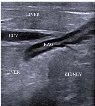
The right gland had a more variable shape, most often S-shaped (n= 14, 47%) (Fig 5), sometimes with a curved appearance (n= 6, 20%), rarely linear (n= 3, 10%). The right gland was located between the cranial pole of the kidney and the impressio renalis of the liver that sometimes became visible dorsocranially to the gland with its caudate process, together with the caudal cava vein (Fig. 5).
In both glands, a hypoechoic peripheral zone was identified as the cortex, while the medulla appeared as an inner, centrally located hyperechoic strip (Fig. 1, 2, 4 and
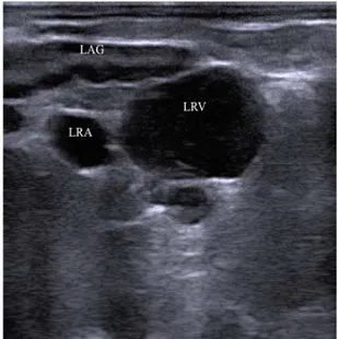
Fig. 1: Caudal part of the left adrenal gland (LAG). Left renal vein (LRV) and artery (LRA) are seen adjacent to the gland. The hypoechoic peripheral cortex, and the inner hyperechoic medulla are well distinguishable. The display depth is 8 cm and cranial is to the left.
Fig. 2: Measurement of left adrenal gland (LAG). D1, caudal pole thickness. D2, caudal pole medullary thickness. The display depth is 8 cm and cranial is to the left.
Fig. 3: The most common aspect of the left adrenal gland (LAG) is linear. In older donkeys a less defined separation between the cortex and the medulla can be noticed. The display depth is 8 cm and cranial is to the left.
Pak Vet J, 2016, 36(1): 21-24. 23
Fig. 4: Cranial pole of the left adrenal gland. Note the large portal vein and the pancreatic parenchyma located cranially to the gland. The display depth is 8 cm and cranial is to the left.
Fig. 5: The slightly S-shaped right adrenal gland (RAG) is embedded between renal and hepatic parenchyma and adjacent to the caudal cava vein (CCV). The display depth is 8 cm and cranial is to the left. Table 1: Mean (±SD) values of left and right adrenal gland measures
Measures Left gland (cm) Right gland (cm) Cranial pole thickness 0.69 ±0.13 0.71±0.11 Cranial pole medullary thickness 0.25 ±0.09 0.24±0.09 Midpoint thickness 0.66 ±0.13 0.65±0.13 Midpoint medullary thickness 0.23 ±0.09 0.21±0.07 Caudal pole thickness 0.57 ±0.15 0.56±0.17 Caudal pole medullary thickness 0.26 ±0.09 0.25±0.07
Length 5.15 ±1.10 5.49±1.90
5). In 3 donkeys (10%), all older than 12 years, the corticomedullary junction of both glands was indistinct, making an accurate measurement difficult (Fig. 3). In one donkey (3%), some round anechoic structures in the cortex of both left and right adrenal glands were detected.
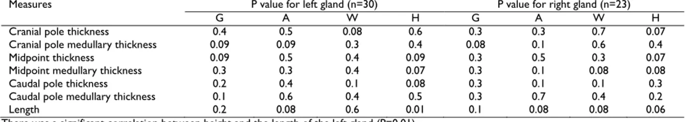
The mean and standard deviation of all measurement are shown in Table 1. Statistical correlations between measurement and gender, age, body weight and height are reported in Table 2. There was a significant correlation between height and the length of the left gland (P=0.01).
DISCUSSION
The method for visualization of left adrenal gland was similar to that used in horses: landmarks as the renal vein and artery for the caudal pole and the cranial mesenteric artery for the cranial pole could be used in the same way described for horses (Laus et al., 2008; Durie et al., 2010). The portal vein was visualized in the pancreatic parenchyma, which resulted in its inclusion in pancreatic ring (anulus pancreatis), an anatomical features typical of Equidae (König and Liebich, 2007).
On the right side, the renal artery was more clearly visible at the point where it branched from the aorta at the level of the caudal pole of the right gland. The right renal artery could be followed in cranial direction and, at the level of the mid-length of the adrenal glands, the arteria adrenalis caudales could be sometimes seen to branches from it and to be directed toward the mid-ventral aspect of the right gland.
Problems in imaging the glands caused by the presence of perirenal fat have been sometimes encountered in horses (Durie et al., 2010). This was not the case of donkeys, probably due to the low presence of this tissue. In the donkey, the left gland was more rectilinear than in horse, while the right one had more often the characteristic S-shape. This could be due to the compression, which the right gland is subjected, because it is partly embedded between the renal and hepatic parenchyma.
As previously reported in horses and older donkeys showed less defined separation between the cortex and the medulla (Durie et al., 2010). Although it has been proved that adrenal glands experience a decline in function with age in horses (Liburt et al., 2013), further studies will be necessary to investigate the relationship between ultrasonographic appearance and functional alteration due to age in these species.
In the present study, ultrasonography of the right gland was not feasible in 23% of donkeys. This appears to be related to donkey size, since in animals with body height less than 116 cm it was not possible to visualize the right gland. This was due to the inability to insert the arm deep enough without inducing discomfort caused by excessive dilation of the anus. In a previous study in horses, it was not possible to visualize the right adrenal gland in animals higher than 151 cm (Durie et al., 2010). In another report, it was possible to visualize correctly the right adrenal gland only in 16 out of 150 horses (10.7%) and all of them were lower than 150 cm in height (Laus et al., 2008). According to the authors’ opinion, this could be due to the most cranial position of the right adrenal gland and to the presence of the cecum. Since in this report the maximum height of the animal was 139 cm, we cannot establish the upper limits of height in donkeys to visualize the right gland. Although it can be supposed that it is similar to that of horses, further studies are needed to investigate this issue.
Pak Vet J, 2016, 36(1): 21-24. 24
Table 2: Statistical correlation between adrenal glands measurements and gender (G), age (A), weight (W) and height (H)
Measures P value for left gland (n=30) P value for right gland (n=23)
G A W H G A W H
Cranial pole thickness 0.4 0.5 0.08 0.6 0.3 0.3 0.7 0.07
Cranial pole medullary thickness 0.09 0.09 0.3 0.4 0.08 0.1 0.6 0.4
Midpoint thickness 0.09 0.5 0.4 0.09 0.3 0.5 0.3 0.07
Midpoint medullary thickness 0.3 0.3 0.4 0.07 0.3 0.1 0.08 0.08
Caudal pole thickness 0.2 0.4 0.1 0.08 0.3 0.1 0.1 0.3
Caudal pole medullary thickness 0.1 0.6 0.4 0.5 0.3 0.7 0.4 0.2
Length 0.2 0.08 0.6 0.01 0.1 0.08 0.08 0.06
There was a significant correlation between height and the length of the left gland (P=0.01).
Very few data are available about adrenal glands anatomy in donkeys. In a recent report based on post-mortem measurements, left adrenal gland of donkey was reported to be 4.15-4.74 cm in length and 0.47-0.76 cm in thickness, while the right one was reported to be 4.31-5.65 cm in length and 0.47-0.77 cm in thickness (Karakurum et al., 2008). Sonographic ranges found in this study (left length: 4.21-6.00 cm; left thickness: 0.49-0.78 cm; right length: 4.59-6.38 cm; right thickness: 0.49-0.79 cm) are nearly in agreement with direct post mortem measurement with the exception of the gland lengths for which the sonographic ranges appear slightly higher. As also previously suggested, differences in size and shape could be due to the different position of the glands in vivo compared with post -mortem samples, and to the quick degeneration of the adrenal glands in cadaveric specimens (Durie et al., 2010).
Conclusions: Ultrasonographic investigation of the adrenal glands is possible on both glands in donkeys with body height more than 120 cm. The investigation is also operator-dependent and limitations for correct visualization of the glands are related both to the animal size (small donkeys) and the operator arm diameter. The study should also be intended as a contribution for veterinary practitioners because it describes for the first time the technique to visualize adrenal glands in donkeys. Further specific studies and a more routinely use of ultrasonography, will allow diagnosis of diseases, so far not fully investigated in this important species.
Author’s contribution: FL performed the ultrasono-graphy, supervised all stages of the work, made statistical analysis and wrote the draft. SA supervised all stages of the work presented in this report and contributed to drafting. VF and EP performed the ultrasonography. CM and AF contribute in writing the draft and to the statistical analysis. BT has been involved in drafting and revising the manuscript and gave final approval of the version to be published. All authors interpreted the data, critically revised the manuscript for important intellectual contents and approved the final version.
REFERENCES
Abramson JH, 2011. WINPEPI updated: computer programs for epidemiologists, and their teaching potential. Epidemiol Perspect Innov, 8:1-9.
Barone R and Simoens P, 2012. Anatomia comparata dei mammiferi domestici. Vol. 7/2: Neurologia. Sistema nervoso periferico,
ghiandole endocrine, estesiologia. Edagricole, Milano, Italy, pp: 744.
Breuer W, Grabner A, Hänichen T, Schmidt P and Hermanns W, 1993. Phäochromozytom als Ursache des plötzlichen Todes eines ferdes. Pferdeheilkunde. 9: 301-304.
Budras KD, Sack WO and Rock S, 2009. Anatomy of the Hhorse. 5th
Ed. Schlutersche Verlagsgesellschaft mbH & Co, Hannover, Germany, pp: 78.
Durie I, van Loon G, Vermeire S, De Clercq D, Vanschandevijl K et al., 2010. Transrectal ultrasonography of the left adrenal gland in healthy horses. Vet Radiol Ultrasound, 51: 540-544.
Dybdal NO and McFarlane D, 2009. Adrenal glands. In: Large Animal Internal Medicine. Smith BP (ed). 4th Ed, Mosby Elsevier, St. Louis,
USA, pp: 1345-1347.
Germann SE, Rütten M, Derungs SB and Feige K, 2006. Multiple-endocrine neoplasia-like syndrome in a horse. Vet Rec, 159: 530-532.
Hart KA and Barton MH, 2011. Adrenocortical insufficiency in horses and foals. Vet Clin North Am Equine Pract, 27: 19-34.
Herbach N, Nagel L, Zwick T and Hermanns W, 2014. Multiple glucagon-producing pancreatic neuroendocrine tumors in a horse (Equus caballus). Vet Pathol, 51: 607-611.
Jellyman JK, Valenzuela OA, Allen VL, Forhead AJ, Holdstock NB et al., 2015. Neonatal glucocorticoid overexposure programs pituitary-adrenal function in ponies. Domest Anim Endocrinol. 50: 45-49. Karakurum E, Özgel Ö and Dursun N, 2008. Morphology and arterial
vasculature of donkey (Equus asinus L.) adrenal gland. Turk J Vet Anim Sci, 32: 469-473.
König HE and Liebich HG, 2007. Veterinary Anatomy of Domestic Mammals: Textbook and Colour Atlas. Schattauer, Stuttgard, Germany.
Laus F, Paggi E, Grabner A, Klaus C, Spaterna A et al., 2008. Ultrasonography of adrenal glands in the horse by transrectal visualization. Pferdeheilkunde, 24: 688-692.
Liburt NR, McKeever KH, Malinowski K, Smarsh DN and Geor RJ, 2013. Response of the hypothalamic-pituitary-adrenal axis to stimulation tests before and after exercise training in old and young Standardbred mares. J Anim Sci, 91: 5208-5219.
Liburt NR, McKeever KH, Malinowski K, Smarsh DN and Geor RJ, 2013. Response of the hypothalamic-pituitary-adrenal axis to stimulation tests before and after exercise training in old and young Standardbred mares. J Anim Sci. 91: 5208-5219.
Muraro MA, Giampietro PG and Galli E, 2002. Soy formulas and nonbovine milk. Ann Allergy Asthma Immunol, 89(6 suppl 1): 97-101.
Perna A, Intaglietta I, Simonetti A and Gambacorta E, 2015. Donkey Milk for Manufacture of Novel Functional Fermented Beverages. J Food Sci. 80 : 1352-1359.
Selvaggi M, D'Alessandro AG and Dario C, 2015. Comparative characteristics of DNA polymorphisms of κ-casein gene (CSN3) in the horse and donkey. Genet Mol Res, 14: 14567-14575. Toribio RE, 2004. The adrenal glands. In: Equine Internal Medicine
(Reed SM, ed). 2nd Ed, Saunders, St. Louis, USA, pp: 357-360.
Trawford A, 2011. Donkey welfare internationally - current research. In: Proceedings of the 50th British Equine Veterinary Association Congress: 7-10 sept. 2011; Liverpool. Edited By BEVA, pp: 258. Trinchese G, Cavaliere G, Canani RB, Matamoros S, Bergamo P et al.,
2015. Human, donkey and cow milk differently affects energy efficiency and inflammatory state by modulating mitochondrial function and gut microbiota. J Nutr Biochem, 26: 1136-1146.