Received 13 Apr 2016
|
Accepted 22 Nov 2016
|
Published 16 Jan 2017
A mast cell-ILC2-Th9 pathway promotes lung
inflammation in cystic fibrosis
Silvia Moretti
1
, Giorgia Renga
1
, Vasilis Oikonomou
1
, Claudia Galosi
1
, Marilena Pariano
1
, Rossana G. Iannitti
1
,
Monica Borghi
1
, Matteo Puccetti
1
, Marco De Zuani
2
, Carlo E. Pucillo
2
, Giuseppe Paolicelli
1
, Teresa Zelante
1
,
Jean-Christophe Renauld
3
, Oxana Bereshchenko
4
, Paolo Sportoletti
5
, Vincenzina Lucidi
6
, Maria Chiara Russo
7
,
Carla Colombo
7
, Ersilia Fiscarelli
8
, Cornelia Lass-Flo¨rl
9
, Fabio Majo
6
, Gabriella Ricciotti
8
, Helmut Ellemunter
10
,
Luigi Ratclif
11
, Vincenzo Nicola Talesa
1
, Valerio Napolioni
1
& Luigina Romani
1
T helper 9 (Th9) cells contribute to lung inflammation and allergy as sources of interleukin-9
(IL-9). However, the mechanisms by which IL-9/Th9 mediate immunopathology in the lung
are unknown. Here we report an IL-9-driven positive feedback loop that reinforces allergic
inflammation. We show that IL-9 increases IL-2 production by mast cells, which leads to
expansion of CD25
þtype 2 innate lymphoid cells (ILC2) and subsequent activation of Th9
cells. Blocking IL-9 or inhibiting CD117 (c-Kit) signalling counteracts the pathogenic effect of
the described IL-9-mast cell-IL-2 signalling axis. Overproduction of IL-9 is observed in
expectorates from cystic fibrosis (CF) patients, and a sex-specific variant of IL-9 is predictive
of allergic reactions in female patients. Our results suggest that blocking IL-9 may be a
therapeutic strategy to ameliorate inflammation associated with microbial colonization in the
lung, and offers a plausible explanation for gender differences in clinical outcomes of patients
with CF.
DOI: 10.1038/ncomms14017
OPEN
1Department of Experimental Medicine, University of Perugia, 06132 Perugia, Italy.2Department of Medical and Biological Science, University of Udine,
33100 Udine, Italy.3Ludwig Institute for Cancer Research, Brussels Branch, B-1200 Brussels, Belgium.4Department of Medicine, Section of Pharmacology,
University of Perugia, 06132 Perugia, Italy.5Institute of Haematology-CREO (Centro di Ricerche Emato-Oncologiche), Ospedale S. Maria Misericordia,
06132 Perugia, Italy.6Unit of Endocrinology and Diabetes, Bambino Gesu` Children’s Hospital, 00165 Rome, Italy.7Fondazione IRCCS Ca’ Granda,
Ospedale Maggiore Policlinico, University of Milan, 20122 Milan, Italy.8Bambino Gesu` Children’s Hospital IRCCS, 00165 Rome, Italy.9Division of Hygiene
and Medical Microbiology, Innsbruck Medical University, 6020 Innsbruck, Austria.10CF Centre, Medical University Innsbruck, 6020 Innsbruck, Austria.
11Servizio di Supporto Fibrosi Cistica, Istituto Ospedale G. Tatarella, Foggia, 71042 Cerignola, Italy. Correspondence and requests for materials should be
I
nnate lymphoid cells (ILCs) perform a variety of immune
functions at barrier surfaces
1. Three types of ILCs have been
reported, which differ on the basis of the cytokines produced.
ILC1 encompass natural killer cells and interferons
(IFN)-g-releasing cells; ILC2 release IL-5, IL-9 and IL-13, and ILC3 release
IL-17A and IL-22. ILC2 preferentially localize to the interface
between the host and the environment (lung, intestine and skin)
and perform a variety of biological functions in mice
2and
humans
3. In the lung, ILC2 and their cytokines play
pro-inflammatory roles in allergic inflammation
2,4,5, but also
protective roles in airway epithelial cell repair and control of
tissue inflammation linked to pathogens
6,7. Thus, ILC2 may affect
the course of airways diseases, resulting in either pathological or
protective outcomes. Lung ILC2 rapidly produces IL-5 and IL-13
on exposure to IL-33 (ref. 5), an effect potentiated by IL-25 and
thymic stromal lymphopoietin (TSLP)
5, and IL-9 on the exposure
to IL-2 (ref. 8). By promoting ILC2 survival
8, IL-9 provides a
positive feedback loop that amplifies ILC2 cytokine production
and the ensuing allergic airway inflammation
9. However, IL-9
also dampens the pathogenic activities of Th17 cells
10and
mediates tolerance imparted by regulatory T cells (Treg) via mast
cells (MC)
11. Produced by MC, in addition to ILC2 and Th9, IL-9
in turn affects the expansion
12and function
13of MC, which are
known to have positive, as well as negative, immunomodulatory
roles in vivo
13–16. Thus, IL-9, like ILC2, may have different roles
in lung immune homeostasis.
In patients with cystic fibrosis (CF), the primary source of
morbidity and mortality is due to a vicious cycle of airway
infection and inflammation eventually resulting in lung damage.
The inflammatory response in CF is dysregulated at several levels,
resulting in inefficient microbial clearance and contributing
to lung damage
17. This is supported by several studies that
have documented an altered balance of
inflammatory/anti-inflammatory cytokines in CF (ref. 17), providing evidence that
targeting specific inflammatory/anti-inflammatory pathways is a
valid therapeutic strategy in CF (ref. 18). This balance is essential
for the efficient control of Aspergillus fumigatus diseases in CF
(ref. 18), where the colonization by the fungus is common
and may lead to fungal sensitization, bronchitis and allergic
broncho-pulmonary aspergillosis (ABPA)
19as well as worse
forced expiratory volume in the first second (FEV1) (ref. 20). In
CF patients, the expression of IL-9 and IL-9R is increased and is
associated with mucus overproduction, but whether and how IL-9
contributes to immunity and pathology in response to the fungal
infection in CF is not known.
In the present study, we determine the contribution of IL-9 to
Aspergillus infection and allergy in murine and human CF, and
assess the therapeutic effectiveness of targeting IL-9-dependent
pathways and the diagnostic potential of this approach. We find
that IL-9-driven IL-2 production by MC expands CD25
þILC2,
which in turn activate Th9 cells, leading to an amplified allergic
inflammation. Overproduction of IL-9 is observed in expectorates
from CF patients and a genetic variant of IL-9 shows a
sex-specific association with IgE levels in female patients.
Blocking IL-9 or inhibiting CD117 (c-Kit) signalling counteracts
the pathogenic potential of the IL-9-MC-IL-2 axis, thus providing
a therapeutic angle to ameliorate the pathological consequences
of microbial colonization in CF.
Results
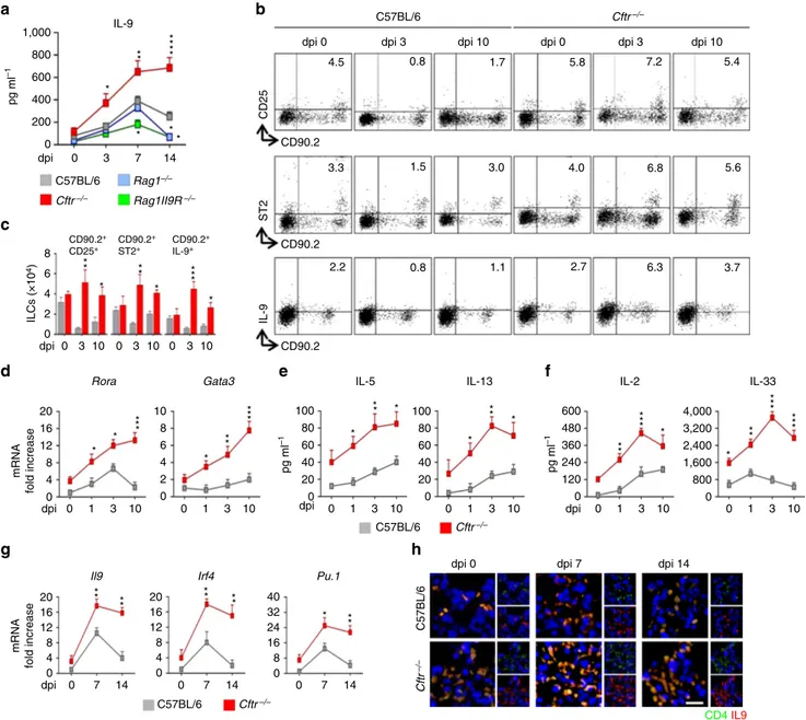
IL-9 production and ILC2-Th9 activation during aspergillosis.
We infected C57BL/6 or Cftr
" / "mice intranasally with
A. fumigatus and measured IL-9 production, ILC2 and Th9 cell
activation in infection. We have already shown that Cftr
" / "mice
are susceptible to Aspergillus infection (from 2.5±0.7 to
3.9±1.0 log colony forming unit (cfu)±s.d. per lung, C57BL/6
versus Cftr
" / "mice, respectively) and allergy (from 9.2±0.7 to
22.4±1.3 ng ml
" 1total serum IgE in C57BL/6 and Cftr
" / "mice, respectively). A peak production of IL-9 occurred during
the first week of the infection in C57BL/6 mice to decline
thereafter as opposed to Cftr
" / "mice in which levels of IL-9
were sustained throughout the infection (Fig. 1a). Peak
produc-tion of IL-9 was also observed in Rag1
" / ", and less in
Rag1
" / "/Il9R
" / ", mice early but not late in infection (Fig. 1a),
a finding suggesting that early IL-9 production is
IL-9R-depen-dent and late is T-cell-depenIL-9R-depen-dent. We looked therefore for the
presence of IL-9
þILC2 and Th9 cells in infection by
character-izing IL-9-producing Lin
"and CD4
þT cells in the lung. ILC2
are marked by expression of the IL-33R as well as the common g
chain (gc) cytokine receptors for IL-2 and IL-7 (ref. 2). Flow
cytometry analysis revealed that CD90.2
þILC2 expressing IL33R
or CD25 were present in the lung of naive C57BL/6 (4.5 and 3.3%
for CD25
þand ST2
þcells, respectively) and Cftr
" / "mice (5.8
and 4.0% for CD25
þand ST2
þcells, respectively; Fig. 1b,c). In
C57BL/6 mice, and similarly in Rag1
" / "mice (Supplementary
Fig. 1a), ST2
þILC2 cells decreased early in infection to return to
baseline level 10 days later while CD25
þILC2 stably decreased
(Fig. 1b,c). In contrast, in Cftr
" / "mice, both types of ILC2
steadily increased throughout the infection (Fig. 1b) along with
the expression of the ILC2 transcription factors, Rora, and Gata3
(Fig. 1d) and the production of ILC2 effector cytokines, IL-5 and
IL-13 (Fig. 1e). IL-9-producing CD90.2
þILC2 were also
expan-ded in Cftr
" / "mice but not in C57BL/6 (Fig. 1b,c) and
Rag1
" / "mice (Supplementary Fig. 1a), as revealed by flow
cytometry. In terms of Th9 cell activation, CD4
þIL-9
þT cells
appeared in C57BL/6 mice a week after the infection to decline
thereafter (Fig. 1h), consistent with the short retention of Th9 at
the inflammatory sites
21. The expansion was instead sustained in
Cftr
" / "mice (Fig. 1h) along with the expression of Il9, Pu.1
(purine-rich box 1) and Irf4 (interferon regulatory factor 4)
transcription factors (Fig. 1g). These data indicate that
IL-9
þILC2 and Th9 cells are all increased in Cftr
" / "mice
during A. fumigatus infection.
Given that ILC1 through IFN-g (ref. 22) and ILC3 through
IL-22 (ref. 23) may affect ILC2 expansion, the differential
expansion of ILC2 could reflect the ILCs dynamics in the lung.
However, NKp46
þNK1.1
þILC1 cells producing IFN-g did not
expand and ILC1-promoting cytokines IL-15 and IL-18 were not
produced
in
Cftr
" / "as
opposed
to
C57BL/6
mice
(Supplementary Fig. 2a,b). Similarly, despite expanded in
Cftr
" / "mice, CCR6
þRORgt
þILC3 produced IL-17A more
than IL-22 (Supplementary Fig. 2c,d). Thus, while confirming the
defective production of IFN-g and IL-22 in Cftr
" / "mice
18,
these findings suggest that the expansion of ILC2 in Cftr
" / "mice
is not dependent on ILCs dynamics in the lung but rather on the
production of ILC2 promoting cytokines. This appeared to be the
case, as the levels of cytokines promoting ST2
þILC2, IL-33 and
CD25
þILC2, IL-2, were constantly elevated in Cftr
" / "mice
whereas a peak production was only observed at an early time
point in C57BL/6 mice (Fig. 1f).
IL-9 contributes to inflammatory pathology in infection. To
assess the role of IL-9 in A. fumigatus infection and allergy, we
resorted to Il9R
" / "mice that, given the crucial role of the IL-9R,
a member of the gc receptor family, for the survival of lung
ILC2 (ref. 8), also have a decreased ILC2 (ref. 8). Mice were
either acutely infected with Aspergillus conidia intranasally or
subjected to fungal allergy (ABPA) by repeated sensitization with
Aspergillus culture filtrate extracts (5.5±0.7 ng ml
" 1versus
respectively). We found that the absence of IL-9R signalling
conferred resistance to both infection and allergy, as indicated by
the reduced fungal load (Fig. 2a) and decreased inflammatory
lung pathology in infection as well as in ABPA (Fig. 2b). The
numbers of lung CD25
þand ST2
þILC2 were decreased in these
mice as revealed by immunofluorescence staining (Fig. 2c).
Concomitantly, Th9 and, partially, Th2 cells—revealed by
Stat6 expression and STAT5 phosphorylation (Supplementary
Fig. 1b,c)—were decreased in both infection and allergy
(Fig. 2d,e), while Th17 and Treg cells were unaffected and Th1
cells increased (Supplementary Fig. 3a,b). Corroborating these
findings, IL-9 neutralization in C57BL/6 or Cftr
" / "mice greatly
ameliorated lung pathology in response to the fungus, both in
terms of inflammatory cell recruitment (Fig. 2f) and fibrosis as
shown by Masson’s trichrome staining (insets of Fig. 2f) and
production of TGF-b (Fig. 2g), a mediator of pulmonary
fibrosis
24. Together, these results indicate that the IL-9/IL-9R
signalling pathway is required for the expansion of pathogenic
ILC2 and Th9 cells in response to the fungus. However, whether
ILC2 promote Th9 cell activation via IL-9R signalling is not
known. To directly assess this, we did criss-cross experiments in
which CD4
þT cells from either C57BL/6 or Cftr
" / "mice were
assessed for IL-9 production and Th9 transcription factor
expression on co-cultivation in a transwell permeable support
with lung Lin
"cells exposed to A. fumigatus and either IL-2 or
IL-33. We found that Th9 cell activation was observed upon
1,000 IL-9 4.5 CD90.2 CD90.2+ CD25+ CD90.2 CD90.2 + ST2+ CD90.2 CD90.2+ IL-9+ CD25 ST2 IL-9 3.3 2.2 0.8 1.1 2.7 6.3 3.7 1.5 3.0 4.0 6.8 5.6 0.8 1.7 5.8 7.2 5.4 ** * * * * * ** ** ** ** ** ** * ** * *** * ** * * * * * ** ** ** ** * ** * ** * * * ** * ** ** * ** ** ** 800 600 pg ml –1 400 200 0 8 ILCs ( × 10 4) 6 4 2 0 20 10 8 6 4 2 pg ml –1 pg ml –1 0 100 600 480 360 240 120 80 60 40 20 0 100 80 60 40 20 0 0 0 4,000 3,200 2,400 1,600 800 mRNA fold increase mRNA fold increase 16 12 8 4 0 20 16 12 8 4 0 20 16 16 12 8 8 4 0 0 40 32 24
Il9 Irf4 Pu.1
dpi
dpi 0
dpi 0 dpi 7 dpi 14
CD4IL9
dpi 3 dpi 10 dpi 0 dpi 3 dpi 10
0 3
dpi 0 3
Rora Gata3 IL-5 IL-13 IL-2 IL-33
10 dpi dpi 0 0 7 14 0 7 14 0 7 14 3 1 10 0 1 3 10 dpi 0 1 3 10 0 1 3 10 dpi 0 1 3 10 0 1 3 10 0 3 10 0 3 10 7 14 C57BL/6 C57BL/6 Cftr–/– C57BL/6 C57BL/6 Cftr–/– C57BL/6 Cftr–/– Cftr –/– Cftr–/– Rag1–/– Rag1II9R–/–
a
b
c
d
e
f
g
h
Figure 1 | IL-9 production and ILC2-Th9 cells activation in Aspergillus fumigatus infection. (a) Time course of IL-9 production at various days post
infection (dpi) in mice (six per group) infected intranasally with live A. fumigatus conidia. (b) Detection of CD90.2þCD25þ, CD90.2þST2þ and
CD90.2þIL-9þlung type 2 ILCs by flow cytometry (numbers refer to percentages of positive cells) and immunofluorescence staining. (c) Absolute
number of lung ILC2; (d) ILC2–specific transcript on lineage negative lung cells; (e,f) ILC2 effector and activating cytokines; (g) Il9 and Th9-cell specific
transcripts on lung CD4þ T cells and (h) immunofluorescence staining of lung CD4þ IL-9þ T cells. Photographs were taken with a high-resolution
microscope (Olympus DP71) equipped with a # 40 objective; scale bar, 100 mm. Mean values±s.d. cytokines were determined on lung homogenates by
ELISA, Il9 and transcripts assessed by PCR with reverse transcription. 0, uninfected mice. *Po0.05, **Po0.01, ***Po0.001, ****Po0.0001, knockout
versus C57BL/6 mice (data represent pooled results or representative images from three experiments, Two-way ANOVA, Bonferroni post test). Gata3, GATA binding protein 3; Irf4, interferon regulatory factor 4; Pu.1, purine-rich box 1; Rora, RAR-related orphan receptor alpha.
Naive Infected ABPA
Naive
Infected ABPA Naive Infected ABPA Naive Infected ABPA Naive Infected ABPA Naive Infected ABPA
Log cfu/lung 10 8 6 4 2 ** C57BL/6 pg ml –1 pg ml –1 1,000 800 600 400 200 0 100 80 60 40 20 0 10 8 6 4 2 0 20 16 12 8 4 0
mRNA fold increase mRNA fold increase
pg ml –1 100 80 60 40 20 0 10 8 6 4 2 0
mRNA fold increase
pg ml –1 100 80 60 40 20 0 ** ** ** ** ** **** *** *** *** ** ** ** ** ** ** * * * 10 8 6 4 2 0
mRNA fold increase
40 32 24 16 8 0 C57BL/6 Il9R–/– C57BL/6 Lin– C57BL/6 C57BL/6 C57BL/6 C57BL/6 Lin– Cftr–/– Untreated Untreated Untreated αIL-9 αIL-9 αIL-9 C57BL/6 TGF-β pg m l –1 600 480 360 240 120 0 Cftr–/– Cftr–/– Il9R–/– Il9R–/– Il9R–/– Il9R–/– Il9R–/– Il9R–/– CD4+ CD4+ Cftr–/– Cftr–/– CD4+ Cftr–/–
None IL-2 IL-33
CD4+
Pu.1 Irf4 IL-9 Stat6
Pu.1 Irf4 IL-9 Pu.1 Irf4 IL-9
IL-4 Il9R –/– C57BL/6 Il9R –/– CD90.2 CD25 CD90.2 ST2 *** ** * ** * ** * ** * * ** ** * ** ** dpi 0 7 14 0 7 14 C57BL/6 Il9R–/– C57BL/6 Il9R–/–
a
b
c
d
e
f
g
h
Figure 2 | IL-9R signaling contributes to inflammation and allergy. C57BL/6 and Il9R" / " mice (six per group) were intranasally infected with live
Aspergillus fumigatus conidia or subjected to ABPA and assessed for (a) lung fungal growth (log10cfu, mean±s.d.); (b) lung histology (periodic acid " Schiff
staining); (c) expression of CD90.2þCD25þ, CD90.2þST2þ lung ILC2 by immunofluorescence; (d,e) Th-cell specific transcripts and cytokine
production. (f) Lung histology (periodic acid–Schiff and, in the inset, Masson’s trichrome staining) and (g) TGF-b production in C57BL/6 or Cftr"/ "mice
infected as above and treated with IL-9 neutralizing antibody for a week. Days post infection (dpi). (h) Th-cell specific transcripts and IL-9 production of
lung CD4þT cells from naive mice co-cultured with lung lineage negative (Lin") cells in the presence of A. fumigatus conidia, IL-2 or IL-33. Photographs
were taken with a high-resolution microscope (Olympus DP71) equipped with a # 20 objective; scale bars, 200 mm and a # 40 objective (insets of f,
scale bars, 100mm). Results are mean values±s.d., ELISA was done on lung homogenates and culture supernatants for cytokines and PCR with reverse
transcription on CD4þ lung cells. *Po0.05, **Po0.01, ***Po0.001, Il9R" / ", Cftr"/ "versus C57BL/6 mice; IL-9-treated versus control isotype-treated
mice; stimulated versus unstimulated (none) cells and Il9R" / "versus C57BL/6 or Cftr"/ "CD4þT cells. Naive, uninfected mice. Data represent pooled
results or representative images from three experiments, Two-tailed Student’s t-test (a) or Two-way ANOVA (d,e) Bonferroni post test. Gata3, GATA binding protein 3; Irf4, interferon regulatory factor 4; Pu.1, purine-rich box 1.
co-cultivation of CD4
þT cells with Lin
"cells in the presence of
IL-2 more than IL-33, an effect magnified in Cftr
" / "as
compared to C57BL/6 mice (Fig. 2h), and requiring the
presence of IL-9R on responder CD4
þT cells being
significantly negated with Il9R
" / "responder cells (Fig. 2h and
Supplementary Fig. 4). These results indicate that ILC2, and
particularly CD25
þILC2, may account for the sustained IL-9
production and Th9 activation responsible for pathology in CF.
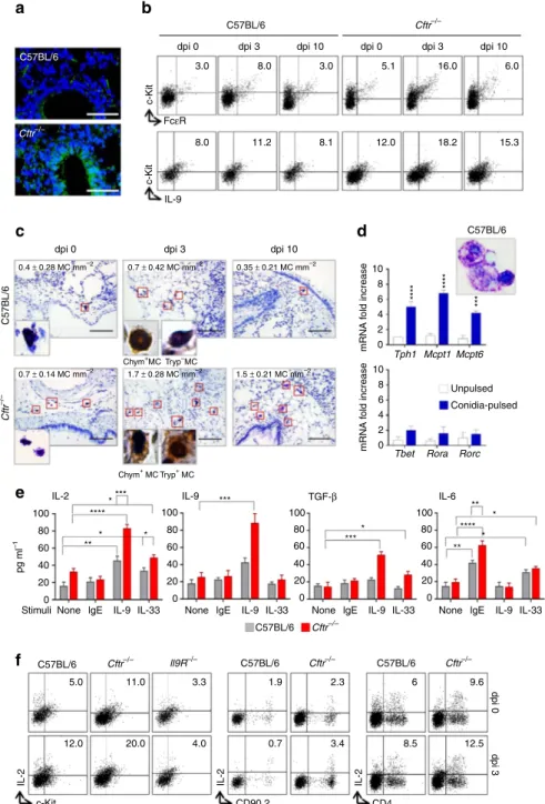
IL-9 activates mast cells to produce IL-2. The sustained
production of IL-33 and IL-2 in Cftr
" / "mice prompted us to
investigate mechanisms behind this production. IL-33 is
constitutively expressed at epithelial barrier surfaces where it is
rapidly released from cells during tissue injury
25. However,
tight regulation of IL-33 following its release to dampen
ST2-dependent
inflammation
to
fungi
has
also
been
described
26. This likely occurred in C57BL/6 but not in Cftr
" /"
mice in which the high levels of epithelial damage observed
upon the infection (Fig. 3a) likely accounted for the sustained
IL-33-dependent ST2
þILC2 expansion. For IL-2, predictably
high in CF, given the sustained NFAT activity
27, in addition to
CD4
þT cells
8and dendritic cells
28, MC are known to produce it
in the lung
7. We looked therefore for MC presence and activity in
the lung of C57BL/6 and Cftr
" / "mice after the infection. MC
were much expanded in Cftr
" / "mice, as seen by flow cytometry
(Fig. 3b) and toluidine staining (Fig. 3c). MC are distinguished by
their granule content whose expression is tissue-dependent
29. In
the lung, chymase-positive MC numbers positively correlated
with better lung function
30, whereas chymase- and
tryptase-positive MC were expanded in areas of fibrosis in CF lungs and
positively correlated with the degree of fibrosis and lung
function
31. Immunohistochemistry revealed that while
tryptase-positive cells could not be detected, chymase-tryptase-positive MC
were present in C57BL/6 mice (insets of Fig. 3c). In contrast,
chymase-positive and tryptase-positive MC were observed in
Cftr
" / "mice (insets of Fig. 3c).
As MC are known to produce cytokines through different
receptor mechanisms
32, we evaluated cytokine production on
magnetically purified c-Kit
þMC (as characterized by
morphometry and MC specific transcripts, Fig. 3d) upon
stimulation with IgE, IL-9 or IL-33. We found that IL-2
production was induced by IL-33 and, more, by IL-9 and not
by IgE, mostly in MC from Cftr
" / "mice (Fig. 3e). As IL-9 also
induced IL-9 production (Fig. 3e) and IL-9
þMC could also be
detected in vivo (Fig. 3b), this suggests that an autocrine loop
appears to mediate the IL-9-dependent IL-2 release by MC. As a
matter of fact, IL-2 production (39±6 ng/ml versus 127±22,
IL-2 in lung homogenates at 3 dpi in Il9R
" / "versus C57BL/6
mice, respectively) and IL-2
þMC (Fig. 3f) were greatly reduced
in Il9R
" / "mice, thus contributing to the defective expansion of
CD25
þILC2 in these mice. Of interest, IL-9 stimulation also
induced TGF-b in MC from Cftr
" / "mice but not IL-6 (Fig. 3e),
a finding suggesting that the autocrine IL-9 stimulation appears
to be specific for IL-2 and TGF-b (Fig. 3e). These data indicate
that MC may contribute to IL-2 production eventually leading to
CD25
þILC2 expansion in Cftr
" / "mice. This appears to be the
case, as IL-2
þMC, more than IL-2
þCD90.2 or IL-2
þCD4
þT cells, were expanded in vivo, early in infection, particularly in
Cftr
" / "mice (Fig. 3f).
To directly prove this, we assessed susceptibility to
inflamma-tory allergy of MC-deficient C57BL/6-Kit
W/W-vmice or Cftr
" / "mice treated with the tyrosine kinase inhibitor imatinib known to
inhibit IL-9-driven mastocytosis in the lung
12. Airway
mastocytosis was reduced in MC-deficient Kit
W/W-vmice
(Fig. 4a) along with reduced levels of IgE, IL-2, IL-9 and
TGF-b (Fig. 4b). Concomitantly, the number of CD25
þILC2
were also decreased in the lung but promptly restored upon MC
engraftment or exogenous IL-2 administration (Fig. 4a). Thus,
MC appear to be able to control CD25
þILC2 expansion in the
lung during the infection via IL-2. Similar results were obtained
upon treatment of Cftr
" / "mice with imatinib. Both
inflammation (Fig. 4c), collagen deposition (insets of Fig. 4c),
IL-2, IL-9 and TGF-b production (Fig. 4d) and Th9 cell activation
(Fig. 4e) were attenuated. Interestingly, imatinib apparently
increased early inflammation in C57BL/6 mice (Supplementary
Fig. 5), a finding suggesting that c-Kit
þcells could contribute to
pathogen resistance early in infection. As a matter of fact,
MC-deficient Kit
W/W-vmice displayed increased susceptibility to the
infection as compared with C57BL/6 mice, as indicated by the
increased fungal load (Supplementary Fig. 6a), neutrophils
recruitment (Supplementary Fig. 6b) and a degree of lung
inflammation (Supplementary Fig. 6c).
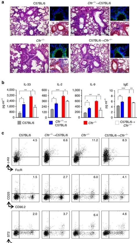
CFTR deficiency contributes to inflammation. The above
results suggest that a circuit involving IL-33, IL-2 and IL-9 and
different types of cells is pathogenically amplified in Cftr
" / "mice. The finding that IL-33, IL-2 and IL-9 are also elevated in CF
patients
33,34prompted us to evaluate the contribution of cystic
fibrosis
transmembrane
conductance
regulator
(CFTR)
dysfunction on the activation of the inflammatory circuit.
Given that CFTR dysfunction on both epithelial and myeloid
cells impacts on lung inflammation
35, we assessed chimeric mice
with CFTR unresponsive myeloid or epithelial cells for lung
damage and inflammation, production of IL-33, IL-2 and IL-9
and MC/ILC2 activation upon Aspergillus infection. We found
that epithelial cell damage and lung inflammation (Fig. 5a), levels
of cytokines and IgE (Fig. 5b), MC and ILC2 expansion (Fig. 5c)
were all attenuated or decreased in condition of CFTR deficiency
in epithelial cells but CFTR sufficient myeloid cells, a finding
suggesting that myeloid, and perhaps lymphoid, deficiency could
contribute to the activation of the inflammatory circuit in CF.
This seems to be the case as the opposite findings were observed
in recipient C57BL/6 mice receiving CFTR deficient myeloid cells
(Fig. 5a,c) These mice, however, showed an intermediate
inflammatory phenotype as compared to Cftr
" / "mice, a
finding suggesting that, although to a different extent, CFTR
deficiency on epithelial and myeloid cells may predispose to lung
inflammation in response to microbial and non-microbial stimuli.
The IL9 rs2069885 SNP correlates with high IgE levels in CF
females. To assess whether IL-9 may contribute to allergy in CF
patients, we determined the effect of the non-synonymous IL9
p.Thr117Met (c.350C4T, rs2069885) polymorphism, known to
be associated with lung function and sensitization
36,37on total
and Aspergillus-specific IgE levels in CF patients (Supplementary
Table 1). Previous association studies demonstrated the existence
of sex dimorphism linked to this polymorphism
36,37. Therefore,
we carried out association testing separately in males and
females. The distribution of the total IgE was skewed but after
natural logarithmic transformation, the distribution adequately
approximated a normal distribution. IL9 rs2069885 genotype
distribution did not deviate from Hardy–Weinberg equilibrium
(w
2test, P ¼ 0.776) and it displayed a minor allele frequency of
0.129, comparable to the European population from 1000
Genome Consortium, minor allele frequency ¼ 0.128 (ref. 38).
IL9 rs2069885 genotype distribution did not differ between males
and females (w
2test, P ¼ 0.243), but a significant rs2069885-sex
interaction on total IgE levels was found (general linear model,
P ¼ 0.004), where female T allele carriers showed high IgE
levels (linear regression: females, b ¼ 0.624, P ¼ 0.043; males,
b ¼ " 0.666, P ¼ 0.034; Fig. 6a). In a subset of CF patients
(N ¼ 114; 57 males and 57 females), Aspergillus-specific IgE were
also significantly higher in IL9 rs2069885-T carriers (general
linear model, P ¼ 0.002; Fig. 6b). Notably, in line with the results
obtained on total IgE, the association of IL9 rs2069885-T allele
with higher Aspergillus-specific IgE levels was observed in females
(linear regression, b ¼ 1.417, P ¼ 0.002) more than males
(linear regression, b ¼ 0.489, P ¼ 0.279). We also tested other
C57BL/6
Cftr–/–
C57BL/6 Cftr–/–
dpi 0 dpi 3 dpi 10 dpi 0 dpi 3 dpi 10
3.0 8.0 3.0 5.1 16.0 6.0 8.0 11.2 8.1 12.0 18.2 15.3 c-Kit FcεR c-Kit IL-9
dpi 0 dpi 3 dpi 10
0.35 ± 0.21 MC mm–2 0.7 ± 0.42 MC mm–2 0.4 ± 0.28 MC mm–2 C57BL/6 Chym+MC Tryp–MC 1.7 ± 0.28 MC mm–2 0.7 ± 0.14 MC mm–2 1.5 ± 0.21 MC mm–2 Chym+ MC Tryp+ MC 10 8 6 4 2 0 mRNA f old increase *** * *** * *** C57BL/6 Tph1 Mcpt1 Mcpt6 Unpulsed Conidia-pulsed
Tbet Rora Rorc
10 8 6 4 2 0 mRNA f old increase Cftr –/– IL-2 100 80 60 40 20 0
None lgE IL-9 IL-33
pg ml –1 Stimuli *** **** * ** * * 100 80 60 40 20 0 100 80 60 40 20 0 100 80 60 40 20 0
None lgE IL-9 IL-33 None lgE IL-9 IL-33 None lgE IL-9 IL-33 IL-9 *** **** TGF-β IL-6 ** **** * * ** Cftr–/– C57BL/6 ll9R–/– C57BL/6 Cftr–/– C57BL/6 Cftr–/– 5.0 11.0 3.3 1.9 2.3 6 9.6 12.0 20.0 4.0 0.7 3.4 8.5 12.5 dpi 3 dpi 0 IL-2 c-Kit IL-2 CD90.2 IL-2 CD4 C57BL/6 Cftr–/–
a
b
c
d
e
f
Figure 3 | IL-9 activates mast cells to produce IL-2. C57BL/6 or Cftr"/ "mice (six per group) infected intranasally with live A. fumigatus conidia were
evaluated at different days after infection (dpi) for (a) deposition of DNA on lung epithelial cells by TUNEL, resulting in bright DNA staining; (b) detection
of c-KitþFceRþ and c-KitþIL-9þlung mast cells (MC) by flow cytometry (numbers refer to percentages of positive cells); (c) toluidine blue, relative MC
number mm" 2and, in the inset, immunohistochemical staining for chymase- and tryptase-positive MC in lung section. Photographs were taken with a
high-resolution microscope (Olympus DP71) equipped with a # 40 objective and (in the inset) a # 100 objective and with EVOS FL Color Imaging System with a # 60 objective (immunohistochemical staining). (d) Toluidine blue stain and transcription factors expression (PCR with reverse transcription) of
c-Kitþ cells magnetically isolated from lung of uninfected C57BL/6 mice and pulsed with live A. fumigatus conidia. (e) Cytokine production (mean
values±s.d., ELISA on culture supernatants) by purified lung c-Kitþcells, pulsed with A. fumigatus and stimulated with IgE, IL-9 and IL-33; (f) detection of
c-KitþIL-2þ, CD90.2þIL-2þ and CD4þIL-2þ lung cells by flow cytometry (numbers refer to percentages of positive cells). *Po0.05, **Po0.01,
***Po0.001, ****Po0.0001, conidia-pulsed versus unpulsed c-Kitþ cells, stimulated versus unstimulated c-Kitþ cells and Cftr"/ "versus C57BL/6
c-Kitþ cells (data represent pooled results or representative images from three experiments, Two-way ANOVA, Bonferroni post test). Mcpt1, mast cell
protease 1; Mcpt6, tryptase beta 2; Rorc, retinoic acid receptor–related orphan receptor C; Rora, RAR-related orphan receptor alpha; Tbet, T box expressed in T cells; Tph1, tryptophan hydroxylase 1.
0.5 FcεR CD90.2 IgE pg ml –1 IL-2 IL-9 TGF-β ** ** ** ** ** * ** * ** * ** * ** * ** ** ** ** ** ** * ** C57BL/6 1.5 1.2 0.9 0.6 0.3 0.0 1,000 800 600 400 200 0 20 16 12 8 4 0 0 7 14
mRNA fold increase
0 7 14 0 7 14 dpi pg ml –1 0 7 14 0 7 14 40 32 24 16 8 0 1,000 800 600 400 200 0 600 480 360 240 120 0 dpi 0 3 10 0 3 10 0 3 10 0 3 10 600 480 360 240 120 0 600 480 360 240 120 0 600 480 360 240 120 0 5,000 4,000 3,000 2,000 1,000 0 5 4 3 2 1 0 dpi 0 3 10 BMMC IL-2 KitW/W-v C57BL/6 None Imatinib dpi 7 dpi 14 Cftr –/– KitW/W-v CD25 c-Kit 2.3 0.3 2.2 0.6 2.1 6.5 4.0 1.2 3.6
dpi 0 dpi 3 dpi 10 BMMC IL-2 MC mm
–2
CD90.2+CD25+ (×104)
*
*
IL-2 IL-9 TGF-β Pu.1 Irf4
None Imatinib
a
b
c
d
e
Figure 4 | IL-9 activates mast cells to produce IL-2. MC-deficient C57BL6-KitW/W-vmice (six per group) were infected intranasally with live A. fumigatus
conidia, engrafted intravenously with wild-type bone marrow-cultured mast cells (BMMC) or treated intraperitoneally with IL-2 for a week and assessed for
(a) c-KitþFceRþlung mast cells (MC) and CD90.2þCD25þ lung ILC2 by flow cytometry (numbers refer to percentages of positive cells) with relative
cell number and (b) IgE and cytokine production. (c) Lung histology (periodic acid–Schiff and Masson’s trichrome staining, in the insets); (d) cytokine
production and (e) Th9-cell specific transcripts expression in Cftr"/ " mice infected as above and treated with imatinib intraperitoneally for a week.
Photographs were taken with a high-resolution microscope (Olympus DP71) equipped with a # 20 objective, scale bars, 200 mm and a # 40 objective
(insets of c, scale bars, 100mm). Results are mean values±s.d., ELISA on lung homogenates for cytokines and PCR with reverse transcription on lung
CD4þT cells. *Po0.05, **Po0.01, ***Po0.001, ****Po0.0001, MC-deficient C57BL6-KitW/W-vversus C57BL/6 mice or imatinib-treated versus untreated
(none) mice (data represent pooled results or representative images from three experiments, Two-way ANOVA, Bonferroni post test). Irf4 ¼ interferon regulatory factor 4; Pu.1 ¼ purine-rich box 1.
IL9 tagSNPs (rs2069882, rs31564, rs1859430, rs1799962;
Supplementary Tables 2 and 3), encompassing the whole IL9
gene region, but none associated with IgE levels. Linkage
disequilibrium (LD) analyses of the five SNPs in IL9 failed to
reveal the presence of significant LD blocks (Supplementary
Fig. 7). Haplotype analysis yielded no significant result
C57BL/6 C57BL/6 Cftr–/–→ C57BL/6 Cftr–/–→C57BL/6 C57BL/6→ Cftr–/– C57BL/6→Cftr–/– 1,000 800 600 400 200 0 11.2 8.3 6.6 4.5 6.0 4.1 2.7 1.5 6.4 4.6 3.7 2.0 FcεR CD90.2 CD90.2 c-Kit CD25 ST2 10 8 6 4 2 0 600 480 360 240 120 0 pg ml –1 pg ml –1 4,000 3,200 2,400 1,600 800 0 C57BL/6 Cftr–/–→C57BL/6 C57BL/6→Cftr–/– Cftr–/– Cftr–/– Cftr–/– IgE IL-9 IL-2 IL-33 ** * * * * * *** ** ** ** *** **
b
c
a
Figure 5 | Epithelial and myeloid CFTR deficiency contribute to the inflammatory phenotype. C57BL/6, Cftr"/ " and chimeric C57BL/6 and Cftr"/ "
mice (10 per group) received 10 # 106viable bone marrow cells 4 weeks before the intranasal infection with A. fumigatus. Chimeric mice were evaluated 7
days after the infection for (a) lung histology (periodic acid–Schiff and, in the insets, Masson’s trichrome and TUNEL staining); (b) cytokines and IgE levels
(mean values±s.d., ELISA on lung homogenates); (c) detection of c-KitþFceRþ mast cells, CD90.2þCD25þ and CD90.2þST2þ type 2 ILCs by flow
cytometry (numbers refer to percentages of positive cells in the lung). Photographs were taken with a high-resolution microscope (Olympus DP71) equipped with a # 20 objective; scale bars, 200 mm and a # 40 objective (insets of panel a, scale bars, 100 mm). *Po0.05, **Po0.01, ***Po0.001, Cftr"/ " versus C57BL/6, chimeric C57BL/6 versus C57BL/6, chimeric Cftr"/ " versus Cftr"/ " mice, Two-way ANOVA, Bonferroni post test.
(Supplementary Table 4), although the most common haplotype
C–A–A–C–A (frequency 43.2%) turned out to be associated with
higher IgE levels in males (linear regression, b ¼ 0.567,
P ¼ 0.022).
We next determined whether the reported sex-specific
associations could be attributable to a differential expression of
IL9 and its receptor, which is located on the pseudoautosomal
region Xq/Yq, between males and females. A significant IL9
rs2069885 by sex interaction was found when analysing the
IL9/IL9R expression ratio (general linear model, P ¼ 0.025) where
T allele carriers showed opposite effects according to sex (linear
regression, females, b ¼ 0.375, P ¼ 0.116; males, b ¼ " 0.426,
P ¼ 0.375; Fig. 6c) paralleling the finding on specific IgE (Fig. 6a)
and on IL-9 levels in expectorates (Fig. 6d). No significant
difference was noticed between males and females when IL9 or
IL9R gene expression levels were analysed independently.
Although these results are to be considered with caution given
the small sample size–despite the sufficient power (b ¼ 0.20,
Power ¼ 0.8; Supplementary Table 6)—which demands for a
definitive validation using a larger new cohort of CF patients, the
present findings confirm the existence of a sex dimorphism at IL9
and IL9R loci, as already reported in several respiratory-related
human phenotypes
36,37.
Discussion
We have shown that IL-9 may expand pro-inflammatory CD25
þILC2/Th9 cell in CF, an activity involving the production of IL-2
by MC. Human MC co-localize near ILC2 in the human lung and
could directly promote ILC2 responses in vitro
39. We found here
that, in addition to CD4
þT cells, known to contribute to
CD25
þILC2 expansion via IL-2 (ref. 8), lung MC may also affect
the expansion of CD25
þILC2 thought to contribute to chronic
inflammation through multiple mechanisms
3. Failure to expand
CD25
þILC2 occurred indeed in MC-deficient mice or mice
treated with imatinib.
MC hyperplasia during chronic allergen challenge is associated
with remodeling of airways
40. In a mouse model of
ovalbumin-induced airway inflammation, the influx of MC into lung peaks
early after allergen challenge to mature over 14 days into cells
expressing lower level of c-Kit, FceRI and integrins
41. MC
hyperplasia was observed in the lung of Cftr
" / "mice along with
the detection of tryptase-positive and chymase-positive MC,
known to be expanded in asthmatic patients
42and in disease
areas of CF lung
31. Tryptases and chymases contribute to
inflammation and tissue remodeling through the selective
proteolysis of matrix proteins and the activation of
protease-activated receptors and matrix metalloproteinases
29. Consistent
with the finding that MC from CF patients are not high in FceR1
expressing
43, we found that lung MC from these mice poorly
responded to IgE in terms of IL-6 production but released IL-2, in
addition to TGF-b, in response to IL-9.
IL-9 is a pleiotropic cytokine that has multiple effects on
structural as well as numerous hematopoietic cells, which are
central to the pathogenesis of asthma
44,45. The important role for
the IL-9-MC axis in the pathology associated with chronic allergic
inflammation has been already described
46. IL-9 not only
stimulates MC growth and expansion but also stimulates
changes in gene expression that might alter responsiveness to
other stimuli
47. In vivo, IL-9 governed allergen-induced MC
numbers in the lung, and anti-IL-9 antibody-treatment protected
from airway remodeling, decreased expression of the profibrotic
mediators TGF-b and improved lung function
48. The correlation
between a reduction in MC numbers and decreased airway
remodeling, after IL-9 inhibition, is consistent with reports that
MC-deficient mice demonstrate significantly attenuated fibrosis
and inflammation after silica
49, ozone
50, or bleomycin injury
51.
We found that not only were IL-9 production and MC
expansion significantly increased in CF mice but that the
IL-9-MC axis contributed to the expansion of CD25
þILC2
leading to Th9 cell activation that further contributed to the
allergic inflammatory pathology (Fig. 7). IL-9 promoted IL-2
production by lung MC from Cftr
" / "mice, a finding that may
explain the increased and persistent expansion of CD25
þILC2 in
these mice. CD25
þILC2 were indeed not expanded in condition
of IL-9 ablation or MC-deficiency, a finding suggesting that the
IL-9/MC/IL-2 axis drives the expansion of CD25
þILC2. In
addition, as IL-2 stimulated lung CD25
þILC2 to produce IL-9
(ref. 9), this may have a positive feedback effect on ILCs, since
lung ILCs cultured with IL-9 increased the production of type 2
cytokines
9and up-regulated the anti-apoptotic protein BCL-3,
thereby promoting ILC2 survival
8. Of great interest, Lin
"cells
from CF lung also promoted Th9 cell activation in vitro, an
activity that required IL-9R expression on responding CD4
þT
cells. Thus, the pathogenic role of IL-9 in promoting allergic
inflammation may go beyond CD25
þILC2 expansion to include
the activation of Th9 cells. That Th9 cells are a major source of
IL-9 in models of allergic inflammation and play an important
role in MC accumulation and activation has been reported
52. By
producing IL-9, Th9 cells may in turn serve as a positive loop
amplifying the IL-9/MC/ILC2 axis, promoting a deleterious
vicious circle in which the production of profibrotic TGF-b by
IL-9-stimulated MC plays a plausible important role. The
presence of TGF-b dependent signalling in areas of prominent
fibrosis in CF has been already documented
24along with its
inhibition of chloride channel activities
53and the association of
the TGF-b genetic variants with more severe lung disease
54.
In Aspergillus -specific IgE (mean) In IgE (mean) IL9/IL9R relative
expression ratio (mean)
pg m –1 –1.50 0.00 –0.50 –1.00 –2.00 C/C T+ C/C Male Female T+ C/C T+ C/C T+ IL-9 IL9 rs2069885 IL9 rs2069885 IL9 rs2069885 4.20 4.00 3.80 3.60 * 100 80 60 40 20 0 1.00 0.80 0.60 0.40
a
b
c
d
Figure 6 | The IL9 rs2069885 polymorphism correlates with high IgE levels in CF females. (a,b) IL9 rs2069885 sex interaction on total or Aspergillus-specific IgE levels and (c) IL9/IL9R expression ratio measured on CF patients. (d) Determination IL-9 (mean±s.d., ELISA) in expectorates from CF patients carrying diverse genotypes at rs2069885.
Of note, together with IL-4, is able to promote Th9 cell
development in vitro
55, a finding highlighting the potential role
of TGF-b in reinforcing Th9 activity in vivo. However, TGF-b
production by MC in response to IL-9 may also serve a
regulatory, anti-inflammatory role
11,56.
In addition to IL-9, IL-33 is also a crucial regulator of MC
functions and both IL-33 and MC have been influentially
associated to the pathophysiology of allergic diseases and
inflammation
57. IL-33 is expressed in epithelia from patients
with CF and potentiates neutrophil recruitment
58as well as in
type-2 pneumocytes on allergic lung inflammation
59. In this
study, IL-33 was increased in CF mice and likely correlated with
the expansion of ST2
þILC2. At variance with IL-2, IL-33 did not
stimulate MC for IL-9/TGF-b production, a finding indicating a
minor contribution of the IL-33/MC axis in promoting
inflammatory allergy and pathology in response to the fungus.
As a matter of fact, the IL-33/MC/IL-2 axis was found
to suppress, rather than promote, papain-induced allergic
inflammation by promoting Treg
7.
Airway inflammation and recurrent pulmonary infections play
a central role in the progression of CF lung disease. It is still an
open question whether CFTR deficiency per se may enhance the
inflammatory response to the different environmental cues
60. Our
data would suggest that CFTR dysfunction on both epithelial and
myeloid cells may impact on the inflammatory circuit leading to
the activation of the inflammatory IL-9/Th9 pathway: namely,
among others
35, on both epithelial damage eventually leading to
overproduction of IL-33 and on the MC propensity to respond to
IL-9 with IL-2. However, PU.1 translocation into the nucleus has
also been shown to be significantly higher in CF monocytes than
in controls
61. It seems that defective CFTR impacts on the
mechanisms of communication between innate and adaptive
immune response.
The potential contribution of IL-9 to CF pathogenesis in
humans is unknown. Elevated levels of IL-9 were observed in the
expectorates from CF patients, likely accounting for the
expansion of TGF-b-producing MC in diseased lung areas
31. It
is of interest that in human asthmatic lung tissue, MC were the
main IL-9R expressing population
46. It seems therefore that the
IL-9/MC/IL-2 axis may have a pathogenic role in CF patients and
that its targeting could lead to a reduction in chronic
inflammation and improved lung function of these patients.
Studies have indeed highlighted the importance of Th9 cells in
allergic lung inflammation by promoting epithelial alterations,
goblet-cell hyperplasia, mucus production and infiltration of MC
and eosinophils
46. Considering that the costimulatory signal
OX40 is required for Th9 activation
62, it is intriguing that OX40
ligand was critical in driving Th2-allergic responses to A.
fumigatus in peripheral CD4
þT cells isolated from CF patients
with ABPA (ref. 63). It is clear that the inflammatory response in
the lung involves different Th cell types whose specific role in CF
remain unclear. Intriguingly, there are conditions where
promoting IL-9 might be therapeutically beneficial
11and ILC2
and MC could be exploited for pathogen immunity and tissue
repair
64. In this regard, the fact that imatinib apparently
exacerbated signs of inflammation during infection points to a
some beneficial role c-Kit
þcells may have in the control
of Aspergillus infection. MC indeed exhibited conidiocidal
activity (Supplementary Fig. 6d), a finding suggesting that MC
could serve as tissue sentinels modulating antifungal immune
responses, as suggested
65.
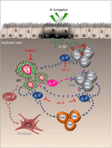
In conclusion, IL-9 and MC may have an important role in
the pathogenesis of lung disease and inflammation in CF.
Considering the inherent resistance to steroids of MC in
asthmatic patients
66, a better understanding of cellular and
molecular pathways leading to inflammation and impaired lung
functions may inspire new treatment avenues in patients with CF.
Our study would suggest that imatinib, known to inhibit lung
fibrosis
67, could be therapeutically exploited in CF patients with
an exalted IL-9/Th9 responses. In addition, it is of great interest
that the IL9 rs2069885 polymorphism, linked to high IgE levels,
was associated with females more than males with CF, a finding
offering an explanation for the, as yet unexplained, ‘gender gap’ in
mortality between females and males in CF
68and fostering
gender medicine in CF.
Methods
General experimental approaches
.
Mice were randomized and assigned to groupallocation at the time of purchase to minimize any potential bias. No blinding was applied on harvesting cells after the treatments.
Mice
.
C57BL/6 (wild-type, WT), Rag1" / "and MC-deficient C57BL/6-KitW/W-vmice, 6–8 week old, of both sexes, were purchased from Charles River (Calco,
Italy). Genetically engineered homozygous Cftr" / "mice69were bred at the
CF core animal facility at San Raffaele Hospital, Milan, Italy. Il9R" / "and
RagIl9R" / "mice were from the Ludwing Institute for Cancer Research, Brussells.
Fungal infection allergy and treatments
.
Anaesthetized (by inhalation of 3%isoflurane (Forane Abbot) in oxygen)) mice were infected by the intranasal
instillation of 2 # 107resting conidia/20 ml saline. For allergic broncho-pulmonary
aspergillosis, A. fumigatus culture filtrate extract in incomplete Freund’s adjuvant A. fumigatus IL-33 Epithelial cells IL-9 IL-9 Th9 Th9 Th9 IL-2 IL-9 Fibroblast TGF-β Imatinib MC ILC2 ILC2 ILC2 ILC2 ILC2 ILC2 CD25 αIL-9 ST2 αIL-9
Figure 7 | Proposed model for the role of IL-9 in promoting a mast cells/ILC2/Th9 fibrotic pathway in CF. IL-9, produced by IL-33-expanded ILC2, activates MC for IL-2 production leading to the expansion of
CD25þILC2 that promote Th9 cell activation. The resulting increased
production of IL-9 further amplifies the inflammatory loop by promoting ILC2 survival and type 2 cytokines production and by activating MC for the
production of fibrotic TGF-b. IL-9 ablation or MC inhibition (imatinib) are
potential drugable pathways through which inflammation and allergy could
(Sigma-Aldrich) was given (100 mg) to intact mice intraperitoneally (i.p.), subcutaneously and then intransally (20 mg), twice a week apart. A week after
the last intranasal challenge, mice received 107Aspergillus resting conidia and
evaluated a week later. Murine monoclonal anti-IL-9 antibody (MM9CI from
BioXcell), or control isotype IgG, were administered i.p. at the dose of 500 mg kg" 1
for a week starting the day of the infection. The levels of IL-9 after antibody
treatment were 65±15 versus 295±16 pg mg" 1for C57BL/6 and 103±27 versus
719±14 pg mg" 1for Cftr" / "mice, treated versus untreated mice. IL-2 at the
dose of 1 mg per mouse was given i.p. for a week. Imatinib mesylate (Glivec, ST1571
Novartis, Basel) were administered i.p. at the dose of 1 mg kg" 1for a week starting
the day of the infection.
Mast cell engraftment
.
Selective engraftment of MC in MC-deficient C57BL/6-KitW/W-vmice was performed as follow. Briefly, bone marrow cells derived from
6-week-old female C57BL/6 mice were cultured in WEHI-3–conditioned medium (ATCC number TIB-68), as a source of IL-3, for 4–5 weeks to obtain MC populations (BMCMC) which purity was higher than 95%. Via the tail vein,
5 # 106BMCMC were injected into each mouse, and the recipients were used
for experiments 4 weeks later.
Generation of bone marrow chimeras
.
Femurs and tibias were removedaseptically from donor C57BL/6 and Cftr" / "euthanized mice. Bone marrow was
retrieved by flushing with cold Dulbecco’s modified Eagle’s medium supplemented
with 10% heat-inactivated foetal calf serum and 2 mML-glutamine (Invitrogen).
Cells were washed twice with PBS without calcium and magnesium supplemented
with 1% foetal calf serum. Recipient C57BL/6 and Cftr" / "mice were irradiated
with 9 Gy and reconstituted no later than 6 h after the last irradiation with
10 # 106T cells by intravenous injection. Mice were given sulfamethoxazole
(150 mg ml" 1) and trimethoprim (30 mg ml" 1) in drinking water for the first 3
weeks of reconstitution. Mice were used no earlier than 4 weeks after transplan-tation. Before use in experiments, all mice were bled from the retro-orbital plexus, and the peripheral blood lymphocytes were analysed for the stable donor-type chimerism by reverse transcription-PCR of Cftr.
In vivo staining analysis
.
For histology, paraffin-embedded tissues were stainedwith Periodic acid-Schiff, Masson’s trichrome or Toluidine Blue staining to investigate inflammation, collagen deposition and MC infiltration, respectively. For
immunofluorescence, lungs were incubated at 4!C with phycoerythrin-conjugated
(PE) anti-CD25 (Miltenyi Biotec clone 7D4, 1:60), anti-T1-ST2 (BioLegend clone DIH9, 1:400), anti-IL-9 (Milenyi Biotec clone RM9A4, 1:60) and fluorescein iso-thiocyanate-conjugated (FITC) anti-mouse CD90.2 (Miltenyi Biotecclone 30-H12, 1:60) and anti-CD4 (BioLegend clone GK1.5, 1:1,000). Nuclei were counterstained with 4,6-diamidino-2-phenylindole. Immunostaining with appropriate irrelevant antibodies did not give positive staining of the lung. For immunohistochemistry, the lung sections were incubated overnight with polyclonal anti-chymase (Bioss, 1:100) or monoclonal anti-tryptase (Abcam clone EPR8476, 1:500) followed by the secondary biotinylated antibodies. Cells were counterstained with haematoxylin. Photographs were taken using a high-resolution Olympus DP71 microscope with a
# 20 and # 40 objective or EVOS FL Color Imaging System with a # 60 objective. For immunoblotting, blots of lung lysates were incubated with polyclonal antibodies against STAT5 and phospho-STAT5 (both from Cell Signaling, 1:1,000) and normalized on b-actin (clone AC-15 from Sigma). The ChemiDocTM XRS þ Imaging system (Bio-Rad) was used to detect chemiluminescence on the addition of the LiteAblotPlus chemiluminescence substrate (Euroclone S.p.A). Quantification was done by densitometry image analysis using Image Lab 5.1 software (Bio-Rad). The uncut blot is shown in Supplementary Fig. 8.
TUNEL assay of lung sections
.
Sections of lungs fixed in 4% bufferedparaformaldehyde, pH 7.3, for 36 h and embedded in paraffin, were deparaffinized, rehydrated, treated with 0.1 M citrate buffer, pH 6.0, washed and blocked in 0.1 M Tris-HCl buffer, pH 7.5, supplemented with 3% bovine serum albumin and 20% foetal calf serum. The slides were then incubated with fluorescein-coupled dUTP and terminal deoxynucleotidyltransferase–mediated deoxyuridine triphosphate nick-end labelling (TUNEL) enzyme (Roche Diagnostics) in the presence of terminal deoxynucleotidyltransferase. Unspecific binding was removed by washing
with phosphate-buffered saline for 10 min at 70!C. The sections were mounted and
analysed by fluorescence microscopy, using a # 40 objective.
Cell isolation and culture
.
Lungs were finely minced, digested in 16 mg ml" 1Collagenase P (Roche) for 30 min and meshed through a 70-mm cell strainer. ILCs were isolated from total lung cells by magnetic depletion of Lineage Positive cells
(Miltenyi Biotec). CD4þT cells and c-Kitþcells were purified from total lung
cells after incubation of CD4 microbeads and with PE-labelled anti-c-Kit followed by anti-PE MicroBeads respectively (both from Miltenyi Biotec). FcERIa-APC and c-Kit-PE (Miltenyi Biotec) staining and morphological examination after
toluidine blue staining on the cytospin slides were used fir c-Kitþcell phenotyping.
For Lin"-CD4þT cell co-culture, 2 # 106CD4þT cells were co-cultured with
1 # 106Lin" cells with or without 50 ng ml" 1recombinant IL-33, 40 ng ml" 1
recombinant IL-2 and pulsed with A. fumigatus conidia. Three days later, IL-9 levels in culture supernatants were analysed by ELISA and Th9 transcription
factors by real-time PCR. To separate Lin"and CD4þT cell, we used the
Transwell culture system (Costar, 0.4 mm pore size; Corning) with CD4þT cells in
the lower wells and Lin"in the upper wells7. For c-Kitþcells culture, 5 # 105cells
were cultured overnight in RPMI medium and pulsed with A. fumigatus conidia
with or without 10 mg ml" 1IgE, 100 ng ml" 1IL-33 and 100 ng ml" 1IL-9.
Flow cytometry
.
Flow cytometry on enriched Lin"cells was performed with acombination of the following fluorescence-conjugated mAbs (all from Miltenyi Biotec unless specified otherwise): APC-conjugated anti-NKp46 (29A1.4.9), anti-CD90.2 (30-H12), anti-Rorg (t; REA278), anti-FcRIa (MAR-1), anti-CD4 (GK1.5); PE-conjugated anti-NK1.1 (PK136), anti-CD25 (7D4), anti-T1-ST2 (DIH9, from Biolegend), anti-CD117 (3C11), anti-IL-9 (RM9A4) and anti-IL-2 (JES6-5H4). For intracellular staining, phorbol 12-myristate 13-acetate (PMA)/ ionomycin-stimulated cells were added of brefeldin, and then permeabilized with the CytoFix/CytoPerm kit (BD Biosciences) for intra-cytoplasmic detection of IL-9
and IL-2. Flow cytometry was done at 4!C on cells first exposed to Fc receptor
mAb (2.4G2). Cells were analysed with a BD LSRFortessa flow cytometer equipped with BD FACSDiva 7.0 software.
ELISA and real-time PCR
.
The levels of cytokines and IgE in lung homogenates,culture supernatants or expectorates were determined by ELISA kits (R&D Systems) following manufacturer’s instructions. Real-time PCR with reverse transcription was performed using CFX96 Touch Real-Time PCR Detection System and SYBR Green chemistry (Bio-Rad) on total RNA reverse transcribed with the cDNA Synthesis Kit (Bio-Rad). The PCR primers were as listed in Supplementary Table 5. Amplification efficiencies were validated and normalized
against Gapdh. The thermal profile for SYBR Green real-time PCR was at 95!C
for 3 min, followed by 40 cycles of denaturation for 30 s at 95!C and an
annealing/extension step of 30 s at 60!C. The messenger RNA-normalized data
were expressed as relative gene messenger RNA in treated versus untreated groups or cells.
Human study
.
A cohort of 347 patients of Caucasian origin with a provendiagnosis of CF (CFTR genotyping, sweat testing and clinical phenotype) was enroled in a prospective multicenter longitudinal genetic association study, See Supplementary Table 1 for clinical data including age, gender, lung function testing, measures of nutrition, microbiological findings and vital status of the patients’ cohort.
SNPs selection and genotyping
.
DNA was isolated from blood with the QIAampDNA Mini (Qiagen, Milan, Italy) system and stored at " 20 !C. IL9 SNPs were
selected based on literature review37and their ability to tag surrounding variants in
the HapMap-CEU population of the International HapMap project, NCBI build B36 assembly HapMap phase III (http://www.hapmap.org). Haplotype-based tagging SNPs were selected by assessing LD blocks from the genes of interest with a
pairwise correlation coefficient r2of at least 0.80 and a minor allele frequency
higher than 5% in the HapMap-CEU population. Five IL9 SNPs complied with the selection criteria: rs2069885, rs2069882, rs31564, rs1859430 and rs1799962. The applied Biosystems 7500 Fast qPCR system (Life Technologies) was used for SNP genotyping by KASPar assays (KBiosciences, Hertfordshire, UK). Each genotyping set comprised randomly selected replicates of sequenced samples and negative controls. Agreement between original and duplicate samples was Z99% for all SNPs. Laboratory personnel were blind to the sample status.
Statistical analysis
.
Data are expressed as mean±s.d. Horizontal barsindicate the means. Statistical significance was calculated by two-way ANOVA (Bonferroni’s post hoc test) for multiple comparisons and by a two tailed Student’s t-test for single comparison. The distribution of levels tested by Kolmogorov– Smirnov normality test turned out to be non-significant. Values of P not 40.05 were considered significant. The data reported are either representative from two or three experiments (FACS data, histology, immunofluoresce and TUNEL assay) or pooled otherwise. The in vivo groups consisted of 6 mice per group. Data were analysed by GraphPad Prism 4.03 programme (GraphPad Software). No statistical method was used to predetermine sample size. Genetic association testing was carried out considering additive and dominant models by linear regression implemented in Plink v1.07 (ref. 70), adjusting for age at sampling. Haplotype-based association tests were performed by general linear model using Plink v1.07 (ref. 70). LD analysis was performed using Haploview, and defining LD blocks based on the solid spine of LD algorithm. Analyses were conducted stratifying the study population according to sex since previous evidence highlighted the existence
of sex dimorphism at IL9 locus36,37. IL9 rs2069885 by sex interaction was tested by
general linear model using SPSS v.21. Two-tail P values are reported. Bonferroni’s correction for multiple testing was not performed since we are assessing specific questions on a candidate gene and we are not searching for associations without a priori hypotheses. Power calculation (using QUANTO v1.2.4) was performed to determine whether the sample study had sufficient power to detect a significant