Treatment with acetyl-L-carnitine exerts a
neuroprotective effect in the sciatic nerve following
loose ligation: a functional and microanatomical study
*Correspondence to:
Lorenzo Di Cesare Mannelli, Ph.D.,
orcid:
0000-0001-8374-4432 (Lorenzo Di Cesare Mannelli)
doi: 10.4103/1673-5374.230297 Accepted: 2018-01-19
Daniele Tomassoni1, Lorenzo Di Cesare Mannelli2, *, Vincenzo Bramanti3, Carla Ghelardini2, Francesco Amenta4, Alessandra Pacini5 1 School of Bioscience and Veterinary Medicine, University of Camerino, Via Gentile III da Varano, Camerino, Italy
2 Department of Neuroscience, Psychology, Drug Research and Child Health - Neurofarba - Pharmacology and Toxicology Section, University of Florence, Viale Pieraccini 6, Firenze, Italy
3 Department of Biomedical and Biotechnological Sciences, University of Catania, Via S. Sofia 87, Catania, Italy
4 Section of Human Anatomy, School of Medicinal and Health Products Sciences, University of Camerino, Via Madonna delle Carceri 9, Camerino, Italy
5 Department of Experimental and Clinical Medicine - DMSC - Section of Anatomy and Histology, University of Florence, Florence, Italy
Abstract
Peripheral neuropathies are chronic painful syndromes characterized by allodynia, hyperalgesia and altered nerve functionality. Nerve tissue degeneration represents the microanatomical correlate of peripheral neu-ropathies. Aimed to improve the therapeutic possibilities, this study investigated the hypersensitivity and the neuromorphological alterations related to the loose ligation of the sciatic nerve in rats. Effects elicited by treatment with acetyl-L-carnitine (ALCAR) in comparison to gabapentin were assessed. Axonal injury, reduction of myelin deposition and accumulation of inflammatory cells were detected in damaged nerve. A decrease of phosphorylated 200-kDa neurofilament (NFP) immunoreactivity and a redistribution in small clusters of myelin basic like-protein (MBP) were observed in ipsilateral nerves. Treatment with ALCAR (100 mg/kg intraperitoneally - i.p.) and gabapentin (70 mg/kg i.p.) administered bis in die for 14 days induced a significant pain relieving effect. ALCAR, but not gabapentin, significantly countered neuromorphological changes and increased axonal NFP immunoreactivity. These findings indicate that both ALCAR and gab-apentin significantly decreased the hypersensitivity related to neuropathic lesions. The observation of the positive ALCAR effect on axonal and myelin sheath alterations in damaged nerve supports its use as neu-rorestorative agent against neuropathies through mechanism(s) consistent to those focused in this study.
Key Words: peripheral nerve; chronic constriction injury; neurorestoration; morphology; neurofilament;
myelin basic protein; neural regeneration
Introduction
Peripheral neuropathies are defined as a heterogeneous category of diseases in which lesions of the nervous system can induce dysfunctional pain signalling and altered sensory mechanisms. The treatment of these pathologies is very dif-ficult and available drugs inhibit hyperalgesic symptomatol-ogy of neuropathy. Neuroprotective and/or neurorestorative effects of pharmacological treatments were reported rarely (Di Cesare Mannelli et al., 2010).
Animal models mimicking peripheral nerve injury have been developed to study chronic neuropathic pain, and one of the most widely used is Bennett and Xie’s (1988) unilater-al sciatic nerve chronic constriction injury (CCI). This mod-el is characterized by a painful syndrome with hyperalgesia beginning approximately 3 days after nerve injury, reaching a plateau between 7 and 15 days, and then decreasing (Ben-net and Xie, 1988). In CCI, hyperalgesia is accompanied by apoptosis phenomena in manipulated nerves, starting since the second week after nerve ligation (Di Cesare Mannelli et al., 2007).
Prolonged treatment of rats undergoing CCI with ace-tyl-L-carnitine (ALCAR) relieves pain sensations and pre-vents apoptosis in ligated nerves (Chiechio et al., 2007; Di Cesare Mannelli et al., 2007, 2009). ALCAR improved pe-ripheral nerve function by increasing nerve conduction ve-locity, reducing sensory neuronal loss, and promoting nerve
regeneration (Chiechio et al., 2007; Karsidag et al., 2012). It has been also reported that ALCAR raises pain thresh-old, displayed an anti-hyperalgesic effect both under acute (Ghelardini et al., 2002; Galeotti et al., 2004) and chronic conditions as well as in clinical settings (diabetes, anticancer and antiretroviral treatment) (Ghirardi et al., 2005; Sima et al., 2005; Osio et al., 2006; Traina, 2016).
Clinical studies in diabetic neuropathy showed that AL-CAR accelerated nerve conduction velocities, improved neuroregeneration, and reduced painful symptomatology (Evans et al., 2008). Moreover, it is effective in relieving opiate-withdrawal hyperalgesia in animal models. In hu-man beings it is effective on withdrawal symptoms such as muscular tension, muscular cramps, and insomnia (Janiri et al., 2009). Actually ALCAR is currently used for the treat-ment of neuropathic pain. Its long-term analgesic effects are dependent on epigenetic modifications, such as reversible modifications in gene activity (Traina, 2016). Thus ALCAR represents a consistent therapeutic option for peripheral neuropathies. Its complex neurotrophic and analgesic effects open new strategies in the study of peripheral nerve disease management (Traina et al., 2016).
ALCAR prevents nerve growth factor (NGF), glial cell line-derived neurotrophic factor (GDNF) and Artemin level changes in the CCI model of neuropathy. In particular, AL-CAR increased Artemin levels in a pathology-independent
manner and induced Artemin expression in dorsal root ganglia and spinal cord of sham animals (Vivoli et al., 2010). This candicates ALCAR as an agent affecting positively pain-ful symptomatology and rescuing damaged nerves poten-tially interfering with the progression of this multifactorial disease (Traina et al. et al., 2011, 2016).
The present research was designed to assess if treatment with ALCAR may have a neuroprotective activity in a mod-el of peripheral neuropathy induced by CCI of rat sciatic nerve. The anticonvulsant agent gabapentin, which is widely used for treating painful symptoms of neuropathies (Gilron et al., 2006) was also used as a reference drug.
Materials and Methods
AnimalsMale Sprague-Dawley rats (Harlan-Italia, Varese, Italy) were used. Animals were housed in number of 4 per cage (size 26 cm × 41 cm). The animals were kept at 23 ± 1°C with a 12 hour light/dark cycle, light at 7 a.m. and fed with standard laboratory diet and tap water ad libitum. Twenty-four hours before the test, the animals were placed in the experimental room for acclimatization. Animal manipulations were car-ried out according to the National and European Commu-nity guidelines for animal care (DL 116/92, of application of the European Communities Council Directive 86/609/ EEC) and of ethical guidelines of the University of Florence, consistent with the Guide for the Care and Use of Labora-tory Animals of the US National Institutes of Health (NIH Publication No. 85-23, revised 1996; University of Florence assurance number: A5278-01). Experiments involving ani-mals have been reported according to ARRIVE guidelines. All efforts were made to minimize animal suffering and to reduce the number of animals used.
Rats were randomly assigned to three groups: ALCAR group (n = 12, CCI followed by ALCAR treatment), gabapen-tin group (n = 12, CCI followed by gabapengabapen-tin treatment), and control group (n = 12, CCI followed by saline alone).
Rat models of peripheral mononeuropathy
Neuropathy was induced in rats anaesthetized with 400 mg/kg chloral hydrate intraperitoneally (i.p.), according to the pro-cedure described by Bennett and Xie (1988). Under aseptic conditions, the right common sciatic nerve was exposed at the level of the middle thigh by blunt dissection. Connective tissue surrounding the nerve was carefully removed proximal to its trifurcation, and four chromic cat gut ligatures (4-0, Ethicon, Norderstedt, Germany) were tied loosely around the nerve with about 1 mm spacing. After hemostasis was con-firmed, incision was closed in layers. After a period of recov-ery from surgrecov-ery, animals were housed one per cage with free access to water and standard laboratory chow.
Treatment
ALCAR was obtained from Sigma-Tau (Pomezia, Italy) and administered at 100 mg/kg. Gabapentin (Sigma-Aldrich, St. Louis, MO, USA) was administrated at 70 mg/kg. Both compounds were solubilized in saline and injected intraper-itoneally (i.p.), twice a day for 14 days starting from the day of the operation. The control group rats were injected
intra-peritoneally (i.p.), twice a day for 14 days with saline (volume was 10 mL per kg weight). Each group consisted of 12 rats analyzed in two different experimental sets.
Paw pressure test
One hour after the last drug administration, with an anal-gesimeter (Ugo Basile, Varese, Italy), the nociceptive thresh-old was determined (Leighton et al., 1988). Using a blunt conical probe by a mechanical device to a small area of the dorsal surface of the paw, a constantly increasing pressure was applied. Mechanical pressure was increased until vocal-ization or a withdrawal reflex occurred while rats were light-ly restrained. Vocalization or withdrawal reflex thresholds were expressed in grams. Rats scoring below 40 g or over 75 g during the test before drug administration (25%) were discarded. An arbitrary cut-off value of 250 g was adopted. The paw pressure test was repeated in a second session at 24 hours after the first experiment. Data were collected by re-searchers who did not know treatment given.
Plantar test
The Hargreaves radiant heat method was carried out as pre-viously demonstrated (Hargreaves et al., 1988). The rats were placed individually in clear plastic chambers of Ugo Basile plantar test apparatus (Varese, Italy) for 20 minutes prior to the experiment for adaptation. Heat stimulation was applied at IR 60 (infrared intensity 50) on the paw with a 30-second cut-off time. The paw withdrawal latency comprised the time from the start of the beam light until the animal withdrew the paw from the heat stimulus (reaction time) was measured.
Tissue processing
One hour after completion of the 2nd plantar test, animals (n
= 6 treated with ALCAR; n = 6 treated with gabapentin; n = 6 treated with saline alone) were sacrificed by cervical dislo-cation. The right sciatic nerve was exposed and excised and the portion containing the ligature was removed. Controlat-eral nerves were also dissected out, and a portion equivalent to that of ligated nerve was removed. The weight of tissue collected per animal/limb was approximately 20 mg.
Osmic acid staining
After animal sacrifice, sciatic nerves were fixed in situ with 4% formalin in phosphate buffered saline (pH 7.4). Nerves were then fixed in a 4% buffered neutral formalin solution and processed for osmic acid staining and paraffin embed-ding. Portions of nerve were osmicated in a 1% solution of osmium tetroxide for 2 hours under continuous stirring. After osmication, tissues were repeatedly rinsed in 0.1 M sodium cacodylate at pH 7.4. After gradual dehydratation in ethanol, nerve samples were embedded in paraffin (Diapath, Milan, Italy). Transverse 5 μm thick sections were cut on a Reichert microtome (Leica, Rijswijk, The Netherlands), and mounted with semi-synthetic mounting medium and ob-served under a light microscopy (Nikon, Milan, Italy).
Morphometric analysis was performed on osmium-fixed sciatic nerve sections taken 10 mm downstream (distal) from the ligation, or at the corresponding plane in non-li-gated control nerves. Sections were viewed, at a final magni-fication of × 400, with a light microscope connected to the
screen of an IAS 2000 image analyzer. The total area of sec-tions and the morphometry of myelinated fibers (see below) were measured. In high magnification fields, nonoverlap-ping areas covering 50–75% of the total cross-sectional area
of the nerve were randomly selected. The number of small (diameter < 6 μm) and large (diameter ≥ 6 μm) fibers was counted. Axon diameter and myelin sheet thickness were measured for nerve fibers (Pacini et al., 2010). Myelin thick-ness was calculated by subtracting values of axon perimeter from those of nerve fiber perimeter. Measurements were made in blind by two researchers independently.
Histochemistry and immunohistochemistry
The 1st, 2nd, 7th, 8th, 13th, 14th, 19th, 20th consecutive paraffin
sections (10 μm thick) were stained alternatively with: i) Masson’s trichromic staining, to investigate the morphol-ogy of different nerve components and the occurrence of oedema and of inflammatory infiltrates; ii) Luxol fast blue, to assess myelin deposition. Sections were viewed at a light microscope connected with the above IAS image analyzer at a final × 400 magnification.
The 3rd, 4th, 9th, 10th, 15th, 16th, 21st, 22th consecutive paraffin
sections (10 μm thick) were processed for 200 kDa neurofil-ament protein (NF) immunoreactivity using a mouse mono-clonal antibody raised against NF (Millipore, Milan, Italy; Cat. No. MAB5262) at the dilution of 1:500 (incubation over-night at 4°C). The 5th , 6th, 11th, 12th, 17th, 18th, 23rd, 24th
con-secutive sections were processed overnight at 4°C for myelin basic protein (MBP) immunohistochemistry using a mouse monoclonal antibody (Calbiochem, Milan, Italy; Cat. No. NE 1019) diluted at 1:1,000 with 0.3% PBS-Triton X-100. After three washes with PBS, sections were then incubated for 1 hour at room temperature in a mouse-biotinylated secondary antibody (Millipore; Cat. No. AP124B). The product of the immune reaction was then revealed using a biotin-streptavi-din immunostaining kit (Vectastain ABC Kit Elite, Vector, Cat. No. PK 6100) with 0.7 mg/mL 3,3′-diaminobenzidine (DAB) in 1.6 mg/mL urea as a chromogen (Sigma FastTM,
Sigma Aldrich, Milan, Italy), Cat. No. D4168). After wash-ing, sections were then dehydrated in ethanol, mounted in mounting medium and observed under a light microscope. Control sections (second of the above series) were processed in the same way, but using a non-immune mouse IgG instead of the primary antibody. These sections did not develop spe-cific immunostaining (data not shown).
Sections processed for immunohistochemistry were viewed under a light microscope connected to the screen of the above image analyzer and used for evaluating the area of the Luxol fast blue staining and the intensity of NFP or MBP immunostaining. The intensity of axonal NFP immu-nostaining and the intensity of MBP immuimmu-nostaining de-veloped in myelin sheaths were assessed microdensitometri-cally with an image analysis system calibrated taking “zero” as the background developed in sections incubated with a non-immune serum and “100” as the conventional value of maximum intensity of staining.
Statistical analysis
Data were collected by researchers who did not know
treat-ment given. All data of different parameters are expressed as the mean ± SEM, calculated from single animal data, and the group means are then obtained from single animal values. The significance of differences between means was analyzed by analysis of variance (ANOVA) followed by the Student-Newman-Keuls test for calculating the significance of differences between means.
Results
Pain measurements
Fourteen days after surgery in response to the paw pres-sure test, the nociceptive threshold of rats undergoing right sciatic nerve ligation was 31.5 ± 3.1 g, compared with the controlateral part that presents a threshold of 58.9 ± 4.5 g (P = 0.006) (Figure 1; control, vehicle-treated rats). Treatment with ALCAR for 14 days starting from the day of the op-eration significantly increased the threshold of nociceptive response to mechanical stimuli. The effect was observed both in the left paw with an analgesic effect (117.4 ± 5.9 g,
P < 0.01, vs. control), but not in the right paw (55.2 ± 6.0 g)
(Figure 1). Gabapentin administration induced an anti-hy-peralgesic effect increasing the nociceptive threshold in the right paw (59.1 ± 3.2 g) (Figure 1). These effects were no-ticeable in tests performed 60 minutes after pharmacological treatment and 24 hours after the last ALCAR, gabapentin or vehicle administration (data not shown).
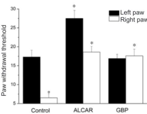
Similar results were found in experiments involving ther-mal hyperalgesic stimuli (Figure 2). Fourteen days after ligation, the ipsilateral paw tolerated the hot stimulus for 6.5 ± 1.1 seconds, and the normal contralateral paw toler-ated the hot stimulus for 17.3 ± 1.8 seconds (P = 0.008, vs. ligated). ALCAR increased the paw withdrawal threshold to 18.6 ± 1.5 seconds and 27.5 ± 2.1 seconds on the ipsilateral and contralateral paws, respectively (P < 0.01 vs. respective control). Gabapentin did not modify contralateral paw with-drawal threshold but significantly increased the ipsilateral paw withdrawal threshold (17.6 ± 1.8 seconds, P = 0.007, vs. respective control).
Morphological results
CCI of the sciatic nerve induced a massive degeneration of myelinated and non-myelinated axons distal to the ligation site. Compared to the left (control) sciatic nerve (Figure 3A), axons of the right sciatic nerve distal to ligation displayed signs of a typical Wallerian degeneration, with less compact and edematous axons and accumulation of inflammatory cells (Figure 3B). Periaxonal myelin sheaths were absent or dam-aged and the myelin-axon border was not clearly identifiable (Figure 3B). Microanatomical changes were less pronounced in the portion of sciatic nerve proximal to the ligation (data not shown). Changes of sciatic nerve distal to ligation were inhibited in part by treatment with ALCAR (Figure 3C), whereas treatment with gabapentin had no effect on the mor-phology of the damaged nerve (Figure 3D).
Nerve morphometry
Morphometric analysis revealed that the total number of nerve fibers of the sciatic nerve was decreased in the distal portion of the ligated nerve compared to the opposite control
Figure 1 Mechanical hyperalgesia.
Paw pressure test in response to noxious stimuli on the paw ipsilateral to ligation in comparison to the contralateral unoperated control paw. Animals were left untreated (control) or treated with acetyl-l-carnitine (AL-CAR, 100 mg/kg, intraperitoneally) or gabapentin (GBP; 70 mg/kg, intra-peritoneally) twice a day for 14 days starting from the day of operation. Tests were performed 60 minutes after the last injection of compounds. Values represent the mean of 12 rats per group analyzed in two differ-ent experimdiffer-ental sets. °P < 0.01, vs. the left paw of control rats; *P < 0.01, vs. the corresponding paw of control rats (analysis of variance followed by the Student-Newman-Keuls test).
side (Figure 4). Treatment with ALCAR rather than with ga-bapentin inhibited numerical changes of nerve fibers (Figure
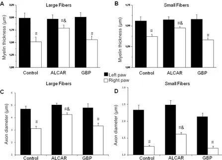
4). Axon area decreased in large and small fibers distal to the
right nerve ligation compared to the homologous nerve fibers of controlateral unoperated nerve (Figure 5). In the distal portion of the ligated nerve, myelin sheaths displayed a less homogeneous deposition of staining (data not shown) with decreased thickness of sheaths irrespectively of the fiber size (Figure 5). Treatment with ALCAR inhibited the reduction of axon and myelin thickness in nerve fibers of the lesioned nerve (Figure 5), whereas gabepentin had no effect (Figure 5).
Molecular analysis results
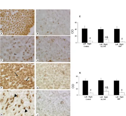
Sections of sciatic nerve treated for NFP immunohistochemis-try developed a dark brown axonal staining, with an immuno-reaction more intense in the external part of axons. Reduced NFP immunoreactivity was observed in the sciatic nerve distal to ligation in control rats (Figure 6B) compared to the con-tralateral unoperated nerve (Figure 6A). Quantitative analysis showed that treatment with ALCAR increased significantly ax-onal NFP immunoreactivity (P = 0.001, vs. control rats) in the distal part of the sciatic nerve (Figure 6C and E).
MBP immunostaining showed a normal pattern of my-elin organization in the left contralateral unoperated sciatic nerve with dark brown immunoreactivity in the myelin sheaths (Figure 6F). A remarkable reduction of MBP immu-noreactivity was noticeable in the distal part of the ligated nerve irrespectively of fiber size and degree of myelination (Figure 6G). Treatment with ALCAR increased significantly MBP immunoreactivity (P = 0.001, vs. control rats) in the distal part of ligated nerve (Figures 6H and K).
Discussion
Peripheral neuropathies are syndromes characterized by nerve fiber degeneration (Valat et al., 2010). Results from this study revealed that microanatomical changes appear after loose
ligation of the sciatic nerve. In this model, either axonal com-ponents of the nerve and myelin sheaths were affected with remarkable loss of myelinated and unmyelinated nerve fibers. These findings support and extend our previous investigations reporting the occurrence of apoptosis in nerve trunks with DNA fragmentation in the nuclei of Schwann cells and axo-nal degeneration in the part distal to the ligation significantly higher than that in the proximal part and the anti-hyperalgesic effect of ALCAR (Di Cesare Mannelli et al., 2009).
This study reported the neuroprotective activity of AL-CAR. The preclinical data deserve further research in hu-mans. ALCAR, the short chain ester of carnitine L-isomer, depending on the country, is available either as a registered drug or as a nutraceutical. It is included in the list of nutri-tional agents, producing cognitive benefits for middle-aged and elderly people (Salvioli et al., 1994; Malaguarnera et al., 2012; Traina et al., 2016). In fact, ALCAR is actively transported across the blood-brain barrier (BBB) and it is important for energetic balance in the brain. ALCAR, in the cells, transports fatty acids from the cytoplasm into the mi-tochondria where they provide substrate for ATP generation
via oxidative phosphorylation (Kidd et al., 2008). It has also
free radical scavenging properties (Mansour et al., 2006) and is thought to influence the cholinergic system by promoting the synthesis and release of acetylcholine (Imperato et al., 1989). In addition to the above functions, ALCAR buffers potentially toxic acyl-CoA metabolites and modulates the ratio of acyl-CoA/CoA facilitating the uptake of acetyl-CoA into the mitochondria during fatty acid oxidation. In the central nervous system, ALCAR participates in the main-tenance and repair processes of neurons, upregulating the expression of NGF and of its receptor, p75NGFR (Piovesan et
al., 1994; Foreman et al., 1995). ALCAR is also able to at-tenuate the mortality rate of neurons (Manfridi et al., 1992), enhance the response to NGF (Taglialatela et al., 1991), and decrease the neurotoxicity evoked by mitochondrial uncou-pling factors or inhibitors (Virmani et al., 1995).
Paw pressure (g) Left paw Right paw * * * °
Paw withdrawal threshold
Left paw Right paw *
* *
Figure 2 Thermal hyperalgesia.
Plantar test in response to noxious stimuli on the paw ipsilateral to li-gation (right one) versus the controlateral unoperated control paw (left one). Animals were left untreated (control) or treated with acetyl-l-car-nitine (ALCAR, 100 mg/kg, intraperitoneally) or gabapentin (GBP; 70 mg/kg, intraperitoneally) twice a day for 14 days starting from the day of operation. Tests were performed for 60 minutes after the last injection of compounds. Values represent the mean of 12 rats per group analyzed in two different experimental sets. °P < 0.01, vs. the left paw of control rats; *P < 0.01, vs. the corresponding paw of control rats (analy-sis of variance followed by the Student-Newman-Keuls test).
Myelin thickness (μm) Myelin thickness (μm)
Axon diameter (μm) Axon diameter (μm)
Left paw Right paw # # # # # # # #& # #& #& #&
Figure 5 Morphometric analysis of myelin thickness and axon diameter for larger (A, C) and small fibers (B, D) in control unoperated left sciatic nerve in the distal portion to ligation of the right sciatic nerve of control, acetyl-l-carnitine (ALCAR)-treated or
gabapentin-treated (GBP) rats.
Analysis was performed as detailed in the materi-als and methods section using osmium fixed tis-sues in six animals per group. #P < 0.01, vs. left un-operated nerve nerve. &P < 0.01, vs. right ligated sciatic nerve of control untreated rats (analysis of variance followed by the Student-Newman-Keuls test).
Our findings revealed that ALCAR elicits a neuroprotec-tive effect on CCI rats. In rat models of neuropathy, ALCAR was able to relieve pain sensation, showing analgesic and anti-hyperalgesic properties. This activity is documented by the increase of pain threshold elicited by ALCAR both in the paw ipsilateral to ligation (anti-hyperalgesic effect) and in controlateral unoperated control paw (analgesic effect). The level of anti-hyperalgesic activity of ALCAR was similar to that elicited by GAB, an anticonvulsant drug largely used for the treatment of painful symptoms of neuropathy (Gil-ron et al., 2006; Teasell et al., 2010; Chaparro et al., 2012). On the other hand, GAB showed no analgesic effects in the animal models used. Moreover, ALCAR exerted a protec-tive effect on the peripheral nerve portion most affected by ligation, the distal one. This neuroprotective activity is not
shared by GAB. It cannot be excluded that the activity on hyperalgesia after treatment with ALCAR may depend in part by the effects that the compound induced on sciatic nerve morphology. On the other hand, ALCAR showed no antihyperalgesic effect after a single administration (data not shown). CCI caused changes of the myelin sheaths and axonal damage which are likely responsible for a transient loss of hindpaw motor function and trigger the pathological pain signal (Kingery et al., 1994; Pacini et al., 2010). After recovery of the axonal lesion (40–60 days), when the distal
axon regeneration occurs, both motor function impairment and hyperalgesia are resolved (Kingery et al., 1994).
The complex mechanism of action of ALCAR leads to a difficulty to hypothesize the pathways through which it exerts neuroprotective effects documented in the present
Figure 3 Micrographs of sections of rat sciatic nerve.
Masson’s Trichromic staining to verify microanatomical details. The morphology of the left unoperated nerve (A) is normal. Ligation (B–D) caused a morphological disarrangement of the nerve, which was re-versed in part by treatment with acetyl-l-carnitine (C) but not with ga-bapentin (D). (A) Sciatic nerve of the left unoperated side of a control rat. (B) Right sciatic nerve close to the ligation site of a damaged rat. (C) Right sciatic nerve close to the ligation area of an ALCAR-treated rat. (D) Right sciatic nerve close to the ligation area of a gabapentin-treated rat. Scale bars: 12.5 μm.
Number of fibers
# #&
#
Figure 4 Quantitative analysis of the total number of nerve fibers of sciatic nerve in control unoperated left sciatic nerve in the distal portion of the right sciatic nerve of control acetyl-l-carnitine (ALCAR)-treated or gabapentin-treated (GBP) rats.
Analysis was performed as detailed in the materials and methods sec-tion using osmium fixed tissues in six animals per group. #P < 0.01, vs. left unoperated nerve. &P < 0.01, vs. right ligated nerve of control untreated rats (analysis of variance followed by the Student-New-man-Keuls test).
Sections processed for neurofilament protein (A–D) and myelin basic protein immunohistochemistry (F–J). In the left unoperated sciatic nerve of control rats (A), an obvious axonal neurofilament protein (NFP) immunore-activity was observed (white asterisk), whereas pale and non-immunoreactive myelin sheaths surrounded axons. Very low immunoreactivity was noticeable in the por-tion distal to ligapor-tion of the right damaged sciatic nerve of control rats (B). Increased immunoreactivity (black asterisk) was observed in ligated nerves of acetyl-l-car-nitine (ALCAR)-treated rats (C), but not in gabapentin (GBP)-treated rats (D). Scale bars: 12.5 mm. Microden-sitometric analysis was shown in panel (E), and data represent the optical density (OD) of immunoreactivity expressed in arbitrary units (mean ± SEM; n = 6). In the sections processed for myelin basic protein immunohis-tochemistry, an immune reaction in the myelin sheaths (arrows) was evident in the left unoperated sciatic nerve of control rats (F). A sharp decrease of immunoreactivity was observed in the right ligated nerve of control rats distal to ligation (G). Myelin basic protein immunoreac-tivity (arrowheads) in ligated nerve was largely decreased after treatment with ALCAR (H) and slightly decreased after GBP treatment (J). Scale bars: 12.5 mm. Microden-sitometric analysis was shown in panel (K), and data represent the optical density (OD) of immunoreactivity expressed in arbitrary units (mean ± SEM; n = 6). #P < 0.01, vs. left unoperated nerve; &P < 0.01, vs. right ligated nerve of untreated control rats (analysis of variance fol-lowed by the Student-Newman-Keuls).
Figure 6 Neurofilament protein 200 kDa and myelin basic protein immunohistochemistry in the rat sciatic nerve.
# #& #
# #& #
OD
OD
study. As working hypotheses, the stimulatory role of AL-CAR on acetyl-CoA (Pettegrew et al., 2000) and acetylcho-line synthesis (Imperato et al., 1989) and/or the ALCAR-in-duced increase in trophic factors (Vivoli et al., 2010) are the most probable. Acetyl-CoA is involved in redox reactions eliminating reactive oxygen species, the increase of which is related to the pathophysiology of neuropathy (Naik et al., 2006). In vivo and in vitro studies have demonstrated that acetylcholine as a neurotransmitter has a neuroprotec-tive role in the central, peripheral and autonomic nervous systems (Ryan et al., 2001; Takada et al., 2003). In addition to their physiological role, i.e., cellular energy metabolic actions to facilitate β-oxidation of fatty acids and carbohy-drate metabolism (stimulation of pyruvate dehydrogenase), carnitines have been shown to display in vitro antioxidant activities, possibly through their action against free hydroxyl radicals (OH•) formation in Fenton-type reactions (Gülcin et al., 2006). Trophic factors are involved both in pain per-ception and neurorestoration. NGF and other members of the neurotrophin family act as pain mediators. The admin-istration of neurotrophin in rats determined a pronounced mechanical and thermal hyperalgesia (Levin et al., 1994). Members of GDNF family and in particular GDNF and Ar-temin, in addition to their ability to support regeneration after nervous tissue damage (Chen at al., 2001; Wang et al., 2008), can normalize pain threshold and therefore exert an anti-hyperalgesic effect (Boucher et al., 2001; Gardel et al., 2003; Wang et al., 2008). A recent study of our group has reported that in the CCI rat models of neuropathy, neuro-trophic factor changes consisted of increased expression of NGF and decreased expressions of GDNF and Artemin (Vivoli et al., 2010). In the same model used in the present study, ALCAR restored altered levels of the above
neuro-trophic factors with a pronounced activity on Artemin ex-pression, which is increased also in sham-operated animals and in a pathology-independent manner (Vivoli et al. 2010). This suggests that the compound may stimulate rescue of damaged nerves by acting on neurotrophic factors.
To conclude, further studies are necessary to clarify the mechanism(s) of neuroprotective activity of ALCAR in the neuropathy models investigated, relevant features in micro-natomical restoration are shown. The compound deserves specific clinical trials aimed at investigating its role in the prevention of peripheral nerve damage.
Author contributions: LDCM performed in vivo experiments. DT
and AP performed histological evaluations. DT and LDCM drafted the paper. VB, CG and FA conceived the study and planned its design. All authors approved the final version of this paper.
Conflicts of interest: Acetyl-l-carnitine was used in the method section,
but the authors declare that there are no competing interests.
Financial support: This research was funded by the Italian Ministry of
Instruction, University and Research (MIUR) and by the Universities of Florence and Camerino.
Research ethics: This study was performed in accordance with the
Eu-ropean Community guidelines for animal care (DL 116/92, EuEu-ropean Community Council Directive 86/609/EEC) and with the ethical guide-lines of the University of Florence, consistent with the Guide for the Care and Use of Laboratory Animals of the US National Institutes of Health (NIH Publication No. 85-23, revised 1996; University of Florence assur-ance number: A5278-01).
Data sharing statement: Datasets analyzed during the current study
are available from the corresponding author on reasonable request.
Plagiarism check: Checked twice by iThenticate. Peer review: Externally peer reviewed.
Open access statement: This is an open access journal, and articles are
distributed under the terms of the Creative Commons Attribution-Non-Commercial-ShareAlike 4.0 License, which allows others to remix, tweak, and build upon the work non-commercially, as long as appropriate credit is given and the new creations are licensed under the identical terms.
Pol-icy and Inequalities Research, USA; Gabriele Siciliano, Universita degli Studi di Pisa, Neurological Clinic, Clinical and Experimental Medicine, Italy.
Additional file: Open peer reviewer reports 1 and 2.
References
Bennett GJ, Xie YK (1988) A peripheral mononeuropathy in rat that pro-duces disorders of pain sensation like those seen in man. Pain 33:87-107. Boucher TJ, McMahon SB (2001) Neurotrophic factors and neuropathic
pain. Curr Opin Pharmacol 1:66-72.
Chaparro LE, Wiffen PJ, Moore RA, Gilron I (2012) Combination phar-macotherapy for the treatment of neuropathic pain in adults. Cochrane Database Syst Rev (7):CD008943.
Chen ZY, Chai YF, Cao L, Lu CL, He C (2001) Glial cell line-derived neu-rotrophic factor enhances axonal regeneration following sciatic nerve transection in adult rats. Brain Res 902:272-276.
Chiechio S, Copani A, Gereau RW 4th, Nicoletti F (2007) Acetyl-L-carni-tine in neuropathic pain: experimental data. CNS Drugs 21 Suppl 1:31-38. Di Cesare Mannelli L, Ghelardini C, Calvani M, Nicolai R, Mosconi L, Vivoli E, Pacini A, Bartolini A (2007) Protective effect of acetyl-l-carni-tine on the apoptotic pathway of peripheral neuropathy. Eur J Neurosci 26:820-827.
Di Cesare Mannelli L, Ghelardini C, Calvani M, Nicolai R, Mosconi L, Toscano A, Pacini A, Bartolini A (2009) Neuroprotective effects of ace-tyl-L-carnitine on neuropathic pain and apoptosis: a role for the nicotin-ic receptor. J Neurosci Res 87:200-207.
Di Cesare Mannelli L, Ghelardini C, Toscano A, Pacini A, Bartolini A (2010) The neuropathy-protective agent acetyl-L-carnitine activates protein kinase C-gamma and MAPKs in a rat model of neuropathic pain. Neuro-science 165:1345-1352.
Evans JD, Jacobs TF, Evans EW (2008) Role of acetyl-L-carnitine in the treatment of diabetic peripheral neuropathy. Ann Pharmacother 42:1686-1691.
Foreman PJ, Perez-Polo JR, Angelucci L, Ramacci MT, Taglialatela G (1995) Effect of Acetyl-L-carnitine treatment and stress exposure on the nerve growth factor receptor (p75NGFR) mRNA level in the central nervous system of aged rats. Prog Neuropsychopharmacol Biol Psych 19:117-133. Galeotti N, Bartolini A, Calvani M, Nicolai R, Ghelardini C (2004) Ace-tyl-L-carnitine requires phospholipase C-IP3 pathway activation to in-duce antinociception. Neuropharmacology 472:286-294.
Gardell LR, Wang R, Ehrenfels C, Ossipov NH, Rossomando AJ, Miller S, Buckle C, Cai AK, Tse A, Foley SF, Gong B, Walus L, Carmillo P, Warley D, Huang C, Engber T, Pepinsky B, Cate RL, Vanderah TW, Lai J, Sah DW, Porreca F (2003) Multiple actions of systemic artemin in experi-mental neuropathy. Nat Med 9:1383-1389.
Ghelardini C, Galeotti N, Calvani M, Mosconi L, Nicolai R, Bartolini A (2002) Acetyl-l-carnitine induces muscarinic antinocieption in mice and rats. Neuropharmacology 43:1180-1187.
Ghirardi O, Lo Giudice P, Pisano C, Vertechy M, Bellucci A, Vesci L, Cundari S, Miloso M, Rigamonti LM, Nicolini G, Zanna C, Carminati P (2005) Acetyl-L-Carnitine prevents and reverts experimental chronic neurotoxicity induced by oxaliplatin, without altering its antitumor properties. Anticancer Res 25:2681-2687.
Gilron I, Watson NPC, Cahill CM, Moulin DE (2006) Neuropathic pain: a pratical guide for the clinician. CMAJ 175:265-275.
Gülcin I (2006) Antioxidant and antiradical activities of L-Carnitine. Life Sci 78:803-811.
Hargreaves K, Dubner R, Brown F, Flores C, Joris J (1988) A new and sen-sitive method for measuring thermal nociception in cutaneous hyperal-gesia. Pain 32:77-88.
Imperato A, Ramacci MT, Angelucci L (1989) Acetyl-L-carnitine enhances acetylcholine release in the striatum and hippocampus of awake freely moving rats. Neurosci Lett 107:251-255.
Janiri L, Martinotti, G, Tonioni F, Ghelardini C, Nicolai R, Galeotti N, Mosconi L, Calvani M, Bartolini A, Iannoni E (2009) Acetyl-L-carnitine in the management of pain during methadone withdrawal syndrome. Clin Neuropharmacol 32:35-40.
Karsidag S, Akcal A, Sahin S, Karsidag S, Kabukcuoglu F, Ugurlu K (2012) Neurophysiological and morphological responses to treatment with ace-tyl-L-carnitine in a sciatic nerve injury model: preliminary data. J Hand Surg Eur 37:529-536.
Kidd PM (2008) Alzheimer’s disease, amnestic mild cognitive impairment, and age-associated memory impairment: current understanding and progress toward integrative prevention. Altern Med Rev 13:85-115.
Kingery WS, Lu JD, Roffers JA, Kell DR (1994) The resolution of neuro-pathic hyperalgesia following motor and sensory functional recovery in sciatic axonotometic mononeuropathies. Pain 58:157-168.
Leighton GE, Rodriguez RE, Hill RG, Hughes J (1988) k-opioid agonist produce antinociception after i.v. and i.c.v. but not intrathecal adminis-tration in the rat. Br J Pharmacol 93:553-560.
Levin GR, Rueff A, Mendell LM (1994) Peripheral and central mechanisms of NGF-induced hyperalgesia. Eur J Neurosci 6:1903-1912.
Malaguarnera M (2012) Carnitine derivatives: clinical usefulness. Curr Opin Gastroenterol 28:166-176.
Manfridi A, Forloni GL, Arrigoni-Martelli E, Mancia M (1992) Culture of dorsal root ganglion neurons from aged rats: effects of acetyl-L-carnitine and NGF. Int J Dev Neurosci 10:321-329.
Mansour HH (2006) Protective role of carnitine ester against radiation-in-duced oxidative stress in rats. Pharmacol Res 54:165-171.
Naik AK, Tandan SK, Dudhgaonkar SP, Jadhav SH, Kataria M, Prakash VR, Kumar D (2006) Role of oxidative stress in pathophysiology of pe-ripheral neuropathy and modulation by N-acetyl-L-cysteine in rats. Eur J Pain 10: 573-579.
Osio M, Muscia F, Zampini L, Nascimbene C, Mailland E, Cargnel A, Mar-iani C (2006) Acetyl-l-carnitine in the treatment of painful antiretroviral toxic neuropathy in human immunodeficiency virus patients: an open label study. J Peripher Nerv Syst 11:72-76.
Pacini A, Di Cesare Mannelli L, Bonaccini L, Ronzoni S, Bartolini A, Ghelardini C (2010) Protective effect of alpha7 nAChR: behavioural and morphological features on neuropathy. Pain 150:542-549.
Pettegrew JW, Levine J, McClure RJ (2000) Acetyl-L-carnitine physi-cal-chemical, metabolic, and therapeutic properties: relevance for its mode of action in Alzheimer’s disease and geriatric depression. Mol Psych 5:616-632.
Piovesan P, Pacifici L, Taglialatela G, Ramacci MT, Angelucci L (1994) Acetyl-L-carnitine treatment increases choline acetyltransferase activity and NGF levels in the CNS of adult rats following total fimbria-fornix transection. Brain Res 633:77-82.
Ryan RE, Ross SA, Drago J, Loiacono RE (2001) Dose-related neuroprotec-tive effects of chronic nicotine in 6-hydroxydopamine treated rats and loss of neuroprotection in alpha4 nicotinic receptor subunit knockout mice. Br J Pharm 132:1650-1656.
Salvioli G, Neri M (1994) L-Acetylcarnitine treatment of mental decline in the elderly. Drugs Exp Clin Res 20:169-176.
Sima AA, Calvani M, Mehra M, Amato A; Acetyl-L-Carnitine Study Group (2005) Acetyl-L-carnitine improves pain, nerve regeneration, and vibra-tory perception in patients with chronic diabetic neuropathy: an analysis of two randomized placebo-controlled trials. Diabetes Care 281:89-94. Taglialatela G, Angelucci L, Ramacci MT, Werrbach-Perez K, Jackson GR,
Perez-Polo JR (1991) Acetyl-L-carnitine enhances the response of PC12 cells to nerve growth factor. Brain Res Dev Brain Res 59:221-230. Takada Y, Yonezawa A, Kume T, Katsuki H, Kaneko S, Sugimoto H,
Akaike A (2003) Nicotinic acetylcholine receptor-mediated neuroprotec-tion by donepezil against glutamate neurotoxicity in rat cortical neurons. J Pharm Exp Ther 306:772-777.
Teasell RW, Mehta S, Aubut JA, Foulon B, Wolfe DL, Hsieh JT, Townson AF, Short C (2010) Spinal Cord Injury Rehabilitation Evidence Research Team. A systematic review of pharmacologic treatments of pain after spinal cord injury. Arch Phys Med Rehabil 91:816-831.
Traina G (2016) The neurobiology of acetyl-L-carnitine. Front Biosci (Landmark Ed) 21:1314-1329.
Traina G, Federighi G, Macchi M, Bernardi R, Durante M, Brunelli M (2011) Modulation of myelin basic protein gene expression by acetyl-L-carni-tine. Mol Neurobiol 44:1-6.
Valat JP, Genevay S, Marty M, Rozenberg S, Koes B (2010) Sciatica. Best Pract Res Clin Rheumatol 24:241-252.
Virmani MA, Biselli R, Spadoni A, Rossi S, Corsico N, Calvani M, Fatto-rossi A, De Simone, C, Arrigoni-Martelli E (1995) Protective actions of L-carnitine and acetyl-L-carnitine on the neurotoxicity evoked by mito-chondrial uncoupling or inhibitors. Pharmacol Res 32:383-389. Vivoli E, Di Cesare Mannelli L, Salvicchi A, Bartolini A, Koverech A,
Nico-lai R, Benatti P, Ghelardini C (2010) Acetyl-l-carnitine increases artemin level and prevents neurotrophic factor alterations during neuropathy. Neuroscience 165:1168-1174.
Wang R, King T, Ossipov MH, Rossomando AJ, Vanderah TW, Harvey P, Cariani P, Frank E, Sah DW, Porreca F (2008) Persistent restoration of sensory function by immediate or delayed systemic artemin after dorsal root injury. Nat Neurosci 11:488-496.