R E S E A R C H A R T I C L E
Open Access
Clinical investigation on
Theileria equi and Babesia
caballi infections in Italian donkeys
Fulvio Laus
1*, Andrea Spaterna
1, Vanessa Faillace
1, Fabrizia Veronesi
2, Silvia Ravagnan
3, Francesca Beribé
1,
Matteo Cerquetella
1, Marina Meligrana
1and Beniamino Tesei
1Abstract
Background: Interest in the welfare and diseases of donkeys is constantly increasing in several countries. Despite this, clinical research into donkeys needs to be in continual development since they show different reactions compared to horses in many conditions, including infectious diseases, and need specific clinical and therapeutic approaches. No reports are currently available on clinical and clinical pathology data regarding donkeys with natural piroplasms infection.
Results: Venous blood samples were taken from one hundred and thirty eight donkeys and underwent indirect fluorescent antibody test (IFAT) to detect IgG antibodies against Theileria equi and Babesia caballi and real-time polimerase chain reaction (PCR) to detect Babesia spp. and Theileria spp. Clinical examinations, haematological analyses and serum bilirubin evaluation were also performed and compared with positive or negative status. A seroprevalence of 40.6% and 47.8% was found for T. equi and B. caballi, respectively; double positivity was detected in 19.6% of the animals. PCR results showed that 17.4% of the animals tested positive for T.equi and 3.6% for B. caballi with no double positivity. Twelve donkeys (8.7%) had clinical signs consistent with chronic forms of the disease and no acute forms were detected. Fifty-eight donkeys had haematological and serum bilirubin alterations and 56 (96.6%) of them were IFAT and/or PCR positive. Changes in erythrocyte number, packed cell volume, hemoglobin concentration, mean corpuscular hemoglobin, platelets number and total bilirubin were significantly associated with positive and symptomatic animals.
Conclusion: Nonspecific clinical presentation seems to be very common in donkeys and several clinical pathology alterations persist after natural infection. Therefore, apparently healthy donkeys can have masked but severe clinical pathology alterations. Acute forms are very seldom observed in donkeys. Clinical monitoring of chronically infected donkeys is recommended since such animals represent a risk both for transmission to other animals and for their own health; furthermore, their production performances could be reduced. The study should also be intended as a contribution for veterinary practitioners because it describes the most usual clinical presentations and laboratory findings of equine piroplasmosis in naturally infected donkeys in endemic areas.
Keywords: Donkey, Piroplasmosis, Tick borne diseases, Theileria equi, Babesia caballi Background
Interest in the welfare and diseases of donkeys is con-stantly increasing in several countries, mostly due to the recent rediscovery of donkey milk as an alternative food source for milk-intolerant children. Clinical research on donkeys needs to be in continual development since they have different reactions compared to horses in
many conditions (e.g. resistance to pain in case of colic), including infectious diseases, making it more difficult to recognize the symptoms normally observed in horses [1,2]. Their current popularity is also due to their use as pets, in addition to recreational purposes, sports activ-ities, donkey-assisted therapy and, to a lesser extent, as pack/draught animals and for meat production [3].
Equine piroplasmosis (EP) is a tickborne disease
caused by the protozoa Babesia caballi and Theileria
equi. EP is endemic in most equine populations in trop-ical and subtroptrop-ical areas of the world and affects all
* Correspondence:[email protected]
1
Scuola di Bioscienze e Medicina Veterinaria, Università di Camerino, Via Circonvallazione 93/95, 62024 Matelica, (MC), Italy
Full list of author information is available at the end of the article
© 2015 Laus et al.; licensee BioMed Central. This is an Open Access article distributed under the terms of the Creative Commons Attribution License (http://creativecommons.org/licenses/by/4.0), which permits unrestricted use, distribution, and reproduction in any medium, provided the original work is properly credited. The Creative Commons Public Domain Dedication waiver (http://creativecommons.org/publicdomain/zero/1.0/) applies to the data made available in this article, unless otherwise stated.
equid species, including horses, donkeys, mules, and ze-bras [4,5]. Chronic cases are more common in donkeys than horses and are usually characterized by nonspecific clinical signs such as lethargy, partial anorexia, poor work performance and body weight loss [6]. In acute forms of EP, donkeys can show fever, listlessness, depression, noticeable thirst, swelling of the eyelids, con-stipation, presence of yellow mucous covering feces, yellowish coloration of urine, and splenomegaly [6]. Donkeys can also show an asymptomatic form of the in-fection and, in comparison to horses, they also show a lower parasitemia [6]. The most common hematological alteration found in horses is decreased packed cell volume, hemoglobin and erythrocyte number, in addition to hyperbilirubinemia and thrombocytopenia [3]. After subclinical, chronic or acute infection, donkeys usually re-main asymptomatic carriers with positive antibody titers throughout life [6].
Several data are available for the epidemiology of equine piroplasmosis (EP) in horses [7,8] and informa-tion on its epidemiology in Italy has also been reported [9-13]. However, few reports exist regarding the preva-lence of these parasites in donkeys [14-17] and, to the authors’ knowledge, only few surveys have been carried out in Italy [18-20]. The present study is the first investi-gation aimed at evaluating and comparing the direct (by
PCR) and indirect (by IFAT) presence of B. caballi and
T. equi with clinical signs and clinical pathology data in naturally infected donkeys in Italy.
Methods
One hundred and thirty eight mixed breed donkeys (109 females, 7 stallions and 22 geldings) ranging from 1 to 22 years of age (mean 7.6, d.s. = 4.7) belonging to 8 dif-ferent farms (mean herd size 17 donkeys, d.s. 6 donkeys) in central Italy were included in the study. The area was chosen due to the high prevalence of tickborne patho-gens previously found in equids [12,13,18,21,22], the proven presence of the tick vectors [23] and because vet practitioners have frequently reported heavy tick infesta-tions in equids. All the animals were born and reared in Italy and had never been moved out of the country. The survey was performed between March and October 2013 in farms of varying nature and size, including herds for milk production (n = 5), onotherapy centers (n = 2) and private facilities (n = 1) where animals were reared for leisure. De-worming and topical ectoparasite repellents were regularly administered to all the animals who were free from ticks at the moment of evaluation.
A general clinical examination was performed on each donkey; the evaluation also included a body condition score (BCS) estimation, following the scheme of Pearson and Quassat (1996) [24]. Donkeys showing clinical signs not attributable to EP (e.g. lameness) were excluded
from the study to avoid interference on blood analysis. Venous blood samples were collected from each donkey from the jugular vein into sterile tubes with (two tubes) and without (one tube) ethylenediaminetetraacetic acid (EDTA) and maintained at +4°C. The samples with EDTA were submitted for a complete blood count (CBC), which included: erythrocytes count (RGB), packed cell volume (PCV), hemoglobin (Hb), mean corpuscular volume (MCV), mean corpuscular hemoglobin (MCH), mean corpuscular hemoglobin concentration (MCHC), total leukocytes, neutrophils, lymphocytes, monocytes, eo-sinophils, basophils and platelets (Cell Dyn 3500, Abbott).
Moreover, an aliquot of 200 μl was destined to genomic
DNA extraction using the QIAamp DNA Blood Mini kit (QIAGEN S.p.A., Milan, Italy) according to the manufac-turer’s instructions. To ensure the effectiveness of the nu-cleic acid extraction, a PCR targeting the 18S rRNA was applied [25]. The extracted DNA was submitted to a Real Time PCR Sybr Green assay to detect 509 base pairs of 18S rRNA gene ofBabesia spp. and Theileria spp. using the
pri-mer BJ1 and BN2 described by Casati et al. (2006) [26].
The method shows a sensitivity of 10^3 DNA copies/μl. The species identity was determined by subsequent ampli-con sequencing. All PCR products were sequenced using the Big Dye Terminator v 3.1 cycle sequencing kit (Applied Biosystem, Foster City, CA, USA) in a 16-capillary ABI PRISM 3130 × l Genetic Analyzer (Applied Biosystem, Foster City, CA, USA). Sequence data were assembled and edited with SeqScape software v 2.5 (Applied Biosystem, Foster City, CA, USA), aligned and compared with repre-sentative sequences available in GenBank [27].
Samples without EDTA were centrifuged at 4000 rpm for 10 minutes; the separated sera were collected and di-vided into two aliquots. The first aliquot was used for dosage of total bilirubin (TB) (Targa 3000 plus, Biotec-nica Instruments); the second was utilized to determine
the presence of IgG antibodies against T. equi and B.
caballi using a commercial indirect fluorescent antibody test (IFAT) (MegaScreen®, 112 DIAGNOSTIK MEGA-CORE Laboratories, Horbranz, Austria).
Statistical analysis
Prevalence and 95% binomial confidence intervals (CI) were calculated [28] for the serologic and molecular test results.
Hematological parameters and serum bilirubin were tested for normality by Kolmogorof-Smirnov test and then analyzed by ANOVA or Mann–Whitney U-test for comparison between positive (both to PCR and/or IFAT) and negative animals and between donkeys with and without clinical signs. Comparison of the clinical path-ology results with normal reference values [29] was car-ried out using a t-student’s test. The Chi-square test was performed to evaluate the differences between IFAT and
PCR prevalence forB. caballi and T. equi.. Statistical sig-nificance was assessed at the 0.05 probability level in all analyses.
All statistical analyses were performed using the WIN-PEPI (WIN-PEPI-for-Windows) computer program (Epidemiol. Perspect. Innov. 1:6. Available from: http://www.biomed-central.com/1742-5573/1/6).
Ethical statement
Tha autor state that the work has been carried out in compliance with relevant guidelines regarding ethical use of animals, approved by the Universitary Ethical Commettee for Animal Protection and in adherence to a high standard (best practice) of veterinary care.
Results
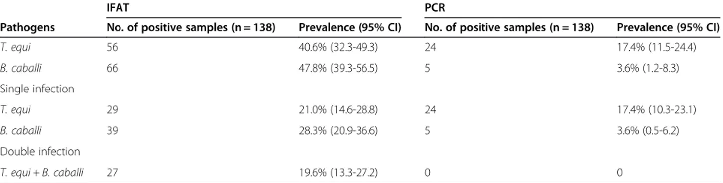
Ninety-five (68.8%) and 29 (21.0%) donkeys tested posi-tive by IFAT and PCR respecposi-tively. The results of the serological and molecular tests performed on blood sam-ples are reported in Table 1, as well as the prevalence (%) and 95% confidence interval (CI) of single and mixed-infections.
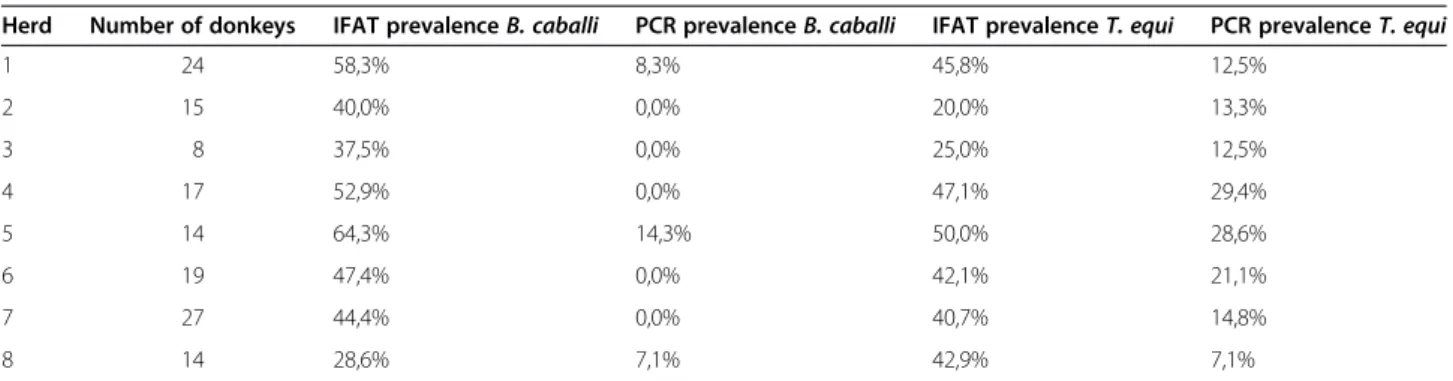
All herds (100%) resulted positive at IFAT for bothT. equi and B. caballi and at PCR for T. equi. Three herds
(37.5%) resulted positive for B. caballi at PCR. The
prevalence rates within herd are reported in Table 2.
The seroprevalence of B. caballi resulted higher than
that of T. equi but the difference was not statistically significant (P = 0.3). The percentage of PCR positive animals resulted statistically higher for T. equi than B. caballi (P < 0.001).
Nine (6.5%, 95% CI: 3.0-12.90%) animals were
simul-taneously IFAT and PCR positive for T. equi while 15
(10.9%, 95% CI: 6.2-17.30%) were only PCR positive. None of the IFAT positive donkeys resulted PCR positive forB. caballi.
Abnormal clinical pathology data with respect to nor-mal ranges were detected in 58 (42.0%) samples. Fifty-six (96.6%) of these donkeys resulted IFAT and/or PCR positive. Hematological alterations included decreased
RGB (n = 49), decreased PCV (n = 24), decreased Hb (n = 31), increased MCH (n = 16), increased MCHC (n = 9) increased WBC (n = 6), increased neutrophils (n = 7), in-creased eosinophils (n = 5), dein-creased platelets (n = 20), and increased bilirubin (n = 19). Among IFAT positive donkeys, 46 (48.4%) had one or more hematological and/or bilirubin alteration: 18 (39.1%) proved positive for B.caballi, 18 (39.1%) for T. equi and 10 (21.7%) were double positives. Among PCR positive donkeys, 19 (65.5%) had one or more hematological and/or bilirubin
alteration: 1 (5.3%) was positive for B. caballi and 18
(94.7%) were positive for T. equi. The statistical differ-ences between positive (both to PCR and/or IFAT) and negative animals are reported in Table 3.
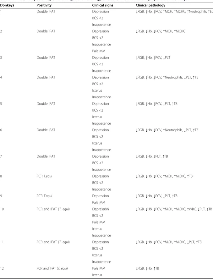
Twelve (10.4%) of the positive donkeys presented signs related to chronic piroplasm infection at the moment of evaluation and all of them were positive for at least one test (Table 4). Detected clinical signs included mild depression (n = 11), body condition score≤ 2 (n = 10), in-appetence (n = 10), pale mucous membranes (n = 4) and mild icterus (n = 6). MCH, MCHC, and TB were statisti-cally higher in symptomatic than in negative donkeys while RGB, PCV, Hb, and platelets were lower. When the blood parameters of symptomatic donkeys were compared to asymptomatic/positives, Hb, MCH and TB resulted in being the only statistically different parame-ters (P = 0.001, P = 0.0005 and P = 0.005, respectively). Discussion
Although not statistically significant, the seroprevalence
of B. caballi (47.8%) was higher than that of T. equi
(40.6%) in accordance with the results obtained in don-keys in Italy [18-20] or in other countries [14,16,17]. The percentage of PCR positive animals resulted
statisti-cally higher for T. equi (17.4%) than B. caballi (3.6%)
proving that also in donkeysT. equi can persist in a sub-clinical form for longer thanB. caballi [30].
In general, the chronic and subclinical natural infec-tion in donkeys included in this study seems to be asso-ciated with decreased RGB, PCV, Hb and PLT and with
Table 1 Number of donkeys, prevalence and confidence interval of the equine tick-borne infections investigated using serological and molecular testing
IFAT PCR
Pathogens No. of positive samples (n = 138) Prevalence (95% CI) No. of positive samples (n = 138) Prevalence (95% CI)
T. equi 56 40.6% (32.3-49.3) 24 17.4% (11.5-24.4) B. caballi 66 47.8% (39.3-56.5) 5 3.6% (1.2-8.3) Single infection T. equi 29 21.0% (14.6-28.8) 24 17.4% (10.3-23.1) B. caballi 39 28.3% (20.9-36.6) 5 3.6% (0.5-6.2) Double infection T. equi + B. caballi 27 19.6% (13.3-27.2) 0 0
Table 2 Intra-herd prevalence ofBabesia caballi and Theileria equi
Herd Number of donkeys IFAT prevalenceB. caballi PCR prevalenceB. caballi IFAT prevalenceT. equi PCR prevalenceT. equi
1 24 58,3% 8,3% 45,8% 12,5% 2 15 40,0% 0,0% 20,0% 13,3% 3 8 37,5% 0,0% 25,0% 12,5% 4 17 52,9% 0,0% 47,1% 29,4% 5 14 64,3% 14,3% 50,0% 28,6% 6 19 47,4% 0,0% 42,1% 21,1% 7 27 44,4% 0,0% 40,7% 14,8% 8 14 28,6% 7,1% 42,9% 7,1%
IFAT, indirect fluorescent antibody test; PCR, Polymerase Chain Reaction.
Table 3 Mean, standard deviation and statistical association with negative donkeys of hematobiochemical paramethers
Negative IFAT positive PCR positive PCR and IFAT positive
Parameters Mean (sd) B. caballi mean (sd) T. equi mean (sd) Double positives mean (sd) B. caballi mean (sd) T. equi mean (sd) T. equi mean (sd) RGB (106/μl) 6.7 (0.8) 5.1 (0.8) 5.0 (0.9) 5.1 (1.0) 5.9 (1.1) 4.7 (0.8) 4.1 (0.4) P < 0.001 P < 0.001 P < 0.001 P = 0.1 P < 0.001 P < 0.001 PCV % 35 (5) 32 (4) 32 (5) 31 (7) 33 (2) 29 (3) 31 (6) P = 0.004 P = 0.06 P = 0.02 P = 0.3 P < 0.001 P = 0.02 Hb (g/dl) 12.1 (1.3) 10.7 (1.4) 10.5 (2.4) 10.4 (2.2) 10.9 (1.1) 9.1 (1.4) 7.9 (0.9) P < 0.001 P = 0.007 P = 0.002 P = 0.06 P < 0.001 P < 0.001 MCV (fl) 52.5 (3.8) 55.1 (3.5) 54.0 (8.0) 56.0 (3.8) 51.3 (4.3) 54.2 (3.1) 55.6 (4.2) P = 0.7 P = 0.6 P = 0.3 P = 0.5 P = 0.5 P = 0.3 MCH (pg) 19.1 (1.5) 20.7 (2.0) 20.3 (1.4) 21.0 (2.0) 19.0 (1.7) 23.7 (4.3) 22.3 (1.8) P = 0.002 P = 0.01 P < 0.001 P = 0.9 P < 0.001 P < 0.001 MCHC (g/dl) 34.2 (1.5) 35.0 (1.7) 34.9 (1.4) 35.2 (2.2) 35.3 (1.9) 34.9 (1.5) 36.1 (2.0) P = 0.06 P = 0.1 P = 0.07 P = 0.2 P = 0.2 P = 0.006 WBC (103/μl) 8.9 (2.9) 9.1 (2.7) 10.1 (2.6) 8.9 (2.4) 9.9 (2.9) 8.7 (2.3) 9.9 (1.7) P = 0.8 P = 0.2 P = 0.9 P = 0.5 P = 0.8 P = 0.3 Neutrophils (103/μl) 5.1 (2.4) 4.3 (1.7) 4.4 (0.9) 4.3 (1.9) 5.2 (1.7) 4.8 (1.6) 6.5 (2.9) P = 0.4 P = 0.6 P = 0.3) P = 0.3 P = 0.3 P = 0.007 Lymphocytes (103/μl) 4.6 (1.9) 4.8 (1.9) 5.4 (2.1) 5.2 (1.9) 5.4 (1.7) 5.6 (1.6) 5.9 (0.9) P = 0.6 P = 0.2 P = 0.4 P = 0.3 P = 0.1 P = 0.06 Monocytes (103/μl) 0.2 (0.2) 0.2 (0.2) 0.3 (0.2) 0.2 (0.2) 0.0 (0.1) 0.3 (0.2) 0.1 (0.2) P = 0.6 P = 0.05 P = 0.3 P = 0.1 P = 0.1 P = 0.9 Eosinophils (103/μl) 0.4 (0.3) 0.5 (0.4) 0.4 (0.3) 0.6 (0.4) 0.5 (0.3) 0.6 (0.4) 0.6 (0.5) P = 0.3 P = 0.4 P = 0.09 P = 0.1 P = 0.1 P = 0.07 Basophils (103/μl) 0.1 (0.1) 0.0 (0.1) 0.1 (0.1) 0.1 (0.0) 0.1 (0.0) 0.1 (0.1) 0.0 (0.1) P = 0.3 P = 0.7 P = 0.3 P = 0.07 P = 0.9 P = 0.6 Platelets (103/μl) 326 (65) 277 (102) 267 (83) 209 (77) 229 (67) 254 (63) 271 (79) P = 0.04 P = 0.01 P < 0.001 P = 0.006 P = 0.002 P = 0.05 TB (mg/dl) 0.3 (0.1) 0.3 (0.14) 0.1 (0.2) 0.3 (0.2) 0.2 (0.0) 0.5 (0.3) 0.3 (0.1) P = 0.3 P = 0.6 P = 0.3 P = 0.2 P = 0.005 P = 0.3
IFAT, indirect fluorescent antibody test; PCR, Polymerase Chain Reaction; RGB, erythrocytes; PCV, packed cell volume; Hb, hemoglobin; MCV, mean corpuscular volume; MCH, mean corpuscular hemoglobin, MCHC, mean corpuscular hemoglobin concentration; WBC, white blood cell; PLT, platelet; TB, total bilirubin.
Table 4 IFAT/PCR positivity and changed hematology and serum bilirubin in symptomatic donkeys
Donkeys Positivity Clinical signs Clinical pathology
1 Double IFAT Depression ↓RGB, ↓Hb, ↓PCV, ↑MCH, ↑MCHC, ↑Neutrophils, ↑Eosinophils BCS <2
Inappetence
2 Double IFAT Depression ↓RGB, ↓Hb, ↓PCV, ↑MCH, ↑MCHC
BCS <2 Inappetence Pale MM
3 Double IFAT Depression ↓RGB, ↓Hb, ↓PCV, ↓PLT
BCS <2 Inappetence
4 Double IFAT Depression ↓RGB, ↓Hb, ↓PCV, ↑Neutrophils, ↓PLT, ↑TB
BCS <2 Icterus Inappetence
5 Double IFAT Depression ↓RGB, ↓Hb, ↓PCV, ↓PLT, ↑TB
BCS <2 Icterus Inappetence
6 Double IFAT Depression ↓RGB, ↓Hb, ↓PCV, ↑Neutrophils, ↓PLT, ↑TB
BCS <2 Icterus Inappetence
7 Double IFAT Depression ↓RGB, ↓Hb, ↓PLT, ↑TB
BCS <2 Inappetence 8 PCR T.equi Depression ↓RGB, ↓Hb, ↓PCV, ↑MCH, ↑MCHC, ↑TB BCS <2 Inappetence 9 PCR T.equi Depression ↓RGB, ↓Hb, ↓PCV, ↓PLT, ↑TB Pale MM
10 PCR and IFAT (T. equi) Depression ↓RGB, ↓Hb, ↓PCV, ↑MCH, ↑MCHC, ↑WBC, ↓PLT, ↑TB BCS <2
Pale MM Icterus Inappetence
11 PCR and IFAT (T. equi) Depression ↓RGB, ↓Hb, ↓PCV, ↑MCH, ↑MCHC, ↓PLT, ↑TB BCS <2
Icterus Inappetence
12 PCR and IFAT (T. equi) Pale MM ↓RGB, ↓Hb, ↑TB
Icterus
IFAT, indirect fluorescent antibody test; PCR, Polymerase Chain Reaction; RGB, erythrocytes; Hb, hemoglobin; PCV, packed cell volume; WBC, white blood cell; PLT, platelet; TB, total bilirubin; MCH, mean corpuscular hemoglobin, MCHC,mean corpuscular hemoglobin concentration, MM mucous membranes.
increased MCH. A systematic comparison with previous studies carried out on donkeys is not reliable since they are experimental trials aimed at investigating pathogenic mechanisms or the efficacy of drugs or vaccines often on few splenectomised donkeys [31-36].
Almost all the donkeys having one or more hematological disorder resulted IFAT positive forB. caballi and/or T. equi; the only two negative animals had increased WBC, neutro-phils or eosinoneutro-phils but similar alterations are not usually related to piroplasm chronic infections [37]. These findings are of remarkable importance since these animals (repre-senting almost half of the IFAT positive donkeys), could not have cleared the parasites from their blood after natural infection, but could only have reduced the level beyond the sensitivity of the PCR test [16]. This consideration is also supported by the fact that 8 (66.7%) of the 12 symptomatic donkeys were IFAT but not PCR positive. Most of the alter-ations were related to hematological signs of anaemia, thrombocytopenia and hemolysis, could suggest the pres-ence of a direct and immune-mediated pathogenic activity of the parasites.. As a consequence of this kind of subclin-ical infection, the donkeys could have a reduction of their work or production performance, although further specific investigation are needed to verify such occurrence. A simi-lar situation can also occur in horses, in which slight anemia caused by chronic infection can result in poor ath-letic performances [3]. However, because of the naturally more quiet behavior of donkeys, their resistance to diseases and the more rural type of farming, in this species it could be difficult to recognize some subtle nonspecific alteration (e.g. reduced milk production, slight decrease in work activ-ity) without a careful evaluation. Piroplasmosis should be considered a differential diagnosis in these animals, which should therefore be monitored for risk of stress (e.g. heavy work load, separation from the foal for the lactating jenny). Furthermore, these animals should also be considered po-tential asymptomatic carriers.
T. equi infected donkeys (e.g. PCR positives) showed a higher likelihood of having hematological alterations
compared with B. caballi infected animals. The only
alteration found in donkeys proving positive for B.
caballi was a decrease in PLT in one subject. This is in contrast with findings reported for horses, where anemia, thrombocytopenia and leukopenia are reported to have a high incidence also in B. caballi positive sub-jects [38]: the low number of positive animals (n = 5) in the present and in the cited paper could have influenced such statistical results. However, none of the B. caballi infected donkeys showed clinical symptoms and it is pos-sible to speculate that T. equi has a higher pathogenicity thanB. caballi in donkeys as suggested for horses [16].
T. equi infected donkeys also have a higher TB serum level compared both to negative and to other positive donkeys. The recent infection causing hemolytic anemia
could be the reason for this condition since donkeys simul-taneously PCR and IFAT positive which are supposed to have a less recent infection since they have already devel-oped antibodies, showed no such association.
Neutrophilia was found to be related only to PCR/ IFAT positive to T. equi (P = 0.006). Acute experimental infection in donkeys can be characterized by a high ab-solute neutrophil count [6] but, although these donkeys resulted to be infected, none of them showed signs of the acute form of the disease. More investigations are needed to confirm if similar alterations of white blood cells relate to natural piroplasm infection or are due to concomitant subclinical diseases as we suppose.
In the present study, only Hb, MCHC and TB resulted to be statistically different between symptomatic and non-symptomatic animals. It could be possible to specu-late that donkeys can control the clinical symptoms after natural infection in endemic areas, but, at the same time, they can have some residual hematochemical alterations similar to those of symptomatic animals, exposing them to the risk of disease or poor performance.
It should also be highlighted that 19 of the 24 donkeys
who resulted PCR positive for T. equi were found to be
free from clinical signs. These animals confirm that sub-clinical forms are widespread among donkeys reared in endemic areas, as observed in horses [13] Such findings also support the existence of lifelong carriers, which are persistently infected subjects, potentially capable of en-hancing the spread of these pathogens.
Conclusions
The high prevalence of piroplasms and associated hema-tochemical alterations in non-symptomatic donkeys or in donkeys with minimal clinical evidence found in this study could be more usual than previously considered, especially in areas where piroplasmosis is endemic. Such animals should be monitored for red cells, red cell re-lated parameters and thrombocytopenia because un-apparent carriers can occasionally exhibit relapses of the clinical disease associated with stress, strenuous exercise, immunosuppression, and steroid administration. Further-more, these animals can act as a source of piroplasms for ticks, increasing the probability of transmission to other an-imals, including horses. Since the effects of this unapparent infection and its possible consequences on the production performance of the donkeys (milk quantity and quality, weight gain, work performance) are not yet known, further studies to investigate such relationships should be per-formed. Currently there is no suitable pharmacotherapy available to clear theT. equi infection from affected don-keys [6]. It has therefore become urgent to act with surveil-lance plans and preventive therapy. The study should also be intended as a contribution for veterinary practitioners because it describes the most usual clinical presentations
and laboratory findings of EP in naturally infected donkeys in Italian endemic areas.
Abbreviations
EP:Equine piroplasmosis; IFAT: Indirect fluorescent antibody test; PCR: Polymerase chain reaction; CBC: Complete blood count; RGB: Erythrocytes; PCV: Packed cell volume; Hb: Hemoglobin; MCV: Mean corpuscular volume; MCH: Mean corpuscular hemoglobin]; MCHC: Mean corpuscular hemoglobin concentration; WBC: White blood cell; PLT: Platelet; TB: Total bilirubin; EDTA: Ethylenediaminetetraacetic acid.
Competing interests
The authors declare that they have no competing interests.
Authors’ contributions
FL carried out the sampling, clinical evaluation and drafted the manuscript. AS and VF carried out the sampling and clinical evaluation. FV and SR carried out the IFAT and PCR analysis. FB and MC carried out hemato-biochemical analyses. MM participated in the design of the study and performed the statistical analysis. BT conceived the study, and participated in its design and coordination and helped to draft the manuscript. All the authors read and approved the final manuscript.
Acknowledgements
The authors thank Antonella Giuri for her revision of the proper format of the manuscript and for data organization; the authors also thank all students and herd staff for their help in handling animals.
Author details
1
Scuola di Bioscienze e Medicina Veterinaria, Università di Camerino, Via Circonvallazione 93/95, 62024 Matelica, (MC), Italy.2Dipartimento di Medicina
Veterinaria, Università di Perugia, 06100 Perugia, Italy.3Istituto Zooprofilattico Sperimentale delle Venezie, 35020 Legnaro, (PD), Italy.
Received: 4 November 2014 Accepted: 16 April 2015
References
1. Trawford A: Donkey welfare internationally - current research. In Proceedings of the 50th British Equine Veterinary Association Congress: 7–10 sept. 2011; Liverpool. Edited By BEVA, 2011:258.
2. Laus F, Paggi E, Cerquetella M, Spaziante D, Spaterna A, Tesei B. Guttural pouch mycosis in a donkey (Equus asinus): a case report. Vet Med. 2010;55:561–5.
3. Wise LN, Kappmeyer LS, Mealey RH, Knowles DP. Review of equine piroplasmosis. J Vet Intern Med. 2013;27:1334–46.
4. Schein E. Equine babesiosis. In: Ristic M, editor. Babesiosis of Domestic Animals and Man. Boca Raton: CRC Press; 1988. p. 197–208.
5. Friedhoff KT, Tenter AM, Muller I. Haemoparasites of equines: impact on international trade of horses. Rev Sci Tech. 1990;9:1187–94.
6. Kumar S, Kumar R, Sugimoto C. A perspective on Theileria equi infections in donkeys. Jpn J Vet Res. 2009;56:171–80.
7. Camacho AT, Guitian FJ, Pallas E, Gestal JJ, Olmeda AS, Habela MA, et al. Theileria (Babesia) equi and Babesia caballi infections in horses in Galicia, Spain. Trop Anim Health Prod. 2005;37:293–302.
8. Chan KY, Wang CH, Wu YL. Serological survey of equine piroplasmosis, equine granulocytic anaplasmosis, and equine Lyme disease in Taiwan. Taiwan Vet J. 2010;36:261–7.
9. Mancianti F, Nardoni S, Cecconi M, Bonanno L. Prevalenza di anticorpi anti-Babesia in cavalli da corsa della Toscana. Ippologia. 2000;1:29–33. 10. Grandi G, Molinari G, Tittarelli M, Sassera D, Kramer LH. Prevalence of
Theileria equi and Babesia caballi infection in horses from northern Italy. Vector Borne Zoonotic Dis. 2011;11:955–6.
11. Moretti A, Mangili V, Salvatori R, Maresca C, Scoccia E, Torina A, et al. Prevalence and diagnosis of Babesia and Theileria infections in horses in Italy: a preliminary study. Vet J. 2010;184:346–50.
12. Veronesi F, Laus F, Moretta I, Piergili Fioretti D, Spaterna A, Tesei B, et al. Prevalenza di Babesia caballi e Theileria equi in cavalli di razza tolfetana. Ippologia. 2010;21:3–9.
13. Laus F, Veronesi F, Passamonti F, Paggi E, Cerquetella M, Hyatt D, et al. Prevalence of tick borne pathogens in horses from Italy. J Vet Med Sci. 2013;75:715–20.
14. Chahan B, Zhang S, Seo JY, Nakamura C, Zhang G, Bannai H, et al. Seroepidemiological evidence for the possible presence of Babesia (Theileria) equi and Babesia caballi infections in donkeys in western Xinjiang, China. J Vet Med Sci. 2006;68:753–5.
15. Acici M, Umur S, Guvenc T, Arslan HH, Kurt M. Seroprevalence of equine babesiosis in the Black Sea region of Turkey. Parasitol Int. 2008;57:198–200. 16. Machado RZ, Toledo CZ, Teixeira MC, André MR, Freschi CR, Sampaio PH.
Molecular and serological detection of Theileria equi and Babesia caballi in donkeys (Equus asinus) in Brazil. Vet Parasitol. 2012;186:461–5.
17. García-Bocanegra I, Arenas-Montes A, Hernández E, Adaszek L, Carbonero A, Almería S, et al. Seroprevalence and risk factors associated with Babesia caballi and Theileria equi infection in equids. Vet J. 2013;195:172–8. 18. Veronesi F, Morganti G, Ravagnan S, Laus F, Spaterna A, Diaferia M, et al.
Molecular and serological detection of tick-borne pathogens in donkeys (Equus asinus) in Italy. Vet Microbiol. 2014;173:348–54.
19. Piantedosi D, D’Alessio N, Di Loria A, Mariani U, Neola B, Santoro M, et al. Seroprevalence and risk factors associated with Babesia caballi and Theileria equi infections in donkeys from Southern Italy. Vet J. 2014;2014(202):578–82. 20. Torina A, Vincente J, Alongi A, Scimeca S, Turia R, Nicosia S, et al. Observed
prevalence of tick-borne pathogens in domestic animals in Sicily, Italy dur-ing 2003–2005. Zoonoses Public Health. 2007;54:8–15.
21. Veronesi F, Passamonti F, Morganti G, Laus F, Spaterna A, Moretti A, et al. Evaluation of the performance of a rapid Enzyme Linked Immunosorbent Assay in the detection of Anaplasma phagocytophilum antibodies in horse. Vector Borne Zoonotic Dis. 2014;14:317–23.
22. Veronesi F, Laus F, Passamonti F, Tesei B, Piergili Fioretti D, Genchi C. Occurrence of Borrelia lusitaniae infection in horses. Vet Microbiol. 2012;160:535–8.
23. Passamonti F, Veronesi F, Cappelli K, Capomaccio S, Coppola G, Marenzoni ML, et al. Anaplasma phagocytophilum in horses and ticks: a preliminary survey of Central Italy. Comp Immunol Microbiol Infect Dis. 2010;33:73–83. 24. Pearson RA, Quassat M. Estimation of the live weight and body condition of
working donkeys in Morocco. Vet Rec. 1996;138:229–33.
25. Smith J, McElhinney LM, Heaton PR, Black EM, Lowings JP. Assessment of template quality by the incorporation of an internal control into a RT-PCR for the detection of rabies and rabies-related viruses. J Virol Methods. 2000;84:107–15.
26. Casati S, Sager H, Gern L, Piffaretti JC. Presence of potentially pathogenic Babesia sp. for human in Ixodes ricinus in Switzerland. Ann Agric Environ Med. 2006;13:65–70.
27. Altschu SF, Gish W, Miller W, Myers EW, Lipman DJ. Basic local alignment search tool. J Mol Biol. 1990;215:403–10.
28. Clopper CJ, Pearson ES ES. The use of confidence or fiducial limits illustrated in the case of the binomial. Biometrika. 1934;26:404–13.
29. Laus F, Spaterna A, Faillace V, Paggi E, Serri E, Vullo C, et al. Reference values for hematological and biochemical parameters of mixed breed donkeys (Equus asinus). Wulfenia. 2015;22:294–304.
30. Rothschild CM. Equine piroplasmosis. J Equine Vet Sci. 2013;33:497–508. 31. Frerichs WM, Allen PC, Holbrook AA. Equine piroplasmosis (Babesia equi):
therapeutic trials of imidocarb dihydrochloride in horses and donkeys. Vet Rec. 1973;93:73–5.
32. Singh BP, Gautam OP, Banerjee DP. Activity of imidocarb dipropionate against experimental Babesia equi infection in donkeys. Indian J Parasitol. 1980;4:51–3. 33. Oladosu LA. Effects of intravenous corticosteroid on the pathogenicity of Babesia equi infection of donkeys (Equus asinus). Zentralbl Veterinarmed B. 1988;35:509–14.
34. Ambawat HK, Malhotra DV, Kumar S, Dhar S. Erythrocyte associated haemato-biochemical changes in Babesia equi infection experimentally produced in donkeys. Vet Parasitol. 1999;85:319–24.
35. Kumar S, Malhotra DV, Dhar S, Nichani AK. Vaccination of donkeys against Babesia equi using killed merozoite immunogen. Vet Parasitol. 2002;106:19–33. 36. Kumar S, Gupta AK, Pal Y, Dwivedi SK. In-vivo therapeutic efficacy trial with
artemisinin derivative, buparvaquone and imidocarb dipropionate against Babesia equi infection in donkeys. J Vet Med Sci. 2003;65:1171–7. 37. Rothschild C, Knowles D, et al. Equine piroplasmosis. In: Equine Infectious
Diseases. Saunders: St. Louis; 2007. p. 465–73.
38. Zobba R, Ardu M, Chessa B, Manna L, Cocco R, Pinna Parpaglia ML. Clinical and laboratory findings in equine piroplasmosis. J Equine Vet Sci. 2008;28:301–8.