is published by the American Chemical Society. 1155 Sixteenth Street N.W., Washington, DC 20036
Matteo Falsini, Daniela Catarzi, Flavia Varano, Costanza Ceni, Diego Dal Ben, Gabriella Marucci, Michela Buccioni, Rosaria Volpini, Lorenzo Di Cesare Mannelli, Elena Lucarini, Carla Ghelardini, Gianluca Bartolucci, Marta Menicatti, and Vittoria Colotta
J. Med. Chem., Just Accepted Manuscript • DOI: 10.1021/acs.jmedchem.9b00778 • Publication Date (Web): 27 Aug 2019
Downloaded from pubs.acs.org on September 2, 2019
Just Accepted
“Just Accepted” manuscripts have been peer-reviewed and accepted for publication. They are posted online prior to technical editing, formatting for publication and author proofing. The American Chemical Society provides “Just Accepted” as a service to the research community to expedite the dissemination of scientific material as soon as possible after acceptance. “Just Accepted” manuscripts appear in full in PDF format accompanied by an HTML abstract. “Just Accepted” manuscripts have been fully peer reviewed, but should not be considered the official version of record. They are citable by the Digital Object Identifier (DOI®). “Just Accepted” is an optional service offered to authors. Therefore, the “Just Accepted” Web site may not include all articles that will be published in the journal. After a manuscript is technically edited and formatted, it will be removed from the “Just Accepted” Web site and published as an ASAP article. Note that technical editing may introduce minor changes to the manuscript text and/or graphics which could affect content, and all legal disclaimers and ethical guidelines that apply to the journal pertain. ACS cannot be held responsible for errors or consequences arising from the use of information contained in these “Just Accepted” manuscripts.
Antioxidant-conjugated 1,2,4-Triazolo[4,3-a]pyrazin-3-one
Derivatives: Highly Potent and Selective Human A
2AAdenosine Receptor Antagonists Possessing Protective
Efficacy in Neuropathic Pain
Matteo Falsini,a Daniela Catarzi,a Flavia Varano,a Costanza Ceni,a Diego Dal Ben,b Gabriella
Marucci,b Michela Buccioni,b Rosaria Volpini,b Lorenzo Di Cesare Mannelli,c Elena Lucarini,c
Carla Ghelardini,c Gianluca Bartolucci,a Marta Menicatti,a Vittoria Colottaa*
aDipartimento di Neuroscienze, Psicologia, Area del Farmaco e Salute del Bambino, Sezione di Farmaceutica e
Nutraceutica, Università degli Studi di Firenze, Via Ugo Schiff, 6, 50019 Sesto Fiorentino, Italy.
bScuola di Scienze del Farmaco e dei Prodotti della Salute, Università degli Studi di Camerino, via S. Agostino 1,
62032 Camerino (MC), Italy.
cDipartimento di Neuroscienze, Psicologia, Area del Farmaco e Salute del Bambino, Sezione di Farmacologia e
Tossicologia, Università degli Studi di Firenze, Viale Pieraccini 6, 50139 Firenze, Italy.
Key words: G protein-coupled receptors, A2A adenosine receptor antagonists,
1,2,4-triazolo[4,3-a]pyrazin-3-one, neuropathic pain, ligand-adenosine receptor modeling studies.
5 6 7 8 9 10 11 12 13 14 15 16 17 18 19 20 21 22 23 24 25 26 27 28 29 30 31 32 33 34 35 36 37 38 39 40 41 42 43 44 45 46 47 48 49 50 51 52 53 54 55 56 57 58 59 60
ABSTRACT
New 8-amino-6-aryl-2-phenyl-1,2,4-triazolo[4,3-a]pyrazine-3-ones were designed to obtain dual antioxidant-human A2A adenosine receptor (hA2A AR) antagonists. Two sets of compounds were
synthesized, the first featuring phenol rings at the 6-position, the second bearing the lipoyl and 4-hydroxy-3,5-di-terbut-benzoyl residues appended by different linkers on the 6-phenyl ring. Several new triazolopyrazines (1-21) were potent and selective hA2A AR antagonists (Ki= 0.17-54.5 nM). Compounds 11, 15 and 21, featuring antioxidant moieties, and compound 12, lacking the antioxidant functionality, reduced oxaliplatin–induced toxicity in microglia cells, the most active being the lipoyl-derivative 15 and the (4-hydroxy-3,5-di-tert-butyl)phenyl- analogue 21 which were effective in reducing the oxygen free radical level. The lipoyl-derivative 15 was also able to revert oxaliplatin-induced neuropathy in mouse. In vivo efficacy of 15 makes it a promising neuroprotective agent in oxidative stress-related diseases.
5 6 7 8 9 10 11 12 13 14 15 16 17 18 19 20 21 22 23 24 25 26 27 28 29 30 31 32 33 34 35 36 37 38 39 40 41 42 43 44 45 46 47 48 49 50 51 52 53 54 55 56 57 58 59 60
INTRODUCTION
The endogenous nucleoside adenosine affects many pathophysiological conditions through activation of G protein-coupled receptors classified as A1, A2A, A2B and A3 receptors. A2A AR
stimulation increases adenylate cyclase activity and cAMP production, thus activating protein kinase A and the mitogen-activated protein kinases p38, ERK1/ 2 and JNK1/2.1,2
The A2A AR subtype is expressed in the central nervous system (CNS) showing the greatest density
in the striatum, olfactory tubercle and nucleus accumbens while lower levels are present in the cortex and hippocampus. In periphery, the A2A AR is abundant in heart, lung, blood vessels and in
the immune system.
The A2A AR plays a key role in the regulation of inflammatory processes both in the CNS and in
periphery.1-4 At peripheral level, it activates an anti-inflammatory cascade through a reduced
functionality of the immune system cells and inflammatory cells. In particular, the A2A AR
decreases the functions of neutrophils, T cells activation, migration of mast cells and macrophages and the release of cytokines. The A2A AR-mediated immune suppressive effect accounts for the
profitable role of the A2A stimulation in inflammatory processes. However, in some types of solid
cancer, in which hypoxia enhances adenosine concentration, this effect can exacerbate. Hence, suppression of the immune responses in the tumor microenvironment, in particular those T cell-mediated, produces deleterious effects since protects cancer cells from death, thus promoting tumor growth and metastasis. As a consequence, A2A AR antagonists, being effective in removing the
adenosine-mediated immune escape, are considered as novel therapeutic agents in the immunotherapy of cancer.5
In the CNS, A2A AR activation can exert opposite effects to the peripheral ones. The A2A AR is
present on both pre- and post-synaptic neurons and also in glial cells where it stimulates inflammatory functions, particularly by inducing activation of both microglia and astrocytes in pro-inflammatory phenotype.1,4 Under physiological conditions, the A
2A AR expression in microglia and
5 6 7 8 9 10 11 12 13 14 15 16 17 18 19 20 21 22 23 24 25 26 27 28 29 30 31 32 33 34 35 36 37 38 39 40 41 42 43 44 45 46 47 48 49 50 51 52 53 54 55 56 57 58 59 60
astrocyte is usually low while it increases after brain insults, nerve injury and inflammatory signals.6,7 Induction of glial A
2A AR expression takes part in an important feed-forward mechanism
to locally control neuroinflammatory responses in the brain.8,9 Activation of A
2A ARs in microglia
has mixed effects on proliferation of these cells, and clearly shows a facilitating action on the release of pro-inflammatory cytokines, such as IL-1, TNF, IL-2 and IL-6, and of ROS, all associated with neuronal damage occurring in Parkinson’s (PD) and Alzheimer’s (AD) diseases.9
Consequently,the blockade of the A2A AR by antagonists induces neuroprotection in these CNS
disorders in which neuroinflammatory and oxidative processes play a significant role.1-3
Neuroprotection attributed to A2A AR antagonists have been associated also with their ability to
reduce glutamate levels by decreasing its release10-12and enhancing its glial uptake.6,13,14
A2A AR is also involved in the development of neuropathic pain and its blockade confers
protection.15 Neuropathic pain is a common type of chronic pain, which occurs in several disorders
and results in several factors leading to impairment in nerve function. Its pathophysiology is quite complex and involves both central and peripheral mechanisms with alterations in the ion channel expression, neurotransmitter release, and pain pathways.16 Although the molecular basis of
neuropathic pain is not completely understood, oxidative stress might contribute to its development.17-19 In pain following spinal cord injury, beside dysfunction of neurons, other
pathogenic events occur, including microglia activation and enhanced extracellular glutamate which, in turn, activates intracellular pathways such as ROS formation.20 Platinum-based anticancer
drugs can cause peripheral neuropathy involving sensory nerves and it has been demonstrated that the treatment with this kind of drugs induces, among others, ROS generation, damage at nuclear and mitochondrial DNA, loss in antioxidant enzymes, and nerve tissue impairment.20 In accordance,
systemic administration of antioxidant17,18 or ROS scavenger17 produces pain relief in different
animal model of neuropathic pain.
5 6 7 8 9 10 11 12 13 14 15 16 17 18 19 20 21 22 23 24 25 26 27 28 29 30 31 32 33 34 35 36 37 38 39 40 41 42 43 44 45 46 47 48 49 50 51 52 53 54 55 56 57 58 59 60
The role of the A2A AR in pain is still controversial because several studies support both its pro- and
anti-nociceptive role, depending on the receptor localization and the type of pain.21 Coherent with a
pro-nociceptive role it was observed that after peripheral nerve injury, A2A AR stimulation induces
both activation and proliferation of microglia and astrocytes responsible of inflammation occurring in neuropathic pain, while genetic deletion of the A2A AR decreases all the behavioral and
histological signs of pain.15 Several studies also showed that systemic22,23 and spinal24
administration of the selective A2A AR antagonist
2-(furan-2-yl)-7-phenethyl-7H-pyrazolo[4,3-e]-1,2,3-triazolo[1,5-c]pyrimidin-5-amine (SCH58261) produced antinociception in several preclinical models. Moreover, potent hA2A inverse agonists belonging to our thiazolo[5,4-d]pyrimidine series
showed an anti-nociceptive effect equal to or greater than morphine in acute pain models.25
Taking into account these premises, over the last few years we have directed a part of our research to obtaining new A2A AR antagonists belonging to bicyclic25-33 and monocyclic34 heterocyclic
classes. Among the former, the 8-amino-2-phenyl-1,2,4-triazolo[4,3-a]pyrazine-3-one series27,33
was investigated and several potent hA2A AR antagonists were identified, some of which proved to
be neuroprotective in PD27 and AD33 in vitro models. In this paper we describe new
1,2,4-triazolo[4,3-a]pyrazines designed as hA2A AR antagonists bearing an unsubstituted phenyl ring at
position 2 and different moieties at position 6 (1-21). The former group was chosen since it proved to be an important feature to obtain an efficient hA2A receptor-ligand interaction,27 while the
6-substituents were mostly selected to obtain dual acting antioxidant-A2A AR antagonists. Compounds
endowed with this mixed activity have attracted our attention since they would possess a potentially increased protective effect both in neurodegenerative diseases and in neuropathic pain. The new triazolopyrazines can be subdivided into two sets, depending on the type of the 6-substituent.
5 6 7 8 9 10 11 12 13 14 15 16 17 18 19 20 21 22 23 24 25 26 27 28 29 30 31 32 33 34 35 36 37 38 39 40 41 42 43 44 45 46 47 48 49 50 51 52 53 54 55 56 57 58 59 60
N N N N NH2 O R4O R2 R3 R5 R2= H, OMe
R3= R5= H, OMe, Me, t-Bu R4= Me R3-R4= O-CH2 1-6 N N N N NH2 O HO R2 R3 R5 7-11 HO HO Me HO OH Me HO t-Bu OMe HO HO HO
Figure 1. New 8-amino-2-phenyl-1,2,4-triazolo[4,3-a]pyrazin-3-ones 1-11.
In the first set (1-11, Figure 1), derivatives 7-11 bear phenolic and polyphenolic rings at the 6-position. These kinds of substituents were chosen since they are a common feature of both natural and synthetic antioxidant agents. Among them, naturally occurring hydroxycinnamic acids, such as caffeic and ferulic acids, and resveratrol (Figure 2) were proven to exert diverse bioactivities affording neuroprotective effects.35-37 3,5-Di-tert-butyl-4-hydroxytoluene (BHT) is a synthetic
antioxidant used for food and pharmaceuticals.37 Like other hindered phenols, BHT can exert
biological functions for its ability to intercept and react with free radicals through atom transfer.38
Figure 2. Some natural and synthetic antioxidant agents.
Considering our triazolopyrazines 7-11, electron-donating groups were also introduced on the 6-(4-hydroxyphenyl) ring, in particular at the ortho position of the hydroxy group (Me, tert-But). These
COOH HO RO R= H Caffeic acid R= Me Ferulic acid Me HO BHT OH HO HO OH Resveratrol 5 6 7 8 9 10 11 12 13 14 15 16 17 18 19 20 21 22 23 24 25 26 27 28 29 30 31 32 33 34 35 36 37 38 39 40 41 42 43 44 45 46 47 48 49 50 51 52 53 54 55 56 57 58 59 60
substituents might have a role in improving radical scavenging activity, which may be mainly related to their ability to delocalize/stabilize the resulting phenoxyl radical.37
The second set of triazolopyrazines (12-21, Figure 3) was synthesized to obtain derivatives 15-17,
20, 21, in which antioxidant moieties were spaced by different linkers from the para position of the
6-phenyl ring.
Figure 3. New 8-amino-2-phenyl-1,2,4-triazolo[4,3-a]pyrazin-3-ones 12-21.
As antioxidant pendants, we selected -lipoic and 3,5-di-tert-butyl-4-hydroxybenzoic acid residues. The latter was chosen for its structural similarity to BHT, the former because, besides being a naturally occurring compound present in food and used as dietary integrator, it emerged in preclinical studies as a promising agent for the treatment and/or prevention of neurodegenerative disorders.39-41 At a molecular level, -lipoic acid is effective in scavenging free radicals and
reducing oxidative stress. It also increases or maintains cellular glutathione levels by acting as a transcriptional inducer of genes governing glutathione synthesis.Clinical studies investigating the effect of -lipoic acid on diabetic neuropathy have revealed its efficacy in relieving neuropathic pain symptoms.42,43 N N N N Ph NH2 O Antioxidant moiety linker N H O N H 2 N H O H N 2 O HO O S S 15-17, 20, 21 N N N N Ph NH2 O R 12-14, 18, 19 R= see Table 2 5 6 7 8 9 10 11 12 13 14 15 16 17 18 19 20 21 22 23 24 25 26 27 28 29 30 31 32 33 34 35 36 37 38 39 40 41 42 43 44 45 46 47 48 49 50 51 52 53 54 55 56 57 58 59 60
All the newly synthesized triazolopyrazines 1-21 were evaluated for their affinity at ARs. These derivatives include not only the target compounds, bearing antioxidant moieties, but also their synthetic precursors and some derivatives prepared to broaden SAR studies.
RESULTS AND DISCUSSION
Chemistry. The 1,2,4-triazolopyrazin-3-one derivatives 1-21 were prepared as depicted in Schemes
1-3. Compounds 1-11 (Scheme 1) were obtained starting from ethyl 5-oxo-1-phenyl-4,5-dihydro-1H-1,2,4-triazole-3-carboxylate 2244 which was regioselectively N4-alkyated with the suitable
-bromoketones 23-28. Of the latter, 23-26 were previously reported,45-48 while 27 and 28 were newly
synthesized in the same conditions employed to obtain 23-26, i.e. by brominating the corresponding acetophenone derivatives 29 and 30. Compound 29 was commercially available while 30 was synthesized by methylation of (4-hydroxy-3,5-di-tert-butyl)phenylethanone.49 The N
4-alkyltriazole
derivatives 31-36 were cyclized with ammonium acetate, by heating in a sealed tube, to give the 1,2,4-triazolo[4,3-a]pyrazine-3,8-dione derivatives 37-42 which were chlorinated with phosphorus oxychloride, under microwave irradiation, to give the related 8-chloro derivatives 43-48. Their treatment with a saturated ethanolic solution of ammonia gave the desired 8-amino-1,2,4-triazolo[4,3-a]pyrazine-3-one derivatives 1-6. Reaction of the 6-(2,4-dimethoxy)phenyl derivative 1
with BBr3 1M dichloromethane solution produced demethylation of the methoxy group at position
4, yielding to the 4-hydroxy-2-methoxyphenyl derivative 7. Its structure was established by NOESY experiment, indicating the spatial proximity between the methoxy group and the sole aromatic proton at position 3.
5 6 7 8 9 10 11 12 13 14 15 16 17 18 19 20 21 22 23 24 25 26 27 28 29 30 31 32 33 34 35 36 37 38 39 40 41 42 43 44 45 46 47 48 49 50 51 52 53 54 55 56 57 58 59 60
Scheme 1a
aReagents and conditions: (a) K
2CO3, DMF/CH3CN, rt; (b) Br2, CHCl3/Et2O, 0 °C-
rt; (c) CH3I, K2CO3, 2-butanone, reflux; (d) NH4OAc, 140 °C sealed tube; (e) POCl3, mw 170 °C;
(f) NH3, absolute EtOH; (g) BBr3, anhydrous CH2Cl2, 0 °C- rt.
Demethylation of the (methoxyphenyl) derivatives 2 and 4, 5 with BBr3 (1M dichloromethane
solution) gave the corresponding hydroxyphenyl-substituted compounds 8-10. These conditions did
R6 R6
1, 23, 31, 37, 43 C6H4-2,4-diOCH3 7 C6H4-2-OCH3-4-OH
2, 24, 32, 38, 44 C6H4-3,4-diOCH3 8 C6H4-3,4-diOH
3, 25, 33, 39, 45 C6H4-3,4-OCH2O 9 C6H4-3,4,5-triOH
4, 26, 34, 40, 46 C6H4-3,4,5-triOCH3 10 C6H4-4-OH-3,5-diCH3
5, 27, 35, 41, 47 C6H4-4-OCH3-3,5-diCH3 11 C6H4-4-OH- tBu
6, 28, 36, 42, 48 C6H4-4-OCH3-3,5-di-tBu Ph N N HN EtO O O Ph N N N EtO O O HN N N N R6 O Ph O N N N N R6 Cl O N N N N R6 NH2 Ph O R6 O Ph a b d e 1-6 7-11 f 22 23-28 31-36 37-42 + O R6 Br 43-48 O CH3 H3CO R R 29 R= CH3 30 R= t-But g HO t-But t-But O CH3 c 5 6 7 8 9 10 11 12 13 14 15 16 17 18 19 20 21 22 23 24 25 26 27 28 29 30 31 32 33 34 35 36 37 38 39 40 41 42 43 44 45 46 47 48 49 50 51 52 53 54 55 56 57 58 59 60
not work to demethylate the 6-(3,5-di-tert-butyl-4-methoxy)phenyl derivative 6, probably due to the steric bulk of the two tert-butyl groups. Reaction was successful in more drastic conditions, i.e. with
48% aqueous HBr at reflux which, however, caused the removal of a tert-butyl substituent. Thus, the 3-tert-butyl-4-hydroxy derivative 11 was obtained, instead of the desired 4-hydroxy-3,5-di-tert-butyl derivative. Scheme 2 depicts the synthesis of the triazolopyrazines 12-16, of which 15 and 16 were the target compounds linking antioxidant moieties.
Scheme 2a N N N N NH2 Ph O HO 49 N N N N NH2 Ph O O 12 R= CN R 13 R= CONH2 a N N N N NH2 Ph O O R-HN b O O OH S S 16 R= 15 R= 14 R= H c
aReagents and conditions: a) NC-CH
2-Cl or NH2-CO-CH2-Cl, K2CO3, anhydrous acetone, reflux; b)
LiAlH4, anhydrous THF, 0 °C; c) (R, S) lipoic acid or 3,5-di-tert-butyl-4-hydroxybenzoic acid,
1-(3-(dimethylamino)-propyl))-3-ethylcarbodiimide hydrochloride, NEt3, 1-hydroxybenzotriazole,
anhydrous DMF, rt.
The starting material was the previously reported 6-(4-hydroxy)phenyl-triazolopyrazine 4927 which
was O-alkylated with the suitable alkyl halides to give the corresponding 6-(4-O-alkylated)
5 6 7 8 9 10 11 12 13 14 15 16 17 18 19 20 21 22 23 24 25 26 27 28 29 30 31 32 33 34 35 36 37 38 39 40 41 42 43 44 45 46 47 48 49 50 51 52 53 54 55 56 57 58 59 60
compounds 12 and 13. The cyano derivative 12 was reduced at rt with LiAlH4 to afford the 6-(4-(2-aminoethoxy)phenyl compound 14 which was reacted with (R,S) lipoic acid and
3,5-ditertbutyl-4-hydroxybenzoic acid, in anhydrous DMF and in presence of 1-hydroxybenzotriazole, 1
-(dimethylamino)-propyl))-3-ethylcarbodiimide hydrochloride and triethylamine, to yield the desired derivatives 15 and 16.
The synthesis of the triazolopyrazines 17-21 is shown in Scheme 3. The 6-(4-lipoylaminophenyl) derivative 17 was obtained by reacting the previously reported 6-(4-aminophenyl) derivative 5033
with (R,S) lipoic acid, in the same conditions described above to prepare 15 from 14.
Scheme 3a N N N N NH2 Ph O H2N 50 N N N N NH2 Ph O N H N N N N NH2 Ph O N H b c O O OH S S 20 R= O a R-HN 21 R= N N N N NH2 Ph O N H 18 O 19 R= H O SS 17 a
aReagents and conditions: a) (R, S) lipoic acid or 3,5-di-tert-butyl-4-hydroxybenzoic acid, 1-(3-(dimethylamino)-propyl))-3-ethylcarbodiimide hydrochloride, NEt3, 1-hydroxybenzotriazole,
5 6 7 8 9 10 11 12 13 14 15 16 17 18 19 20 21 22 23 24 25 26 27 28 29 30 31 32 33 34 35 36 37 38 39 40 41 42 43 44 45 46 47 48 49 50 51 52 53 54 55 56 57 58 59 60
anhydrous DMF, rt; b) Cl-(CH2)2-COOH, 1-(3-(dimethylamino)-propyl))-3-ethylcarbodiimide
hydrochloride, NEt3, anhydrous DMF, rt; c) NH3 gas/ absolute EtOH, sealed tube, 130 °C.
When the same experimental conditions were employed to react compound 50 with 3-chloropropionic acid, the 6-(4-acrylamidophenyl) derivative 18 was obtained which was allowed to react with a saturated solution of ammonia in absolute ethanol to afford the 6-(4-(3-aminopropanamido)phenyl-derivative 19. This intermediate was transformed into derivatives 20 and 21 by acylation with (R,S) lipoic acid and 3,5-ditertbutyl-4-hydroxybenzoic acid, respectively, in the same conditions described above to obtain 15 from 14.
Binding and cAMP assays
All the newly synthesized triazolopyrazines 1-21 were evaluated for their affinity at hA1, hA2A and
hA3 ARs, stably transfected in Chinese hamster ovary (CHO) cells, and were tested at the hA2B AR
subtype by determining their inhibitory effects on NECA-stimulated cAMP levels in hA2B CHO
cells (Tables 1 and 2). Derivatives 11, 12, 15, 20 and 21, showing high hA2A AR affinity and
selectivity, were selected to assess their antagonistic profile. Hence, their ability to inhibit or stimulate the hA2A AR was determined by evaluating their effect on cAMP production in CHO
cells, stably expressing hA2A ARs (Table 3).
Structure-Affinity Relationship Studies
The results reported in Tables 1 and 2 displayed that several of the targeted triazolopyrazines featuring potential antioxidant moieties (7-8, 10, 11 and 15, 17, 20, 21) showed nanomolar hA2A
AR affinity and different degrees of selectivity. Within the first set of compounds (Table 1), the 6-(4-hydroxy-3-terbutyl)-phenyl derivative 11 was the most selective for the hA2A AR (Ki= 8.5 nM).
5 6 7 8 9 10 11 12 13 14 15 16 17 18 19 20 21 22 23 24 25 26 27 28 29 30 31 32 33 34 35 36 37 38 39 40 41 42 43 44 45 46 47 48 49 50 51 52 53 54 55 56 57 58 59 60
The other phenolic derivatives (7, 8 and 10) showed nanomolar affinity at the hA2A AR, compound
10 being the most active (Ki= 2.5 nM), but scarce selectivity since they were able to bind the hA1
subtype with significant affinity (Ki= 21.3-42.6 nM).
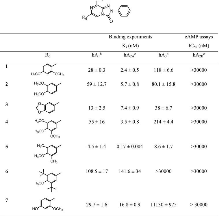
Table 1. Biological activity of compounds 1-11 at hARs.a
Binding experiments Ki (nM) cAMP assays IC50 (nM) R6 hA1b hA2Ac hA3d hA2Be 1 H3CO OCH3 28 ± 0.3 2.4 ± 0.5 118 ± 6.6 >30000 2 H3CO H3CO 59 ± 12.7 5.7 ± 0.8 80.1 ± 15.8 >30000 3 O O 13 ± 2.5 7.4 ± 0.9 38 ± 6.7 >30000 4 H3CO OCH3 H3CO 55 ± 16 3.5 ± 0.8 214 ± 4.4 >30000 5 H3CO CH3 H3C 4.5 ± 1.4 0.17 ± 0.004 8.6 ± 1.7 >30000 6 H3CO 108.5 ± 17 141.6 ± 34 >30000 >30000 7 HO OCH3 29.7 ± 1.6 16.8 ± 0.9 11130 ± 975 > 30000 N N N N NH2 O R6 5 6 7 8 9 10 11 12 13 14 15 16 17 18 19 20 21 22 23 24 25 26 27 28 29 30 31 32 33 34 35 36 37 38 39 40 41 42 43 44 45 46 47 48 49 50 51 52 53 54 55 56 57 58 59 60
aData (n= 3-5) are expressed as means ± standard errors. bDisplacement of specific [3H]-CCPA binding
at hA1 AR expressed in CHO cells. cDisplacement of specific [3H]-NECA binding at hA2A AR expressed
in CHO cells. dDisplacement of specific [3H]-HEMADO binding at hA
3 AR expressed in CHO cells. eIC
50 values of the inhibition of NECA-stimulated adenylyl cyclase activity in CHO cells expressing
hA2B AR.
Derivatives 1-6, including the methoxy synthetic intermediates and the 6-(3,4-methylenedioxyphenyl) derivative 3, on the whole, showed high affinities for the hA2A AR,
spanning one-digit nanomolar values, and also for the hA1 subtype. The most active compound at
the hA2A AR was derivative 5 (Ki= 0.17 nM), featuring the 6-(3,5-dimethyl-4-methoxyphenyl)
substitution, while its 6-(3,5-di-tert-butyl-4-methoxyphenyl) analogue 6 showed significantly lower hA2A AR binding activity (Ki= 141.6 nM), probably due to the steric bulk of the two tert-butyl
groups. Compounds 5 and 3 also possess significant affinity for the hA3 subtype.
In the second set of triazolopyrazines (12-21), -lipoic acid and 4-hydroxy-3,5-ditertbutylbenzoic acid were selected as antioxidant portions and linked by different chains at the para position of the 6-phenyl ring. The choice of this position was based on the results of previous molecular docking
8 HO HO 42.6 ± 9.6 5.2 ± 0.5 950 ± 200 >30000 9 HO HO OH 175.5 ± 3 94.5 ± 21 5575 ± 989 17330 ± 3365 10 HO CH3 H3C 21.3 ± 7 2.5 ± 0.8 100 ± 0.7 >30000 11 HO >30000 8.5 ± 1.4 >30000 >30000 5 6 7 8 9 10 11 12 13 14 15 16 17 18 19 20 21 22 23 24 25 26 27 28 29 30 31 32 33 34 35 36 37 38 39 40 41 42 43 44 45 46 47 48 49 50 51 52 53 54 55 56 57 58 59 60
studies of this class of compounds at the hA2A AR, highlighting that the presence of hindering
substituents on the 6-phenyl ring favored a binding pose with this moiety pointing towards the extracellular side of the receptor. Hence, we envisaged that long substituents at the para position could be well tolerated because they could point away from the binding pocket. The selected chains were linked through an ethereal (compounds 15, 16) or an amide function (compounds 17, 20 and
21).
Table 2. Biological activity of derivatives 12-21 and the reference compounds 49 and 50, at hARs.a
Binding experiments Ki (nM) cAMP assays IC50 (nM) R hA1b hA2Ac hA3d hA2Be 12 N O > 30000 8.2 ± 2.3 > 30000 > 30000 13 H2N O O 391.7 ± 104 26 ± 1.7 604 ± 94 > 30000 14 H2N O 288.7 ± 54 14.9 ± 0.1 2131 ± 173.5 > 30000 15 O N H O S S 378.6 ± 91 2.4 ± 0.3 4097 ± 812 >30000 16 HO O N H O 13670 ± 275 14750 ± 270 >30000 >30000 N N N N NH2 O R 5 6 7 8 9 10 11 12 13 14 15 16 17 18 19 20 21 22 23 24 25 26 27 28 29 30 31 32 33 34 35 36 37 38 39 40 41 42 43 44 45 46 47 48 49 50 51 52 53 54 55 56 57 58 59 60
aData (n= 3-5) are expressed as means ± standard errors. bDisplacement of specific [3H]-CCPA binding
at hA1 AR expressed in CHO cells. cDisplacement of specific [3H]-NECA binding at hA2A AR expressed
in CHO cells. dDisplacement of specific [3H]-HEMADO binding at hA
3 AR expressed in CHO cells. eIC
50 values of the inhibition of NECA-stimulated adenylyl cyclase activity in CHO cells expressing
hA2B AR. fRef. 27. gRef. 33.
The binding data (Table 2) proved us right. On the whole, all the substituents appended on the 4-hydroxy- and 4-amino group of derivatives 49 and 50, respectively, increased affinity and /or selectivity for the hA2A AR (compare derivatives 12-16 to 49 and compounds 17-21 to 50), with the
only exception being compound 16, which showed a dropped hA2A AR binding activity.
The lipoyl derivatives 15, 17 and 20 resulted in high affinity hA2A AR ligands (Ki= 2.4-36.4 nM)
with different degrees of selectivity versus the hA1 AR, depending on the nature of the linker.
17 8.4 ± 0.4 5 ± 0.6 >30000 >30000 18 H2C O NH 262.7 ± 1.9 1.8 ± 0.09 >30000 >30000 19 O NH H2N 479.2 ± 89 0.59 ± 0.2 509 ± 90 9658 ± 1431 20 N H O S S O NH 1359 ± 284 36.4 ± 8.2 >30000 >30000 21 HO O N H O NH >30000 54.5 ± 7.1 >30000 >30000 49f OH 45 ± 10 45 ± 12 53± 13 >30000 50g NH 2 33.5 ± 6.7 22.9 ± 0.2 253.7 ± 67.6 >30000 O S S NH 5 6 7 8 9 10 11 12 13 14 15 16 17 18 19 20 21 22 23 24 25 26 27 28 29 30 31 32 33 34 35 36 37 38 39 40 41 42 43 44 45 46 47 48 49 50 51 52 53 54 55 56 57 58 59 60
Compound 17, bearing the lipoyl residue directly attached on the para-amino group, showed a high affinity not only for the hA2A receptor but also for the hA1 AR subtype. Very interesting results
were obtained when the lipoyl moiety was spaced from the para-position through the flexible oxyethylamino chain (-O-(CH2)2-NH-). In fact, the resulting compound 15 possessed a very high
affinity for the targeted hA2A receptor (Ki= 2.4 nM) and also high selectivity, being significantly
less active at the hA1 AR (Ki= 378.6 nM). When the oxyethylamino spacer was replaced by the
longer and more rigid carboxyamidoethylamino spacer (-NH-CO-(CH2)2-NH-), a selective ligand
for the hA2A AR was still obtained (compound 20) but its affinity and selectivity were lower, with
respect to those of 15. The same two spacers were employed to link the (4-hydroxy-3,5-di-tert-butyl)benzoyl pendant to the 6-phenyl ring (derivative 16 and 21) but in this case an opposite effect was obtained since the best results, both in terms of A2A AR affinity and selectivity, were obtained
with the carboxyamidoethylamino spacer. In fact, compound 21 showed good affinity for the hA2A
AR (Ki= 54.5 nM) and high selectivity, while it analogue 16 was almost inactive at all ARs. The
significant difference between hA2A affinities of the two compounds obviously depends on the
different spacer. The bit longer carboxyamidoethylamino spacer seems to direct the terminal bulky aryl ring in a more favorable pose in the receptor binding site (see modeling analysis). Also derivatives 12-14 and 18, 19, which were not our primary target compounds, resulted in interesting ligands, showing nanomolar affinity and good to high selectivity for the hA2A AR. In particular,
derivative 19, bearing the carboxyamido-ethylamine substituent at the para position of the 6-phenyl ring, was the most active (Ki= 0.59 nM). Derivatives 12 and 18, featuring at the para-position a
cyanomethoxy (Ki= 8.2 nM) and an acrilamido group (Ki= 1.8 nM), respectively, displayed high
hA2A AR affinities and selectivity. Reduction of the cyano residue of compound 12 afforded the
amino derivative 14 which maintained the ability to target the hA2A AR with nanomolar affinity
(Ki= 14.9 nM) but lower selectivity. Transformation of the cyano in amide group (compound 13)
also retained affinity but reduced selectivity for the target hA2A receptor.
5 6 7 8 9 10 11 12 13 14 15 16 17 18 19 20 21 22 23 24 25 26 27 28 29 30 31 32 33 34 35 36 37 38 39 40 41 42 43 44 45 46 47 48 49 50 51 52 53 54 55 56 57 58 59 60
Finally, compounds 11, 12, 15, 20 and 21, showing high hA2A AR affinity and selectivity, were
selected to be further pharmacologically profiled in in vitro studies. Previously, we ascertained their antagonistic profile by evaluating their effect on cAMP production in CHO cells, stably expressing hA2A ARs (Table 3). The compounds proved to be able to counteract NECA-stimulated cAMP
production, thus behaving as hA2A AR antagonists.
Table 3. Potencies of the selected triazolopyrazines 11, 12, 15, 20 and 21 at hA2A AR.
aIC
50 values of the inhibition of NECA-stimulated adenylyl cyclase activity in CHO cells expressing
hA2A AR. Data are expressed as means ± standard errors.
Molecular modeling studies. The binding mode of the synthesized compounds at the hA2A AR
cavity was simulated with docking analysis by using the MOE (Molecular Operating Environment 2014.09) software and the CCDC Gold docking tool.50,51 The MOE software analysis was carried
out by selecting the induced fit docking and optimization protocol (schematically, a preliminary docking analysis providing a set of ligand conformations then energy minimized with the side chains of the receptor residues in proximity). For the docking tasks, a crystal structure of the hA2A
AR in complex with the antagonist/inverse agonist ZM241385 was employed (http://www.rcsb.org; pdb code: 5NM4; 1.7-Å resolution52). For a subset of compounds, the binding modes at the hA
1 AR
crystal structure (pdb code: 5N2S; 3.3-Å resolution53) were also simulated with the same tools and
protocols. hA2A AR (IC50 nM)a 11 179 ± 53 12 157 ± 43 15 116 ± 31 20 296 ± 66 21 263 ± 58 5 6 7 8 9 10 11 12 13 14 15 16 17 18 19 20 21 22 23 24 25 26 27 28 29 30 31 32 33 34 35 36 37 38 39 40 41 42 43 44 45 46 47 48 49 50 51 52 53 54 55 56 57 58 59 60
The docking conformations generally obtained for the new derivatives at the hA2A AR are similar to
those observed for our previously reported triazolopyrazines and is shown in Figure 4A.27 In this
binding mode, the bicyclic core is positioned between the side chains of Phe168 (EL2) and Leu2496.51 and engages non-polar interactions with these residues. The exocyclic amine group
makes H-bond contacts with Asn2536.55 and Glu169 (EL2), while the 2-phenyl substituent is
located in the depth of the cavity. The R6 group is positioned at the entrance of the binding site and
oriented toward the extracellular environment. Such compound orientation and interaction resemble the binding mode of the co-crystallized 4-(2-[7-amino-2-(2-furyl[1,2,4]-triazolo[2,3-a][1,3,5]triazin-5ylamino]ethyl)phenol (ZM241385) in the employed crystal structure52 but also in
other previously reported hA2A AR X-ray structures.54,55
The presence of substituents on the 6-phenyl ring modulates the interaction with the receptor residues at the entrance of the cavity and leads to various degrees of affinity for the hA2A AR (see
Tables 1 and 2). For previously reported analogues,27 it was observed that a non-polar
para-substituent on this ring was more efficient to improve the hA2A AR affinity than a polar one.
Considering the meta-substituents of the 6-phenyl ring, the affinity data showed that the hA2A AR
affinity was not significantly influenced by the chemical-physical profile of the substituent. Compounds of this new set of triazolopyrazines, differing in polarity of para- or meta-substituent, present various hA2A AR affinity. Considering derivatives bearing a small 4-substituent on the
6-phenyl moiety, again a non-polar para-substituent on this ring appears more efficient to improve the hA2A AR affinity than a polar one. As an example, compounds 5, the most active of the herein
reported derivatives, featuring a 4-methoxy and 3,5-di-methyl groups on the 6-phenyl ring, is endowed with 15-fold higher affinity at the hA2A AR than 10, which bears a 4-hydroxy and
3,5-di-methyl groups. 5 6 7 8 9 10 11 12 13 14 15 16 17 18 19 20 21 22 23 24 25 26 27 28 29 30 31 32 33 34 35 36 37 38 39 40 41 42 43 44 45 46 47 48 49 50 51 52 53 54 55 56 57 58 59 60
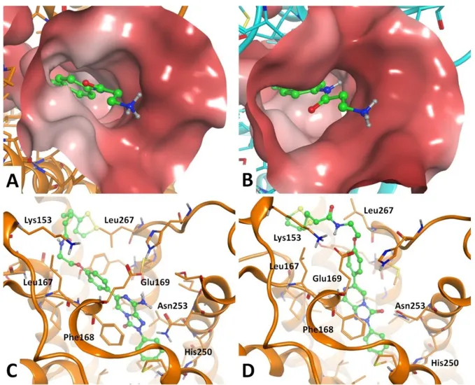
Figure 4. (A) General binding mode of the synthesized compounds at the hA2A AR (pdb: 5NM4)
binding cavity, with indication of some key receptor residues; compound 5 is showed. (B) Top-view of the hA2A AR residues at the entrance of the binding cavity and potentially giving interaction with
substituents on the 6-phenyl ring. (C) Molecular surface representation of the entrance of the hA2A
AR binding cavity; dark-to-light color indicates hydrophilic-to-hydrophobic scale.
The substituents inserted on the 6-phenyl ring are located in proximity of Ile662.64, Ser672.65,
Leu167 (EL2), Leu267 (EL3), Met2707.35 and Tyr2717.36 (Figure 4B-C). Considering the volume,
Figure 4C shows a molecular surface representation of the entrance of the hA2A AR binding cavity.
From this figure it can be seen that small substituents are allowed in the ortho- and meta-position of the R6 group, but the space is too limited to allow the insertion of two or more bulky moieties at the
same positions. In fact, compound 6, bearing two tert-butyl groups at the meta-position of the 6-phenyl ring, has a significantly reduced hA2A AR affinity compared to the other analogues (Table
1). The non-polar profile of the above cited amino acids surrounding the R6 group allows a
5 6 7 8 9 10 11 12 13 14 15 16 17 18 19 20 21 22 23 24 25 26 27 28 29 30 31 32 33 34 35 36 37 38 39 40 41 42 43 44 45 46 47 48 49 50 51 52 53 54 55 56 57 58 59 60
favorable interaction with non-polar substituents rather than polar ones. Combinations of para- and meta-substituents or para- and ortho-substituents lead to slight modulation of hA2A AR affinity.
Figure 5. (A) General binding mode of the synthesized compounds at the hA1 AR (pdb: 5N2S)
binding cavity, with indication of some key receptor residues; compound 19 is showed. (B) Top-view of the hA1 AR residues at the entrance of the binding cavity and potentially giving interaction
with substituents on the R6 aryl ring.
Docking results obtained at the hA1 AR crystal structure are highly similar to the ones obtained at
the hA2A AR (Figure 5). Considering compounds bearing small substituents at the meta- and
para-position of the 6-phenyl group, docking conformations suggest analogue considerations as above for the impact on the hA1 AR affinity given by these substituents. This is in agreement with
biological evaluation results of compounds 1-5,7-10, which show similar trends of binding affinity
5 6 7 8 9 10 11 12 13 14 15 16 17 18 19 20 21 22 23 24 25 26 27 28 29 30 31 32 33 34 35 36 37 38 39 40 41 42 43 44 45 46 47 48 49 50 51 52 53 54 55 56 57 58 59 60
values at hA1 AR and hA2A AR. As for the hA2A AR, compound 5 is the most active of the whole set
of derivatives at the hA1 AR, with 4-fold higher affinity than 10, its para-hydroxy substituted
analogue. Even in this case, compounds bearing tert-butyl groups are endowed with lower affinity. Still considering compounds bearing small substituents at the meta- and para-position of the R6
group, affinities at the hA2A AR are generally higher than those observed at the hA1 AR. The set of
hA2A AR residues in proximity with the para-substituent is globally more hydrophobic than the hA1
AR one, due to the presence of Leu167 (EL2), Leu267 (EL3), Met2707.35 in the hA
2A AR instead of
the Glu170 (EL2), Ser267 (EL3) and Thr2717.35 residues in the respective positions of the hA 1 AR.
This factor could play a key role in providing a slight hA2A AR selectivity (versus the hA1 AR) for
the compounds described above.
Considering the compounds presenting only small para-substituents on the 6-phenyl ring (12-14,
18, 19), the hA2A AR affinities appear significantly higher than the hA1 AR ones. The different
affinity for the two AR subtypes could be due to both the chemical-physical profile of the AR residues in proximity of the substituent and how much the substituent itself is exposed to the external environment. Figure 6A-B shows a surface representation of the entrance of the binding cavities, in proximity to the para-position of the 6-phenyl ring. The light-to-dark regions indicate a hydrophobic-to-hydrophilic profile of the residues. The more polar profile of the hA1 AR residues
with respect to the hA2A AR ones is due in particular to the presence of a negatively charged
glutamate residue (Glu170, Figure 5B) in hA1 AR, while the hA2A AR bears a non-polar leucine
(Leu167, Figure 4B) in the same position. The presence of Glu170 in hA1 AR leads to a repulsive
effect between this residue and the carbonyl group of the compounds bearing a carboxyamidoethyl spacer (19, 20, 21). This effect is evident from the comparison of the activities of the latter compounds with the higher affinity data at the hA1 AR of the corresponding analogues bearing an
oxyethyl spacer (14, 15, 16, respectively). At the hA2A AR, both the carboxyamidoethyl and
oxyethyl spacers generally lead to nanomolar affinities.
5 6 7 8 9 10 11 12 13 14 15 16 17 18 19 20 21 22 23 24 25 26 27 28 29 30 31 32 33 34 35 36 37 38 39 40 41 42 43 44 45 46 47 48 49 50 51 52 53 54 55 56 57 58 59 60
Figure 6. A-B Top-view of the binding mode of compound 19 at the hA2A AR (A, pdb: 5NM4) and
hA1 AR (B pdb: 5N2S) binding sites. Molecular surface representations of both binding cavities are
represented. Light-to-dark colors of surface correspond to hydrophobic-to-hydrophilic regions.
C-D. Binding modes suggested for compounds bearing large para-substituents in the 6-phenyl ring;
compound 15 at the hA2A AR (pdb: 5NM4) is shown. These molecules may adopt the general
binding mode above described (C) or an alternative, energetically more stable, docking conformation that makes the para-substituent externally oriented without clashes with receptor residues (D).
Docking studies performed for the compounds presenting small para-substituents (12-14, 18, 19) show that these molecules may adopt a binding mode similar to the one described above. The
5 6 7 8 9 10 11 12 13 14 15 16 17 18 19 20 21 22 23 24 25 26 27 28 29 30 31 32 33 34 35 36 37 38 39 40 41 42 43 44 45 46 47 48 49 50 51 52 53 54 55 56 57 58 59 60
interaction of the para-substituent with hA2A AR residues is modulated by the chemical-physical
profile of the substituent itself and the receptor residues in proximity. The positively charged amine function of compound 19 gets located in proximity of both the hydroxyl group of Tyr2717.36 and the
carbonyl group of Ser672.65 (Figures 4B and 6A), thus providing subnanomolar affinity for the hA 2A
AR. Compounds bearing a large para-substituent (15-17, 20-21) may adopt as well as the above-described binding mode (Figure 6C, compound 15), even if the large para-substituent gets located too close to the receptor residues, giving a steric clash with the protein atoms. Docking results for these compounds suggest also an alternative binding mode, with the bicyclic scaffold upside-down oriented to point the 3-carbonyl group toward the amine function of Asn2536.55 (Figure 6D,
compound 15). This binding mode lacks some hydrophilic interactions with the receptor that are observed in the general compound orientation described above (i.e. with Glu169 at hA2A AR); on
the other hand, the alternative binding mode favors the pointing of the para-substituent toward the external environment with a more energetically stable compound conformation. This may explain the favorable docking scores obtained by the alternative binding mode arrangement for compounds bearing large para-substituents in the 6-phenyl ring. Considering the largest compounds (15, 16, 20,
21), the lowest activity belongs to derivative 16, which is the one presenting a para-substituent of
large volume and the shortest oxyethylamino spacer linking the 6-phenyl ring. In contrast, its corresponding analogue with the longer carboxyamidoethyl spacer (21) presents a 300-fold improvement of the hA2A AR affinity. On the other hand, both derivatives presenting a less bulky
lipoyl group (15 and 20) are endowed with significantly higher affinity. This suggests that the length and volume of the large para-substituents appear critical for the receptor affinity, for an energetically stable accommodation of the substituent itself within the receptor residues.
Neuroprotection Studies on oxaliplatin-induced neurotoxicity in microglia cells. Based on the affinity data, compounds 11, 12, 15, 20 and 21, potent and selective hA2A AR antagonists, were
5 6 7 8 9 10 11 12 13 14 15 16 17 18 19 20 21 22 23 24 25 26 27 28 29 30 31 32 33 34 35 36 37 38 39 40 41 42 43 44 45 46 47 48 49 50 51 52 53 54 55 56 57 58 59 60
selected for further pharmacological evaluation. In particular, their protective effect against the neurotoxicity of the anticancer drug oxaliplatin on rat microglia cells was determined. Neuropathy induced by oxaliplatin is a common side effect in patients treated with this drug and consists in paresthesia, dysesthesia, and pain. Such a condition adversely affects quality of life and can lead to discontinuation of therapy.56 It is well-known that glia cells play a key role in the CNS homeostasis
and are strongly involved in the responses to nerve injury. Microglia and astrocytes activate several mechanisms, such as production of trophic factors, regulation of transmitter and ion concentrations, which tend to decrease neuronal injury. Nevertheless, in pathological conditions, the maladaptive plasticity of glial cells strongly sustains negative symptoms like chronic pain. In particular, microglia functional modifications have the potential to induce neuronal dysfunction, playing a pivotal role in oxaliplatin neuropathy development.57,58
The selected compounds were chosen taking into account their high affinity and selectivity toward the hA2AAR but also the presence in all of them, except compound 12, of antioxidant moieties
which were thought to counteract oxaliplatin neurotoxicity. In fact, although the molecular basis underlying the oxaliplatin neuropathy is unclear, some experimental evidence pointed out a correlation between oxidative stress damage and neuropathic pain onset,20 also highlighting efficacy
of the antioxidant silibinin and α-tocopherol in reducing oxaliplatin-dependent pain induced by mechanical and thermal stimuli.59 Compound 12, lacking the antioxidant portion, was tested to
evaluate how the lone blockade of the A2A AR could affect oxaliplatin toxicity.
Primary rat microglia cells were treated with oxaliplatin in the absence or in the presence of the tested compounds. Oxaliplatin damage was evaluated as cell viability and oxidative stress, the latter previously described as the main damage evoked by oxaliplatin.60 The new synthesized compounds
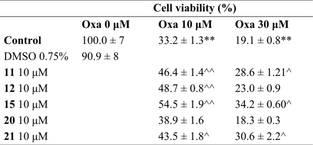
were tested at 10 μM, the maximum soluble concentration. Oxaliplatin, concentration-dependently, strongly reduced microglia viability (MTT test) after 24 h incubation (33% and 19% viability with 10 and 30 μM, respectively, in comparison to 100% of control condition).
5 6 7 8 9 10 11 12 13 14 15 16 17 18 19 20 21 22 23 24 25 26 27 28 29 30 31 32 33 34 35 36 37 38 39 40 41 42 43 44 45 46 47 48 49 50 51 52 53 54 55 56 57 58 59 60
The obtained results showed that the lipoic acid-conjugated derivative 15 was the most active in preventing the oxaliplatin damage, also when incubated at the high oxaliplatin concentration (30 M). Compound 12 was instead effective against 10 μM oxaliplatin. Regarding the other tested compounds, the 6-phenol derivatives 11 and 21 showed partial activity at both oxaliplatin concentrations, whereas the lipoic derivative 20, contrary to our expectations, turned out to be ineffective (Table 4). This latter result might be due to a possible instability of 20 in the microglia assay medium, as reported in the “Chemical stability study” section.
Table 4. Compound effects on microglial cell viabilitya
a Primary rat microglia cells were plated 4000 cells/well and 24 hours later cells were treated with
oxaliplatin (Oxa) 10 and 30 μM in presence of 11, 12, 15, 20 and 21 at 10 μM for 24 hours. Cell vitality was assessed via MTT assay. Viability is expressed as % in comparison to the control cells (arbitrarily set 100% of viable cells). Data are presented as mean ± SEM of three experiments. *P<0.05 and **P<0.01 versus control; ^P<0.05 and ^^P<0.01 versus oxaliplatin.
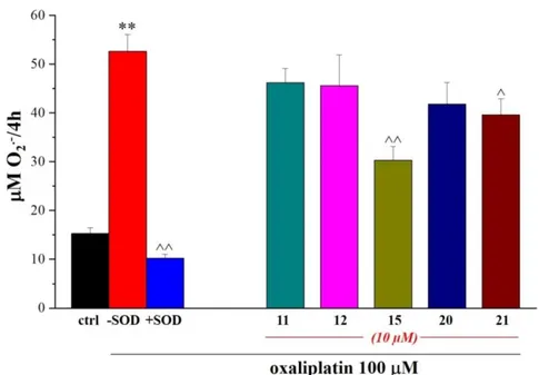
Further investigations were carried out on these compounds by evaluating their ability to prevent the oxaliplatin-dependent increase of the SOD-inhibitable superoxide anion (cytochrome C assay). According to the obtained data, compounds 15 and 21 proved to be effective in significantly
Cell viability (%)
Oxa 0 μM Oxa 10 μM Oxa 30 μM Control 100.0 ± 7 33.2 ± 1.3** 19.1 ± 0.8** DMSO 0.75% 90.9 ± 8 11 10 μM 46.4 ± 1.4^^ 28.6 ± 1.21^ 12 10 μM 48.7 ± 0.8^^ 23.0 ± 0.9 15 10 μM 54.5 ± 1.9^^ 34.2 ± 0.60^ 20 10 μM 38.9 ± 1.6 18.3 ± 0.3 21 10 μM 43.5 ± 1.8^ 30.6 ± 2.2^ 5 6 7 8 9 10 11 12 13 14 15 16 17 18 19 20 21 22 23 24 25 26 27 28 29 30 31 32 33 34 35 36 37 38 39 40 41 42 43 44 45 46 47 48 49 50 51 52 53 54 55 56 57 58 59 60
decreasing the oxygen free radical level thus suggesting a direct antioxidant activity or, hypothetically, a protective property against mitochondrion (Figure 7).
Figure 7. Compound effects on SOD-inhibitable O2.− concentrations in rat microglia cells.
Microglia cells (5×10⁵ cells/well) were exposed to 100 μM oxaliplatin for 4h in the absence or
presence of tested compounds (10 μM). O2.− concentration was evaluated by cytochrome c assay.
The nonspecific absorbance was measured in the presence of SOD (300 mU/ml) and subtracted from the total value. Values are expressed as the mean ± SEM of three experiments. *P<0.05 and **P<0.01 versus control; ^P<0.05 and ^^P<0.01 versus oxaliplatin.
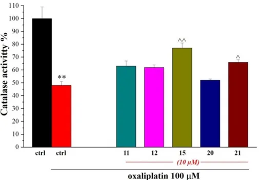
The activity of the detoxifying enzyme catalase was also measured to study the potential effect of new compounds on peroxisomes, the other intracellular organelle involved in the redox balance. As shown in Figure 8, oxaliplatin impaired peroxisome functionality, reducing catalase activity while 15 and 21 significantly prevented the damage.
5 6 7 8 9 10 11 12 13 14 15 16 17 18 19 20 21 22 23 24 25 26 27 28 29 30 31 32 33 34 35 36 37 38 39 40 41 42 43 44 45 46 47 48 49 50 51 52 53 54 55 56 57 58 59 60
Figure 8. Compound effects on catalase activity. Microglia cells (5·105 cells/well) were treated
with oxaliplatin (10 µM) in the absence or in the presence of the new compounds (10 µM). Activity was measured after 24h incubations. Values are expressed as the mean ± S.E.M. of three experiments. Control condition was arbitrarily set as 100%. *P<0.05 and **P<0.01 versus control; ^P<0.05 and ^^P<0.01 versus oxaliplatin.
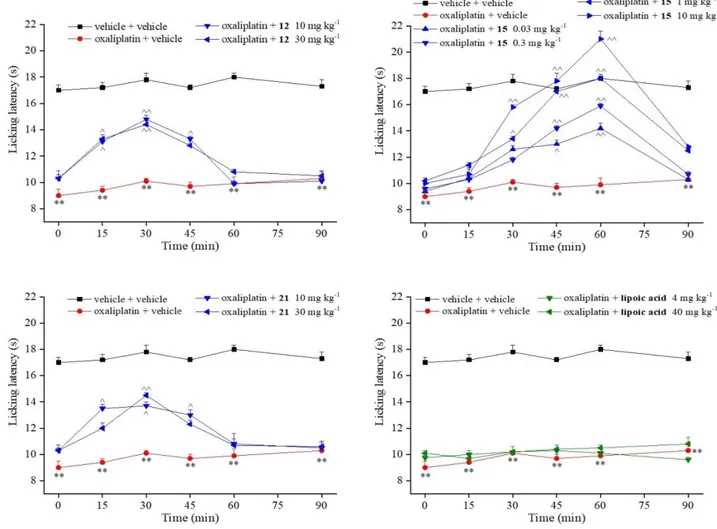
Behavioral studies in the oxaliplatin-induced neuropathy model. On the basis of the results
obtained from in vitro tests, we selected compounds 12, 15 and 21 for an in vivo study in a mouse model of oxaliplatin-induced neuropathy. On day 14, after a repeated treatment with the anticancer drug injected at a clinically-relevant dose,61 the hypersensivity to a cold non noxious stimulus (cold
plate test) was significantly established (Figure 9). The pain-relieving effects of new synthesized compounds were tested after a single per os administration. Compounds 12 and 21 (10 and 30 mg kg-1) were able to increase the pain threshold between 15 and 45 min after treatment. Interestingly,
compound 15 induced significant relieving effects starting from the 0.03 mg kg-1 dose. The
efficacy, dose-dependently, increased till it completely reverted oxaliplatin-induced neuropathic
5 6 7 8 9 10 11 12 13 14 15 16 17 18 19 20 21 22 23 24 25 26 27 28 29 30 31 32 33 34 35 36 37 38 39 40 41 42 43 44 45 46 47 48 49 50 51 52 53 54 55 56 57 58 59 60
pain when administered at 1 and 10 mg kg-1. The effect of 15 began 30 min after treatment, and the
compound was still fully active at 60 min. In the same model, lipoic acid, administered per os at an equimolar dose (4 mg kg-1) to 10 mg kg-1 of compound 15, was completely inactive. Also when
tested at 10-fold higher dose (40 mg kg-1), lipoic acid was ineffective (Figure 9).
Figure 9. Compound effects against neuropathic pain. Mice were repeatedly treated with
oxaliplatin (2.4 mg kg-1; dissolved in 5% glucose solution and i.p. administered). On day 14,
compounds were suspended in carboxymethylcellulose and administered p.o. Pain-related behavior (i.e. lifting and licking of the hind paw) was observed and the time (in seconds) of the first sign was recorded. **P<0.01 vs vehicle + vehicle treated animals; ^P<0.05 and ^^P<0.01 vs oxaliplatin + vehicle treated animals. Each value represents the mean of 10 mice performing in two different experimental sets. 5 6 7 8 9 10 11 12 13 14 15 16 17 18 19 20 21 22 23 24 25 26 27 28 29 30 31 32 33 34 35 36 37 38 39 40 41 42 43 44 45 46 47 48 49 50 51 52 53 54 55 56 57 58 59 60
The significant difference between the potency of the lipoic-conjugated triazolopyrazine 15 and lipoic acid could be due both to diverse pharmacokinetic properties of the two compounds and to the presence of the hA2A AR antagonist component in derivative 15. This hypothesis is supported
by data obtained in different animal model of neuroprotection37,59,60 indicating that lipoic acid is
well absorbed per os but is subject to pre-systemic elimination by the liver, and in rat only about 27-34% lipoic acid administered orally is available for absorption by the tissue.59 Moreover, the
complexity of neuropathic pain signaling does not allow consideration of the redox imbalance as the unique pathological signature.54 The importance of the hA
2A AR antagonist component in reducing
oxaliplatin-induced neuropathy is underlined by the pain-relieving effect of compound 12, lacking the antioxidant portion. It is worth noting that the lipoic-conjugated compound 15, showing the best activity in the microglia assays, was the most active also in this in vivo model. Hence, we hypothesized that these findings might be ascribed, at least in part, to the higher affinity of compound 15 for the A2A AR in rodents, with respect to those of 12 and 21, as could be envisaged
on the basis of the A2A AR affinities obtained for the human species (Table 2). To confirm our
prediction, binding studies at the rat (r) A2A AR were carried out on the three derivatives. The
achieved results (Table 5) confirmed the expected trend, because the lipoic derivative 15 displayed the highest binding value, being about 4-fold and 10-fold more active than compounds 12 and 21, respectively.
Table 5. Binding activity of compounds 12, 15, 21, and ZM241385 as reference ligand, at rA2A
AR.a rA2A Ki nMb 12 39 ± 5.6 15 9 ± 1.7 21 86 ± 9.1 ZM241385 2.8 ± 0.3 5 6 7 8 9 10 11 12 13 14 15 16 17 18 19 20 21 22 23 24 25 26 27 28 29 30 31 32 33 34 35 36 37 38 39 40 41 42 43 44 45 46 47 48 49 50 51 52 53 54 55 56 57 58 59 60
aData (n= 3-5) are expressed as means ± standard errors. bDisplacement of specific [3H]-NECA
binding at rA2A AR expressed in CHO cells.
Obviously, besides the different rA2A AR affinity, other molecular features, such as the antioxidant
character and the pharmacokinetic properties, can be responsible of the diverse in vivo activity of these triazolopyrazines. The higher potency of derivative 15, compared to 21, suggests that the lipoic acid residue, with respect to the BHT-analogue group, might confer enhanced in vivo properties, for both its antioxidant activity and its positive effect on pharmacokinetics. To address this issue further studies have been planned to gain more insight into the interesting protective profile of compound 15.
Chemical stability study. Compounds 11, 12, 15, 20 and 21, selected for pharmacological
evaluation, feature antioxidant moieties, amide functions, or a cyano group. All these functionalities might have some lability, hence we thought it interesting to ascertain their stability towards spontaneous or enzymatic degradation in 50 mM tris(hydroxymethyl)aminomethane hydrochloride buffer solution (50 mM Tris buffer, pH= 7.4) and human plasma, respectively.
The instrumental conditions are reported in the Experimental Procedure section.
The solution stability of each studied compound was verified by monitoring the variation of its concentration at different incubation times in 50 mM Tris buffer and human plasma samples. By plotting these data (natural logarithm of analyte concentration versus the incubation time) the respective degradation profiles were obtained (Figures 10 and 11 and Figures 1S-4S in Supporting Information) which demonstrated that all the compounds were stable in 50 mM Tris buffer and most of them also in human plasma. In fact, only the degradation plots of the 6-(3-ter-butyl-4-hydroxyphenyl) derivative 11 and the lipoyl derivative 20 (Figures 10 and 11) in human plasma
5 6 7 8 9 10 11 12 13 14 15 16 17 18 19 20 21 22 23 24 25 26 27 28 29 30 31 32 33 34 35 36 37 38 39 40 41 42 43 44 45 46 47 48 49 50 51 52 53 54 55 56 57 58 59 60
showed a significant decay rate (k value, defined in Supporting Information) and their calculated half-life values (t1/2) are 122 ± 4 min and 41 ± 13 min respectively, as displayed in Table 4.
-4 -3.5 -3 -2.5 -2 -1.5 -1 -0.5 0 0.5 1 -5 0 5 10 15 20 25 30 35 40 45 50 55 60 65 70 75 80 85 90 95 100 105 110 115 120 125 Time (min.) ln (c o n c. )
Initial Concentration TRIS H-pl
Figure 10. Degradation plots of compound 11 in 50 mM Tris buffer solution (blue square) and
human plasma (red triangle).
5 6 7 8 9 10 11 12 13 14 15 16 17 18 19 20 21 22 23 24 25 26 27 28 29 30 31 32 33 34 35 36 37 38 39 40 41 42 43 44 45 46 47 48 49 50 51 52 53 54 55 56 57 58 59 60
-4 -3.5 -3 -2.5 -2 -1.5 -1 -0.5 0 0.5 1 -5 0 5 10 15 20 25 30 35 40 45 50 55 60 65 70 75 80 85 90 95 100 105 110 115 120 125 Time (min.) ln (c o n c. )
Initial Concentration TRIS H-pl
Figure 11. Degradation plots of compound 20 in 50 mM Tris buffer solution (blue square) and
human plasma (red triangle).
The half-life value of ketoprofene ethylester (KEE), used as reference compound, demonstrated that the employed human batch was enzymatically active (half-life < 2 h).64 The k values of the stable
compounds were close to 0; consequently for these derivatives, extremely high t1/2 values can be
calculated. Since under the proposed experimental conditions a half-life over 240 min is not correctly evaluated, it is reasonable to consider that their half-life values could be equal to or greater than 240 min. The 50 mM Tris buffer and human plasma half-lifes of other studied compounds are reported in Table 5.
Table 5. Half-life (t1/2) of studied compounds in 50 nM Tris buffer and human plasma samples.
50 mM Tris buffer t1/2 ± error (min) Human-plasma t1/2 ± error (min) 5 6 7 8 9 10 11 12 13 14 15 16 17 18 19 20 21 22 23 24 25 26 27 28 29 30 31 32 33 34 35 36 37 38 39 40 41 42 43 44 45 46 47 48 49 50 51 52 53 54 55 56 57 58 59 60
anot determined
To summarize, these experiments demonstrated that the tested compounds did not suffer significant degradation process under the proposed conditions. Only derivatives 11 and 20 showed a clear degradation rate in human plasma, but with large different of t1/2 values (122 ± 4 and 41 ± 13 respectively). The behavior of 20 might suggest a possible explanation of its inactivity on microglia cell viability test, where all the other compounds proved to be effective. Hence, inactivity of 20 might be partly ascribed to its possible decomposition in the microglia assay medium.
CONCLUSION
This study has produced new highly potent and selective antagonists for the hA2A AR, some of
which possess hindering antioxidant functions. Insertion of these functions on the 6-aryl group, notwithstanding their high steric bulk, maintained a nanomolar hA2A AR affinity and high
selectivity. Molecular docking studies highlighted that the 6-aryl moiety of these new triazolopyrazines is positioned at the entrance of the hA2A AR binding site and that both
lipophilicity and the volume of the substituent inserted on this ring modulate affinity and selectivity. On the whole, non-polar para-substituents are more efficient than polar groups in improving hA2A
receptor-ligand interaction due to the presence of non-polar amino acid residues surrounding the 6-aryl pendant. Further pharmacological studies were carried out on selected triazolopyrazines showing high hA2A affinity and selectivity. Compounds 11, 15 and 21, featuring antioxidant
KEE nda 107 ± 16 11 ≥240 122 ± 4 12 ≥240 ≥240 15 ≥240 ≥240 20 ≥240 41 ± 13 21 ≥240 ≥240 5 6 7 8 9 10 11 12 13 14 15 16 17 18 19 20 21 22 23 24 25 26 27 28 29 30 31 32 33 34 35 36 37 38 39 40 41 42 43 44 45 46 47 48 49 50 51 52 53 54 55 56 57 58 59 60
moieties, and compound 12, lacking the antioxidant functionality, reduced oxaliplatin–induced toxicity in microglia cells, the most active being the lipoyl derivative 15. This compounds and, to a lesser extent, the BHT analogue 21 proved to be effective in reducing the oxygen free radical level, thus suggesting a direct antioxidant activity. Derivatives 12, 15 and 21, further investigated in vivo, were able to reduce oxaliplatin-induced neuropathy in mouse. Also in these tests, the lipoyl-derivative 15 displayed the best results, being able to completely revert oxaliplatin-induced pain when administered at 1 and 10 mg kg-1. The in vivo efficacy of derivative 15 makes it a promising
neuroprotective candidate in oxidative stress-related pathologies.
EXPERIMENTAL PROCEDURE
Chemistry. The microwave-assisted syntheses were performed using an Initiator EXP Microwave
Biotage instrument (frequency of irradiation: 2.45 GHz). Analytical silica gel plates (0.20 mm, F254, Merck, Germany) and silica gel 60 (Merck, 70-230 mesh) was used for analytical and column chromatography, respectively. All melting points were determined on a Gallenkamp melting point apparatus and are uncorrected. Elemental analyses were performed with a Flash E1112 Thermofinnigan elemental analyzer for C, H, N and the results were within 0.4% of the theoretical values. All final compounds revealed purity not less than 95%. The IR spectra were recorded with a Perkin-Elmer Spectrum RX I spectrometer in Nujol mulls and are expressed in cm-1. NMR spectra
were recorded on a Bruker Avance 400 spectrometer (400 MHz for 1H- and 100 Mz for 13C- NMR).
The chemical shifts are reported in δ (ppm) and are relative to the central peak of the solvent which was CDCl3 or DMSOd6. The following abbreviations are used: s= singlet, d= doublet, t= triplet, q=
quartet, m= multiplet, br= broad and ar= aromatic protons. The high resolution mass spectrometry (HRMS) analysis was performed with a Thermo LTQ Orbitrap mass spectrometer equipped with an electrospray ionization source (ESI). The analysis were carried out in positive ion mode monitoring the [M+H]+ species by using a proper dwell time acquisition to achieve 60,000 resolving power
5 6 7 8 9 10 11 12 13 14 15 16 17 18 19 20 21 22 23 24 25 26 27 28 29 30 31 32 33 34 35 36 37 38 39 40 41 42 43 44 45 46 47 48 49 50 51 52 53 54 55 56 57 58 59 60
units at Full Width at Half Maximum of the m/z signal. Elemental composition of compounds were calculated on the basis of their measured accurate masses, accepting only results with an attribution error less than 5 ppm and a not integer double bond/ring equivalents value, in order to consider only the protonated species.65
Compounds were named following IUPAC rules as applied by ChemDrawUtra 9.0.
General procedure for the synthesis of 8-Amino-6-aryl-2-phenyl-1,2,4-triazolo[4,3-a]pyrazin-3(2H)-ones (1-6). A suspension of the 8-chloro-triazolopyrazine derivatives 43-48 (1.0 mmol), in a
saturated ethanolic solution of NH3 (50 mL), was heated at 140 °C in a sealed tube for 16 h. The
mixture was cooled at rt, the suspended solid was collected by filtration, washed with water (about 5-10 mL), and purified by recrystallization or column chromatography.
8-Amino-6-(2,4-dimethoxyphenyl)-2-phenyl-1,2,4-triazolo[4,3-a]pyrazin-3 (2H)-one (1). Yield
43%; mp 255-257 °C (EtOH/2-methoxyethanol). 1H NMR (DMSO-d
6) 8.07 (d, 2H, ar, J = 8.4 Hz),
8.02 (d, 1H, ar, J = 8.6 Hz), 7.80 (s, 1H, H-5), 7.57 (t, 2H, ar, J = 8.4 Hz), 7.43 (br s, 2H, NH2), 7.35
(t, 1H, ar, J = 7.4 Hz), 6.64-6.68 (m, 2H, ar), 3.92 (s, 3H, CH3), 3.82 (s, 3H, CH3). Anal. Calcd for C19H17N5O3: C, 62.80; H, 4.72; N,19.27. Found: C, 62.67; H, 4.68; N, 19.36. ESI-HRMS (m/z)
calculated for [M+H]+ 364.1404, found 364.1409.
8-Amino-6-(3,4-dimethoxyphenyl)-2-phenyl-1,2,4-triazolo[4,3-a]pyrazin-3(2H)-one (2). Yield
65%; mp 212-214 °C (EtOH/2-methoxyethanol). 1H NMR (DMSO-d
6) 8.08 (d, 2H, ar J = 7.7 Hz),
7.76 (s, 1H, H-5), 7.52-7.58 (m, 5H, 3ar + NH2), 7.36 (t, 1H, ar, J = 7.4 Hz), 7.00 (d, 2H, ar, J = 8.4
Hz), 3.86 (s, 3H, OCH3), 3.80 (s, 3H, OCH3). 13C-NMR (DMSO-d6) 149.42, 149.20, 147.63,
137.99; 135.92, 131.54, 129.64, 129.57, 126.72, 119.85, 118.63, 112.05, 109.59, 100.99, 56.09, 55.98. IR 3348, 3340-3300, 1714, 1699. Anal. Calcd for C19H17N5O3:C, 62.80; H, 4.72; N, 19.27.
5 6 7 8 9 10 11 12 13 14 15 16 17 18 19 20 21 22 23 24 25 26 27 28 29 30 31 32 33 34 35 36 37 38 39 40 41 42 43 44 45 46 47 48 49 50 51 52 53 54 55 56 57 58 59 60
Found: C, 62.98; H, 4.83; N, 19.45. ESI-HRMS (m/z) calculated for [M+H]+ 364.1404, found
364.1407.
8-Amino-6-(3,4-methylendioxyphenyl)-2-phenyl-1,2,4-triazolo[4,3-a]pyrazin-3(2H)-one (3).
Yield 96%; mp > 300 °C (AcOH/DMF). 1H NMR (DMSO-d
6) 8.07 (d, 2H, ar, J = 7.8 Hz), 7.72 (s,
1H, H-5), 7.54-7.58 (m, 6H, 4ar + NH2), 7.35 (t, 1H, ar, J = 7.4 Hz), 6.96 (d, 1H, ar, J = 7.9 Hz),
6.06 (s, 2H, CH2). 13C-NMR (DMSO-d6) 148.12, 147.67, 147.63, 147.60, 137.97, 135.62, 131.54,
131.09, 129.65, 126.74, 119.92, 119.86, 108.63, 106.26, 101.58, 101.09. Anal. Calcd for C18H13N5O3: C, 62.24; H, 3.77; N, 20.16. Found: C, 62.46; H, 3.54; N, 20.34. ESI-HRMS (m/z)
calculated for [M+H]+ 348.1091, found 348.1090.
8-Amino-6-(3,4,5-trimethoxyphenyl)-2-phenyl-1,2,4-triazolo[4,3-a]pyrazin-3 (2H)-one (4).
Yield 95%; mp 231-232 °C. Purified by column chromatography (eluent CHCl3 9.5/MeOH 0.5). 1H
NMR (DMSO-d6) 8.08 (d, 2H, ar, J = 7.7 Hz), 7.90 (s, 1H, H-5), 7.56-7.58 (m, 4H, 2ar + NH2),
7.35 (t, 1H, ar, J = 7.4 Hz), 7.28 (s, 2H, ar), 3.87 (s, 6H, CH3), 3.70 (s, 3H, CH3). 13C-NMR (DMSO-d6) 153.42, 147.67, 147.59, 138.07, 137.97, 135.74, 132.53, 131.56, 129.66, 126.76,
119.89, 103.48, 102.11, 60.53, 56.46. Anal. Calcd for C20H19N5O4: C, 61.06; H, 4.87; N, 17.80.
Found: C, 61.24; H, 4.62; N, 17.98. ESI-HRMS (m/z) calculated for [M+H]+ 394.1510, found
394.1512.
8-Amino-6-(4-methoxy-3,5-dimethylphenyl)-2-phenyl-1,2,4-triazolo[4,3-a]pyrazin-3(2H)-one
(5). Yield 70%; mp 228-229 °C (EtOH). 1H NMR (DMSO-d
6) 8.07 (d, 2H, ar, J = 7.8 Hz),
7.71-7.66 (m, 3H, ar), 7.58-7.54 (m, 4H, ar + NH2), 7.35 (t, 1H, ar, J = 7.4 Hz), 3.68 (s, 3H, CH3), 2.27
(s, 6H, CH3).13C-NMR (DMSO-d6) 157.12, 147.75, 147.62, 137.96, 135.79, 132.02, 131.51,
130.67, 129.66, 126.76, 126.38, 119.91, 101.15, 59.79, 16.43. IR 3400, 3298, 1699. Anal. Calcd for C20H19N5O2: C, 66.47; H, 5.30; N, 19.36. Found: C, 66.34; H, 5.63; N, 19.58. ESI-HRMS (m/z) calculated for [M+H]+ 362.1612, found 362.1609.
5 6 7 8 9 10 11 12 13 14 15 16 17 18 19 20 21 22 23 24 25 26 27 28 29 30 31 32 33 34 35 36 37 38 39 40 41 42 43 44 45 46 47 48 49 50 51 52 53 54 55 56 57 58 59 60
![Figure 1. New 8-amino-2-phenyl-1,2,4-triazolo[4,3-a]pyrazin-3-ones 1-11.](https://thumb-eu.123doks.com/thumbv2/123dokorg/5405200.58228/7.892.92.713.133.419/figure-new-amino-phenyl-triazolo-a-pyrazin-ones.webp)
![Figure 3. New 8-amino-2-phenyl-1,2,4-triazolo[4,3-a]pyrazin-3-ones 12-21.](https://thumb-eu.123doks.com/thumbv2/123dokorg/5405200.58228/8.892.90.740.380.615/figure-new-amino-phenyl-triazolo-a-pyrazin-ones.webp)