UNIVERSITÀ DEGLI STUDI DI FOGGIA
DIPARTIMENTO DI SCIENZE AGRARIE, DEGLI ALIMENTI E
DELL'AMBIENTE
Tesi di Dottorato in
“Biotecnologie dei Prodotti Alimentari”
XXVII ciclo
Polyphasic characterization of exopolysaccharides produced by
Lactobacillus plantarum Lp90 strain
TUTOR:
prof. Giuseppe Spano
CO-TUTOR:
prof.ssa Milena Sinigaglia
COORDINATORE DEL DOTTORATO:
prof. Matteo Alessandro Del Nobile
DOTTORANDO:
Graziano Caggianiello
2
INDEX
1. INTRODUCTION………...……12
1.1 Probiotics………12
1.1.1 The origins of probiotis………..12
1.1.2 Probiotics nowadays.……….13
1.2 Lactic acid bacteria……….……...15
1.2.1 Carbohydrates metabolism of lactic acid bacteria………..…16
1.2.2 Lactic acid bacteria in wine………...…17
1.2.3 The malolactic fermentation (MLF)……….…17
1.2.4 Lactobacillus plantarum……….18
1.2.5 Lactobacillus plantarum Lp90………...20
1.3 Prebiotics………22
1.4 Microbial exopolysaccharides (EPS)………23
1.4.1 Exopolysaccharides produced by LAB………23
1.4.2 The potential prebiotics properties of exopolysaccharides………29
1.4.3 Exopolysaccharides in food industry………30
1.5 Bacterial resistance to the oro-gastro-intestinal transit……….31
1.5.1 The role of exopolysaccharides during the in vitro gastro-intestinal transit………33
1.6 Bacterial adhesion to the intestinal mucosa and displacement of pathogen bacteria...34
1.6.1 Caco-2 cell in vitro model adhesion………..36
1.6.2 Zebrafish in vivo model adhesion……….37
1.7 Host cells and probiotics interaction………38
1.7.1 Host cell response and immune-modulation………42
1.8 Lactic acid bacteria and stress tolerance……….46
1.8.1 Tolerance to ethanol………...46
1.8.2 Tolerance to acid………48
1.8.3 Tolerance to sulfur dioxide………49
1.8.4 Tolerance to lysozyme………50
1.8.5 Tolerance to bile……….51
2. AIMS OF THE RESEARCH………..53
3
3.1 Bacteria………...54
3.1.1 Bacterial strains………..54
3.1.2 Bacterial culture conditions………..55
3.2 Transmission Electron Microscopy………..56
3.3 Exopolysaccharides produced by L. plantarum Lp90………56
3.3.1 Exopolysaccharides isolation………56
3.3.2 EPS quantification by phenol-sulfuric acid method………...57
3.3.3 Determination of monosaccharide composition………..57
3.4 Genome sequencings and annotation of Lactobacillus plantarum Lp90………...58
3.4.1 Genomic DNA isolation……….58
3.4.2 Genome sequencing and assembly………58
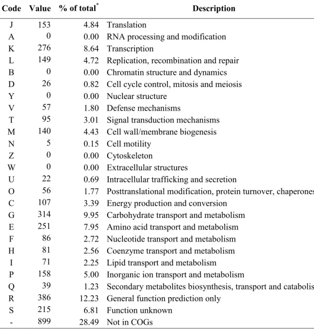
3.4.3 Genome annotation………59
3.5 Construction of genes-deletion Lactobacillus plantarum Lp90 mutant strain…………..59
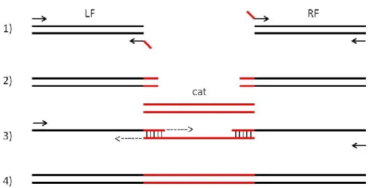
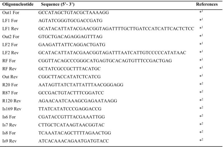
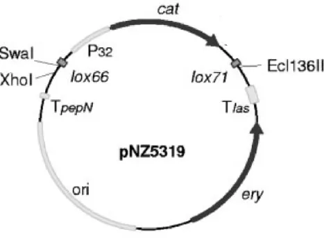
3.5.1 Generation of mutagenesis plasmids………59
3.5.2 E. coli transformation procedure……….64
3.5.3 Electrocompetent cells and electroporation of L. plantarum Lp90………...65
3.6 Caco-2 cells: in vitro assays………..…….66
3.6.1 Caco-2 cells growing condition………...…...66
3.6.2 Caco-2 cell culture, adhesion and competition assays for adhesion between E. coli and Lactobacillus plantarum……….………….66
3.6.3 Caco-2 cells immune stimulation assay………...…….68
3.6.4 RNA isolation and cDNA synthesis transcript profiling………...68
3.6.5 Quantitative Real Time (PCR) and transcriptional profiling………...69
3.7 Zebrafish in vivo model………...71
3.7.1 Transfer of pRCR12 to L. plantarum strains...71
3.7.2 mCherry protein fluorescence determination during bacterial growth...71
3.7.3 Determination of pRCR12 plasmid copy number...72
3.7.4 Zebrafish processing...73
3.7.5 Challenge test and enumeration of L. plantarum strains transformed with pRCR12 in infected zebrafish larvae...73
3.8 Biofilm formation………...74
3.9 Lactobacillus plantarum strains during in vitro gastro-intestinal tract condition……....74
3.10 L. plantarum Lp90 (EPS producing) in yogurt: oro-gastro-intestinal an immune-stimulation in vitro assays………...75
4
3.10.1 Yogurt production………75
3.10.2 Chemical analysis……….76
3.10.3 Microbiological analysis………..76
3.10.4 Lactobacilli oro-gastrointestinal tolerance in vitro assay in yogurt matrix…….76
3.10.5 Human monocytoid leukemia-derived cells (THP-1) growth conditions…………79
3.10.6 Immune-stimulation of THP-1 cells with lactobacilli………...79
3.10.7 Propidium monoazide (PMA) treatment and microbial DNA extraction………..80
3.10.8 THP-1 RNA extraction and cDNA synthesis………...…..81
3.10.9 qPCR analysis………...81
3.11 Tolerance of Lactobacillus plantarum strains to ethanol, acid, sulfur dioxide, lysozyme, and bile stress………82
3.12 Microvinification assays………..83
3.13 Statistical analysis………84
4. RESULTS AND DISCUSSION...85
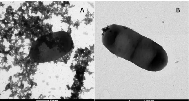
4.1 L. plantarum Lp90 cells: Transmission Electron Microscopy imaging……….85
4.2 Exopolisaccharides of L. plantarum Lp90………86
4.2.1 Exopolisaccharides yield………86
4.2.2 Chemical characterization of exopolysaccharides produced L. plantarum Lp90…86 4.3 Non contiguous-finished genome sequence of Lactobacillus plantarum strain Lp90…...87
4.3.1 L. plantarum Lp90 genome properties……….87
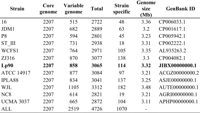
4.3.2 Comparison with other L. plantarum genomes…………...89
4.3.3 Comparison of Lp90 cps clusters and homologous clusters in L. plantarum species...92
4.4 Genes-deletion of Lactobacillus plantarum Lp90: Lp90Δcps2 and Lp90Δcps2.5 two non-ropy mutant strains………...99
4.4.1 pNZ8220 and pNZ8221 mutagenesis plasmids and E. coli transformation………..99
4.4.2 L. plantarum Lp90 transformation with pNZ8220 and pNZ8221 mutagenesis plasmids………..101
4.5 Lactobacilli and Caco-2 cells in vitro interactions………105
4.5.1 Lactobacilli adhesion on Caco-2 cells……….105
4.5.2 Competition against Escherichia coli O157: H7 in adhesion assays on Caco-2 cells...107
5
4.6 Zebrafish gut in vivo colonization by mCherry-labelled L. planatrum strains………...113
4.6.1 Fluorescent labeling of Lactobacillus strains with pRCR12 and detection of the mCherry protein………...113
4.6.2 Zebrafish larvae colonization by L. plantarum strains tagged with mCherry…...118
4.7 Biofilm formation on abiotic surface……….123
4.8 Lactobacilli survival during in vitro gastro-intestinal (GI) tract condition………126
4.9 Immune-stimulation of macrophage-differentiated THP-1 cells with in vitro oro-gastro-intestinal digested yogurt containing L. plantarum Lp90………128
4.9.1 Preliminary chemical analysis of yogurt………128
4.9.2 Viability of Lactobacillus plantarum strains in yogurt………..130
4.9.3 Tolerance of L. plantarum strains inoculated in yogurt during an in vitro oro-gastro-intestinal assay………..131
4.9.4 Stimulation of THP-1 cells with lactobacilli and expression of cytokine-related genes……….133
4.10 Tolerance to stress……….140
4.10.1 Tolerance of L. plantarum strains to ethanol stress………140
4.10.2 Tolerance of L. plantarum strains to acidic stress………...141
4.10.3 Tolerance of L. plantarum strains to sulfur dioxide stress……….143
4.10.4 Tolerance of L. plantarum strains to lysozyme stress……….144
4.10.5 Tolerance of L. plantarum strains to bile stress………...146
4.11 Bacterial survival and malolactic fermentation in microvinification assays………...147
5. CONCLUSIONS..………..152
6. REFERENCES………..157
7. APPENDIX……….186
7.1 List of scientific publications………..186
7.2 Participation to national and international congresses………...186
7.3 Experiences in other research centers……….………..187
6
ABSTRACT
Introduction
Lactic acid bacteria (LAB) occur in a variety range of fermented foods. Lactobacillus plantarum is a widespread LAB species which is encountered in diverse niches and some L. plantarum strains have been recognized as probiotics.
Several LAB are able to secrete exopolysaccharides (EPS), which can be either highly adherent or loosely bound to the microbial cell surface, thus distinguished into capsular and secreted forms, respectively; they are thought to provide protection against adverse environment.
The ability to produce EPS by LAB has been reported to be strictly correlated to the presence of specific eps/cps gene clusters.
EPS and EPS-producing LAB have been investigated in relation to their application in food industry and in bacteria-host interaction. Moreover, the prebiotic and pro-technologicals functions of exopolysaccharides produced by LAB are topics of growing interest.
Aims of the research
In this study, a polyphasic characterization of exopolysaccharides (EPS) produced by a
Lactobacillus plantarum strain, named Lp90, was performed. The strain was previously isolated
from wine and selected for a typical ropy phenotype.
Materials and methods
EPS produced by L. plantarum Lp90 were purified and quantified by phenol-sulfuric acid method. Furthermore, chemical characterization was performed by gas-liquid chromatography (GLC).
The genome of L. plantarum Lp90 was sequenced using the Illumina GAIIx platform and annotated by RAST (Rapid Annotation using Subsystem Technology) server, allowing a comparative genome analysis with L. plantarum strains already sequenced.
7 Knockout of genes responsible for the ropy phenotype was performed and L. plantarum Lp90 non-ropy mutant strains obtained.
Host-lactobacilli (EPS producing) interaction was performed in order to understand the probiotic potential of L. plantarum Lp90 and the possible prebiotic actions of exopolysaccharides produced by this strain. Bacterial survival during the simulation of the gastro-intestinal tract was assayed. The ability of L. plantarum strain Lp90 to adhere and compete for adhesion sites with
E. coli O157: H7 on Caco-2 cells, and the colonization of L. plantarum strain Lp90 fluorescently
labeled on enterocytic cells of zebrafish larvae, was performed. The potential immune-modulation effects of Lp90 on Caco-2 cells as well as on macrophage-differentiated THP-1 cells with digested yogurt containing this bacterial strain were also evaluated. Moreover, the affinity with abiotic surfaces was observed by the biofilms formation on glass tubes.
The potential role of exopolysaccharides produced by L. plantarum Lp90 in relation to its original habitat (wine) was analysed during microvinification assays and in presence of typical wine stresses, such as ethanol, pH and sulfur dioxide.
Results
Transmission Electron Microscopy (TEM) images clearly showed the presence of exopolysaccharides around the cell wall of Lactobacillus plantarum Lp90. Moreover, the chemical analysis suggested that they are hetero-polysaccharides, composed by rhamnose, glucose, galactose, glucosamine and galactosamine.
L. plantarum Lp90 genome is about 3,324,076 bps long with a total of 3,273 predicted genes.
Four different cps/eps gene clusters involved in exopolysaccharides biosynthesis were identified; in particular the cps2 gene cluster presented three glycosyltransferase genes apparently unique in Lp90 but homologous to Lactobacillus fabifermentans T30PCM01.
Following the entire or partial cps2 cluster deletion, we obtained two non-ropy mutant strains, (Lp90Δcps2 and Lp90Δcps2.5 respectively), thus suggesting that ropy phenotype of L.
8 EPS produced by L. plantarum Lp90 do not seem to promote in vitro and in vivo bacterial adhesion on intestinal epithelium, as well as the immune-modulation after the interaction of Caco-2 cells, while their inhibitory effect on E. coli adhesion on Caco-2 was observed. Furthemore, L. plantarum Lp90 showed a moderate survival during in vitro models of the gastro-intestinal tract, which is an added value for this strain considering its origin habitat.
Exopolysaccharides produced by L. plantarum strain Lp90 mask the ability of this strain to form biofilm on glass surface.
Exopolysaccharides produced by L. plantarum strain Lp90 confer increased tolerance to certain stressful conditions (ethanol, low pH, sulfur dioxide, lysozyme) usually encountered during winemaking.
Finally, preliminary analysis of yogurt produced with L. plantarum strain Lp90, showed a positive technological features and immune-modulation of cytokine-mediating genes.
Keywords: exopolysaccharides, ropy phenotype, Lactobacillus plantarum, probiotic, prebiotic.
SOMMARIO
Introduzione
I batteri lattici sono presenti in un’ampia gamma di alimenti fermentati. Lactobacillus plantarum è una specie diffusa di batteri lattici, riscontrabile in diverse nicchie e alcuni di essi sono stati riconosciuti come probiotici.
Diversi batteri lattici sono in grado di secernere esopolisaccaridi (EPS), che possono essere molto aderenti o debolmente legati alla superficie della cellula microbica, rispettivamente classificati come capsulari o dispersi; inoltre si ritiene che essi forniscano protezione contro ambienti avversi.
9 È stato dimostrato che la capacità dei batteri lattici di produrre EPS è strettamente correlata a specifici “cluster” genetici (eps/cps).
Gli esopolisaccaridi e i batteri lattici che li producono sono stati studiati per le loro applicazioni nel settore alimentare e nell’interazione con l’organismo ospite. Inoltre, le funzioni prebiotiche e protecnologiche degli esopolisaccaridi prodotti dai batteri lattici sono argomenti di crescente interesse.
Obiettivi della ricerca
In questo studio, è stata effettuata una caratterizzazione polifasica di esopolisaccaridi (EPS) prodotti da un ceppo di Lactobacillus plantarum, nominato Lp90. In precedenza, questo ceppo è stato isolato da vino e selezionato per il suo tipico fenotipo “ropy” (viscoso).
Materiali e metodi
Gli esopolisaccaridi prodotti da L. plantarum Lp90 sono stati purificati e quantificati con il metodo felono-acido solforico. Inoltre, la caratterizzazione chimica è stata eseguita mediante cromatografia gas-liquido (GLC).
Il genoma di L. plantarum Lp90 è stato sequenziato mediante “Illumina GAIIx platform” e annotato utilizzando RAST (Rapid Annotation using Subsystem Technology) server, permettendo così l'analisi comparativa con il genoma di altri L. plantarum.
È stata effettuata la delezione dei geni responsabili del fenotipo “ropy”, al fine di ottenere ceppi mutanti di L. plantarum Lp90 cosiddetti “non-ropy”.
Sono stati compiuti alcuni esperimenti sull’interazione organismo ospite-lattobacilli (produttore di EPS) al fine di comprendere le potenzialità probiotiche di L. plantarum Lp90 e le possibili azioni prebiotiche degli esopolisaccaridi prodotti da questo ceppo. È stata inoltra analizzata la sopravvivenza batterica mediante un sistema che simula il tratto gastro-intestinale. Inoltre, è stata studiata la capacità di Lp90 di aderire e competere per i siti di adesione con E. coli O157: H7 su cellule Caco-2 in vitro, come anche la colonizzazione in vivo di Lp90 marcato con una proteina fluorescente su enterociti di larve di “zebrafish”. Sono stati valutati i potenziali effetti
immuno-10 modulatori di Lp90 su cellule Caco-2, nonché sui macrofagi THP-1 differenziati in seguito all’esposizione con yogurt digerito contenente questo ceppo batterico. È stata analizzata l'affinità con le superfici abiotiche mediante formazione di biofilm su tubi di vetro.
Sono state effettuate analisi per comprendere il ruolo degli esopolisaccaridi prodotti da L.
plantarum Lp90 in relazione al suo habitat di origine (vino). A tal proposito, sono stati eseguiti
esprimenti di microvinificazione e studi sulla resistenza a diversi stress tipici dell’ambiente vino (etanolo, pH bassi, anidride solforosa, lisozima).
Risultati
Le immagini ottenute dalla microscopia elettronica in trasmissione hanno mostrato chiaramente la presenza di esopolisaccaridi intorno alla parete cellulare di Lactobacillus plantarum Lp90. Inoltre, le analisi chimiche hanno suggerito che sono etero-polisaccaridi, composti da ramnosio, glucosio, galattosio, glucosamina e galattosamina.
Il genoma di L. plantarum Lp90 ha una lunghezza di 3.324.076 bps con un totale di 3.273 geni predetti. Sono stati identificati quattro diversi “cluster” di geni cps/eps coinvolti nella biosintesi esopolisaccaridi; in particolare il cluster cps2 ha presentato tre glicosiltransferasi apparentemente uniche in Lp90 ma omologhe in Lactobacillus fabifermentans T30PCM01.
In seguito alla delezione intera o parziale del cluster cps2, sono stati ottenuti due ceppi mutanti “non-ropy” (rispettivamente Lp90Δcps2 e Lp90Δcps2.5), suggerendo così che il fenotipo “ropy” di L. plantarum Lp90 è inerente al “cluster” cps2.
Gli esopolisaccaridi prodotti da L. plantarum Lp90 non sembrano promuovere l’adesione batterica in vitro o in vivo sull’epitelio intestinale, così come l’immuno-modulazione dopo l'interazione con cellule Caco-2, invece è stato osservato un loro effetto inibitorio sull’adesione di E. coli su cellule Caco-2. Inoltre, L. plantarum Lp90 ha mostrato una modesta sopravvivenza durante i modelli in vitro del tratto gastro-intestinale, il che rappresenta un valore aggiunto per questo ceppo considerando il suo habitat originale.
11 Gli esopolisaccaridi di Lp90 mascherano l’abilità di questo ceppo nella formazione di biofilm su superficie di vetro.
Gli EPS prodotti da L. plantarum Lp90 conferiscono una maggiore resistenza a determinate condizioni di stress (etanolo, pH basso, anidride solforosa, lisozima) tipiche del processo di vinificazione.
Infine, le analisi preliminari sullo yogurt prodotto con L. plantarum Lp90, hanno mostrato risultati positivi sulle proprietà tecnologiche e sull’immuno-modulazione di geni coinvolti nella mediazione di citochine.
Parole chiave: esopolisaccaridi, fenotipo “ropy”, Lactobacillus plantarum, probiotico,
12
1. INTRODUCTION
1.1 Probiotics
1.1.1 The origins of probiotis
The first scientist who realized a positive function played by bacteria that colonize the human body, was the Nobel laureate Eli Metchnikoff. Considering that the aging process is caused by toxic substances such as phenols, indoles and ammonia produced by proteolytic microbes in the large intestine, he suggested the possibility to replace the harmful bacteria with beneficial ones. He also observed that some rural peoples in Europe, who used to drink milk fermented, had a relatively long life, and that milk fermented by lactic acid bacteria (LAB), inhibited the growth of proteolytic bacteria due to the low pH value. Subsequently, Metchnikoff introduced the use of fermented sour milk, using a bacterial species that he later called ‘Bulgarian bacillus’ (Vaughan, 1965). Tissier (1900) first isolated a Bifidobacterium from a breast-fed infant, at first called
Bacillus bifidus communi and later renamed Bifidobacterium bifidum. He concluded that this
species was predominant in the microflora of breast-fed infants and recommended it for feeding babies suffering from diarrhea (Tissier, 1900).
In 1917, professor Alfred Nissle isolated the bacterium Escherichia coli from the feces of a World War I soldier who did not develop enterocolitis during a severe outbreak of shigellosis. He successfully used this strain to treat intestinal diseases such as shigellosis and salmonellosis (Nissle, 1918). At that time antibiotics were not discovered yet. The probiotic E. coli Nissle 1917 is still in use today and recent studies have demonstrated its direct interaction with the host adaptive immune system (Molin, 2001).
In 1920, professor Leo F. Rettger showed that ‘Bulgarian Bacillus’, later known as Lactobacillus
13 Metchinikoff’s theory was disputed and the idea of fermented food died out (Cheplin and Rettger, 1920).
The word “probiotic” was coined from the Greek, “προ” plus “βιοτος” meaning literally “for life”.
Probably the first author that used the term “probiotic” has been Kollath in 1953, describing it as organic and inorganic supplement necessary to restore health to patients suffering a form of malnutrition resulting from eating too much highly refined food (Kollath, 1953). In 1954 Vergin suggested that antibiotics can upset the microbial balance of the body, and that this can be restored by a proper diet of probiotics, including fermentation products (Vergin, 1954). Lilly and Stillwell in 1965 following their observations give a more limited use of this word, giving the name probiotics to ‘growth promoting factors produced by microorganisms’ (Lilly and Stillwell, 1965). While, in 1973 Fujii and Cook defined as “compounds that build resistance to infection in the host but do not inhibit the growth of microorganisms in vitro”, referring to synthetic chemicals that protected mice against infection with Staphylococcus aureus (Fujii and Cook, 1973). Instead, Parker in 1974 seems to have been the first to use the word in relation to the interactions of micro-organisms with the whole animal or human host (Parker, 1974). Subsequently, Fuller in 1989 defined as probiotic ‘a live microbial food supplement which beneficially affects the animal host by improving its intestinal microbial balance’ (Fuller, 1989).
1.1.2 Probiotics nowadays
Currently, the probiotics are defined as ‘live microorganisms which when administered in adequate amounts, confer a health benefit on the host’ (FAO/WHO, 2002).
In order to be defined probiotics, microorganisms have to fulfill specific requisites. These characteristics include documented clinical efficacy, safety for human consumption, ability to reach, survive and colonize, at least transiently, the human gut, where probiotics exert their beneficial effects (Owehand et al., 2002).
14 Deterrence and reversion of intestinal dysbioses, enhancement of immune defenses, prevention of food allergies and infections, reinforcement of the gut barrier are among the beneficial effects ascribed to probiotics (Deshpande et al., 2011).
Specific probiotic strains are known to (i) normalize altered gut microecology and intestinal permeability; (ii) attenuate mucosal hypersensitivity and inflammatory reactions; (iii) stimulate non-specific host resistance to microbial pathogens and favour their eradication (Isolauri et al., 2004).
The positive impact of Lactobacillus plantarum and others probiotic LAB is thought to be mediated by various mechanisms including enhancement of the epithelial barrier, increased adhesion to intestinal mucosa and concomitant inhibition of pathogen adhesion, competitive exclusion of pathogenic microorganisms, production of anti-microbial substances and modulation of the immune system (Marco et al., 2006; Bermudez-Brito et al., 2012).
FAO/WHO developed Operating Standards establishing guidelines for all companies producing probiotic products (FAO/WHO, 2002; Reid, 2005).
These guidelines include:
- guidelines for the use of probiotics;
- phase I, II and III of clinical trials to prove health benefits;
- good manufacturing practice and production of high quality products; - studies to identify mechanism of action in vivo;
- informative labelling;
- development of probiotic organisms that can deliver vaccines to hosts;
- expansion of proven strains to benefit the oral cavity, nasopharynx, respiratory tract, stomach, vagina, bladder and skin as well as for cancer, allergies and recovery from surgery or injury. Resistance to the extreme conditions of the oro-gastrointestinal (OGI) tract, including highly acidic gastric juices and pancreatic bile salt secretions, is an essential criterion for the selection of orally delivered (food-borne) probiotics. The viability of probiotics is extremely important in
15 order to guarantee high bacterial loads into the main site of action (e.g., the intestine) and their optimal functionality.
All along the different OGI sections, bacteria are challenged also by the action of diverse digestive enzymes, including lysozyme (in the oral cavity); pepsin (stomach), pancreatin, chimotrypsin, and carboxypeptidases (intestine). These enzymes can remarkably compromise bacterial cell structures, by attacking and degrading surface-exposed macromolecules (Frenhani and Burini, 1999).
Bacterial cells are naturally equipped with various defence mechanisms to enhance survival in hostile environments (Van de Guchte et al., 2002; Spano and Massa 2006; Fiocco et al., 2007; Fiocco et al., 2010). These include chaperone proteins, which assist the folding of misfolded proteins, proteases which degrade irreversibly damaged proteins, transport systems to maintain correct osmolarity, catalases and superoxide dismutases to tackle reactive oxygen species, as well as proton pumps, decarboxylases and transporters to counteract intracellular pH decreases (Sugimoto et al., 2008).
1.2 Lactic acid bacteria
The denomination of “lactic acid bacteria” refers to bacteria involved in milk fermentation and capable to produce lactic acid from lactose. The family name Lactobacteriacea was applied by Orla-Jensen (1919). Today the main LAB genera include: Lactobacillus, Leucocostoc,
Pediococcus and Streptococcus (Schroeter and Klaenhammer, 2009).
Lactic acid bacteria are heterogeneous group of Gram-positive, low-GC, acid-tolerant, generally asporigen, rod- or cocci-shaped, catalase-negative, microaerophilic bacteria. Being a gram-positive bacterium, the cell envelope is a multilayered structure, which is mainly composed of peptidoglycan with embedded teichoic acids, proteins, and polysaccharides and which is essential to for the cellular integrity and shape (Silhavy et al., 2010). The common feature of
16 LAB is the production of lactic acid as the major metabolic end-product of carbohydrate fermentation (Carr et al., 2002). Several species are inhabitants of the human oro-gastrointestinal (OGI) tract. They are naturally associated with the mucosal surfaces, particularly the Gastro-Intestinal (GI) tract, mouth and vagina of mammals, and are also indigenous to food-related habitats, including plants (fruits, vegetables, and cereal grains), wine, milk, and meat. LAB are important in food industry: they are used as microbial starters to drive several fermentation processes, contributing to determine texture, organoleptic properties, and shelf-life of the final products. Moreover, LAB commonly used in the formulation of functional probiotic foods (Bron and Kleerebezem, 2011) and specific LAB are marketed as health-promoting organisms or probiotics (FAO/WHO 2002).
1.2.1 Carbohydrates metabolism of lactic acid bacteria
The main pathways in lactic acid bacteria for the metabolism of glucose are: - Glycolysis/Embden-Meyerhof-Parnas (EMP) pathway;
- 6-phosphogluconate/phosphoketolase (6-PG/PK) pathway (phosphoketolase- or pentose phosphate pathway) (Fugelsang and Edwards, 1997).
Before that glucose enters into one of these two pathways is transported into the cell where it is phosphorylated by hexokinase, an ATPdependant reaction.
The final products of EMP pathway are lactic acid and CO2. This pathway is also known as homolactic fermentation in LAB and it is divided into two steps: glycolysis, whereby pyruvate is produced from glucose, followed by the conversion of pyruvate to produce lactic acid (Ribéreau-Gayon et al., 2006).
The final products of 6-PG/PK pathway, also known as heterolactic fermentation, consist in lactic acid, CO2, ethanol and acetate. Leuconostoc some Lactobacillus species and Oenococcus
17 Other Lactobacillus species are facultative heterofermentors, including L. plantarum and L.
casei. These LAB make use of the EMP pathway for hexose metabolism and the 6-PG/PK
pathway for the metabolism of pentose sugars and other substrates.
Many LAB are able to ferment pentose sugars, which are phosphorylated, converted by epimerases or isomerases to phosphate derivatives ribulose-5-phosphate or xylulose-5-phosphate, subsequently they are metabolised via the bottom half of the 6-PG/PK pathway. The final products of pentoses metabolism are lactic acid, acetic acid and CO2 (Lerm et al., 2010).
1.2.2 Lactic acid bacteria in wine
Oenological lactic acid bacteria have a wide variability due to region, cultivar and vinification procedures. There is a successional growth of several species of LAB during vinification (Wibowo et al., 1985; Fugelsang and Edwards, 1997; Lerm et al., 2010). Oenococcus oeni is the main LAB species associated with MLF in wine. However, several species belong to
Pediococcus and Lactobacillus genera occur during or after MLF is completed (Wibowo et al.,
1985; Powell et al., 2006, Lerm et al., 2010).
The LAB population in the grape must generally range from 103 to 104 CFU/mL after crushing and the start of alcoholic fermentation. The major species of LAB present at this stage include
Lactobacillus plantarum, Lactobacillus casei, Leuconostoc mesenteroides, Pediococcus damnosus and O. oeni, which largely decline at the end of alcoholic fermentation. This could be
due to ethanol concentrations, high SO2 concentrations, low pH, low temperatures, the nutritional status and competitive interactions with the yeast culture (Lerm et al., 2010).
1.2.3 The malolactic fermentation (MLF)
Malolactic fermentation (MLF) is a secondary fermentation process that normally takes places after the alcoholic fermentation by yeasts. It is carried out by one or more species of lactic acid bacteria (Arthurs and Lloyd, 1999), including bacteria from the genera Oenococcus,
18
Lactobacillus, Pediococcus and Leuconostoc, among these, O. oeni is best adapted to the harsh
wine environment, such as high alcohol, low pH and sulphur dioxide (SO2) (Wibowo et al., 1985; Spano and Massa 2006).
MLF plays an important role in the winemaking process as it contributes to the deacidification of wine, microbial stability and has an influence on the aroma profile, mainly in red wines (Lerm et
al., 2010).
The MLF reaction consists in the conversion of malic acid, which is a dicarboxylic acid, to L-lactic acid, a monocarboxylic acid, and the production of CO2.
Lactic acid bacteria have three possible enzymatic pathways for the conversion of L-malic acid to L-lactic acid and CO2 (Lerm et al., 2010):
1. Direct conversion of malic acid to lactic acid via malate decarboxylase, also known as the malolactic enzyme (MLE).
2. Pathway employing the malic enzyme to convert L-malic acid to pyruvic acid, which is reduced by L-lactate dehydrogenase to lactic acid.
3. Reduction of malate by malate dehydrogenase to oxaloacetate, followed by decarboxylation to pyruvate and reduction to lactic acid.
The physiological function of the malate fermentation pathway is mainly involved to generate a proton motive force (PMF) as a means to acquire energy to drive essential cellular processes (Konings, 2002).
At the end of MLF, the remaining LAB can still metabolise residual sugar, which could result in spoilage including volatile acidity (Fugelsang and Edwards, 1997). For this reason, it is essential to check the potential impact of residual LAB populations.
1.2.4 Lactobacillus plantarum
The genus Lactobacillus which belong to the phylum Firmicutes includes a considerable number of different species with high degree of diversity (Stiles and Holzapfel, 1997). Among these,
19
Lactobacillus plantarum is a widespread LAB species which is found in diverse niches
associated to food matrices, vegetables, soil, human body and it is among the most common lactobacilli occurring on the human oral and intestinal mucosa (Ahrne et al., 1998; de Vries et
al., 2006; Siezen et al., 2010), and it contributes to specific organoleptic and nutritional
properties of the final product (Kleerebezem et al., 2003). L. plantarum is a facultative heterofermentative organism closely related to Lactobacillus paraplantarum, Lactobacillus
pentosus and Lactobacillus fabifermentans (Siezen and van Hylckama Vlieg, 2011). Cells are
Gram positive, rod shaped, non-spore-forming and non-motile.
Lactobacillus plantarum produces both isomers (D and L) of lactic acid and it is used for the
production and preservation of fermented foods obtained from different raw materials, in which it is either present as a contaminant or added as a starter to carry out fermentations. Recently, it has been considered as the next generation starter culture for malolactic fermentation in wine, because it is one of the most dominant species of lactobacilli occurring throughout the winemaking process (du Toit et al., 2011; Capozzi et al., 2012).
The genus Lactobacillus has GRAS (Generally Recognized as Safe) status (De Angelis and Gobbetti, 2004), due to its natural occurrence and long history of safe use in food production. Several L. plantarum strains have been investigated for healthy properties and have been recognized as probiotics. Several reports, including human clinical studies, document the potential beneficial effects of lactobacilli, including L. plantarum (de Vries et al., 2006; van Baarlen et al., 2013) and some L. plantarum strains can be found in a variety of marketed probiotic functional foods.
The Lactobacillus plantarum WCFS1, a single colony of L. plantarum NCIMB 8826 (National Collection of Industrial and Marine Bacteria, Aberdeen, UK), isolated from human saliva, has been the first L. plantarum complete genome sequenced and annotated in 2003 (Kleerebezem et
al., 2003). In 2012 the complete genome of WCFS1 was sequenced and annotated again, and 116
20 proteins (Siezen et al., 2012). L. plantarum WCFS1 has become one of the model strains in LAB research since the initial genome publication. Different bioinformatics tools have been used to predict the function of its genes, reconstruct metabolic pathways and gene regulatory networks, and compare its genome with genomes of other LAB. The genomic, phenotypic, and metabolic diversity of L. plantarum has been previously described. Moreover, L. plantarum has been employed as a model for LAB interactions with mammalian gut tissues in studies that provided insights into the microbial adaptation to that habitat and identified candidate probiotic genes (Siezen et al., 2012).
The ecological flexibility of L. plantarum is confirmed by the observation that this species has one of the largest genomes (approximately 3.3 Mb) known among LAB.
The availability of such data has prompted the genetic and molecular dissection of this species, also in relation to its probiotic behavior.
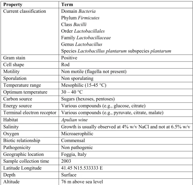
1.2.5 Lactobacillus plantarum Lp90
Lactobacillus plantarum Lp90 is a strain previously isolated from fermented wine in a winery
located nearby Foggia, Italy. This strain is characterized by a distinctive ropy phenotype (Figure
1.1), which was ascribed to its capacity to over-produce exopolysaccharides (EPS) (Caggianiello et al., 2013). This strain was already analysed for different features (Spano et al., 2004; Spano et al., 2005; Fiocco et al., 2007; Siezen et al., 2010). The complete genome of this strain has been
sequenced and it is the first L. plantarum genome coming from a strain of wine origin (Lamontanara et al., 2015).
21
Property Term
Current classification Domain Bacteria Phylum Firmicutes Class Bacilli
Order Lactobacillales Family Lactobacillaceae Genus Lactobacillus
Species Lactobacillus plantarum subspecies plantarum
Gram stain Positive
Cell shape Rod
Motility Non motile (flagella not present)
Sporulation Non sporulating
Temperature range Mesophilic (15-45 °C) Optimum temperature 30 – 40 °C
Carbon source Sugars (hexoses, pentoses)
Energy source Various compounds (e.g., glucose, citrate)
Terminal electron receptor Various compounds (e.g., pyruvate, citrate, malate)
Habitat Apulian wine
Salinity Growth is usually observed at 4% w/v NaCl and not at 6.5% w/v
Oxygen Microaerophilic
Biotic relationship Commensal
Pathogenicity Non pathogenic
Geographic location Foggia, Italy Sample collection time 2003
Latitude Longitude 41.45 N15.533333 E
Depth Surface
Altitude 76 m above sea level
Table 1.1 - Classification and general features of Lactobacillus plantarum Lp90 (adapted from
22
1.3 Prebiotics
A first definition of prebiotic was given by Gibson and Roberfroid (1995) as “a nondigestible food ingredient that beneficially affects the host by selectively stimulating the growth and/or activity of one or a limited number of bacteria in the colon, and thus improves host health”. It has been proposed to refine the original definition to “a prebiotic is a selectively fermented ingredient that allows specific changes, both in the composition and/or activity in the gastrointestinal microflora, that confer benefits upon host wellbeing and health” (Gibson et al., 2004).
Prebiotics can be incorporated into many foodstuffs and they act on the intestinal flora and improve the balance of the flora by enhancing the growth of beneficial intestinal bacteria and/or inhibiting the growth of harmful ones, resulting in scavenging in the intestinal environment. The candidate prebiotics include oligosaccharides, dietary fiber, resistant starch, (Mitsuoka; 2014). The demonstration of a prebiotic effect must be carried out in vivo by validated methodologies to produce sound scientific data (Roberfroid et al., 2007).
23 To determine whether a dietary carbohydrate could be considered a potential prebiotic, need to evaluate several factors: (i) resistance to gastric acidity, to hydrolysis by mammalian enzymes, and to gastrointestinal absorption; (ii) fermentation by intestinal microflora; and (iii) selective stimulation of the growth and/or activity of those intestinal bacteria that contribute to health and well-being (Gibson et al., 2004).
The first two ingredients that fulfill these criteria were inulin and trans-galactooligosaccharides (TOS).
Although still insufficient, promising data on prebiotic activity have been reported for glucooligosaccharides, isomaltooligosaccharides, lactosucrose, polydextrose, soybean oligosaccharides, and xylooligosaccharides. Moreover, there are still many substances for which are being evaluated the possible prebiotic effects, including the microbial exopolysaccharides.
1.4 Microbial exopolysaccharides (EPS)
1.4.1 Exopolysaccharides produced by LAB
The term “exopolysaccharides” (EPS) as proposed by Sutherland (1972) provides a general name for all forms of bacterial polysaccharides found outside the cell wall. Several LAB are able to secrete long-chains of homo- or hetero-polysaccharides, consisting of branched, repeating units of sugars or sugar derivatives (Ruas-Madiedo et al., 2002). Such exopolysaccharides (EPS) can be either highly adherent or loosely bound to the microbial cell surface and are thus distinguished into capsular and secreted forms. EPS-producing LAB could be responsible for a ropy phenotype characterized by a viscous and texture observed in spoiled alcoholic beverages, such as wine especially with a pH > 3.8 (Coulon et al., 2012). This phenomenon has been already observed by Pasteur in 1860. The ropy appearance in wines is due to the presence of exopolysaccharides produced by some lactic acid bacteria, such as Pediococcus parvulus found in Bordeaux wines (Dols-Lafargue and Lonvaud-Funel, 2009), but mainly by Pediococcus
24
damnosus. Moreover, it was found that some ropy strains are more tolerant to ethanol and SO2 stress conditions (Lonvaud-Funel, 1999; Dols-Lafargue et al., 2008).
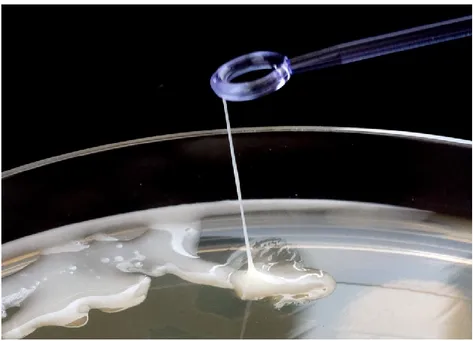
The ability to produce EPS by LAB has been reported to be strictly correlated to the presence of specific gene clusters (eps/cps), located either on plasmids (Van Kranenburg, et al., 1997) or on the main chromosome (Stingele et al., 1996; De Vuyst et al., 1999) (Figure 1.2).
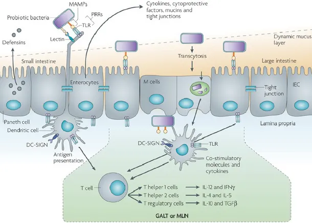
In the chromosomal genome of L. plantarum WCFS1, 4 cps genes clusters are associated with surface polysaccharide production (Remus et al., 2012) (Figure 1.3). The cps1, cps2, cps3 clusters are separated by transposase genes and fragments, encoding proteins involved in biosynthesis and export of extracellular or capsular polysaccharides (Siezen et al., 2011).
Figure 1.2 - Eps genes cluster organization (A) Eps gene cluster involved in the exopolysaccharides
biosynthesis in L. lactis subsp. cremoris NIZO B40 (plasmid-localized) (Van Kranenburg, et al., 1997); (B)
25 Figure 1.3 - Schematic representation of polysaccharide biosynthesis gene clusters in L. plantarum strains (from Remus et al., 2012).
(A) Cps1, Cps2 and Cps3 gene clusters of Lactobacillus plantarum WCFS1 involved in polysaccharide biosynthesis and comparison with the corresponding clusters of L. plantarum strains ST-III, JDM-1 and ATCC 14917. Dark-grey colored connecting blocks indicate regions of high sequence conservation between L. plantarum genomes. (B) Cps4 cluster of L. plantarum WCFS1 involved in polysaccharide biosynthesis.
26 The eps/cps clusters exhibit a conserved modular organization and include genes encoding both regulatory factors and enzymes involved in EPS biosynthesis, polymerization and secretion, including glycosyl-transferases, which are responsible for the assembly of the characteristic EPS-repeating unit (De Vuyst et al., 1999; Welman and Maddox, 2003; Lebeer et al., 2009). Polymer length depends on a tyrosine kinase phosphoregulatory system, whose genes are located in the initial part of the cluster (Figure 1.2).
The repeating units are synthesized in the cytoplasm and assembled on the lipid carrier undecaprenyl phosphate by sequential transfer of monosaccharides from nucleotide sugars by specific glycosyltransferases (De Vuyst et al., 1999).
The polymerization mechanism of the repeating unit and export from the cell in LAB, is not entirely known. A model has been proposed for Lactococcus lactis based on the action of “flippase” which move the lipid-bound repeating units from the cytoplasmic face of the membrane to the periplasmic face (Laws et al., 2001). Other mechanisms have been proposed for
Streptococcus pneumoniae (Bentley et al., 2006) and Lactobacillus rhamnosus (Lebeer et al.,
2009) (Figure 1.4). A polymerase could catalyse the linking of the repeating units and an enzyme could uncouple the lipid-bound polymer and control chain length (Welman and Maddox
27 Figure – 1.4 (A) Schematic representation of the EPS gene cluster of L. rhamnosus GG and comparison with the corresponding gene cluster in L. rhamnosus ATCC 9595. The arrows with the same color of gray
indicate genes encoding similar functions in EPS biosynthesis. The dark gray arrows indicate the genes encoding proteins putatively involved in the regulation of EPS production and polymerization. The light gray arrows indicate the gene putatively encoding the polysaccharide transporter and polymerase. The white arrows indicate the genes encoding the putative glycosyltransferases. The long stripes arrows indicate genes encoding the proteins involved in the biosynthesis of the dTDP-rhamnose precursor. The lightest gray arrows indicate the glf gene, which encode the UDP-galactopyranose mutase. The triangles indicate insertion sequence elements (IS). Gray boxes indicate the genes with high homology. (B) Putative representation of the EPS biosynthesis in L.
rhamnosus GG. The membrane-associated priming glycosyltransferase WelE allows the transfer of a
phosphogalactosyl residue from an activated nucleotide sugar to the undecaprenyl phosphate (UndP)-lipid carrier on the cytoplasmic face of the membrane. Consequently, unique glycosyltransferases WelF to WelJ add the remaining sugars in a sugar and glycosidic linkage-dependent manner. A Wzx flippase allows translocation across the cytoplasmic membrane of a complete subunit EPS, followed by linkage of the repeating units into long polysaccharides by a specific Wzy polymerase. Wze autophosphorylating tyrosine kinase and a Wzb phosphotyrosine protein phosphatase forming a phosphorylation complex could be involved in the regulation of EPS biosynthesis (from Lebeer et al., 2009).
28 EPS biosynthesis can be divided into three main steps: (i) assimilation of a carbon substrate; (ii) intracellular synthesis of the polysaccharides; (iii) EPS exudation out of the cell (Vandamme et
al., 2002).
The physiological role that exopolysaccharides play in the bacterial ecology of probiotics lactic acid bacteria is not yet entirely clear. EPS are thought to protect against biotic stress, like competition, and abiotic stresses that might include temperature, light intensity, pH or osmotic stress. In the cases of acidophilic or thermophilic species, EPS aid in adapting to extreme conditions. It has been suggested that EPS from other bacteria can act as protective agents against desiccation, antimicrobial compounds, bacteriophage attack, and to permit adhesion to solid surfaces (De Vuyst and Degeest, 1999; Forde et al., 1999; Looijesteijn et al., 2001; López
et al., 2004). They can also be involved in adhesion to surfaces and biofilm formation and to cell
adhesion/recognition mechanisms (Ruas-Madiedo et al., 2002; Broadbent et al., 2003; Rozen et
al., 2004), however, the involvement of these biopolymers in bacterial adhesion to the intestinal
epithelium in vivo has not yet been validated (Ruas-Madiedo et al., 2008).
Despite the wide diversity of microbial EPS with physicochemical properties that are industrially promising, only two EPSs are authorised for use as additives in the food industry in the United States and Europe: xanthan (30 000 tons/year) and gellan (Donot et al., 2012).
EPS from microbial sources can be classified into two groups: homopolysaccharides (e.g. cellulose, dextran, mutan, alternan, pullulan, levan and curdlan) and heteropolysaccharides (e.g. gellan and xanthan). Homopolysaccharides consist of repeating units of only one type of monosaccharide (D-glucose or D-fructose) and can be divided into two major groups: glucans and fructans. By contrast, heteropolysaccharides from LAB have repeating units that demonstrate little structural similarity to one another. The molecular mass of these polymers ranges between 4.0×10⁴ and 6.0×10⁶ Da (Welman and Maddox, 2003).
The structural diversity of EPS among lactobacilli may determine strain-specific properties important for probiotic action and technological applications (adhesion, stress resistance).
29 EPS have been reported to possess a number of health benefits, such as immune-stimulatory (Vinderola et al., 2006; Matsuzaki et al., 2014), and antitumoral effects (Kitazawa et al., 1991) lowering blood cholesterol (Nakajima et al., 1992; Maeda et al., 2004b) and prebiotic effects (Dal Bello et al., 2001; O’Connor et al., 2005). Surface polysaccharides may also contribute to protection against intestinal innate immune factors such as the antimicrobial peptide LL-37 (Lebeer et al., 2011). Exopolysaccharides produced by LAB can regulate inflammatory responses in the intestinal lumen (Notararigo et al., 2014). The cell surface-associated exopolysaccharide of the probiotic Bifidobacterium brevis reduces the production of pro-inflammatory cytokines and suppresses the generation of B. brevis-specific antibodies, thus allowing this probiotic to be tolerated in the gut Fanning et al., 2012).
1.4.2 The potential prebiotics properties of exopolysaccharides
Currently, little is known about the prebiotic properties of EPS produced by lactic acid bacteria, although they have received increasing attention in relation to health benefits (i.e., immune stimulation, antimutagenicity, and antitumor activity (Kitazawa et al., 1998; Ruas-Madiedo et
al., 2002; Salazar et al., 2014).
A potential prebiotic effect has been reported for exopolysaccharides produced in whey by L.
plantarum. The EPS produced can be used by the probiotic parent strain, thus suggesting that it
could possess enzymes capable to degrade the EPS (Tsuda and Miyamoto, 2010). An α-d-glucan produced by Lactobacillus plantarum exhibited lowest digestibility by artificial gastric juice and
in vitro prebiotic activities showed increased growth of probiotic bacteria such as Bifidobacterium infantis and Lactobacillus acidophilus, but did not support the growth of
non-probiotic bacteria such as Escherichia coli and Enterobacter aerogenes indicating their potential use as prebiotic additive for food products (Das et al., 2014). The prebiotic properties levan-type EPS from Lactobacillus sanfranciscensis were studied and the bifidogenic effect of the EPS was observed (Dal Bello et al., 2001).
30 EPS from Weissiella cibaria, W. confusa, L. plantarum and P. pentosaceus exhibited high resistance to gastric and intestinal digestions, selective enhancement of beneficial gut bacteria (particularly bifidobacteria group) suggesting their prebiotic potentials (Hongpattarakere et al., 2012). The ingestion of exopolysaccharide-producing lactobacilli improve lipid metabolism, associated with changes in the gut microbiota (London et al., 2014).
A positive effect of the β-D-glucan produced by P. parvulus was observed on the growth of both
L. plantarum and L. acidophilus strains, suggesting that its use as a prebiotic may positively
modulate the growth of probiotic organisms (Russo et al., 2012). Conversely, purified EPS from
P. parvulus did not show prebiotic effect in the mouse model, although ingestion of live
EPS-producing bacterium antagonized Enterobacteriaceae without disturbing the homeostasis of the microbiota (Lindström et al., 2013).
1.4.3 Exopolysaccharides in food industry
Since several decades exopolysaccharides produced by lactic acid bacteria has received increasing interest, regarding their potential use in industrial field (Cerning, 1995). In the food industry, EPS produced by LAB and other microorganisms are used as viscosifiers, stabilizers, emulsifiers, or gelling agents to modify the rheological properties, texture and ‘mouthfeel’ of fermented dairy and non-dairy products (Hassan, 2008; Galle et al., 2012). Most of the strains used in the production of functional dairy food synthesize heteropolysaccharides (Welman and Maddox, 2003; Mende et al., 2012).
Several authors evaluated the affect of EPS produced by LAB on rheological and sensorial properties in yogurt (Hassan et al., 2003; Doleyres et al., 2005; Folkenberg et al., 2006; Yang et
al., 2014), the product of fermentation of milk led by starter cultures of Lactobacillus delbrueckii
subsp. bulgaricus and Streptococcus thermophilus in ratio 1:1. Both bacteria produce EPS from 30 to 890 mg/L for S. thermophilus and from 60 to 150 mg/L for L. delbrueckii subsp.
31 exopolysaccharides in yogurt contribute to improve the viscosity and texture and they do not alter the flavor of the final product (Jolly et al., 2002; Badel et al., 2011).
Several species of lactobacilli are described to produce exopolysaccharide. The best documented species are L. casei, L. acidophilus, L. brevis, L. curvatus, L. delbrueckii bulgaricus, L.
helveticus, L. rhamnosus, L. plantarum and L. johnsonii. L. reuteri 121 has been found to
synthesize several HoPSs in the same culture conditions (van Geel-Schutten et al., 1999) and it is capable to secrete β-(2,1) fructans (inulin like polysaccharide) recognized as prebiotic (van Hijum et al., 2002). The soluble reuteran has been found opportunities in baking industry in association with levan synthesized by L. reuteri and L. sanfranciscensis, as their polysaccharides provides beneficial effect on bread flavour, texture and shelf-life of products derived from sourdough fermentation (Tieking et al., 2005; Badel et al., 2011).
The use of LAB starter cultures which produce EPS in situ during fermentation could be a valid alternative for products whose polysaccharides addition requires the specification of food additives, which is a condition not much appreciated by consumer.
1.5 Bacterial resistance to the oro-gastro-intestinal transit
The human gastrointestinal tract (GIT) is colonized by an enormous and diverse community of microbes which are essential to its proper functioning. These microbes have evolved in concert with their host to occupy specific regions and niches in the GIT. A balanced, complex microflora is necessary for normal digestion and to maintain the homeostasis of intestinal ecosystem (Simon and Gorbach, 1986).
Tolerance to the harsh conditions of the oro-gastro-intestinal transit (OGI), which comprises highly acidic gastric juices and pancreatic bile salt secretions, is a fundamental criterion for the selection of orally delivered probiotics. For this reason, the analysis of potential probiotics in
32 tract is a prerequisite to subsequent in vivo experiments. Development and implementation of such systems are highly encouraged by FAO and WHO (2002) and several recent studies have addressed this issue (Fernández de Palencia et al., 2008; Lo Curto et al., 2011).
The lysozyme and chewing stress represent the first obstacle of the oral tract. The various proposed models, simulate the phenomena that occur during the digestion, from filling to the gradual emptying of the stomach. In the condition of full stomach, bacteria ingested together with the food matrix are subjected to pH values of 5.0-6.0, then undergo more drastic acidic conditions, as there is a lowering of pH at values of 2.0 - 1.5. Bacteria exposure to acids environments, disturb the proton motive force across the membrane, causing an accumulation of protons inside the cell (Corcoran et al., 2008). The emptying of the gastric pouch is an event that takes place gradually, in tandem with the digestion of food. The liquids empty from the stomach is faster than solids and in general food remains in the stomach between 2 and 4 hours, while the transit time through the small intestine takes from 1 to 4 hours. The adverse conditions of the small intestine include the presence of bile and pancreatin in the lumen of the small intestine, pH is around 8.0. Bile salts secreted in the duodenum emulsifies and solubilize lipids and lipid soluble vitamins (Begley et al., 2005). A concentration of 0.15 - 0.3% of bile salts has been recommended as a suitable concentration for selecting probiotic bacteria for human use (Goldin and Gorbach, 1992; Huang and Adams, 2004).
Bacterial cells have various defense mechanisms to resist the hostile environments (Van de Guchte et al., 2002; Mills et al., 2011). The chaperone proteins assist the folding of misfolded proteins, proteases which degrade irreversibly damaged proteins, transport systems to maintain correct osmolarity, catalases and superoxide dismutases to tackle reactive oxygen species, as well as proton pumps, decarboxylases and transporters to counteract intracellular pH decreases (De Angelis and Gobbetti, 2004; Sugimoto et al., 2008).
33
1.5.1 The role of exopolysaccharides during the in vitro gastro-intestinal transit
An important aspect related to the potential prebiotic effect of microbial exopolysaccharides, is the behavior that they have during the gastro-intestinal transit, considering that the low pH stress is usually the hardest obstacle for survival of probiotic bacteria (Both et al., 2010; Bove et al., 2013). Fernández de Palencia et al. (2009) reported that a ropy strain of Pediococcus parvulus and its relative non ropy strain subjected to an in vitro gastric or gastro-intestinal stress, have the same pattern of resistance to stress, indicating that the presence of EPS did not confer to bacterial cells an advantage for survival in the human digestive tract. By contrasty, synthesis of the P.
parvulus β-glucan confers to Lactobacillus paracasei higher survival during gastrointestinal
passage or technological process (Stack et al., 2010). Arena et al. (2014a) reported that
Figure 1.5 - Compartments of the human GI tract and related densities of the residing bacterial population. Food-borne bacteria stress sequential in the acidic environment of the stomach and subsequently
pancreatin and bile into the small intestine. Dietary supplementation of probiotics can generate a relative high abundance of these species in the first tract of the small intestine, where their metabolic activity can be relevant. The ileum, where the probiotic loads tend to decrease with respect to the indigenous microbiota, is the major site of probiotic immune activity. In the large intestine, commensal bifidobacteria and probiotic supplements contribute to catabolize diet- and host-derived glycans, generating a variety of short chain fatty acids that are used as important energy source by the colonic mucosa (adapted from: Kleerebezem and
34 exogenous polysaccharides such as food matrices containing β-glucans, enhanced the oro-gastrointestinal stress tolerance of lactobacillus probiotic strains.
1.6 Bacterial adhesion to the intestinal mucosa and displacement of pathogen bacteria
The ability to adhere to the intestinal mucosa is an advantageous feature of probiotic microorganisms, as it ensures persistence in the intestinal tract, which is necessary for them to come in close contact with host epithelial cells, to control the balance of the intestinal microflora, to antagonize pathogen growth, and to exert immune modulation on the host (Isolauri et al., 2004).
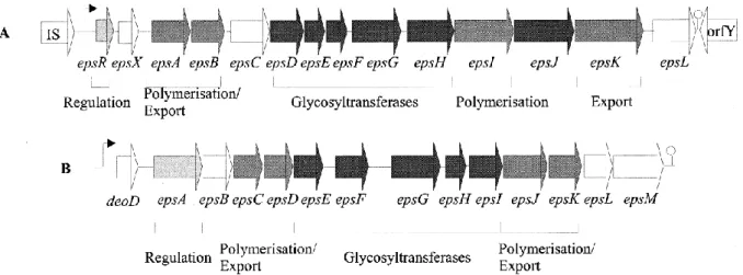
Adhesion to the surface of host epithelial cells is a key pathogenic factor of intestinal pathogens (Scaletsky et al., 2002). Enterohemorrhagic Escherichia coli (EHEC) is a human pathogen that enters the intestinal tract as a result of food contamination and causes hemorrhagic colitis and hemolytic uremic syndrome (HUS) (Kim et al., 2009). Lactobacilli have been shown to possess surface adhesins similar to those on bacterial pathogens (Neeser et al., 2000) and thus they may interfere with pathogen adhesion on the intestinal mucosa. The ability of probiotic bacteria to adhere on the intestinal surface, is an important factor in the displacement of pathogens (Lee and Puong, 2002; Gueimonde et al., 2006). A probiotic should be able to compete with a pathogen for the binding sites, nutrients, production of antimicrobial substances and immune-stimulating compounds.
A first physical barrier to host-cell stimulation by bacteria in the gut, is represented by the mucus layer bound to gastro-intestinal epithelia. This is composed of a continuous gel matrix, which is formed primarily of complex glycoproteins that acts as a protective barrier for the host against harmful antigens and promote luminal motility. The adhesion to mucus layer is therefore the first requirement for probiotic organisms to interact with host cells. The thickness of the human intestinal tract mucus layer is variable. Generally it is greater starting from the small intestine,
35 where the intestinal flora is more abundant, and it is thinner in the rectum (Van Tassell and Miller, 2011).
The polymers that compose intestinal mucin are considered nutritive glycans for commensal bacteria in the promotion of their residence and associated benefits (Carrington et al., 2009). Probiotic persistence and colonization do not permanently exist in the GI tract and they provide host benefits only for brief periods, once finished the administration (Tannock et al., 2000; Garrido et al., 2005). Bacteria at first adhere to gastro-intestinal surfaces by nonspecific physical interactions, which are reversible attachments. Many lactobacilli have large surface proteins with highly repetitive structures that are involved in mucus adhesion (Van Tassell and Miller, 2011). Mucus-binding proteins showing lectin-like interactions have been isolated; they may be conserved in numerous Lactobacillus species, and some of them showed to promote mucus adhesion. Mucus-targeting bacterial adhesins is the mucus-binding protein (MUB), produced by
L. reuteri (Tannock et al., 2000; Roos and Jonsson, 2002), and in L. acidophilus NCFM (Buck et al., 2005) have been identified.
Probably, carbohydrate-protein interactions play a key role in the adhesion of these proteins to mucin-bound oligosaccharides. Numerous MUB homologues and MucBP domain containing proteins have been found, almost exclusively in lactic acid bacteria and mainly in lactobacilli found naturally in intestinal niches (Van Tassell and Miller, 2011). This suggests that MucBP domain containing proteins play an important role in establishing host-microbial interactions in the gut and promoted the evolution of the species as primarily GI organisms (Boekhorst et al., 2006; Dam and Brewer, 2010).
S-layer proteins and glycoproteins can form a monolayer coating the surface of bacterial cells (Boot et al., 1996; Sleytr et al., 1997), they are present in only some Lactobacillus species, and has been ascribed a role in adhesion to host cell and inhibition of pathogen adhesion to the same surface. In Lactobacillus crispatus ZJ001, S-layer proteins are responsible for adhesion to epithelial cells and competitive exclusion of pathogens such as E. coli O157:H7 and Salmonella
36
typhimurium (Chen et al., 2007). Ramiah et al. (2007), found a consistent induction of Mub and
other adhesion proteins in a probiotic strain of L. plantarum, especially when mucins were added to a media simulating gut conditions.
Another mechanism of bacterial adhesion is based on the binding to mannose-containing receptors on epithelial cells. Among probiotic bacteria, L. plantarum is able to recognize mannose-residues. By in silico studies, the predictive sequence of a L. plantarum WCFS1 adhesin gene (lp_1229) was identified. Knockout of this gene resulted in a complete loss of yeast agglutination ability, while its overexpression enhanced this phenotype. Moreover, analysis of the protein showed putative carbohydrate-binding domains, supporting its role in binding mannose residues. Therefore, this gene was designated to encode the mannose-specific adhesin (msa), probably involved in the interaction of L. plantarum with the host along the intestinal tract (Pretzer et al., 2005).
The EPS produced by probiotic strains could be able to adhere to intestinal mucus, the effect being dose and EPS type dependent. This could reflect the adaptation of probiotics to their natural environment. Thereby, EPS could act as adherence factor that may play a role in the transitory colonization of the intestinal mucosa by probiotics. The ubiquity of EPS gene clusters on probiotic genomes suggest that a number of strains from the intestinal microbiota may produce extracellular polymers in this environment and that high EPS concentrations could be locally reached in the gastrointestinal tract (Ruas-Madiedo et al., 2006).
1.6.1 Caco-2 cell in vitro model adhesion
The molecular mechanisms underlying probiotic activities are being disclosed more and more by
in vitro and in vivo studies focused on the interaction between probiotic bacteria and host
intestinal epithelial or immune cells (Marco et al., 2006). Due to obvious difficulties in performing in vivo studies, preliminary studies of potentially adherent strains are mainly based
37 on in vitro adhesion assays. Currently there is not an in vitro adhesion standard protocol, in fact for this reason the results are highly variable (Laparra and Sanz, 2009).
Tissue cultures of the human colon carcinoma cell lines Caco-2 are the most frequently used, and their applications are well documented in the literature. They are considered one of the best representations of the in vivo environment and they can be grown in culture to form a homogeneous polar monolayer of mature enterocytes resembling the tissue of the small intestine (Pinto et al., 1983). Caco-2 cells represent a continuous line of heterogeneous human epithelial colorectal adenocarcinoma cells, developed by the Sloan-Kettering Institute for Cancer Research. Caco-2 cells are capable to initiate spontaneous differentiation and reach confluence under normal culture conditions (e.g., presence of glucose and serum) (Fossati et al., 2008). Over a period of 20 - 30 days of post-confluent culture, Caco-2 cells gradually acquire a morphological polarity comparable with those of mature intestinal absorbing cells. Caco-2 cells also provide a valuable system for immunological studies (Ou et al., 2009).
Moreover, some microorganisms provide essential vitamins (e.g., folate, biotin, vitamin K) and produce short chain fatty acids that are used as energy source by colon cells (Saulnier et al., 2009).
1.6.2 Zebrafish in vivo model adhesion
In recent years, zebrafish (Danio rerio) has been found as an interesting model to study vertebrate development, immunity and disease because of their small size, high fecundity, rapid development, optical transparency of the embryos, amenability to genetic screens, and structural similarities to mammals (Meeker and Trede 2008; Sullivan and Kim 2008). Scientific studies on this model were several: host immune response under a number of microbial infections (van der Sar et al., 2004; Rojo et al., 2007); interactions between the host and its natural gut microbiota (Milligan-Myhre et al., 2011); host-probiotics interactions (Gioacchini et al., 2012; Rendueles et