Contents lists available atScienceDirect
IJP: Parasites and Wildlife
journal homepage:www.elsevier.com/locate/ijppawAir sac trematodes: Morishitium polonicum as a newly identi
fied cause of
death in the common blackbird (Turdus merula)
Livio Galosi
a, Petr Heneberg
b, Giacomo Rossi
a, Jilji Sitko
c, Gian Enrico Magi
a, Stefania Perrucci
d,∗ aSchool of Biosciences & Veterinary Medicine, Matelica (MC), University of Camerino, ItalybCharles University, Third Faculty of Medicine, Prague, Czech Republic cComenius Museum, Moravian Ornithological Station, Přerov, Czech Republic dDepartment of Veterinary Sciences, University of Pisa, Pisa, Italy
A R T I C L E I N F O
Keywords: Air sac trematodes Cyclocoelidae Death Histopathological lesions Morishitium polonicum Turdus merula A B S T R A C T
Necropsy of two free-ranging common blackbirds (Turdus merula) found dead in central Italy revealed the presence of a high number of cyclocoelidflukes in the coelomatic cavity. Cyclocoelid flukes primarily infect avian respiratory system. Histologically, air sac walls were covered with afibrinous exudate containing de-generate heterophils, many trematodes and some colonies of Gram-positive cocci. The superficial bronchi and parabronchi were markedly distended, and the adjacent pulmonary parenchyma was congested and collapsed. Trematodes, surrounded by a mild suppurative to pyogranulomatous inflammatory reaction, were also observed on the pericardial, intestinal, kidney and hepatic serosal surfaces. The death of the two examined birds was likely due to the high parasite load and associated severe lesions. At parasitological examination,flukes showed a tongue-shaped elongate body, tapered anteriorly and rounded posteriorly, of 2,088–2,314 μm in width and 8,268–11,830 μm in length. The mouth was slightly oval and sub-terminal, with a small oral sucker. The oval pharynx measured 250–309 μm, and the two caeca joined posteriorly. Two large (550–702 μm × 450–520 μm) globular testes were situated obliquely to each other, whereas an oval (250 × 300μm in mean) or round (about 334μm in diameter) intertesticular ovary was placed in a longitudinal straight line with the testes. The ootype was about 110μm in diameter, while the brown-yellow eggs measured 131.5 × 73.9 μm in mean. The genital pore was post-pharyngeal, while the symmetrically arranged vitelline glands were not confluent posteriorly. Morphoflogical diagnosis led to the identification of Morishitium polonicum, a cyclocoelid fluke species that typically inhabits the air sacs of blackbirds. The morphological diagnosis was corroborated by molecular phy-logenetic analysis of the mitochondrial (CO1, ND1) DNA loci. The present study provides thefirst report of pathological lesions and death caused by M. polonicum in birds.
1. Introduction
The common blackbird (Turdus merula Linnaeus, 1758) is a pas-serine species of the Turdidae family that is found throughout most of Eurasia and North Africa and has been introduced to Australia and New Zealand (BirdLife International, 2016). Sedentary and migratory po-pulations of this bird species may be present at the same latitude (Sitko and Zaleśny, 2014). Turdus merula is omnivorous, eating a wide range of insects, earthworms, little snails, seeds and berries. It also occa-sionally captures small amphibians, lizards and freshwater fishes (Clement et al., 2000). Animal prey predominates, and is particularly important during the breeding season, with berries and seeds taken more in the autumn and winter (Clement et al., 2000). Among bird
pathogens, parasites are known to be able to affect negatively the po-pulation dynamics of their avian hosts possibly causing retarded growth, weight loss, reduced food consumption, impaired fertility and nesting success, and even mortality, especially among nestlings or when in heavy burdens. Parasites may also cause cyclicfluctuations of wild populations and population crashes and may represent a critical issue in the conservation of threatened species (Hamilton and Zuk, 1982;Snow, 1988;Møller et al., 1990;Loye and Zuk, 1991; Hudson et al., 1998; Thompson et al., 2010;Brym et al., 2018). Although helminth infec-tions are usually not associated with any major health effects, massive infections may in fact result in severe adverse health effects, and death may also occur (Grünberg and Kutzer, 1964; Höfle et al., 2003; Liptovszky et al., 2012).
https://doi.org/10.1016/j.ijppaw.2019.03.021
∗Corresponding author. Department of Veterinary Sciences, University of Pisa, Viale delle Piagge 2, 56124 Pisa. Italy.
E-mail addresses:[email protected](L. Galosi),[email protected](P. Heneberg),[email protected](G. Rossi),[email protected](J. Sitko), [email protected](G.E. Magi),[email protected],[email protected](S. Perrucci).
Among helminths of Turdus spp., cestode, nematode, trematode and acanthocephalan species have been identified in various organs and systems (Slater, 1967;Martínez et al., 1977;Machalska, 1980;Ching, 1993;Perrucci et al., 1997;Mani et al., 1998;Literák and Sitko, 2006; Okulewicz et al., 2010;Hamer and Muzzall, 2013;Calegaro-Marques and Amato, 2014;Sitko and Zaleśny, 2014). In the respiratory system, the air sac cyclocoelid trematode species Cyclocoelum mutabile (Zeder, 1800), Morishitium dollfusi (Timon-David, 1950) and Morishitium polo-nicum (Machalska, 1980) have been reported (Martínez et al., 1977; Machalska, 1980;Sitko et al., 2017).
The morphological and molecular identification of a trematode species found in the coelomatic cavity of two deceased T. merula and the evaluation of associated pathological lesions, represented the main aims of the present study.
2. Materials and methods
Necropsy of two free-ranging females of T. merula, found dead in Jesi (Ancona, central Italy, 43°31′46″ N, 13°14′27″ E) in January 2016 and in Matelica (Macerata, central Italy, 43°15′18″ N, 13°00′41″ E) in March 2017, revealed the presence of a large number of trematodes in the coelomatic cavity (Fig. 1. A, B). Both the two birds were necropsied in a short time after their death. For the identification of flukes at the species level, approximately 50 adult parasites per bird were collected. Most of them werefixed in the medium of Looss (composed of 10% glycerol and 90% of 70% ethanol) and used for parasitological analysis, while the remaining werefixed in 96% ethanol and used for molecular analysis. Pathological lesions associated with fluke infections were evaluated by histopathological analysis.
Adult voucher specimens have been deposited in the collection of Comenius Museum, Přerov, Czech Republic (marked as P-P-1873/5), while DNA samples have been deposited at the Charles University, Third Faculty of Medicine, Prague, Czech Republic (marked as 3LF-4201 and 3LF-4202). Histological slides have been deposited in the archive of the Laboratory of Veterinary Histoptahology, School of Biosciences and Veterinary Medicine, University of Camerino, Matelica, Italy (marked as B16–126 and B17-340).
For parasitological analysis, fixed trematodes were cleared in Amann lactophenol, mounted and microscopically examined. Measurements were taken by means of an ocular micrometer. The
identification of the trematode species was performed based on the description given byMachalska (1980).
For molecular analysis, DNA was extracted and amplified as de-scribed by Sitko et al. (2017). The primers used targeted the mi-tochondrial CO1 and ND1 loci (Bowles et al., 1992;Morgan and Blair, 1998;Bray et al., 1999) and the amplified DNA was subjected to bi-directional Sanger sequencing using an ABI 3130 DNA Analyser (Ap-plied Biosystems, Foster City, CA). The resulting consensus DNA se-quences were submitted to NCBI GenBank under accession numbers MH800193–MH800195. The newly generated sequences, sequences obtained from NCBI GenBank as of October 31, 2018, and sequences of corresponding outgroups were aligned using MUSCLE (gap opening penalty −400, gap extension penalty 0, clustering method UPGMB, lambda 24) in MEGA5. The alignments were manually corrected for any inconsistencies and the aligned sequences were trimmed to ensure that they all represent the same extent of the analyzed locus. Short-length sequences were removed from the alignments, and only trimmed se-quences were utilized for further analyses. The trimmed CO1 locus (partial CO1 coding sequence) corresponded to nt. 49–351 (303 bp) of Philophthalmus gralli Mathis and Leger, 1910 JQ675731 (Fig. S1). The trimmed ND1 locus (partial ND1 coding sequence) corresponded to nt. 1–435 (435 bp) of Parafasciolopsis fasciolaemorpha Ejsmont, 1932 EF612500 (Fig. S2). For each analysis, the maximum likelihoodfits of the 24 nucleotide substitution models (Tables S1 and S2) was calcu-lated. A bootstrap procedure at 1000 replicates and the nearest-neighbor-interchange were used as the maximum likelihood heuristic method of choice to determine the tree inference when the initial tree was formed using a neighbor joining algorithm. Best-fit models for the follow-up maximum likelihood phylogenetic analyses and for the cal-culations of the evolutionary divergence between the analyzed isolates (Tables S3 and S4) were used. For the CO1 locus, the best-fit model included the assumption of evolutionarily invariable rates, which is not allowed for the calculation of evolutionary divergence. Thus, the best-fit model with the second lowest Bayesian information score was used. The same issue was with the ND1 locus as the divergence analysis is not implemented in MEGA5 for the Hasegawa-Kishino-Yano model.
For histopathology, all organs from the coelomatic cavity were collected andfixed in 10% neutral buffered formalin for a period of 24 h, and routinely processed. Two-μm paraffin sections were placed on Superfrost Plus slides (Histoline, Milan, Italy). The slides were then Fig. 1. Necropsy of Turdus merula, female. Gross lesions are represented by heavy parasite colonization of coelomic cavity.A. Many trematodes are clearly seen on dif-ferent serosal membranes. Note the pre-sence of parasites on the liver serosa, air sacs and pericardium (arrow).B. After re-moval of all the organs of the gastroenteric apparatus, an involvement of kidney and lungs serosa is also evident. Note the pre-sence of an inflammatory focus with exu-date at the periphery of the left lung (arrow-head).
dewaxed and stained with hematoxylin and eosin stain (H&E) for mi-croscopic examination.
3. Results
At necropsy, both birds were found heavily infected by trematodes. More specifically, about 270 adult trematodes were counted in the coelomatic cavity the blackbird from Jesi (Ancona, Italy). Six trema-todes were also observed in the choanae. From the coelomatic cavity of the second blackbird (Matelica, Macerata, Italy), 97 parasites were isolated. In both bird subjects, gross lesions were characterized by se-vere parasite colonization of air sacs, lungs and serosae and were as-sociated with opacification and thickening of the air sacs, where the presence offibrinous material was visible (Fig. 1. A). After removal of affected air sacs, the lung parenchyma was found congested and showed areas characterized by the absence of gas (atelectasia) for the presence of inflammatory exudate filling the lung parenchyma (Fig. 1B).
Histologically, air sac walls were covered with a mild fibrinous exudate containing degenerate heterophiles, mononuclear cells, cellular debris, and interspersed bacterial colonies of Gram-positive cocci. In many lesions, sections of trematodes were clearly observed inside the exudate. Diffuse areas of calcification with urate precipitates were also present. The superficial bronchi and parabronchi were markedly dis-tended with mucoid material containing bacterial colonies, and the adjacent pulmonary parenchyma was congested and collapsed. Large numbers of trematodes, surrounded by a mild to moderate suppurative to pyogranulomatous inflammatory reaction, were observed also on the pericardial, intestinal, kidney and hepatic serosal surfaces. (Fig. 2).
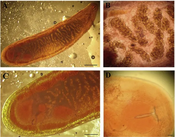
At parasitological examination, flukes showed a tongue-shaped elongate body of 2,088–2,314 μm in with and 8,268–11,830 μm in length, tapered anteriorly and rounded at the posterior end (Fig. 3A). The mouth was slightly oval and sub-terminal with a weakly developed
oral sucker. The ootype was about 110μm in diameter, while the brown-yellow eggs (Fig. 3B) measured 131.5 × 73.9μm in mean (range 122–135 μm in length and 62.4–80.6 μm in width). Two large (550–702 μm × 450–520 μm) and globular testes were situated ob-liquely to each other, while the intertesticular oval (250 × 300μm) or round (about 334μm) ovary was placed in a longitudinal straight line with the testes (Fig. 3C). The two caeca joined posteriorly, while the vitelline glands were arranged symmetrically and were not confluent posteriorly (Fig. 3C). The oval and well-developed pharynx measured 250–309 μm, while the genital pore was post-pharyngeal (Fig. 3D). For morphology and dimensions, thefluke here examined was identified as M. polonicum according toMachalska (1980), who described this spe-cies in T. merula. The diagnosis was also consistent with other pre-viously published data of M. polonicum in turdids (Visconti et al., 1988; Sitko et al., 2017;Jaume-Ramis and Pinya, 2018).
Molecular analyses of mitochondrial (CO1 and ND1) DNA loci confirmed the morphological diagnosis as revealed by the maximum likelihood approach (Fig. 4). The sequence of the CO1 locus was identical with one of previously published M. polonicum sequences (KU877887) and differed by only 0.003 base substitutions per site from other two M. polonicum sequences (Table S3). The newly obtained se-quences of the ND1 locus of both examined infection cases were iden-tical. The ND1 sequences differed by 0.005–0.012 base substitutions per site from previously identified M. polonicum cases from T. merula and Turdus philomelos, while the closest another previously sequenced cy-clocoelid, Cyclocoelum mutabile, differed by 0.370–0.386 base sub-stitutions per site (Table S4). Combined, both analyzed markers unan-imously confirm the diagnosis of the examined isolates as M. polonicum.
4. Discussion
In Europe, M. polonicum infections in Turdidae birds have been previously reported in Poland (Machalska, 1980; Sulgostowska and Fig. 2. Turdus merula female, histology of different coelomic organs. A. Lower power magnification of an area where flukes are adherent to the liver. The presence of in-flammatory infiltrate is observed on the Glissonian capsule (arrow) in the contact area with the parasite. At this magnification a general overview of the parasite is also clear: the cephalic portion (arrow-head), different internal organs and the uterus filled by the ova (asterisk) are appreciated (H&E, scale bar = 0.2 cm).B. Detail of the contact area between the fluke tegument and liver serosa, involved in the in-flammatory reaction (arrow). Note the pyogranulomatous exudate represented by large amounts of mononuclear cells with interspersed heterophils in the area of close contact with the parasite (asterisk). Trematode eggs and an internal gland (arrow-head) are also visible (H&E, scale bar = 200μm). C. Fluke localization on the kidney capsule: note the same inflammatory reaction (arrow) described in the liver, in the areas of more close contact (H&E, scale bar = 2 mm).D. Air sacs inflammation and modification in the site of parasite attach-ment (arrow). Note the previously de-scribed inflammatory infiltrate and the al-tered lung parenchyma (asterisk) in the area of the affected air sac. The pulmonary
par-Fig. 3. Morishitium polonicum from the air sacs of Turdus merula. A. Tongue-shaped specimen of M. polonicum. In the posterior part of the body are clearly visible the two large testes and the ovary lying between them. (scale bar = 200μm). B. Anterior end of M. polonicum showing the eggs inside the uterus. (scale bar = 100 μm). C. Posterior end of M. polonicum showing two globular testes situated obliquely to each other, an intertesticular oval ovary placed in a longitudinal straight line with the testes, two caeca joined posteriorly and two symmetrical vitelline glands not confluent posteriorly. (scale bar = 300 μm). D. The oral sucker, the pharynx, the genital pore of M. polonicum in the anterior end (scale bar = 50μm).
Fig. 4. Maximum likelihood analyses of sequences of mitochondrial DNA loci of the newly isolated Morishitium polonicum and previously sequenced Cyclocoelidae. (A) CO1, (B) ND1. The bars indicate the number of substitutions per nucleotide.
Czaplinska, 1987), Italy (Visconti et al., 1988), Czech Republic (Sitko et al., 2017) and Spain (Jaume-Ramis and Pinya, 2018). In Italy, M. polonicum was previously reported in T. merula from an area neigh-boring that where the blackbirds here examined were found (Visconti et al., 1988). Dimensions and morphology of the species reported by Visconti et al. (1988) are very similar to that of the specimens here examined.
Previously,Dronen (2007)andDronen and Blend (2015)suggested that in the original description byMachalska (1980), the eggs of M. polonicum from T. merula (150 × 81μm) are larger compared to those from Turdus philomelos (maximum egg size 139 × 69μm), which was indicative that they are two different Morishitium species. Currently, there are recognized four Morishitium spp. Infecting thrushes, namely Morishitium bivesiculatum (Prudhoe, 1944), Morishitium dollfusi (Timon-David, 1950), Morishitium petrowi (Oganesov, 1959) and M. polonicum. The available material does not allow to test whether all of them re-present valid species, but based on morphology and measurements, the species occurring in the two birds here examined matches clearly the measurements provided byMachalska (1980)for M. polonicum.
M. polonicum typically inhabits the air sacs of thrushes and its life cycle include terrestrial snails, mainly of the genus Helicella, as inter-mediate hosts (Machalska, 1980;Jaume-Ramis and Pinya, 2018). Adult trematodes reside in the respiratory system and release eggs into the air sacs of the definitive avian host. These eggs are then excreted via re-spiratory secretions. In the environment, eggs hatch into miracidia, which asexually reproduce within the snail intermediate host, after burrowing into its muscular foot. Miracidia then develop into rediae to produce the metacercariae, the infective life stage. The definitive avian host consumes the intermediate host containing the metacercaria, and adultfluke development proceeds (Delaski et al., 2015).
In wild animals, parasites may negatively affect the health, survival, growth and reproduction (Norte et al., 2009; Thompson et al., 2010, 2013;Brym et al., 2018). Moreover, in wild animals a huge diversity of factors, as seasonal migration, environmental changes, human inter-ventions in wild areas, immunology and diets, including changes in food and habits, may influence or increase the risk and susceptibility to parasitic infections at the individual or population level (Thompson et al., 2010,2013;McDonald et al., 2018;Moudgil and Singla, 2013; Hawley and Altizer, 2011; Van Hemert et al., 2014). Many parasitic helminth species often responsible for significant disease in their animal hosts, are transmitted by the ingestion of intermediate hosts. Therefore, host diet and food habit may play an important role in the acquisition of these infections (Leung and Koprivnikar, 2016). Moreover, experi-mental studies have indicated that effects of parasitism can vary with host sex (Granroth-Wilding et al., 2017). Interestingly, it was previously observed that female birds of the family Turdidae are more frequently infected with Moristhitium spp. than males (Okulewicz and Sitko, 2012). To explain thisfinding, authors suppose that this may be linked with differences in feeding preferences between male and female turdids, since females may prefer eating snails for the intake of calcium neces-sary to build eggshells and ingested snails may contain the infective stages of Moristhitium trematodes. Nevertheless, this same reason may also represent the main factor for the high parasite load and the con-sequent death observed in the two female blackbirds examined in this study.
Although suspected (Visconti et al., 1988; Okulewicz and Sitko, 2012), no previous reports deal with disease and death caused by Morishitium spp. trematodes. Nevertheless, severe clinical signs and lesions, debilitation and death associated with infections caused by different air sac cyclocoelid trematodes, have been reported in several avian species, including free-ranging and captive animals (Cole et al., 1995; Delaski et al., 2015). In free-ranging birds, a female snail kite (Rostrhamus sociabilis plumbeus Ridgway, 1874 (Accipitriformes) from
variolaris Fuhrmann, 1904, which was considered the main contributing factors for the death of the bird (Cole et al., 1995). Necropsy of an American coot (Fulica americana) found dead in USA, revealed an in-fection caused by the air sac trematode C. mutabile associated with biliary congestion, hemopericardium, blood-filled air sacs, ruptured aorta and secondary bacterial infection (Branton et al., 1985). C. mu-tabile may also infect thrushes, and a prevalence of about 8% and an average intensity of about 17 parasites per bird were recorded in 90 examined Turdus spp. birds (Cardells et al., 2014). In avian exhibits of zoos and captive enclosures, air sac trematode infections have been frequently accounted as a cause of retardation of molting, reduced breeding performance, debilitation and death in several bird species (Libert et al., 2012;Delaski et al., 2015). Stressing factors linked with captivity are likely to exacerbate the consequences of these infections. Death often occurred after birds showing dyspnea due to physical bronchial obstruction and suffocation (Libert et al., 2012;Delaski et al., 2015). Several difficulties in the treatment and control of air sac tre-matode infections have been also reported (Dronen et al., 2009;Libert et al., 2012;Delaski et al., 2015). The life cycles of these trematodes, that include snail intermediate hosts and frequently including more than a single bird species as vertebrate definitive hosts in the life cycle of a single cyclocoelid trematode species, are considered important factors that in these facilities make the management and control of air sac trematode infections very difficult and a potentially significant problem (Dronen et al., 2006). Passeriformes are considered at greater risk of air sac trematode infections and lesions caused by these parasites than other birds (Dronen et al., 2006, 2009). Among captive Passer-iformes, death associated with air sac trematodiasis caused by Szidati-trema species, have been reported in Irena puella (Latham, 1790) and Thraupis episcopus (Linnaeus, 1766) (Delaski et al., 2015). In other captive avian species, severe clinical signs and death have been re-ported in Lybius dubius (Gmelin, 1788) heavily infected with Szidati-trema yamagutiiDronen et al. (2006)and, mainly, in Momotus momota (Linnaeus, 1766) infected with Circumvitellatrema momotaDronen et al. (2009)(Dronen et al., 2009;Libert et al., 2012;Delaski et al., 2015).
Although infections caused by M. polonicum were previously re-ported in blackbirds and in other European turdids (Machalska, 1980; Sulgostowska and Czaplinska, 1987;Visconti et al., 1988;Sitko et al., 2017;Jaume-Ramis and Pinya, 2018), associated pathological lesions were never reported before. However, as observed in the blackbirds examined in this study, severe cyclocoelid air sac trematode infection with associated airsacculitis, often fibrinous, pyogranulomatous or granulomatous, and with pyogranulomatous bronchitis and peribron-chitis and secondary bacterial infections, have been reported in dif-ferent species of birds, including both Passeriformes and other avian taxa, deceased after infections by cyclocoelid air sac trematodes (Cole et al., 1995;Libert et al., 2012;Delaski et al., 2015). Bronchiectasia, atelectasia and pulmonary edema observed in this study in the two examined blackbirds, were previously reported also in a raptor (Cole et al., 1995).
5. Conclusions
Parasitological, molecular and pathological analysis performed in this study identified the air sac trematode species M. polonicum as the cause for death of these two common blackbirds (T. merula). Until now, the pathogenicity of M. polonicum was poorly understood because of the lack of recorded clinical cases and pathological lesions. Despite in some previous observations Morishitium spp.-infected and deceased birds were found in poor conditions (Visconti et al., 1988;Okulewicz and Sitko, 2012), thesefindings did not definitely allow to identify these flukes as the main cause of the observed signs. Nevertheless, the diffuse and highly severe gross and, mainly, histological lesions found
asso-highlighted in this study, these lesions may also be complicated by other secondary pathogens. Moreover, severe histological lesions here observed in the areas where M. polonicum adults were in contact with the serosae of the two examined blackbirds, may represent a further indication of a direct involvement of this parasite in the etiology of observed lesions. The present study is thefirst report of pathological lesions and death caused by M. polonicum in the common blackbird. Declarations of interest
None.
Conflict of interest
Authors declare no conflict of interest. Acknowledgements
We thank Miroslav Těšínský for expert technical assistance. Appendix A. Supplementary data
Supplementary data to this article can be found online athttps:// doi.org/10.1016/j.ijppaw.2019.03.021.
Financial support
This research did not receive any specific grant from funding agencies in the public, commercial, or not-for-profit sectors.
References
BirdLife International, 2016. "Turdus merula". The IUCN Red List of Threatened Species. IUCN 2016: e.T103888106A87871094.
doi:10.2305/IUCN.UK.2016-3.RLTS.T103888106A87871094.en , Accessed date: 8 October 2018.
Bowles, J., Blair, D., McManus, D.P., 1992. Genetic variants within the genus Echinococcus identified by mitochondrial DNA sequencing. Mol. Biochem. Parasitol. 54, 165–173.
Branton, S.L., Deaton, J.W., Gerlach, H., Ruff, M.D., 1985. Cyclocoelum mutabile infection and aortic rupture in an American coot (Fulica americana). Avian Dis. 29, 246–249.
Bray, R.A., Littlewood, D.T.J., Herniov, E.A., Williams, B., Henderson, R.E., 1999. Digenean parasites of deep-sea teleosts: a review and case studies of intrageneric phylogenies. Parasitology 119, 5125–5144.
Brym, M.Z., Henry, C., Kendall, R.J., 2018. Elevated parasite burdens as a potential mechanism affecting northern bobwhite (Colinus virginianus) population dynamics in the Rolling Plains of West Texas. Parasitol. Res. 117, 1683–1688.
Calegaro-Marques, C., Amato, S.B., 2014. Urbanization breaks up host-parasite interac-tions: a case study on parasite community ecology of Rufous-bellied Thrushes (Turdus rufiventris) along a rural-urban gradient. PLoS One 9, e103144.
Cardells, J., Ortega, J., Martinez-Herrero, M.C., Martischarfhausen, M.R., Villamayor, M., Dominguez, V., Pereira, P., Catalagregori, P., Severino, R., Garijo, M., 2014. Influencia del estado parasitario de los tordos (Turdus spp.) sobre su condición cor-poral. In: Proceedings of the 51 Congreso Científico de Avicultura (Valencia) 2-3 October 2014, . http://www.wpsa-aeca.es/articulo.php?id_articulo=3724.
Ching, H.L., 1993. Helminths of varied thrushes, Ixoreus naevius and robins Turdus mi-gratorius, from British Columbia. Proc. Helminthol. Soc. Wash. 60, 239–242.
Clement, P., Hathway, R., Wilczur, J., 2000. Thrushes (Helm Identification Guides). Christopher Helm Publishers Ltd., London, UK.
Cole, R.A., Thomas, N.J., Roderick, C.L., 1995. Bothrigaster vanolans (Trematoda: Cyclocoelidae) infection in two Florida snail kites (Rostrhamus sociabilis plumbeus). J. Wildl. Dis. 31, 576–578.
Delaski, K.M., Nelson, S., Dronen, N.O., Craig, T.M., Pond, J., Gamble, K.C., 2015. Detection and management of air sac trematodes (Szidatitrema species) in captive multispecies avian exhibits. J. Avian Med. Surg. 29, 345–353.
Dronen, N.O., Craig, T.M., Hammond, E.E., 2006. Szidatitrema yamagutii n. sp. (Digenea: Cyclocoelidae: Ophthalmophaginae) from the bearded barbet, Lybius dubius (Capitionidae), and the white-necked myna, Streptocitta albicollis (Sturnidae), that died at the Audubon Zoo in New Orleans, Louisiana, U.S.A. Zootaxa 1219, 59–68.
Dronen, N.O., 2007. Revision of the family Cyclocoelidae Stossich, 1902 with the pro-posal of two new subfamilies and the description of a new species of Morishitium Witenberg, 1928 from the common snipe, Gallinago gallinago, from Texas, U.S.A. Zootaxa 1563, 55–68.
Dronen, N.O., Greiner, E.C., Ialeggio, D.M., Nolan, T.S., 2009. Circumvitellatrema momota n. gen., n. sp. (Digenea: Cyclocoelidae: Cyclocoelinae) from a captive-hatched blue-crowned motmot, Momotus momota (Momotidae). Zootaxa 2161, 60–68.
Dronen, N.O., Blend, C.K., 2015. Updated keys to the genera in the subfamilies of
Cyclocoelidae Stossich, 1902, including a reconsideration of species assignments, species keys and the proposal of a new genus in Szidatitreminae Dronen, 2007. Zootaxa 4053, 1–100.
Granroth-Wilding, H.M., Daunt, F., Cunningham, E.J., Burthe, S.J., 2017. Between-in-dividual variation in nematode burden among juveniles in a wild host. Parasitology 144, 248–258.
Grünberg, W., Kutzer, E., 1964. Die Pathologie verschiedener Trematodeninfektionen bei Störchen (Ciconia ciconia L., Ciconia nigra L.). Zentralblatt für Veterinarmed. 11B, 712–727.
Hamer, G.L., Muzzall, P.M., 2013. Helminths of American robins, Turdus migratorius, and house sparrows, Passer domesticus (order: Passeriformes), from suburban Chicago, Illinois, USA. Comp. Parasitol. 80, 287–291.
Hamilton, W.D., Zuk, M., 1982. Heritable truefitness and bright birds: a role for para-sites? Science 218, 384–387.
Hawley, D.M., Altizer, S.M., 2011. Disease ecology meets ecological immunology: un-derstanding the links between organismal immunity and infection dynamics in nat-ural populations. Funct. Ecol. 25, 48–60.
Höfle, U., Krone, O., Blanco, J.M., Pizarro, M., 2003. Chaunocephalus ferox in free-living white storks in Central Spain. Avian Dis. 47, 506–512.
Hudson, P.J., Dobson, A.P., Newborn, D., 1998. Prevention of population cycles by parasite removal. Science 282, 2256–2258.
Jaume-Ramis, S., Pinya, S., 2018. First record of Morishitium polonicum (Machalska, 1980) (Trematoda, Cyclocoelidae) parasitizing Turdus philomelos Brehm, 1831 in Mallorca (Balearic Islands, Spain). Boll. Soc. Hist. Nat. Balears 61, 9–15.
Leung, T.L., Koprivnikar, J., 2016. Nematode parasite diversity in birds: the role of host ecology, life history and migration. J. Anim. Ecol. 85, 1471–1480.
Libert, C., Jouet, D., Ferté, H., Lemberger, K., Keck, N., 2012. Air sacfluke
Circumvitellatrema momota in a captive blue-crowned motmot (Momotus momota) in France. J. Zoo Wildl. Med. 43, 689–692.
Liptovszky, M., Majoros, G., Perge, E., 2012. Cathaemasia hians in a Black Stork (Ciconia nigra) in Hungary. J. Wildl. Dis. 48, 809–811.
Literák, I., Sitko, J., 2006. Where in Europe should we look for sources of the cutaneous trematode Collyriclum faba infections in migrating birds? J. Helminthol. 80, 349–355.
Loye, J.E., Zuk, M., 1991. Bird-parasite Interactions. Oxford Ornithology Series. Oxford University Press, Oxford.
Machalska, J., 1980. Cyclocoelum polonicum sp. n. (Trematoda, Cyclocoelidae) from the thrushes Turdus philomelos Br. and T. merula L. Acta Parasitol. Pol. 26, 129–136.
Mani, P., Rossi, G., Perrucci, S., Bertini, S., 1998. Mortalità dei merli (Turdus merula) in Toscana. Selezione Veterinaria 8, 749–753.
Martínez, F., Hernández, S., Calero, R., Becerra, C., Moreno, T., Domínguez de Tena, M., Acosta, M.I., 1977. Parásitos de aves paseriformes en la Provincia de Córdoba. Rev. Iber. Parasitol. 37, 133–141.
McDonald, J.L., Robertson, A., Silk, M.J., 2018. Wildlife disease ecology from the in-dividual to the population: insights from a long‐term study of a naturally infected European badger population. J. Anim. Ecol. 87, 101–112.
Møller, A.P., Allander, K., Dufva, R., 1990. Fitness effects of parasites on passerine birds: a review. In: Blondel, J., Gosler, A., Lebreton, J.D., McCleery, R. (Eds.), Population Biology of Passerine Birds. Springer, Berlin, Heidelberg, pp. 269–280.
Morgan, J.A., Blair, D., 1998. Mitochondrial ND1 gene sequences used to identify echi-nostome isolates from Australia and New Zealand. Int. J. Parasitol. 28, 493–502.
Moudgil, A.D., Singla, L.D., 2013. Role of neglected wildlife disease ecology in emergence and resurgence of parasitic diseases. Trends Parasitol. Res. 2, 18–23.
Norte, A.C., Araujo, P.M., Sampaio, H.L., Sousa, J.P., Ramos, J.A., 2009. Haematozoa infections in a Great Tit Parus major population in Central Portugal: relationships with breeding effort and health. Ibis 151, 677–688.
Okulewicz, A., Sitko, J., 2012. Parasitic helminthes– probable cause of death of birds. Helminthologia 49, 241–246.
Okulewicz, A., Okulewicz, J., Sitko, J., Wesołowska, M., 2010. New records of digenean flukes (Trematoda) in birds in Poland. Wiad. Parazytol. 56, 67–70.
Perrucci, S., Rossi, G., Marconcini, A., Mani, P., 1997. Acantocefali del merlo (Turdus merula). Selezione Veterinaria 8, 827–882.
Sitko, J., Bizos, J., Heneberg, P., 2017. Central European parasiticflatworms of the Cyclocoelidae Stossich, 1902 (Trematoda: Plagiorchiida): molecular and comparative morphological analysis suggests the reclassification of Cyclocoelum obscurum (Leidy, 1887) into the Harrahium Witenberg, 1926. Parasitology 144, 368–383.
Sitko, J., Zaleśny, G., 2014. The effect of urbanization on helminth communities in the Eurasian blackbird (Turdus merula L.) from the eastern part of the Czech Republic. J. Helminthol. 88, 97–104.
Slater, R.L., 1967. Helminths of the robin, Turdus migratorius Ridgway, from Northern Colorado. Am. Midl. Nat. 77, 190–199.
Snow, D.W., 1988. A Study of Blackbirds. London British Museum of Natural History, London, UK.
Sulgostowska, T., Czaplinska, D., 1987. Pasozyty ptakow? Parasiti avium. Zeszyt 1. Pierwotniaki i Przywry. Protozoa et Trematoda. Katalog Fauny Pasozytniczej Polski. Panstwowe Wydawnictwo Naukowe, Wroclaw, pp. 210.
Thompson, R.C.A., Lymbery, A.J., Smith, A., 2010. Parasites, emerging disease and wildlife conservation. Int. J. Parasitol. 40, 1163–1170.
Thompson, R.C.A., 2013. Parasite zoonoses and wildlife: One health, spillover and human activity. Int. J. Parasitol. 43, 1079–1088.
Van Hemert, C., Pearce, J.M., Handel, C.M., 2014. Wildlife health in a rapidly changing North: focus on avian disease. Front. Ecol. Environ. 12, 548–556.
Visconti, S., Giovannetti, L., Canestri-Trotti, G., 1988. Su di un trematode del genere Cyclocoelum Brandes, 1892, parassita dei sacchi aerei di Turdus merula L. Parassitologia 30 (Suppl. 1), 211–212.