Università della Calabria
Dipartimento di Ingegneria per l’Ambiente e il Territorio e
Ingegneria Chimica
Dottorato di Ricerca in Ingegneria Chimica e dei Materiali
SCUOLA DI DOTTORATO " PITAGORA " IN SCIENZE INGEGNERISTICHE
Con il contributo dell’Istituto per la Tecnologia delle Membrane
del Consiglio Nazionale della Ricerca ITM-CNR
CICLO XXVI
BIODEGRADABLE POLYMERIC
MEMBRANE SYSTEMS
FOR TISSUE ENGINEERING APPLICATIONS
Settore Scientifico Disciplinare CHIM07 – Fondamenti chimici delle tecnologie
Coordinatore: Chiar.mo Raffaele Molinari
Supervisori: Dott.ssa Loredana De Bartolo Ing. Efrem Curcio
Dottoranda: Dott.ssa Antonietta Messina
3
Table of contents
General introduction and Aim of the work 4
Chapter 1: Biodegradable polymeric membrane for tissue engineering application
Introduction 7
1.1 Tissue Engineering: history and definition 7 1.2 Biological aspect involved in the Tissue Engineering approach 10
1.3 Key concepts 12
1.4 Scaffolds for Tissue Engineering 13
1.5 Scaffolds production and processing methods 15
1.6 Biomaterials for scaffold fabrication 20
1.7 Polymeric membranes as biomaterials in Tissue Engineering and medical
applications 25
References 34
Chapter 2: Preparation and characterization of polymeric membranes
Introduction 38
2.1 Polymeric membrane preparation 38
2.2 Membrane characterization 45
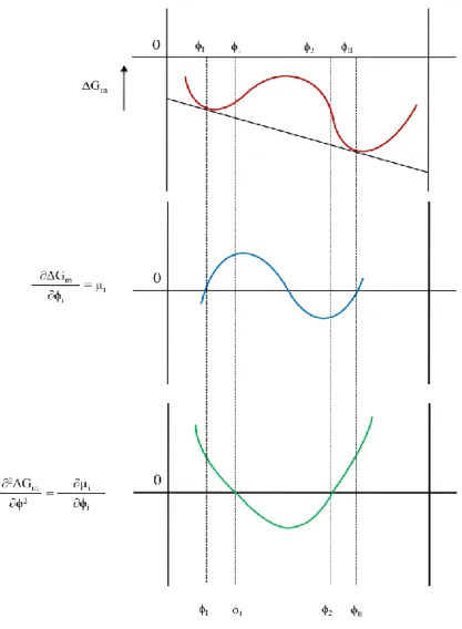
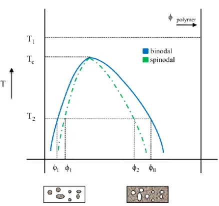
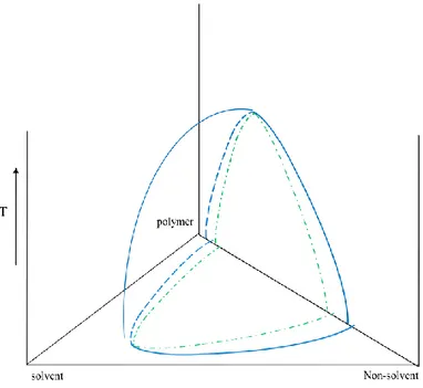
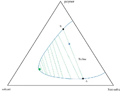
2.3 Thermodynamic principles 50
References 63
Chapter 3: Tissue Engineering scaffold-free: a new approach
Introduction 65
3.1 Self-assembly and self-organization 66
3.2 Energy minimization during the self-assembly process 68
3.3 Tissue fusion in self-assembly tissues 69
3.4 Self-assembly in Tissue Engineering 72
3.5 Self-organization in Tissue Engineering 77
3.6 Conclusion and future direction 78
References
Chapter 4: Development of biodegradable polymeric membranes for TE application
Introduction 81
Materials and methods
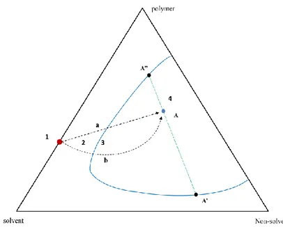
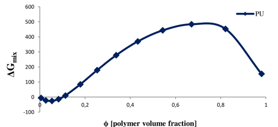
4.1 Thermodynamic analysis 89
4.2 Membrane preparation 90
4.3 Morphological investigation 90
4.4 FT-IR analysis, wettability, surface properties and porosity 91
4.5 Mechanical properties determination 91
4.6 Dissolution profile: Biodegradability 91
Results 92
Discussion 95
References 102
Chapter 5: Neuronal growth and differentiation on biodegradable membranes
Introduction 105
Materials and methods
4
5.2 Membrane characterization 107
5.3 Cell culture 108
5.4 MTT assay 109
5.5 Sample preparation for SEM 109
5.6 Immunostaining for neuronal cells and quantitative analysis 109
5.7 Western blotting 110
Results 110
Discussions 114
References 125
Chapter 6: Bio-hybrid membrane system as temporary support for the self-assembly process of tissue spheroids
Introduction 127
Materials and methods
6.1 Membrane preparation 130
6.2 Inert agarose mould preparation 131
6.3 Membrane characterization 131
6.4 Cell cultures 132
6.5 Spheroids culture on polymeric membranes 133
6.6 Morphological analysis 133
6.7 Fusion process evaluation 134
6.8 Glucose consumption and lactate production 134 6.9 Oxygen permeation and central hypoxia evaluation into spheroids 134
Results 135
Discussion 138
References 156
Chapter 7: Polycaprolactone-Hydroxyapatite composite membrane scaffolds for bone tissue engineering
Introduction 157
Materials and methods 158
Results and discussions 160
Conclusions 161
References 162
General conclusion 165
Acknoledgements 167
5
General introduction and Aim of work
Watching the cells grow and building up a new tissue outside the body has been the new insights appeared with the TE development. The deepening study on the interaction of cells and tissues with biomaterials in a specific arrangement contributed new cognitions about the influence of several technical and biological factors involved in the tissue regeneration process and consequently on the scaffolds design and its outcome. Such factors are for example mechanical loading, biomaterial degradation behavior, cell number or the cell type in contact with the material. The main future challenge is the complete investigation not onlu of the tissue properties that needs to be replaced and their transfer to the biomaterial used for the tissue regeneration, but above all the understanting of how the biomaterial properties influence the tissue formation and functionalization, that means definig the perfect biomaterial characteristics for each possible application in TE field and regenerative medicine. Otherwise an alternative approach to the classical scaffold-based TE, named scaffold-free TE has been developed, in order to reproduce the native embryonic condition during the tissue in vitro production, relying on the self assembly potential of cells and their secretion of a specific extracellular matrix network. Tissue spheroids have been used for this purpose, as building blocks for the biofabrication of three-dimensional functional living macrotissues and organ constructs, with no scaffolds or supports request. On the other side, not all the cells are able to self-assemble themselves without external stimuli, and the time requested for the spheroids to be compact enough for the handling, sometime is so long to ensure the perfect vitality of the engineered tissue, and eventually cells go through necrosis. Despite the amazing results reached and published all around the world, both the approaches appear to be still rich in disadvantages: scaffold degradation is rarely synchronized to neotissue formation, making the tissue remodeling and its integration difficult, thus compromising functional properties. Furthermore, toxicity and immunogenicity due to scaffold creation, seeding, or degradation are of concern [Liu et al., 2004], and the presence of a scaffold may also alter the phenotype of cells that come into contact with it [Levy-Mishali et al., 2009]. One can infer that in the near future new pathways able to overcome the limitations for both the TE approaches will be not easy available. This is the reason way, despite the considerable progress in the field of biomaterials and life science a continuous development of new insights is required for the recreation of a tissue engineered system.
6
Thus, this study aimed to the development and the design of new bio-artificial systems able to reproduce in vitro an engineered tissue/organ mimic usable for TE applications and furthermore investigations in the field of the regenerative medicine.
The first section of this report, will think back to the state of the art and the literature references about the two TE approaches (chapter 1-2-3), and then, the following chapters will present the experimental work done in the last three years.
1) (chapter 4) The development of polymeric membranes with biodegradable properties is shown. It is well known how the morphological, physicochemical, mechanical and dissolution properties of a polymeric substrates are dependent on the materials choosen and on the processing methods used for their preparation. Starting from the uncountable literature references available, Chitosan, Polycaprolactone and Polyurethane have been purposely choosen and molded through the phase inversion technique as flat biodegradable membranes and investigated in their properties, in order to understand if they could be eventually used as substrates for tissue engineering applications.
2) (chapter 5) The already set biodegradable polymeric membranes were tested to investigate the efficacy to promote the adhesion and differentiation of neuronal cells. The human neuroblastoma cell line SHSY5Y, a wellestablished system for studying neuronal differentiation, has been used as biological component on the bio-hybrid system. The investigation of viability and specific neuronal marker expression allowed assessment that the correct neuronal differentiation and the formation of neuronal network had taken place in vitro in the cells seeded on different biodegradable membranes.
3) (chapter 6) The biodegradable polymeric membranes were used to highlight if they can act as substrates for sustaining the fusion process of tissue spheroids, nowadays considered as the new building-blocks for the engineered tissue and organ mimics. Conbining the use of substrates with spheroids in a unique system is a completely new approach, in the TE field. Generally in fact a polymeric membrane or a scaffold are used for the development of bio-hybrid system that reproduce only partially the in-vivo regeneration process, being a 2D system; whereas the spheroids are investigated in an environment completely inert only focusing on the cell-cell interaction. On the contrary, in this case spheroids obtained from three defferent cell lines, SH SH 5Y, Fibroblast and Myoblast, and their fusion process, the biological activity and the level of necrosis hypoxia-induced have been investigated on the polymeric membranes
7
developed in order to highlights how the properties of the obtained substrates can influence the tissue maturation in time with respect to the scaffold-free TE approach, for this study represented by an agarose support.
4)
(chapter 7) Polycaprolactone (PCL) and hydroxyapatite (HA) were used in order to develop novel controlled nanostructured biomaterials for bone tissue engineering applications. After preparation, membrane scaffolds were characterized in order to evaluate its morphological, physico-chemical and mechanical properties and then used for the osteoclast cell culture.8
Chapter 1
Biodegradable polymeric membranes for Tissue engineering
and medical applications
Introduction
Repairing and replacing damaged or malfunctioning parts of the human body is an objective with ancient roots in history. Etruscans and Egyptians learned more than 2500 years ago how to operate to substitute teeth, build a bridge with animal bones to sustain limb fractures and create amalgams in order to prevent infections and let wounds heal and regenerate. Since then in every time more sophisticated inventions were done all around the world in order to improve the existing medical discoveries and devices and create a few new ones. But it’s only some decades ago that all the knowledge in medicine and sciences converged in what we all know as Tissue Engineering.
1.1 Tissue Engineering: history and definition
“…an interdisciplinary field that applies the principles of engineering and of life science towards the development of biological substitutes that restore, maintain and/or improve tissue or organ function […], thus the whole principles and methods suitable for establishing the foundations of structure-function relationships between healthy or pathologic mammalian tissues”. That is the first official definition reported by Fox and Skalak to summarize the outlines of the Ist congress NSF held in 1988 in California, and in which the expression “Tissue Engineering” (TE) was coined [Fox et al., 1988]. For the first time then, TE emerged as the first real significant chance to transfer the fruit of the last decades to clinical practice: a new field able to create a potential alternative or complementary solution to transplantation, surgical repair, artificial prosthesis, mechanical device and drug therapy [Chapekar et al 2000]. Few difficulties arise when surgical transplantation is used. Taking a graft from a donor and implanted it in a patient is not easy, and eventually the collateral effects could be unpredictable. Moreover the insufficient donor organs number, pathogen transmission risk, lifetime immunosuppression therapy and rejection of the replaced tissue/organ with consequent fear of a replacement within days to years after the surgery, are just some of these difficulties. Besides artificial mechanical devices or sustenance machines when used as
9
prosthesis for treating injuries or damages on human body are not less dangerous.That’s the reason why the chance to implant natural, synthetic or semi-synthetic tissue and organ mimics, that are fully functional form the start or that can be “planned” to grow into the required functionality, with the opportunity to use patient’s own cells for the creation of an autogenic tissue construct or substitute in vitro is considered an excellent alternative to direct transplantation of donor organs [Langer et al., 1993; Fucks et al. 2001; Lanza et al., 2000; Saltzman et al., 2004]. In that case not dependency on donors is required and some of the limitations of direct transplantation particularly concerning rejection and pathogen transmission could be avoided or overcome.
Eventually the TE approach appeared and it is still today really promising. First sign of this new developing field appear in the 1960s when synthetic fibers were used as skin grafts for burn treatment; first step in the clinical evolution that lead in just a decade to the development of a collagen-based artificial skin used to treat severe burns [Burke J.F. et al. 1981] and to a collagen sponge skin substitute, cross-linked with chondroitin and covered with silicone for oral mucosa injuries treatments [Levin M.P. et al. 1979]. In the last 20 years more areas of tissue engineering have been explored and applied afterwords: skin, heart, vessels, bone and cartilage replacements used in clinical trials and applications are uncountable [Blitterswijk et al., 2008; Fong et al., 2006; Jawad et al., 2007; Nesic et al., 2006]. Ten years ago cardiovascular autografts obtained with patients own cells have been implanted on children with various complex heart diseases. Cells were isolated, cultured and subsequently seeded on a biodegradable polymer scaffold of poly(glycolic acid) combined with poly(lactic acid‐ε‐caprolactone) before the insert operation, and no post‐operative complications were detected [Matsumura et al., 2003]. An allogenic engineered trachea has been designed and implanted in vivo in order to prevent an immune reaction. Macchiarini and colleagues removed from trachea all donor’s cell and antigens and subsequently autogenic cells were re-cultured on the matrix, let multiply and transplanted into the patient’s main bronchus. The tissue engineered trachea became functionalize and transplanted into the patient’s main bronchus [Macchiarini et al., 2008]. The tissue engineered trachea behave perfectly already after only 4 months from the surgery showing a normal appearance and good functional properties. In truth, replacing failing or malfunctioning organs or promoting their partial or complete regeneration after an injuries or a pathology requires more than just a collection of acknowledgements and intuitions, and framing a new trend of medical research in order to coordinate the few advances already achieved in distant areas of science is not an easy task.
10
Engineering, chemistry, physics, biology, biotechnology and medicine must be confronted and engaged in a multi-disciplinary approach to tissue, in order to develop bioactive tissue substitutes as an alternative to inert systems. Therefore, the complexity of all biological tissues in terms of macromolecular composition, ultra-structural organization and interactions between cells and environment, made it hard to switch engineered constructs in the clinical trial. For many native tissues, the in vvo stresses and strains to which they are subjected are not well defined, and furthermore, tissue properties and answers in body vary with age, site and other host factors, making it difficult to match parameters, as mechanical and physicochemical properties, to design and develop a general list of criteria for engineered tissues [Badylak et al., 2002]. Moreover highly specialized structures as articular cartilage and cardiac tissue for example, show unique biomechanical properties required to move the limbs and circulate the blood. The loss of function of these tissue due to injury, disease, or aging origin a significant number of clinical disorders as consequence. Although different in many respects, these tissues share two features quite relevant for the tissue engineering approach: (1) they lack of intrinsic capacity for self-repair and (2) they do not perform biomechanically in vitro as in in vivo conditions (Praemer et al., 1999; Thom et al., 2006).
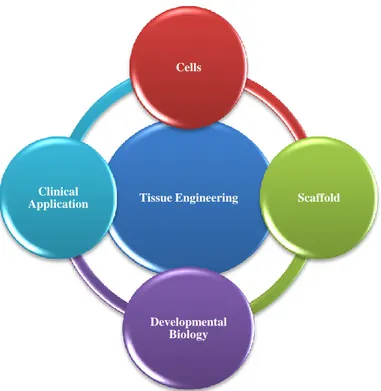
Tissue Engineering Cells Scaffold Developmental Biology Clinical Application
11
1.2 Biological aspects involved in TE approach
Biological and functional tissues basically consist of three key components: cells, signaling systems and extracellular matrix (ECM) [Lanza et al., 2000]. Therefore, combining together the three components and finding the best combination of stimuli and supports could generate an engineered tissue replacement in vitro appreciable as an excellent alternative to direct transplantation of donor organs [Langer et al., 1993; Fucks et al. 2001; Saltzman et al., 2004 ]. The cells, core of the tissue, are the main character of the tissue regeneration and repair, working and living in presence of a few stimuli and regulating factors In order to be used for TE applications, the cells’ source must be accessible and the derived lines easily expandable with physiological and phenotypical properties and functions able to endure during the experimental operations. Due to a lack of human-organ availability, the current main source of hepatocytes for bioartificial systems is exogeneic material (rat, porcine, mouse, hamster), but although they demonstrate the same qualities of human cells, these type of cell sources carry the risk of xenogenic infections and lack of metabolic compatibility. Stem cells have been suggested as interesting alternative to animal source. Stem cells, are derived from a few human tissue, as bone marrow or umbelical cord, and they are the most flexible cells known in nature, being undifferentiated in morphology and biological pathways and expressing a remarkable ability to differentiate into a desired cell type under specific stimuli.
The signaling system consists of genes that secrete transcriptional products when differentially activated, and urgesspecific cues for each process involved in the tissue formation and differentiation [Lanza et al., 2000].
The ECM defined as network‐like substance within the extracellular space, is able to supports cell attachment, cell-cell interactions and promotes cell proliferation [Badylak et al., 2007; Blitterswijk et al., 2008]. The native ECM is composed basically of water, proteins and polysaccharides, but in truth, each tissue has an ECM with a unique composition and topology, generated during tissue development through a dynamic biochemical and biophysical dialogue between the various cellular components and the cellular microenvironment. Indeed, the physical, topological, and biochemical composition of the ECM made it not only tissue-specific, but also highly heterogeneous. Moreover, the ECM is a highly dynamic structure that is constantly remodeled, through a few post-translational modifications. Thanks to these physical and biochemical characteristics the ECM generates the biochemical and mechanical properties requested of each organ, i.e. tensile and compressive strength and elasticity, mediating the extracellular homeostasis and the water
12
retention. Furthermore, the ECMs guides morphological organization and physiological function of a tissue by binding the growth factors (GFs) and interacting with cell-surface receptors to elicit transduction signals and regulating gene transcriptions [Frantz et al., 2010]. Collagen, is a fibrous protein and a major natural extracellular matrix component. It is the most abundant protein in mammals and is the main structural element
In skin, bone, tendon, cartilage and blood vessels and heart valve [Creighton, 1993; Kose et al., 2005; Lee et al., 2000; Taylor et al., 2006]. There are 25 types of collagen differing in their chemical composition and molecular structure have been identified. Te bulk of interstitial collagen is transcribed and secreted by fibroblasts, but a few cells can recruit it from neighboring tissues and collect it in their stroma [De Wever et al., 2008]. Due to the importance of the ECM in so many fundamental cellular processes, a lot of tissue-culture models have been developed to study its biochemical and biophysical properties in order to understand the mlolecular origins and the regulation mediated by ECM. Tissue engineers and biomaterial specialists have generated ECM scaffolds from various tissues [Macchiarini et al., 2008], and once combined with colonies of seeded cells, they showed the ability to reconstitute normal tissues with reasonable fidelity to the native one (Lutolf et al., 2009). ECMs have also been isolated and extracted from various tissues, such as small intestine, skin (from cadavers), pancreas and breast [Rosso et al., 2005], and they have been used to engineer skin grafts, for enhancing wound healing and to study tumor progression through the ECM changes in time [Badylak et al., 2007]. The role played by ECM proteins in tissue development was further demonstrated in relation with the surface chemistry and the topographic properties. The presence of a fibronectin layer adsorbed on a Ca-P thin film improved osteoblast response in terms of adhesion, proliferation and differentiation; moreover on underlying regular Ca-P surface topography cells showed negative statistically significant differences in focal adhesion assembly, protein expression and features [Cairns et al., 2010]. Appeared clear that the chance to reproduce through new biomaterials an ECM-like system would represent the best way to improve the biological answer and the tissue regeneration in
vitro. The considerable progress in the field of biomaterials science led researchers nowadays
to reproduce and then benefit artificial ECMs in different conformation (scaffolds or membanes) and use them as support for engineering a new tissue from isolated cells. Properly designed, these artificial ECMs provide an appropriate environment and the mechanical support for the tissue formation and regeneration [Langer et al., 1993; Abatangelo et al., 2001; Putnam et al., 1996].
13
1.3 Key concepts
TE in vitro approach, showed in figure 1.2, foresees three steps basically: 1) Cells isolation by biopsy from a patient or a donor and their consequent amplification in number with conventional methods in vitro. Cells are cultured under specific conditions and regulated parameters, as humidity, CO2 level, pH, temperature and medium composition (growth factors
and nutrients). 2) Cell seeding or impregnation on a biomaterial in form of scaffold, membrane, hydrogel or carrier where growth, proliferation and differentiation are stimulated and sustained. 3) Maturation of the new engineered construct until functional tissue/organ-like units are obtained. The new tissue answers are eventually monitored, investigated and recorded in order to collect informations about its biochemistry and physiology, main research step in order to use it for further in vitro studies, i.e. pharmacological treatment for the drug response, or the final implantation into the patient for in vivo experimentations, avoiding clinical problems as stress shieding, allergic reactions, wear particles and chronic inplammatory reactions.
14
The ECM-like scaffolds, with specific physical, mechanical and biological properties, act as substrates for cellular growth, proliferation and support for new tissue formation. Then independently on the application it will be used for, the final bio-hybrid device need to imitate in their composition and structure the naïve and physiological condition for specific cells to perform complex biochemical funcions, including adaptive control and the replacement of normal living tissues, once the new mimic is obtained.
1.4 Scaffolds for TE
The design of a scaffold ultimately determines the functionality of the grown tissue. Scaffold design comprehends the material and the processing method used, and additionally the appearance of the construct (shape, size and surface topography). As already explained, since each type of tissue requires particular conditions, the understanding of all the specific natural biological environment requested in vivo must be known to allow optimization of culturing in
vitro. Mass transport and biophysical signaling, showed to improve and control the structure,
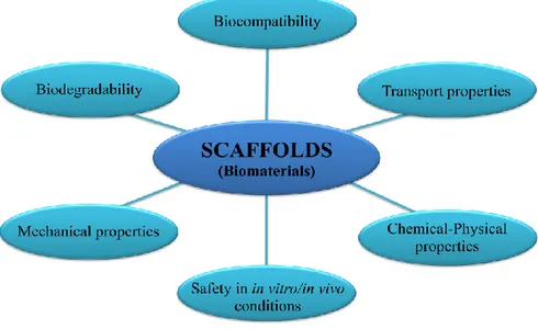
composition, and functional properties of engineered tissues. A scaffold is defined as a solid biomaterials properly designed in order to perform specific functions and can be considered as a surrogate of the ECM, that biologically contributes to mechanical integrity and has important signaling and regularity functions in development, maintenance and regeneration of tissues [Langer et al., 2004; Muschler et al., 2005; Lutolf et al., 2005]. Although the final requirements depend on the specific purpose of the scaffold and its final application, several characteristics (fig. 1.3) are specifically requested for all designs [Hutmacher et al., 2000; Moroni et al., 2008]. The scaffold should be/have: 1) biocompatible; it should induce an appropriate biological response in a specific application and prevent any adverse response of the surrounding tissue [Babensee et al., 1998; Williams et al., 2008]. 2) biodegradable; the scaffold materials should degrade in tandem with tissue regeneration and remodeling its matrix into smaller non‐toxic substances without interfering with the function of the surrounding tissue [Hutmacher et al., 2001]. The decomposition rate of a scaffold directly depends on the chemical-physical characteristics of the biomaterial it is made of, and it can be adjusted by modification of the crystal phase and structure of the starting material and the ratios of all the elements in the system. The degradation rate has to be adapted to the tissue reconstruction going along with the progressive healing and tissue synthesis process, thus the cutback of the scaffold can be varied within the time scale between several weeks to months,
15
with products of degradation that must not disturb the cell and the regeneration process. 3) promote cell attachment, spreading and proliferation; required for the regulation of cell growth and differentiation [Ito et al., 2007]. The adhesion process is for most cell types a prerequisite for a functional differentiation, matrix production and survival. Its quality (e.g. adhesion kinetics and bonding strength between biomaterial and cell) depends on the charge distribution, the wettability and the surface structure in the nanometer scale. The proliferation rate and migration too are higher on smooth materials than on rougher surfaces, then regulating the scaffold surface topography the tissue formation can be sustained and improved in terms of organization and increasing of functions [Wang et al., 2010; De Bartolo et al., 2007, 2008; Papenburg et al., 2007]. 4) suitable mechanical strength; scaffold endurance and stiffness should be comparable to in vivo tissue in the site of implantation; that means, a scaffold requires flexibility or rigidity depending on its final application, i.e. cardiovascular versus bone prostheses [Mitragotri et al., 2009]; 5) good transport properties; if it has to be used as container for cell culture it must ensure sufficient nutrient transport towards the cells and removal of waste products, then good porosity and pore connectivity are essential [Agrawal et al., 2001; Karande et al., 2004]. A sufficient nutrient supply and exchange of metabolic products of cells within a scaffold is the main request in vitro and in vivo conditions, then the basis of a successful tissue engineering product lies in a scaffold able to combine them in its substructure. Large pore sizes with an interconnective pore structure can create the perfect conditions for the mass transport and the diffusion of nutrients and growth factors necessary to keep the cells inside vivid, and lower ones ensure the requested integrity and sustainability when mechanical load is applied [Chirila 2001]. The needed pore properties of a scaffold that enable the ingrowth of functional tissue in a scaffold material depend on the desired tissue type; for the synthesis of vital tissue it has been proved the necessity of a mean pore diameter of about 50 µm for the soft systems, about 100 µm diameter for the ingrowth of rigid material (as the extracellular phase of osteoblasts), and at least 450 µm diameter for a vivily vascularized tissue. 6) easy to connect to the vascular system of the host as soon as implanted; to ensure good nutrient supply throughout the scaffold post‐implantation, the scaffold should be connected to the natural nutrient supplying system [Hutmacher et al., 2000; Agrawal et al., 2001, Kannan et al., 2005].
16
Fig. 1.3 Characteristics requested for a scaffolds (biomaterials) suitable for Tissue Engineering applications
These requirements are fulfilled by choosing the proper biomaterial and a specific production process as described in the following paragraphs.
1.5 Scaffolds production and processing methods
Many methods have been used for the production of scaffolds, membranes or hydrogels to be used as framework materials for the creation of microenvironments that reproduce the metastable tissues surrounding. The TE community has begun to capitalize all the methods available for the material processing since 1980s, in order to highlight the best techniques through which rproduce scaffolds able to mimic the native ECM. This section reports only some of the more representatives between the processing methods for the scaffold preparation, whereas a summary presentation of other techniques is reported in table 1.1.
i. Solid freeform fabrication (SFF) uses a computer-generated models to
layer-manufacturing supports for tissue culture with improved parameters such as pore size, porosity and pore distribution. Layer by layer every section of the scaffold is generated and linked one to another until the final shape and dimension is reached. As results of oxygen and nutrients mass transport into the inner mesh of the scaffold are increased supporting cellular growth in all the regions [Sachlos et al., 2003].
ii. Electrospinning [Pham et al., 2006] allows the production of polymer fibers with
variable diameters [Subbiah et al., 2005]. The process consists of applying to a solution an electric field through a high voltage source inducing in a charge repulsion
17
within the solution. When the generated system is stable a jet stream is initiated and the solvent is induced to evaporate from the solution and the resulting fiber is collected. Controlling and varying the process parameters, as viscosity of the solution, the dielectricconstant of the solvent, its conductivity, the surface tension of the polymer, its molecular weight, the distance between tip and collector, the flow rate, the strength of the electric field, it is possible to obtain a final product with all the specific characteristics required.
iii. The Solvent Casting methods foresees the dissolution of a polymer, in an specific solvent. The homogeneous solution obtained is treated with pore-forming substances before being filled into a mold, and the precipitation of the polymer occur. Salts, saccharose, ice, gelatine or paraffin are just some of the porogens usable [Yannas et al.1989]. The pore builders determine the later pore size, distribution and porosity. After the casting process, the solvent evaporates and a termic treatment or a bath are used to leach or evaporate the pore builders, revealing a porous scaffold. Natural materials like peptides can be use too in order to produce substrates and/or scaffolds for supporting cell culture in TE applications by this method.
iv. Due to their self assembling properties the mostly amphiphilic molecules with a strong
b-sheet configuration in water (or sucrose), a hydrophobic face (e.g. the –CH3 groups
of the Alanin residues) and a hydrophilic face (the –COOH groups of the aspartic acids and the –NH2 groups of the arginine groups) react in a specific way rearranging their natural structures and originating a scaffold. Peptides spontaneously self-assemble in anti-parallel arrangement, forming a network of interweaving fibers of several mm in length (with an average thickness of 10 nm) and pores of 5 to 200 nm in diameter [Garreta et al., 2006] when anion stremgth is increased or pH values are raised to neutrality (e.g. physiological salt concentrations, culture media, buffers).The RAD16-I peptide is such a self assembling protein available in the form of the BD PuraMatrix Peptide Hydrogel®, it is used to form suitable scaffold for osteoblasts and liver cells [Zhang et al., 2003].
v. The Photopolymerization, mainly used for the production of hydrogels, is a processing method carried out using resins as mixtures of simple low-molecular-weight monomers capable of chain reacting to form solid long-chain polymers when activated by radiant energy within specific wavelength range [Yang et al., 2002] vi. Freeze Drying is a commonly used technique to process heat sensitive bioproducts.
18
sublimation and desorption. Even if the structural stability of the final sacaffold and its mechanical properties are not easy to ensured (consequence of the hydratation during the final step), through this technique it is possible to control and determine the porosity level by varying the freezing time and the annealing stage [Liapiz et al., 1996; Hottot et al., 2004].
19
Method Material Application References
Biodegradable porous scaffold fabrication
Solvent casting/salt
leaching method PLLA, PLGA, collagen
Bone and cartilage tissue engineering
Mikos et al., 1993, 1994 Ochi et al., 2003 Ice particle leaching
method PLLA & PLGA Porous 3D scaffolds for bone tissue engineering
Holy et al., 2000 Karp et al., 2003 Kang et al., 2006 Gas foaming/salt leaching
method PLLA, PLGA & PDLLA
Drug delivery and tissue engineering Mooney et al., 1996 Yoon et al., 2001 Murphy et al., 2002 Microsphere fabrication Solvent evaporation
technique PLGA, PLAGA Bone repair
Laurencin et al., 1996 Devin et al., 1996
Woo et al., 2001 Particle aggregated
scaffold Chitosan, HAP
Bone, cartilage, or osteochondral tissue
engineering
Borden et al., 2003 Malafaya et al., 2005, 2008
Freeze drying method PLLA, PGA, PLGA, PPF, Collagen, and Chitosan
Scaffolds for TE Zhang et al., 1999 Ohya et al., 2003, 2004
Hydrogel scaffold fabrication
Micromolding Alginate, PMMA, HA, PEG
Insulin delivery, gene therapy, bioreactor, and
immunoisolation Yeh et al., 2006 Fukuda et al., 2006 Khademhosseini et al., 2006 Photolithography Chitosan, fibronectin, HA, PEG, PNIAAm, PAA, PMMA, PAam,
and PDMAEM
Microdevices, biosensors, growth factors, matrix components, forces, and
cell-cell interactions Beebe et al., 2000 Liu et al., 2002 Dendukuri et al., 2006 Microfluidics PGS, PEG, calcium alginate, silicon and PDMS
Sensing, cell separation, cell-based microreactors, and controlled microreactors, Nisisako et al., 2002 Burdick et al., 2004 Nie et al 2005
Emulsification Gelatin, HA, and collagen
Sustainable and controllable drug delivery therapies
Peppas et al., 1993 Alexakis et al., 1995
Reis et al., 2003
Fibrous scaffold fabrication
Nanofiber electrospinning process
PGA, PLA, PLGA, PCL copolymers, collagen,
elastin, and so forth
Drug delivery, wound healing, soft tissue synthetic skin, and scaffolds for tissue
engineering Doshi et al., 1995 Li et al., 2003 Zheng et al., 2005 Microfiber wet-spinning process
PLGA, PLA, chitosan, and PCL
Solar sails, reinforcement, vascular grafts, nonwetting
textile surfaces, and scaffolds for tissue
Hirano et al., 2000 Ponfret et al., 2000 Okuzaki et al., 2009
Nonwoven fibre by melt-blown
process
Polyesters, PGA, and PDO Filtration, membrane separation, protective military clothing, biosensors, wound Lyons et al., 2004 Ellison et al., 2007 Kin et al., 2009
20
Table 1.1 Scaffolds’ fabrication techniques in tissue engineering applications
dressings, and scaffolds for tissue engineering
Ceramic scaffold fabrication
Sponge replication method PUsponge, PVA, TCP, BCP or calcium sulfate
Bone tissue engineering
Sepulveda et al., 1999 Chen et al., 2006
Shin et al., 2009
Simple calcium phosphate coating method
Coating on: metals, glasses, inorganic ceramics and
organic polymers (PLGA, PS,
PP,
silicone, and PTFE), collagens, fibres of silk, and hairs
Orthopedic application
Li et al., 2002 Chen et al., 2006 Yang et al., 2008
Keratin scaffold fabrication
Self-assembled process Keratin
Drug delivery, wound healing, soft tissue augmentation, synthetic skin, coatings for implants,
and scaffolds for tissue engineering
Tachibana et al., 2002, 2005 Katoh et al., 2004
21
1.6 Biomaterials for scaffold fabrication
Due to the variation in mechanical properties required in ‘soft’ versus ‘hard’ TE applications [Mitragotri et al., 2009], different classes of biomaterials could be used for the design of a scaffold and the production of a tissue engineered constructs. For soft TE applications, e.g. skeletal muscle or cardiovascular substitutes, generally a wide variety of polymers are applied. On the other hand, hard tissue replacements, e.g. bone substitutes, are generally based on more rigid polymers, ceramics and metals. Choosing, modifying and developing a biomaterial able to interface with biological systems is not a simple task. An appropriate substructure, charge conditions, permeability, pore size, degradation rate, mechanical properties (e.g. youngs modulus, aggregate modulus, poisson ratio) and viscoelasticity are the requirements that allow a biomaterial to be processed into a defined shape and to act as a suitable support for the tissue regeneration, driving the spreading of the cells and their rearrangement towards a definite tissue. To serve in the process of tissue reconstruction and regeneration the properties of the biomaterial must ideally have defined characteristics [Eisenbarth et al., 2007], that reflect on the final scaffold properties, already described in section 1.4: a. The biomaterial must support cell processes with a suitable surface chemistry for cell attachment, proliferation and differentiation. b. The crystalline substructure has to maximize the space for cellular adhesion, growth, extracellular matrix secretion, revascularization, adequate nutrition and oxygen supply without compromising mechanical strength, in order to assist the cellular ingrowth and the transport of nutrients and oxygen. c. The biomaterial must degrade along with the reconstruction of the newly build tissue. The degradation products must not be toxic and not affect the tissue regeneration and remodeling process. d. The biomaterial must be sterilizable to avoid toxic contamination through hot or chemical treatment. Biomaterials used for this purpose are polymers, ceramics, bioglass, and metals either of natural or synthetic origin. They can undergo a large variety of processing methods in order to reach a final form with the appropriate properties for a special application. Collagen, Chitosan, Alginate derivates, have been useful to hold up the regeneration of the cartilage tissue [ Cho et al., 2010; Lu et al., 2001; Gutowska et al., 2001; Nettles et al., 2002, Shu et al., 2003], whereas Polyglycolic acid (PGA), Poly(L)-lactat (PLA) and other, are currently applied in skin tissue regeneration and generally for suture applications [Lebourg et al., 2008; Donlan et al., 2002]. The first biomaterials chosen for tissue engineering boast to have natural origin as collagen, fibrin or silk. Later degradable synthetic polymers and hydrogels were used to better meet the special requirements thanks to
22
a better adjustability of their properties. In fact operating on the biomaterial characteristics, as substructure, charge conditions, hydrofilicity, pore size, degradation rate, mechanical properties (e.g. youngs modulus, aggregate modulus, poisson ratio) and viscoelasticity, all the engineeristic requirements can be perfectly fulfilled. The differentiation status of the cells is based on a biochemical support (e.g. growth factors) and the properties of the biomaterial the cells interact with, as already said, in terms of charge conditions, mainly the surface energy, the substructure, the mechanical properties of the material and the degradation kinetics. The adhesion process (e.g. adhesion kinetics and bonding strength between biomaterial and cell) for example is, for the most cell types, a prerequisite for a functional differentiation, matrix production and survival. Table 1.2 summarizes biomaterials, forms, production methods used for tissue engineering.
Biomaterials Form of Applications Processing method Engineered tissue
Natural origin:
Collagen networks, Alginate, Chitosan, Gelatin
Fibrin and Hyaluronic acids
Polymers Hydrogel o Scaffold 3D Membranes Solvent casting Cryo Etching Electrospinning e Mineralization
Cartilage, Heart muscle Nerve cells, spinal cord optical neurites epidermal cells Synthetic origin:
Polyglycolic acid (PGA), Poly Lactic acid (PLA), Poly(L-lactide-co-glycolide) (PLGA),
Poly-hydroxymethyl methacrylate, (PHEMA), Chitosan, Poly-l-lactic acid (PLLA), Polycaprolactone (PCL) Hydrogel Scaffold Polymeric films Membranes
Solid free form fabrication Solvent casting Electrospinning Tendons Intervertebral disk spinal cord smooth muscle epidermal cells Ceramics Calcium phosphates Bioglass Scaffold, Carrier, Coating
Sintering from natural bone Sol-Gel-Process
Scaffold or coating for bone
Carriers for cartilage Metals Tantalum Magnesium Metals Scaffolds Powder metallurgy Casting
Bone or cartilage contact
23
Collagen, one of the main structural element of the ECM, and the most abundant protein in the body, it has been proved supply tensile strength, regulate cel adhesion, support chemotaxis and migration, and direct tissue development [Rozario and De Simone, 2010]. Among the 25 distinct forms identified, type I collagen has been the most investigated for biomedical applications and usd for TE, especially for soft tissue repair such us skin [Kose et al., 2005], but it offers also a suitable environment for the induction of osteoblastic differentiation in
vitro and osteogenesis in vivo. Having high mechanical strength, good biocompatibility and
low antigenicity, it favors cellular attachment as well as being chemotactic to cells. Furthermore, collagen is widely used on its own, or as a component of a composite, for tissue engineering applications [Kikuchi et al., 2004; Ma et al., 2004 a, b; Rodrigues et al., 2003; Rothamel et al., 2005], and even its denatured form (gelatin) has been processed into porous materials for tissue repair [Choi et al., 1999]. Despite these results there are concerns with the use of collagens regarding their immunogenicity where applied to the biomedical devices production [Lynn et al., 2004], in terms of potential pathogen transmission and immune reactions [Ma et al., 2004]. Various efforts such as cross-linking of collagen and hybridization with other biomaterials are being made to overcome these potential drawbacks. Ma et al. [2004a, b] have reported using a water soluble cross-linking agent, that in vitro biodegradation degree of the collagen can be greatly decreased resulting in a more biological stable scaffold. Moreover collagen based composites (either with bioceramics or biopolymers) exhibited the advantages of both the hybrid biomaterial and collagen [Chang and Tanaka 2002a, b; Chen et al., 2001; Kikuchi et al., 2004; Rodrigues et al., 2003; Sukhodub et al., 2004]. These include increased mechanical strength with enhanced cell seeding and promoted cell interactions. Moreover growth factors and other bioactive molecules have been combined with collagen-based systems to prolong their release rate and increase the final therapeutic effects [Greiger et al., 2003; Wallace and Rosenblatt, 2003]. Other ECM components usable as materials for TE are available. The fibronectin, for example, is able to induce cell attachment and spreading through different binding and synergy sites on its structure. Deposited and oriented layer of fibronectin showed to enhance the viability of HUVECs with respect of classical culture systems [Calonder et al., 2005]. On the other side, the fibrin, even not being a regular component of the ECM, it has been found as a temporary matrix during the haemostatic and tissue repair processes; it has been fully ised for skin TE and as cell delivery matrix for cartilage TE, particularly in combination with alginate [Perka et al., 2000] and hyaluronic acid [Park et al., 2005]. Hyaluronic acid, is an important element of the connective tissue, the synovial fluid and the vitreus eyes humor. Being a polysaccharide, it
24
exhibits vicoelastic properties that make it a good lubrificant and a biological absorber easily produced in large scale through microbial fermentation, allowing its use as sponge for the treatment of osteochondral defects [Solchaga et al 2005], flat sheet scaffolds for vascular grafts [Arrigoni et al., 2006] and hydrogels [Leach et al 2003]. Chitosan is another important natural biopolymer obtained by deacetylation of chitin [Chung et al., 2002; Shen et al., 2000; Zhao et al., 2002]. It has been reported to be safe, haemostatic and osteoconductive and to promote wound healing. HA/chitosan-gelatin scaffolds have been fabricated and in vitro examination demonstrated that extracellular matrices could be synthesized and bone like tissue could be formed [Zhao et al., 2002]. Chitosan fibers, sponges, injectable cell delivery vehicles and tubes have been used for bone, cartilage and nerve regeneration [Tuzlakoglu et al., 2004; Nettles et al., 2002; Frieier et al., 2005; Hoemann et al., 2005]. An extensive presentation of all the characteristics and the applications of the CHT is reported in chapter 4. PLA, PGA and their co-polymer, poly(DL-lactic acid-co-glycolic acid) (PLGA) are widely used in scaffold fabrication. Scaffolds made of electrospun PLGA nanofibres placed on a knitted PLGA demonstrated a good mechanical strength and internal hierarchical structure, and facilitated cell attachment and new ECM deposition, whereas non-woven, nano-fibred scaffold, fabricated from PLGA and PLA-PEG block copolymer was used for therapeutic application in gene delivery [Luu et al., 2003]. Poly(ε-caprolactone) (PCL) is a semicrystalline, bioresorbable polymer belonging to the aliphatic polyester family. It is regarded as a soft and hard tissue-compatible bioresorbable material and has been used as scaffold for tissue engineering applications [Burkersroda et al., 2002]. Its degradation makes it attractive for general tissue engineering, making it an appropriate candidate as a long-term drug delivery carrier. It is often combined with other materials, such as bioceramics, to manipulate its Young’s modulus and adjust its biodegradation rate. Important synthetic biodegradable polymers are poly(ortho esters) and polyanhydrides (from nonphysiological monomers), with biocompatibility and well-defined degradation characteristics [Muggli et al., 1999]. Originally designed for controlling drug delivery devices [Burkoth et al., 2000; Hanes et al., 1998; Ibim et al., 1998], they have also been explored for the tissue engineering. Ibim et al. (1998) reported that the biocompatibility of poly(anhydride-co-imide) is equal to PLGA, and that it is able to support cortical bone regeneration. An implantable scaffold made from poly(anhydride-co-imide) has been used in orthopaedic surgery and other weight-bearing applications since 2000s [Burkoth et al., 2000]. Tyrosine-derived polycarbonates based on natural metabolites (the amino acid tyrosine) are another synthetic biopolymer currently used as tissue engineering scaffolds. An in vivo experiment carried out in a canine bone chamber
25
model revealed that tyrosine-derived polycarbonates, are comparable, if not superior, to PLA in terms of biocompatibility [Choueka et al., 1996]. Poly(propylene fumarate) (PPF) is a linear polyester degradable through hydrolysis of the ester bonds [Peter et al., 1998]. Its major advantage over many other biodegradable synthetic polymers is represented injectability, that means the PPF systems are usable for direct application into a patient’s defect site and cross-linking in situ. It is often used also in cooperation with another osteoinductive component, such as b-tricalciumphosphate (b-TCP) [Peter et al., 2000], osteogenic peptides or proteins through the use of PLGA based microparticles [Hed berg et al., 2005 a, b; Kempen et al., 2006; Schek et al., 2006]. Peter et al. (1998) reported that the mechanical properties of PPF/TCP composite scaffold exhibit mechanical properties similar to human trabecular bone and maintained these properties over several weeks of degradation.
Then, the biomaterials usable originate from a wide range of natural as well as synthetic source and many progresses are reported in literature in TE field.
A single material alone might often not have sufficient mechanical strength or chemicalphysical characteristics to ensure all the main requested properties, then in order to generate everytime a better environment for tissue regeneration, the combination of two or more classes of materials is the bestb way for improving results and reinforcing the final products. A combination of two material types thus reduces their drawbacks while benefiting from their respective advantages [Zhao et al., 2008]. Synthetic inorganic materials in these cases are used in combination with polymers in order to increase the final properties of the scaffold. When bioactive ceramics, i.e. hydroxyapatite, are hybridized with biodegradable polymers, the final composite systems created posses a level of flexibility, appropriate mechanical properties, as well as improved biological activity and osteoconductivity [Shor et al., 2007; Lebourg et al., 2010; Kim et al., 2004]. There is a wide variety of biocompatible materials which includes bioceramics, synthetic and natural biopolymers available for tissue engineering. Each material has its own characteristics, and within each family of materials there is a range of properties and characteristics. There seems to be less emphasis on new materials, but rather on the combination, processing or other treatment of established systems in novel ways. The selection of materials therefore depends on the specific requirement dictated by the application and suitable fabrication technique. TCP and HA (Ca10(PO4)6(OH)2), and their combinations are the most frequently used bioceramics in
scaffold manufacturing [Daculsi, 1996; Meenen et al., 1992]. These two bioceramics have excellent biocompatibility with hard tissues, and high osteoconductivity and bioactivity. They have neither antigenicity nor cytotoxicity and can be processed into porous form for use as
26
bone substitutes or scaffolds [Kikuchi et al., 2004; Liu, 1997; Miko and Temenoff, 2000; Sukhodub et al., 2004; Vail et al., 1999]. However, their use is limited because of their brittle nature and the difficulty in processing into highly porous structures with controlled porosity [Liu, 1997; Meenan et al., 2000]. To overcome these disadvantages and to enhance their biocompatibility and cell attachment, these bioceramics (HA and TCP) are usually combined with collagen to make HA/collagen composite scaffolds [Clarke et al., 1993; Rodrigues et al., 2003]. This can be fabricated by dissolving and thoroughly mixing fine HA powder and collagen in an acidic solution. The composite is harvested by centrifugation and then freeze dried. Kikuchi and co-workers [Kikuchi et al., 2004; Zhang and Ma, 1999b] have fabricated a bone-like HA/collagen composite by using a biomimetic co-precipitation method. The bone tissue reactions demonstrated excellent osteoclastic resorption and bone formation which is very similar to the reaction of a transplanted autogenous bone. The in vitro study using human osteoblasts revealed that the cells adhered and spread on both the HA particle surface and the collagen fibres, and was proposed as an ideal scaffold for osteoconduction. In truth, the bioceramics are not the only elements available for the production of composites: metals and carbon are nowadays fully employ too. Nanoparticles of noble metals have been investigated with high interest for biomedical applications since 1971 [Faulk et al., 1971], and from then they where used as probes for electron microscopy, drug delivery and detection, diagnosis and therapy; gold and silver are for sure the most used and well known. The silver in particular has the ability to release its ions in a controlled manner which in turn lead to an amazing antibacterial activity against a large range of bacteria [Falletta et al., 2008; Evanoff et al., 2005] allowing it to arise as a really interesting tool for design of new devices in the TE field and medical applications. Processed in form of nanotubes, the carbon, has the potential in providing the needed structural reinforcement for biomedical scaffolds. By dispersing a small fraction of carbon nanotubes into a polymer, significant improvements in the composite mechanical and electrical properties have been observed.
1.7 Polymeric membranes as biomaterials in TE and medical applications
Owing to their similarity with the extracellular matrix (ECM), polymers over all other biomaterials available, can satisfy the requirements of biomedical applications, also avoiding chronic inflammation or immunological reaction and toxicity. Whether it’s natural or synthetic, a polymer is an organic compounds formed by monomeric units recurring as building blocks and linked together in a specific manner through covalence bonds. These
27
small units, matched with a specific industrial processing method, allow the control of the final polymeric structure in terms of chemistry (linear, branched, reticulate chain) and morphology (amorphous or crystalline phase). All these aspects eventually affect the mechanical-physical properties of the materials and their final applications. Molecular weight, polymerization degree, organization of the multiple chains, and then the response of the polymer to physical-chemical energy in form of temperature, mechanical strength and pH, are the parameters which determine the choice of a particular technique or a processing methods, suitable for obtaining the final form of the biomaterial for a specific biomedical application. Though scaffolds, hydrogel, carrier, are the most famous and used final form obtained from polymers, an amazing increasing in the last twenty years has been recorded in the use of polymers processed in form of membranes (table 1.3). Processes of purification and removal of toxic substances from soils, row or sea water treatment, and also the production of synthetic materials for the textile industry and the packaging for the food, are in truth well-known industrial applications where polymeric membranes have been used, but being easily reproducible and workable made them more interesting day by day in the field of the tissue engineering and regenerative medicine both for in vivo and in vitro study system. Polymeric membranes can be classified according to various parameters, such as the nature of the polymer used for their production (natural or synthetic), their structure (symmetric, asymmetric, porous, dense, etc..), configuration (flat or cylindrical) or physical-chemical properties of the final membrane (hydrophobic or hydrophilic). Despite being produced industrially, the main characteristic that a polymeric membrane has is certainly the ability to resemble the cellular membranes in terms of separation properties of substances and molecules of different kinds and sizes, then its permeability. This is the reason why even in the medical field a wide variety of polymeric membranes are of high importance. The mass transport process through a membrane is the one of the real attractive characteristics, fundamental for the breathing of cells and tissue. Being the driving force for the nutrient supply and the removal of the metabolites from a biological system across the membrane, the mass transport is a non-equilibrium process and is conventionally described by the phenomenological equations [Porter, 1990]. Today, the membranes are used for blood oxygenation and purification in replacement or substitution of pulmonary and renal function, and in hybrid artificial organs (bioartificial liver, bioartificial pancreas) for the therapeutic treatment of patients with deficiencies of type staff who are waiting for organ transplantation or regeneration partially damaged [Langer et al., 1993].
28 Polymer
Thermal and mechanical properties TE applications References Melting point [°C] Glass transition temperature [°C] Polylactic acid PLA 173-178 60-65 Fracture fixation,
Interference screws, suture anchors, meniscus repair Shin et al 2003; Koegler et al 2004; Lu et al 2000; Bendix et al 2008 Polyglycolic acid - PGA 35-40 35-40 Suture anchors, meniscus repair, medical devices, drug delivery Shin et al 2003; Koegler et al 2004; Poly(3-caprolactone) PCL
58-63 -60 Suture coating, dental , orthopaedic implants Lepoittevin et al 2002; Griffith et al 2000 Poly- latic-co-glycolic PLGA Amorfo 50-55 Interference screws, suture anchors, ACL
reconstruction; suture; drug delivery; artificial skin; wound healing
Shin et al 2003; Koegler et al 2004; Lu et al 2000
Poly(Propylene
Fumarate) - PPF 30-50 -60
Orthopaedic implants,
detal,foam coatings, drug delivery
Shi et al 2005; Sitharaman et al 2007
Chitosan - CHT 270 61
Tissue replacements; Wound dressings skin substitutes; wound dressings skin substitutes; drug/growth factor delivery
Liu et al., 2011;
Madihally et al., 1999; Riva et al., 2011; Leedy et al., 2011
Polyurethane - PU 240
Transdermal drug delivery patches; Transient cardiovascular devices; Ventricular assist devices; Intra-aortic balloon pumps; Wound dressings and barrier scaffolds
29
In bioartificial organs, membranes act as immunoselettive barriers, which serve to prevent the contact between the cells and the immunocompetent species present in the blood of the patient, and at the same time permit the transport of nutrients and metabolites to and from the cells [Catapano et al., 1996]. In such devices, the membranes also function as a means for the oxygenation of the cells and as support for the anchorage-dependent cells like the hepatocytes [Bader et al. 1999; De Bartolo et al. 2000; Bader et al., 2000] mimicking in vitro the in vivo conditions and the cellular environment. For this reason membrane characteristics, as selective permeability, biostability, and induction of cell growth, can play a decisive role in the interaction cell-membrane. Therefore the choice of a particular polymeric membrane for a medical device depends on its permeability characteristics as well as its chemicalphysical properties and separation processes [De Bartolo et al., 1999; De Bartolo et al., 2004]. The importance of the morphological and physicochemical properties of the polymer surface in cell interactions has been demonstrated. Surface free energy, electric charge and morphology might all affect the cell attachment and behavior, and when correctly developed, they could support cell processes that build up a new functioning tissue [De Bartolo et al., 2008]. It has been proved also that cells morphology changes depending on the surface properties they adhere to [De Bartolo et al., 2006; Drioli et al., 2006]. Several degradable polymers, including Collagen, Chitosan, Hyaluronic acids, Polyglycolic acid (PGA), Poly(L)-lactat (PLA), Poly(DL)glycolactate (PLGA), Poly-l-lactic acid (PLLA), Polycaprolactone (PCL), Polyurethane (PU), have been used for many engineeristic purposes, due to their characteristics of biodegradability and biocompatibility. Nowadays in particular, synthetic polymeric membranes in various conformations (flat, capillary, hallow fiber, etc.), are widely used in innovative biomedical devices, thanks their stability and permeability characteristics. The ability to engineer specific surface features that will actively promote surface interactions can present an opportunity to control the cell response. Cells are constantly sensing, responding and modifying their behavior in response to their immediate environment, in a relationship “cell-biomaterials” that can be described as dynamic. The PEEK-WC-PU membranes, for example, combine both the starting polymers advantages (biocompatibility, mechanical strength, elasticity) optimizing properties as permeability, selectivity and geometry. Hepatocytes showed a high-profile adhesion when cultured on PEEK-WC-PU membranes, with results comparable to collagen and other natural substrates [Salerno et al., 2009]. In the engineering of liver tissue constructs for diagnostic and therapeutic applications, it is a challenge to support hepatocytes functionality for a long time, indeed they are known to rapidly lose their liver specific functions and to show strong spontaneous alterations in gene
30
expression patterns when maintained under standard in vitro culture conditions. Biodegradable and non-biodegradable polymeric matrices have been studied and used both in
in vivo and in vitro systems, showing that polymers represent an optimal solution to be a
support for hepatocytes, until they are replaced by living tissue. Polyglycolic acid (PGA), polylactic acid (PLA) and other natural and synthetic polymers have been also studied for the liver regeneration and engineering. The tissue engineering of the nervous system is the science of design and plan systems in which nerve cells are organized in a controlled manner to simulate the nervous system. The cellular organization must include the control of the cell-cell interactions and of the surrounding environment, since the functionality and repair of neuronal cells depends on their intrinsic genetic program and from the extracellular environment. In different regeneration context the surface micro-geometry [Aebischer et al., 1990], the molecular weight of polymer and the membranes’ molecular weight cut-off (MWCO) [Aebischer et al., 1989], the electrical properties, and the rate of degradation of materials [Maquet et al., 2001] appear to have a marked influence on the tissue response, and also mechanical and chemical regulation have been proved to be important for the growth and differentiation of cells in biohybrid systems: when neuronal cells interact with polymeric substrates induction of specific cellular responses is revealed, allowing neurons to assume a definite orientation in space with the creation of a network of synaptic connections in an in
vitro system. [De Bartolo et al., 2009]. Moreover recent investigation of viability and specific
neuronal marker expression allowed assessment that neural cell responses depend on the nature of the biodegradable polymer used to develop a membrane, as well as on the dissolution, hydrophilic and above all, mechanical membrane properties. PCL and PU membranes showed to be useful for nerve TE to the point the neuritis outgrowth and synapse development resulted well characterized if neuronal cells were cultured on them or on the PCL-PU membranes, a polymeric blend membrane, exhibiting mechanical properties able to improve the expression of specific neuronal markers [Morelli et al., 2012]. Recent research strongly suggests that the choice of scaffold material and its internal porous architecture significantly affect regenerate tissue type, structure, and function [Hutmacher et al., 2010]. The effects of mean pore size have been extensively studied [Karageorgiou et al., 2005; Zeltinger et al., 2004], and Chang et al. showed that the direction of bone ingrowth was along the long axis of the porous channels [Chang et al., 2000]. In addition to possessing the appropriate material composition and internal pore architecture for regenerating a specific target tissue, scaffolds must also have mechanical properties appropriate to support the newly formed tissue [Hutmacher et al., 2001]. Bone tissue engineering in fact typically involves the
31
use of porous, bioresorbable scffolds to serve as temporary, three-dimensional scaffolds to guide cell attachment, differentiation, proliferation and subsequent tissue regeneration. Conventional single-component polymer materials cannot satisfy these requirements. Although various polymeric materials are available and have been investigated for tissue engineering, sometimes as already explained, a single biodegradable polymer cannot meet all the requirements for bone engineering applications. Therefore, the design and preparation of multi-component polymer systems represent a viable strategy in order to develop innovative multifunctional biomaterials. In particular, polymer–ceramic composites have been developed to combine the intrinsic properties of each component and to optimize the physicochemical and biological properties that the hard tissues required [Wang et al., 2003]. Traditional biodegradable polyesters used in biomedical field, as polylactide (PLA) polyglycolic acid (PGA) or Polycaprolactone (PCL), are easily reabsorbed, show ductile properties, and can be processed to different devices. Bioceramics such as hydroxyapatite (HA) and tricalcium phosphate (TCP) have shown to induce a good response from bony cells and have often been combined with biodegradable polymers to produce bone substitutes because of their structural similarity to the mineral phase of bone and their osteoconductive and bonebinding properties. Indeed Hydroxyapatite (Ca5(PO4)3(OH))n is the main mineral component of bone and because
of its high bioactivity and biocompatibility is commonly used as filler in polymer-based bone substitutes [Azevedo et al., 2003]. This approach also offsets the problems of brittleness and the difficulty of shaping hard ceramic materials to fit bone defects [Zhao et al., 2007]. In fact they are currently in use in orthopedics, despite their fragility, scarce remodeling, low flexibility and moldability. A combination of both material types thus reduces their drawbacks while benefiting from their respective advantages [Zhao et al., 2007]. When bioactive ceramics are hybridized with biodegradable polymers, the composite systems possess a level of flexibility, appropriate mechanical properties, as well as improved biological activity and osteoconductivity [Shor et al., 2007; Lebourg et al., 2010; Kim et al., 2004]. Hydroxyapatite (HA) produced from corals has been reportedly used for orthopedic bone defect reconstruction. These porous coral HA scaffolds are reported to exhibit a hydrothermal exchange reaction thereby converting porous coralline skeletal materials into HA that have similar microstructure as the starting carbonate skeletal material. A bioactive and bioresorbable scaffold fabricated from medical grade PCL (mPCL) and incorporating 20% beta-tricalcium phosphate (mPCL–TCP) has been well characterized and studied by Hutmacher et al. [Lam et al. 2008, 2007; Sawyer et al. 2009; Hutmacher et al. 2008, 2000] and all the results reveal that composite scaffolds made with polymer/ceramics matrix could