A
Al
lm
ma
a
M
Ma
at
t
e
e
r
r
S
St
tu
ud
di
io
or
r
um
u
m
–
–
U
Un
ni
iv
ve
e
r
r
si
s
it
tà
à
d
di
i
B
B
ol
o
lo
og
g
na
n
a
DOTTORATO DI RICERCA IN
SCIENZE BIOMEDICHE
Ciclo XXVSettore Concorsuale di afferenza: 06/D3
Settore Scientifico disciplinare: MED/15
TITOLO TESI
SMO inhibitor specifically targets the Hedgehog
Pathway and reverts the drug-resistance of
Leukemic Stem Cells
Presentata da: VIVIANA GUADAGNUOLO
Coordinatore Dottorato
Relatore
INDEX
1.
INTRODUCTION ... 6
1.1 STEM CELL ...7
1.1.1 SELF RENEWAL...7
1.1.2 POTENCY...8
1.2 HAEMATOPOIETIC STEM CELL ...9
1.2.1 CD34+ HAEMATOPOIETIC STEM CELL ...12
1.3 LEUKEMIC STEM CELL...13
1.4 LEUKEMIA ...14
1.4.1 ACUTE MYELOID LEUKEMIA ...15
1.4.2 CRONIC MYELOID LEUKEMIA ...19ù 1.4.3 ACUTE LYMPHOBLASTIC LEUKEMIA ...22
1.5 BCR-ABL TYROSINE KINASE INHIBITORS ...26
1.5.1 IMATINIB MESYLATE ...26
1.5.2 NILOTINIB...28
1.5.3 DASATINIB ...29
1.5.4 BOSUTINIB ...30
1.6 CYTOSINE β-D-ARABINOFURANOSIDE (ARA-C) ...30
1.7 CHEMORESISTANCE MECHANISMS IN LEUKEMIC CELLS ...31
1.7.1 MECHANISMS OF RESISTANCE TO NILOTINIB ...33
1.7.2 MECHANISMS OF RESISTANCE TO DASATINIB ...33
1.7.3 MECHANISMS OF RESISTANCE TO BOSUTINIB ...34
1.7.4 ADENOSINE TRIPHOSPHATE BINDING CASSETTE (ABC) TRASPORTERs ...34
1.8 HEDGEHOG SIGNALING PATHWAY ...35
1.8.1 HEDGEHOG SIGNALING PATHWAY IN DROSOPHILA ...36
1.8.3 HEDGEHOG SIGNALING PATHWAY AND CANCER ...38
1.8.4 HEDGEHOG SIGNALING PATHWAY AND CHEMORESISTANCE MECHANISMS ...40
1.8.5 MOLECULE INHIBITORS OF THE HEDGEHOG SIGNALING PATHWAY ...42
2.
AIM... 45
3.
MATERIALS AND METHODS ... 48
3.1 CD34+ IMMUNOMAGNETIC SEPARATION ...49
3.1.1 PRINCIPLE OF THE MACS® SEPARATION ...49
3.1.2 BACKGROUND INFORMATION ...49
3.1.3 MAGNETIC SEPARATION ...50
3.1.4 EVALUTATION OF HEMATOPOIETIC PROGENITOR CELL PURITY ...51
3.2 GENE-CHIP HUMAN GENOME U133 PLUS 2.0 ARRAY...51
3.3 DRUGS ...54 3.4 CELL LINES ...54 3.4.1 BV173...54 3.4.2 SUP-B15...55 3.4.3 K562 ...55 3.4.4 HL60 ...55 3.4.5 KG1 ...56 3.4.6 MOLM13 ...56
3.5 PREPARATION OF CULTURE MEDIUM ...57
3.6 COUNTING – TRYPAN BLUE ASSAY –...57
3.7 EVALUATION OF CELL VITALITY ...58
3.7.1 CELL VITALITY...58
3.8 EVALUATION OF TRASNSCRIPT LEVELS ...59
3.8.1 MANUAL EXTRACTION RNA ...59
3.8.2 REVERSE TRANSCRIPTASE PCR (RT-PCR) ...60
3.8.3 REAL-TIME PCR ...61
3.9 WESTERN BLOT ANALYSIS ...62
3.9.1 PROTEIN EXTRACTION ...62
3.9.2 PROTEIN QUANTIFICATION ...63
3.9.3 WESTERN BLOT ...63
4.
RESULTS ... 65
4.1 ‘EX VIVO’ SMO INHIBITOR ORAL TREATMENT ...66
4.1.1 ‘EX VIVO’ SMO INHIBITOR SPECIFICALLY TARGETS THE HEDGEHOG PATHWAY IN LEUKEMIC STEM CELL-ENRICHED CD34+ FRACTION ...66
4.1.2 GAS1 AND KIF27 GENES ARE STRONGLY UPREGULATED BIOMARKERS OF HEDGEHOG PATHWAY INHIBITION IN ‘EX VIVO’ LEUKEMIC STEM CELL -ENRICHED CD34+ FRACTION ...67
4.1.3 CASEIN KINASE I, GLI3 AND β-CATENIN GENES ARE STRONGLY DOWN-REGULATED IN LEUKEMIC STEM CELL-ENRICHED CD34+ FRACTION AFTER SMO INHIBITOR TREATMENT ...68
4.1.4 THE PHARMACOLOGICAL INHIBITION OF HEDGEHOG SIGNALING REDUCES BCL-2 EXPRESSION IN LEUKEMIC STEM CELL-ENRICHED CD34+ FRACTION ...69
4.1.5 THE PHARMACOLOGICAL INHIBITION OF HEDGEHOG SIGNALING REDUCES EXPRESSION LEVELS OF TWO ABC TRASPORTER (ABCG2 AND ABCB1) IN LEUKEMIC STEM CELL-ENRICHED CD34+ FRACTION ...70
4.2 OVER-EXPRESSION OF HEDGEHOG PATHWAY GENES IN PRIMARY ACUTE MYELOID AND LYMPHOBLASTIC CELLS ...71
4.3 ‘IN VITRO’ SMO INHIBITOR TREATMENT ...73
4.3.1 ‘IN VITRO’ SMO INHIBITOR HAS NO EFFICACY ON VIABILITY AND DOES NOT INDUCE APOPTOSIS ...73
4.3.2 ‘IN VITRO’ SMO INHIBITOR SPECIFICALLY TARGETS THE HEDGEHOG PATHWAY AND MODULATES THE EXPRESSION OF TWO ABC TRANSPORTERS ...76
4.
DISCUSSION ... 81
1.1 STEM CELL
Stem cells are immature cells that have not yet developed into specialised cells. Found throughout the body from just after conception right through to adulthood, stem cells are able to generate all the different types of cells in our bodies. The stem cells possess two properties:
Self-renewal: the ability to go through numerous cycles of cell division while maintaining the undifferentiated state.
Potency: the capacity to differentiate into specialized cell types. In the strictest sense,
this requires stem cells to be either totipotent or pluripotent to be able to give rise to any mature cell type, although multipotent or unipotent progenitor cells are sometimes referred to as stem cells. Apart from this it is said that stem cell function is regulated in a feed back mechanism.
1.1.1 SELF RENEWAL
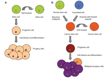
Self-renewal is the process by which a stem cell divides asymmetrically or symmetrically to generate one or two daughter stem cells that have a developmental potential similar to the mother cell. The ability to self-renew is essential for stem cells to expand their numbers during development, to be maintained within adult tissues, and to restore the stem cell pool after injury. Defects in self-renewal mechanisms can lead to developmental defects, premature aging phenotypes, and cancer. The elucidation of self-renewal mechanisms offers the potential for fundamental insights into development, cancer, and aging. Self-renewal is not the same as proliferation, although both processes depend on cell division. Proliferation is a more general term that incorporates all types of stem and progenitor cell divisions, self-renewing and otherwise. Self-renewal requires that at least one of the daughter cells has a developmental potential similar to the mother cell [1-2] (Figure 1).
Fig. 1. Stem cell division and differentiation. In order to maintain self-renewal, stem cells undergo two types of division. During symmetrical division, each stem cell give rise to two identical daughter cells. Asymmetric division gives rise to one identical daughter stem cell and a partially defined progenitor cell..
Genes and pathways involved in the molecular control of self-renewal include the Wnt /β-catenina , Notch and Sonic Hedgehog Pathway.
1.1.2 POTENCY
Potency specifies the differentiation potential (the potential to differentiate into different cell types):
Totipotent stem cells can differentiate into embryonic and extraembryonic cell types. Such cells can construct a complete, viable organism. These cells are produced from the fusion of an egg and sperm cell. Cells produced by the first few divisions of the fertilized egg are also totipotent [3].
Pluripotent stem cells are the descendants of totipotent cells and can differentiate into nearly all cells, i.e. cells derived from any of the three germ layers [4].
Multipotent stem cells can differentiate into a number of cells, but only those of a closely related family of cells.
Oligopotent stem cells can differentiate into only a few cells, such as lymphoid or myeloid stem cells.
Unipotent cells can produce only one cell type, their own, but have the property of self-renewal, which distinguishes them from non-stem cells (e.g., muscle stem cells).
1.2 HAEMATOPOIETIC STEM CELL
In animals, we can identify two broad classes of stem cell: embryonic stem cells (ESCs), and adult (somatic) stem cells (ASCs). ESCs are present in the inner of the embryo and can give rise to the three embryonic germ layers (endoderm, mesoderm and ectoderm) while
ASCs are responsible for normal tissue maintenance. Each organ system of the body has a
hierarchical cellular structure. At the head of hierarchy there is a somatic or ASCs which can self-renew within a specific tissue for indefinite periods; ASCs have also have the unique ability to retain the potential to asymmetrically divide into daughter cells, as well specialised cell types (hematopoietic, neural and mesenchymal stem cells) depending on instructions from the surrounding environment (Fig. 2).
The hematopoietic system is thought to originate from pluripotent hematopoietic stem cells (HSCs) capable of producing a hierarchy of downstream multilineage and unilineage progenitor cells that differentiate into mature cells [5-6]. The first experimental evidence to indicate the existence of HSCs was the discovery in 1961 by Till and McCulloch of a population of clonogenic bone marrow cells capable of generating myelo-erythroid colonies in the spleen of lethally irradiated hosts [7]. HSCs have self-renewal and clonogenic abilities and can differentiate into multiple lineages. HSCs with the capacity for both long-term (LT-HSC) and short-term (ST-(LT-HSC) repopulation of the mouse hematopoietic system have been characterized. These rare cells give rise to progenitor cells, which are destined to generate fully differentiated cells [8].
Stem cells reside in highly regulated microenvironments called niches, which allow them to maintain a balance of self-renewal and differentiation. The concept of the stem cell niche was proposed by R. Schofield in 1978 for the hematopoietic stem cell (HSC) in bone marrow (Schofield 1978). Schofield stated ―The cellular environment which retains the stem cell I
shall call a stem cell ‘niche‘ [9]. Although Schofield is generally credited with the concept of
a stem cell niche, it was Trentin who proposed the idea of an inductive microenvironment. In these classic ―bone-in-spleen‖ experiments, the inductive effect of the immediate local environment was demonstrated and the term hematopoietic inductive microenvironment was coined. The microenvironment is defined as the cells adjacent to or surrounding a stem cell. These cells, together with stem cells, constitute the stem cell niche. The inductive microenvironment provides the mechanical support and extrinsic molecular mechanisms that maintain stem cell fate and inhibit differentiation [10]. These microenvironments are maintained by a constant dialogue between the stem cells and the surrounding niche cells. The niche provides the stem cells shelter from differentiation stimuli, apoptotic stimuli and any other stimuli that might challenge the stem cell stores [11]. The niche must also protect the stem cells from overproduction, which if not properly controlled may lead to cancer [12]. The bone marrow contains multiple stem cell types, including the hematopoietic stem cells (HSCs) and the mesenchymal stem cells (MSCs). The HSCs are the best-characterized adult stem cells and have been purified close to homogeneity. Yet, only recent studies have begun to shed some light on their niche(s). In the adult bone marrow, the HSCs are known to reside in two different niches, an ―endosteal‖ niches, an ―endosteal‖ niche and a ―perivascular‖ niche [11] (Figure 3).
Fig. 3. Schematic representation of the endosteal and pericascular niches of hematopoietic stem cells (HSC) within the bone marrow, and signaling molecules involved in their regulation. Stromal cells, possibly including mesenchymal stem cells (MSCs), surround hematopoietic stem and progenitor cells.
- In the endosteal niche, the hematopoietic stem cells interacts with osteoclasts, osteoblasts and mesenchymal cells, involving transduction pathways of WNT, NOTCH and PTEN. The function of this niche is to ensure quiescence kinetics and the maintenance of self-renewal of HSCs. Furthermore, the osteoclasts are able to maintain the HSC in the niche or to determine its mobilization, under appropriate stimulation.
-In the vascular niche the HSCs interact with sinusoidal endothelial cells and pericytes (perisinusoidal monocytes), via the axis of communication APO1-Tie2; the niche performs its function in the regulation of the maturation process and in the mobilization of circulating HSC. It was in fact observed the ability of the latter to migrate from a niche to another, through the sinusoids.
So there is a compartmentalization of the hematopoietic tissue, which is achieved due to the different distribution of bone marrow cells and growth factors (GFs) secreted by them, within the scaffold by the elements of the extracellular matrix (collagen, fibronectin, glycosaminoglycans).
The interaction of HSCs with the different cell types and with the ECM is through the recognition of specific adhesion molecules that mediate signal transduction, thereby regulating the processes of adhesion, proliferation and differentiation of stem cells.
The haematopoietic growth factors, soluble or bound to the matrix, are instead recognized by specific receptors. called hemopoietin. They are in fact cytokine produced mainly by bone marrow cells, and lymphocytes and macrophages, mostly to paracrine action, which regulate via a system of cooperation differentiation and proliferation of stem cells.
One can distinguish hemopoietin that act by stimulating the cells more immature, from HSCs to progenitors, defined early acting GFs, and growth factors that act instead of precursors already commissioned, said late acting GFs or CSF (Colony Stimulating Factors). SCF (Stem Cell Factor) and Flt3-ligand are the two GFs early acting par excellence, while among the most important CSF were detected multi-CSF, which stimulates the differentiation of different precursors of the myeloid lineage, and the main lineage-CSF specific:
- TPO or thrombopoietin for platelet lineage; - EPO or erythropoietin for erythroid lineage; - GM-CSF for the granulo-monocytic lineage;
- IL-5 and IL-4 respectively for eosinophil and basophil lineage;
Other cytokines play instead a less specific, acting on different cell types (pleiotropic effect). No less important is also the function of the negative regulators of hematopoiesis, including TGF-β, TNF-α, interferon and prostaglandins.
It is therefore clear how the different spatial distribution of these growth factors in the environment medullary stimuli differentially maturation of HSCs.
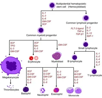
A schematic representation of the maturational process and GFs involved can be given from the tree differentiation hematopoiesis (Fig. 4), which shows how from one cell.
Fig. 4: Diagram showing the development of different blood cells from haematopoietic stem cell to mature cells.
1.2.1 CD34+ HAEMATOPOIETIC STEM CELL
In 1985 were discovered monoclonal antibodies that bind to a portion of the cell surface present on all hematopoietic progenitors, indicated as CD34. This molecule is expressed by a heterogeneous population of human hematopoietic stem cells, cells endothelial progenitor cells, vascular endothelial cells, embryonic fibroblasts and some cells of the fetal nervous tissue and not with a large capacity for proliferation and differentiation. This cellular compartment is in fact very heterogeneous, including primitive stem cells, progenitors and precursors involved in the differentiation morphologically differentiated. The CD34 molecule is an integral membrane glycoprotein. The sequence of 373 aa protein (40 kDa) is heavily glycosylated and not presents homologies identified with any known protein. The gene coding for the CD34 antigen is localized on chromosomal region 1q32, in a region containing a group of genes that encode for the adhesion molecules. The mucin-like structure of CD34 suggests its possible role in cell adhesion, perhaps in the accession of progenitor and stem cells to cells stromal cells. CD34 antibodies are widely used for applications in the field of
research as immunological studies of hematologic malignancies. Particularly significant are the studies to determine the phenotype of the compartment more immature, CD34+ cells, which constitutes only a small fraction of hematopoietic tissue, those stem cells quiescent capable of self-renewal used in experiments with cell cultures and in vivo transplantation. CD34+ cells are pluripotent stem cells that give rise to all cell types in blood. These stem cells are typically found in the bone marrow; fewer than 1% of nucleated cells in the blood are CD34+. CD34+ cells be also be found in umbilical cord blood where they typically make up 1% of the mononuclear cells.
1.3 LEUKEMIC STEM CELL
Some form of normal stem or progenitor cell undergoes a mutation giving rise to an entity that is functionally defined as a leukemic stem cell. The normal stem cells continue to differentiate into the hematopoietic lineage giving rise to erythrocytes, platelets, leukocytes, and granulocytes. The mutated stem cells have properties similar to the normal stem cells and can also differentiate into the hematopoietic lineage carrying the defect/s or can remain and accumulate as immature progenitor cells, also known as blast cells [13-14] (Figure 5).
Figure 5: Asymmetric division in stem cells. (a) Asymmetric division in a normal stem cell. A stem cell can self-renew to give rise to another stem cell (green) but can also divide to form a progenitor cell (pink). (b) Asymmetric division in a cancer stem cell. A cancer stem cell (orange) can also asymmetrically divide to form another cancer stem cell (orange) or give rise to a progenitor cell (brown).
The chemotherapeutic agents used today effectively eradicate the blast cells in many patients. However, those same agents have very little if any activity at the level of the blast progenitor
cell, the leukemic stem cell (LSC), which is biologically distinct from most of the cells that are found in a typical patient. An LSC is a functionally defined entity not necessarily named because it arises from a normal stem cell, but because it fulfills the same criteria used to define normal stem cells. These cells can undergo self renewal, are multipotent, and highly proliferative. The origin of the LSC has been the subject of considerable research in recent years [15]. During normal developmental progression from stem cell to progenitor cell to mature cell, mutations may potentially occur at any point during this evolution, giving rise to a malignant entity. However, there is experimental evidence that suggests that mutations in a progenitor cell that no longer has all the characteristics of a stem cell can also give rise to an entity that can initiate and maintain leukemic disease [16]. For most leukemia, as for most cancers, the target cell of transforming mutations is still unknown. Because normal stem cells and LSCs share the ability to self-renew, as well as various developmental pathways, it has been postulated that LSCs are HSCs that have become leukemic as the result of accu mulated mutations. HSCs have the machinery for self-renewal already activated and therefore may require fewer mutations to maintain it than more differentiated cells would require to activate it ectopically. HSCs also persist throughout life and therefore have much greater opportunities to accumulate mutations than more mature cells, which persist only for a short period. Conversely, LSCs could also be a more restricted progenitor or even a differentiated mature cell, which would have first to reacquire the stem cell capability for self-renewal before becoming tumorigenic to accumulate additional mutations [17]. The development of cancer is a stepwise process in which increasing numbers of somatic mutations give rise to an increasingly transformed clonal population of cells [18]. The multistep model of carcinogenesis was originally postulated to require a clonal event causing increased proliferation, which, together with mutations blocking cellular differentiation, synergizes to cause transformation.
1.4 LEUKEMIA
Leukemia is a monoclonal diseases of the hemopoietic system arising from the mutation of a single stem cell. The neoplastic transformation can affect cell differentiation into different stages: if it hits a multipotent HSC originates a hybrid leukemia, with population biphenotypic or biclonal (both myeloid lymphoid elements), and if the mutation occurs in a stem cell already commissioned for myelopoiesis or lymphopoiesis, originate leukemias with phenotypic myeloid or lymphoid characteristics respectively.
The neoplastic population is characterized by a high polymorphism, linked firstly to the stage of differentiation of the transformed cell and secondly to the maintenance of the same capacity maturation and differentiation.
Can be distinguished two types of leukemia: acute and chronic. In acute leukemia, the mutated cell loses the ability to differentiate and causes the proliferation and accumulation of immature cells in the bone marrow or blast cells. In contrast, the chronic form is characterized by the proliferation of cells with phenotypic characteristics of mature, of one or more cell lines depending on the potential of the cell originally mutated.
The onset of this disease is related to the acquisition of a proliferative advantage of transformed cells than normal cells. Mutations leading to activation of a oncogene or inactivation of a tumor suppressor gene are the main responsible for the survival and proliferation of cancer cells. Etiological factors behind these changes are not fully known but have been identified some risk agents that can contribute in varying degrees to the onset of the disease:
- exposure to ionizing radiation, particularly at high doses;
- the Smoke, for the presence of leukemogenic agents in cigarettes;
- exposure to chemicals such as benzene, urethane, and others, for their ability to bind and permanently damage the DNA;
- treatment with conventional chemotherapy;
- the infection by specific viruses such as retroviruses leukemogenic acute, chronic leukemogenic retroviruses (including HTLV and HIV), and Epstein-Barr virus.
Ongoing studies are also evaluating the possible correlation between acute leukemia and prolonged exposure to electromagnetic fields.
The course of the disease can be very variable depending on the type of leukemia that affects the patient, of molecular and cytogenetic abnormalities that characterize it, and therapeutic approaches must always be more specific for subtype identified. To increase the probability of success is desirable to move towards the study and planning of treatment programs customized.
1.4.1 ACUTE MYELOID LEUKEMIA
Acute myeloid leukaemia (AML) is defined as a clonal disorder caused by malignant transformation of a bone marrow-derived, self-renewing stem or progenitor cell, which demonstrates an enhanced proliferation as well as aberrant differentiation resulting in
haematopoietic insufficiency (i.e. granulocytopenia, thrombocytopenia or anaemia). The clinical signs and symptoms of AML are diverse and nonspecific, but they are usually directly caused by the leukaemic infiltration of the bone marrow, with resultant cytopenia. AML is considered to be a heterogeneous group of disorders with variable underlying abnormalities and clinical behaviour, including responses to treatment. Therefore, classification of the disease is important and several classification systems exist to subdivide AML [19]. Historically, AMLs were divided into subtypes based on the type of cell from which the leukaemia developed and the level of maturation, French-American-British (FAB) classification (Table 1) [20]:
FAB subtype Name Adult AML patients (%)
M0 Undifferentiated acute myeloblastic leukemia 5% M1 Acute myeloblastic leukemia with minimal maturation 15% M2 Acute myeloblastic leukemia with maturation 25% M3 Acute promyelocytic leukemia 10% M4 Acute myelomonocytic leukemia 20% M4eos Acute myelomonocytic leukemia with eosinophilia 5%
M5 Acute monocytic leukemia 10%
M6 Acute erythroid leukemia 5%
M7 Acute megakaryocytic leukemia 5%
Table 1: French-American-British (FAB) classification.
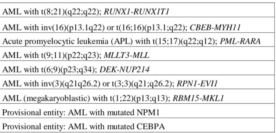
Nowadays, the World Health Organization (WHO) provides a classification system in which morphology, cytogenetics, molecular genetics, and immunological markers are incorporated and interrelated (Table 2) [21]:
AML with t(8;21)(q22;q22); RUNX1-RUNX1T1
AML with inv(16)(p13.1q22) or t(16;16)(p13.1;q22); CBEB-MYH11 Acute promyelocytic leukemia (APL) with t(15;17)(q22;q12); PML-RARA AML with t(9;11)(p22;q23); MLLT3-MLL
AML with t(6;9)(p23;q34); DEK-NUP214
AML with inv(3)(q21q26.2) or t(3;3)(q21;q26.2); RPN1-EVI1 AML (megakaryoblastic) with t(1;22)(p13;q13); RBM15-MKL1 Provisional entity: AML with mutated NPM1
Provisional entity: AML with mutated CEBPA
Recently, for the first time, specific gene mutations (i.e. mutations in CEBPA and NPM1) have been included as ‗provisional entities‘ in the revised WHO 2008 classification for AML [22]. There is growing evidence that these two gene mutations represent primary genetic lesions (so-called class II mutations) that impair haematopoietic differentiation [23]. Mutations in the fms-related tyrosine kinase 3 (FLT3) gene (e.g. FLT3-ITD or FLT3 kinase domain mutations) are considered class I mutations conferring a proliferation and/or survival advantage. AML with FLT3 mutations is not considered a distinct entity, although determining the presence of such mutations is recommended because they have prognostic significance [24].
A number of clinical and biological features that reflect the heterogeneity of AML are used to predict the likelihood that a patient will have a response to treatment or relapse. Adverse prognostic factors in AML include increasing age, a poor performance before treatment, unfavourable cytogenetic abnormalities and a high white blood cell count.
Furthermore, therapy-related AML or AML arising after a myelodysplastic or myeloproliferative syndrome is usually more resistant to standard treatment than de novo AML. Important predictors of disease outcome are the pre-treatment cytogenetic and molecular findings in AML blasts. To date, in AML approximately 200 different structural and numerical aberrations have been described [24-25]. Cytogenetic findings permit patient risk to be categorised as favourable, intermediate or unfavourable, with very different cure rates. Although there may be (subtle) differences in the criteria used to define these risk groups among different study groups, the presence of for instance t(8;21)(q22;q22), t(15;17)(q22;q21) and inv16(p13q22)/t(16;16)(p13;q22) is generally classified as favourable-risk AML (with leucocytes <20 x 109). On the other end of the spectrum is the unfavourable-risk group, which includes blasts showing e.g. monosomies of chromosome 5 or 7, deletion of the long arm of chromosomes 3, 5 and 7 and complex karyotypes. Of note, the monosomal karyotype, defined as non-core-binding factor (CBF) leukaemias with a karyotype with at least two autosomal monosomies or one single autosomal monosomy in the presence of one or more structural cytogenetic abnormalities, is considered to be a better predictor of (very) poor outcome than the traditionally defined complex karyotype [26]. The intermediate-risk group includes AMLs with a normal karyotype and AMLs which are not classified in the other two risk groups. In recent years, the discovery of mutations in e.g. genesencoding FLT3,
NPM1 and CEBPA has shown to be of major importance. Nowadays, it is increasingly
possible to distinguish subsets of patients with differing outcomes from the large cohort with a normal karyotype AML or miscellaneous cytogenetic abnormalities considered as
intermediate-risk cytogenetics. The majority of FLT3 receptor tyrosine kinase gene mutations are internal tandem duplications (ITD); less frequent are mutations involving the tyrosine kinase domain (TKD). Several groups have consistently reported that FLT3-ITD is a major independent adverse risk factor in AML.27-31 The prognostic relevance of FLT3-TKD mutations, however, remains controversial [24]. FLT3-ITD has a prevalence of 20 to 25% in young adults and nearly 35% in the older adult population. The ratio of the FLT3-ITD and the wild-type FLT3 (measured by polymerase chain reaction, PCR) varies from patient to patient, and this difference may have clinical implications. Thiede et al. found that patients with an allelic ratio (AR) above the median (0.78) had significantly shorter overall and disease-free survival, whereas survival in patients with ratios below 0.78 did not differ from those without FLT3 aberrations [27]. CEBPA, a transcription factor involved in normal myelopoiesis, is mutated in ~10% of AML cases and predicts a relatively favourable outcome in paediatric and adult AML, however, only when CEBPA is mutated on both alleles [28-29].
Approximately 50% of adult normal karyotype AMLs harbour an NPM1 mutation, which leads to delocalisation of the NPM1 protein to the cytoplasm.38 NPM1 and FLT3-ITD commonly co-exist in normal karyotype AML suggesting that they may cooperate in generating the leukaemic phenotype. The presence of an NPM1 mutation (in the absence of an FLT3-ITD mutation) is associated with better outcome in terms of higher complete response rates and increased long-term survival compared with patients lacking the mutation [30-33]. Consequently, it has been suggested that cytogenetically normal AML involving the genotype of mutant NPM1 without FLT3-ITD should no longer be classified as intermediate-risk leukaemia but rather should be classified as favourable-intermediate-risk leukaemia [32]. Furthermore, patients with mutant NPM1 without FLT3-ITD may not benefit from related-donor transplantation as first-line treatment [32]. Mutations in the Wilms‘ tumour gene (WT1), present in ~10% of patients with normal karyotype AML, have been found to be associated with poor outcome, especially in combination with an FLT3-ITD [34-37]. RAS mutations, occurring in ~15% of cases, are suggested to be prognostically neutral [30].
Recently, mutations in genes involved in metabolism have been discovered [38-39]. In AML, but also in low-grade gliomas and secondary glioblastoma multiforme (GBM), mutations in cytosolic isocitrate dehydrogenase 1 (IDH1) and its mitochondrial homolog IDH2 have been identified. Both IDH1 and IDH2 are important enzymes in the citrate cycle (Krebs cycle). Two distinct alterations are caused by the tumour-derived mutations in IDH1 or IDH2: loss of its normal catalytic activity in the production of α-ketoglutarate (α-KG) and gain of the
catalytic activity to produce 2-hydroxygulatrate (2-HG). Consequently, less α-ketoglutarate is available for biological processes in which it functions as a co-factor. Remarkably, IDH1/2 mutations, occurring in ~10 to 25% of AML cases, were mutually exclusive with mutations in gene encoding the a-ketoglutarate-dependent enzyme tet oncogene family member 2 (TET2) (occurring in 12 to 20% of AML cases) [40-42]. Loss-of-function mutations in TET2 were associated with similar epigenetic defects as IDH1/2 mutants. Interestingly, a shared proleukaemogenic effect between TET2 mutations and mutations in IDH1 and IDH2 was suggested since α-ketoglutarate is a co-factor for TET2 in the hydroxylation of 5-methylcytosine and thus effects the methylation process [43].
In cytogenetically favourable core binding factor (CBF AML (i.e. AML with t(8;21) or inv(16)/t(16;16)), the presence of a mutation in the KIT receptor tyrosine kinase has been shown to have an unfavourable influence on outcome in retrospective studies. Recently, highly recurrent mutations in the DNA methyltransferase gene DNMT3A have been discovered and were found to be independently associated with poor outcome in AML [44]. Other mutations as those involving protein tyrosine phosphatase, non-receptor type 11 (PTPN11) and runt-related transcription factor 1 (RUNX1) are relatively rare (i.e. <5% of cases), making their relevance to risk-stratified treatment approaches uncertain at the present time [45]. The panel of known molecular markers is continuosly increasing, for example, considering the recently described EZH2, DNMT3A, ASXL1 and IDH1/2 mutations.
Mutations such as ASXL1, RUNX1, EZH2, ETV6/TEL and TP53 have an adverse impact on patient overall survival. Early evidence suggests that some mutations might influence treatment response, necessitating reassessment of the prognostic effect of genetic alterations in the light of every new treatment. The introduction of next generation sequencing will certainly catalyze the molecular characterization of AML.
1.4.2 CRONIC MYELOID LEUKEMIA
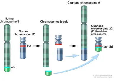
Cronic Myeloid Leukemia (CML) arises as the result of a mutation in a pluripotent stem cell and is characterized by progressive granulocytosis, marrow hypercellularity, and splenomegaly [46-49]. The diagnostic hallmark is the Ph chromosome (Figure 6), [50] which is present in all dividing cells of hematopoietic lineage, as well as in B and T cells in some patients, but is absent in all other cells. Although hematopoiesis is overwhelmed by the Ph -chromosome–positive clone, a normal Ph-chromosome–negative pool of stem cells persists [47, 51]. The goal of treatment is the suppression or elimination of the Ph-chromosome–
positive clone and the restoration of Ph-chromosome–negative hematopoiesis. The diagnosis of the disease is based on the blood count to assess the level of leukocytosis, usually recorded around 20-30 billion cells / L for the massive mobilization of tumor cells from the bone related to the alteration of adhesion. The number of platelets can instead be normal, increased or reduced. The morphological analysis of peripheral blood shows that the cell types responsible for the leukocytosis are mainly neutrophils and immature granulocyte series, from myeloblast to metamyelocyte. Finally, the bone marrow aspiration is essential to confirm the diagnosis: there was a marked hyperplasia of the granulocytic and megakaryocytic series at the expense of the erythroid compartment and the fatty component. CML has a biphasic or triphasic course but is usually diagnosed during the initial, or chronic, phase, in which the granulocytic population expands but remains able to differentiate. The chronic phase in which there are clinical symptoms that allow the diagnosis may occur from 2 to 10 years after the beginning of the biological disease. At this stage, the uncontrolled proliferation of the granulocytic line ultimately leads to suppression of hematopoietic stem cell pool of healthy (Ph-). The chronic phase is relatively stable and responds to therapy, but it eventually evolves into an intermediate, accelerated phase characterized by blastic elements in the peripheral blood, anemia and thrombocytopenia, related to the accumulation of mutations in stem cells neoplastic with progressive loss of the cell maturation. The accelerated phase in which increasing doses of hydroxyurea are needed to control disease, followed by a blast phase. Blast-phase disease resembles acute leukemia. Its phenotype is myeloblastic in 70 to 80% of patients and lymphoblastic in 20 to 30%. The clinical features becomes very similar to that of a LAM and symptoms such as fever, muscle and bone pain, fatigue and weight loss. Typically this stage is of short duration (< 6 months) and leads very quickly to the patient's death due to resistance to conventional treatments gained in the inactivation of tumor suppressor genes such as p53. With conventional treatment the median survival among patients with CML is about five years, but the range is very broad. Blast-phase cells may be more dependent on secondary oncogenic aberrations than on the tyrosine kinase activity of BCR-ABL. As the leukemic clone becomes unable to differentiate, blast cells accumulate, leading inexorably to a blast crisis. Some patients with an aggressive form of chronic-phase disease survive only months, whereas others, who have indolent, chemoresponsive CML, live 10 years or longer. The Ph chromosome is a truncated chromosome 22 that results from a reciprocal exchange of genetic material between the long arms of chromosomes 9 and 22 (Fig. 6). The translocation t(9;22) results in the juxtaposition of 3' DNA sequences derived from the Abelson (ABL) proto-oncogene normally located on
chromosome 9 with 5' sequences of the breakpoint cluster region (BCR) gene on chromosome 22 [52-54]. The ABL proto-oncogene is homologous with the transforming gene present in the Abelson leukemia virus, which causes leukemia in mice [46, 55] ABL encodes a tyrosine kinase that is tightly regulated, whereas the activity of BCR-ABL is autonomous and markedly increased relative to that of normal ABL. The Ph chromosome is present in approximately 95% of patients with classic CML. About half of the remaining 5% of patients have been found to have the BCR-ABL gene when the polymerase chain reaction (PCR) is used for identification and are classified as being Ph-chromosome–negative, BCR-ABL–positive. Although the precise oncogenic mechanism of BCR-ABL is unknown [46, 56], its tyrosine kinase activity leads to the chronic phase of CML [57]. Transplantation of hematopoietic stem cells containing a BCR-ABL gene construct into mice results in a disease resembling CML [58-59]. The Ph chromosome is also detected in about 25% of adults and 5% of children with acute lymphoblastic leukemia (ALL) and is associated with an aggressive course and poor survival [60-62]. Only about one third of patients with Ph-chromosome–positive ALL have the 210-kD BCR-ABL protein characteristic of CML; approximately two thirds have a smaller chimeric BCR-ABL protein of 185 to 190 kD that has more potent tyrosine kinase and oncogenic activity [47, 57, 63] continuously (or constitutively) active BCR-ABL oncoprotein phosphorylates substrates of remarkable diversity, including RAS, that activate multiple signaling pathways [46, 55, 64]. Because RAS serves as a critical control point for signal transduction from cell membrane to nucleus [64-65], the BCR-ABL–mediated overexpression of RAS appears to alter signal transduction in a target stem cell, leading to abnormal mitosis and neoplastic expansion. In addition, BCR-ABL reduces cellular adhesion to stromal matrix [66-69], which may disrupt the interaction between hematopoietic cells and stromal cells and membrane signaling mediated by cytoadhesion molecules, allowing myeloid progenitor cells to remain longer in the proliferative phase before undergoing differentiation [70]. BCR-ABL also diminishes cellular responsiveness to apoptotic stimuli, providing a survival advantage to the leukemic clone [71-74]. In theory, since chronic-phase CML is dependent on the tyrosine kinase activity of BCR-ABL, a potent BCR-ABL inhibitor might eliminate the leukemic clone and restore normal Ph-chromosome–negative hematopoiesis. Although the mechanism for blastic transformation is unknown, possible scenarios have been considered. For example, BCR-ABL promotes genomic instability in the leukemic clone [75], which may lead to secondary mutations (e.g., trisomy 8).
Fig. 6: Translocation leading to the Philadelphia (Ph) Chromosome and the role of BCR-ABL in the pathogenesis of CML.
During the chronic phase it is possible to obtain hematologic remission of 95% of cases thanks to the use of drugs recently discovered that inhibit the activity of tyrosine kinases involved in oncogenesis, such as c-Abl, c-kit and PDGFR. Imatinib mesylate, the Nilotinib, Dasatinib and the Ponatinib are an example. These inhibitors are able to bind the inactive forms of the tyrosine kinase targets (eg. BCR-ABL), preventing binding of ATP and the conversion to the active form. This shuts down the signaling pathways iperattivated and the loss of the leukemic phenotype: although cytogenetic remission is only achieved in 75% of cases, at the molecular level the action of BCR-ABL is however neutralized. An alternative to treatment with current chemotherapy remains the allogeneic HSC after chemotherapy conditioning. In 40% of cases, the transplant can result in complete cure, which is due to the phenomenon of graft-vs-leukemia in which the immune system of healthy donor is directed against leukemia cells are not eliminated by chemotherapy. The low percentage of success is tied to complications during or post-transplant, as for example the graft-vs-host disease in which there is the rejection of donor cells.
1.4.3 ACUTE LYMPHOBLASTIC LEUKEMIA
ALL represents a biologically and clinically heterogeneous group of B/T-precursor-stage lymphoid cell malignancies arising from genetic insults that block lymphoid differentiation and drive aberrant cell proliferation and survival. Incidence and cure rates differ among children and adults. In children, ALL is the commonest malignancy accounting for approximately 25 % of childhood cancer and it has 5-year event-free survival rates ranging between 76 % and 86 % in patients receiving protocol-based therapy. In adults, ALL
is less common and generally carries a worse prognosis with a long-term survival probability less than 35–40 % [76-77]. Although there is remarkable progress made in the treatment of ALL in children and, with less efficacy, in adults, several ALL subtypes continue to have a poor prognosis and in a proportion of longterm surviving patients, treatment is responsible for short and long-term toxicities. Consequently, there is a need in improving the molecular dissection of subtypes, identifying genetic alterations that predict the risk of treatment failure, and developing novel and targeted therapies. Cytogenetics has long been used for diagnosis, risk stratification, and therapeutic implications, however experimental models [78] [79] have established that primary cytogenetic abnormalities alone are insufficient to induce leukemia and that cooperative mutations are required.
The symptoms and signs of the disease depend on the following pathophysiologic mechanisms:
a) reduction of normal hematopoiesis, with consequent anemia, granulocytopenia, and thrombocytopenia;
b) expansion of leukemic cells in bone marrow (normally the blast infiltration in the bone marrow is subtotal or total). Only 4 % of cases have an infiltration < 40% in the peripheral blood and, secondarily, in other extramedullary sites;
c) release of cytokines and inflammatory mediators, by both the leukemia cells that of normal cells (symptoms relatively more frequent in children).
The early simpto anemia, fatigue, pallor, fever, infection, and bleeding (bruising, petechiae, epistaxis, mucosal bleeding). Lymphadenomegaly and splenoepatomegalia occur in approximately half of the cases; mediastinal lymph nodes and abdominal have a volume increased in most of the ALL-T and can cause cardiorespiratory problems. The central nervous system is commonly involved in diagnosis, but his interest can cause neurological disorders, such as headache or cranial nerve palsies, complicating the course of the disease. Poliartromialgie are frequent, often very intense and resistant to anti-inflammatory therapy. The involvement of other organs in a clinically relevant manner occurs in less than 5% of cases and affects the kidneys, skin, eye and retina, lungs, pleura, heart, pericardium, testicles, ovaries, lymph nodes, abdominal or retroperitoneal [76, 78-79].
The development of microarray technologies to profile gene expression and structural genet ic alterations in a genome-wide and high-resolution fashion have revolutionized our ability to identify genetic abnormalities providing important insights into the pathways deregulated in ALL [80]. Moreover, recently the development of next-generation sequencing (NGS) technologies has provided researchers with completely new and effective tools for the
discovery of novel alterations, depicting an exhaustive picture of the leukemia genome complexity.
Although the eziogenesis of the disease is unknown, molecular genetic studies have allowed us to identify different genomic alterations at the base of leukemic transformation in more than 60% of ALL. Most of these alterations leads to activation of proto-oncogenes, or through the deregulation of the same or, more frequently, through the formation of hybrid genes of fusion with aberrant activity. In the LAL with the alteration of the adult phenotype B more frequent (25-30% of cases) is the t (9; 22) that leads to the formation of the fusion gene BCR / ABL on the Philadelphia chromosome. As in CML, constitutive activation of Abl promotes cell proliferation, inhibits apoptosis and alters cell adhesion to bone marrow microenvironment. Unlike CML, however, in the ALL is not formed a fusion product of 210 kDa but a shorter protein, p190, whose transforming power is higher. Furthermore, according to the morphology of blast cells in the circulation at the time of diagnosis, it is possible to distinguish three classes of ALL, according to the FAB classification:
L1: ALL small blasts homogeneous prevalent in children;
L2: ALL blasts heterogeneous in size and cell characteristics, prevalent in the adult; L3: ALL large blasts with basophilic cytoplasm and ipervacuolato.
At the morphological diagnosis is always well associate a diagnosis and immunophenotypic analysis cytogenetic-molecular, so as to be able to program a targeted therapy, and thus more effective, particularly against the subtype of ALL.
The standard treatment of ALL involves three steps:
- Induction of remission by chemotherapy with agents such as corticosteroids, anthracyclines, vincristine, and asparaginase;
- Prophylaxis of leukemia localization in the CNS by intrathecal administration of anticancer drugs or radiation farmci on the skull;
- Intensification and maintenance of remission with chemotherapy.
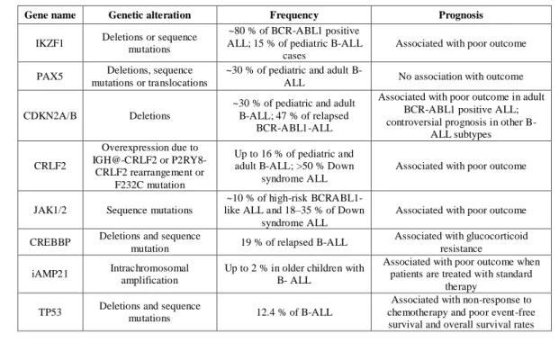
In more severe cases may need to be autologous or allogeneic bone marrow. Specific therapies are available for the subtypes of ALL with a known genetic alteration at the base. The most representative example is that of Philadelphia positive ALL, so we adopt a targeted therapy against BCR-ABL with tyrosine kinase inhibitor drugs, such as Imatinib mesylate. The advent of high-resolution genome-wide analyses of gene expression, DNA copy number alterations (CNA), and loss of heterozygosity have led to the detection of many novel genetic abnormalities refining the prognostic models for ALL-B (Table 3) and ALL-T (Table 4) [81].
Gene name Genetic alteration Frequency Prognosis
IKZF1 Deletions or sequence mutations
~80 % of BCR-ABL1 positive ALL; 15 % of pediatric B-ALL
cases
Associated with poor outcome
PAX5 Deletions, sequence mutations or translocations
~30 % of pediatric and adult
B-ALL No association with outcome
CDKN2A/B Deletions
~30 % of pediatric and adult B-ALL; 47 % of relapsed
BCR-ABL1-ALL
Associated with poor outcome in adult BCR-ABL1 positive ALL; controversial prognosis in other
B-ALL subtypes CRLF2 Overexpression due to IGH@-CRLF2 or P2RY8-CRLF2 rearrangement or F232C mutation Up to 16 % of pediatric and adult B-ALL; >50 % Down
syndrome ALL
Associated with poor outcome
JAK1/2 Sequence mutations
~10 % of high-risk BCRABL1- like ALL and 18–35 % of Down
syndrome ALL
Associated with poor outcome
CREBBP Deletions and sequence
mutation 19 % of relapsed B-ALL
Associated with glucocorticoid resistance iAMP21 Intrachromosomal
amplification
Up to 2 % in older children with B- ALL
Associated with poor outcome when patients are treated with standard
therapy TP53 Deletions and sequence mutations 12.4 % of B-ALL
Associated with non-response to chemotherapy and poor event-free survival and overall survival rates
.
Table 3: Novel recurring genetic alterations occurring in B-progenitor ALL and their correlation with outcome.
Gene name Genetic alteration Frequency Prognosis
NOTCH1 Sequence mutations ~50 % of T-ALL Associated with favorable outcome in children
FBXW7 Sequence mutations ~20 % of T-ALL Associated with favorable outcome in children
PTEN Deletions or sequence mutations 6–8 % of T-ALL
Associated with poor response to chemotherapy and resistance to pharmacological inhibition of NOTCH1
CDKN2A/B Deletions 30–70 % of T-ALL Associated with poor outcome in adult
and children T-ALL CDKN1A Deletions or sequence mutations 12 % of T-ALL To be investigated
6q15-16.1 Deletion 12 % of T-ALL Associated with poor outcome
PHF6 Deletions or sequence mutations
16 % of pediatric T-ALL cases;
38 % of adult T-ALL cases
Associated with reduced overall survival
WT1 Frameshift mutations
13 % of pediatric T-ALL cases;
12 % of adult T-ALL cases
No association with outcome
LEF1 Focal deletions or sequence
mutations 15 % of pediatric T-ALL cases
Associated with younger age and a trend toward a better overall survival JAK1 Sequence mutations 18 % of adult T-ALL cases Associated with reduced disease-free
survival and overall survival FLT3 Internal tandem duplication 4 % of adult T-ALL cases; 3 %
of pediatric T-ALL cases No association with outcome
PTPN2 Deletion 6 % of T-ALL
Down-regulation of PTPN2 expression results in prolonged survival of
ALL-SIL
cells after Imatinib treatment
1.5 BCR-ABL TYROSINE KINASE INHIBITORS
The fusion protein BCR-ABL is necessary and sufficient to induce the phenotype transformant of leukemic cells. If the tyrosine kinase activity of BCR-ABL is turned off, the cell pH is suppressed. Therefore, the leukemic cells Ph+ constitute an ideal target for therapeutic targeting of inhibitors of tyrosine kinase. The treatment of CML and ALL Ph+ patients was revolutionized by the introduction of Imatinib mesylate (IM, Gleevec®), a BCR-ABL tyrosine kinase inhibitor (TKI). The clinical use of specific BCR-BCR-ABL inhibitors has resulted in a significantly improved prognosis, response rate, overall survival, and patient outcome in CML patients compared to previous therapeutic regimens. However, the complete eradication of CML in patients receiving Imatinib was limited by the emergence of resistance mostly due to mutations in the ABL kinase domain and to a lesser extent by molecular residual disease after treatment. The second-generation BCR-ABL TKIs Nilotinib (Tasigna®) and Dasatinib (Sprycel®), showed significant activity in clinical trials in patients intolerant or resistant to Imatinib therapy, except in those patients with the T315I BCR -ABL mutation. Bosutinib is a third generation tyrosine kinase inhibitor. It is being tested in clinical trials and looks very promising. It has been useful in patients whose leukemia is resistant to both first and second generation tyrosine kinase inhibitors.
1.5.1 IMATINIB MESYLATE
In 1996 was developed by the collaboration of Druker and Novartis Pharmaceuticals (Ciba-Geigy, Basel, Switzerland), an investigational drug, now known as Imatinib mesylate (CGP57148B; STI571, Gleevec®) (Fig. 5A), which is a derivative of 2-phenyl-amino-pyrimidine and acts specifically by inhibiting the tyrosine Abl kinase. In 2001 Imatinib receives FDA approval (Food and Drug Administration) and EMEA (European Medicines Evaluation Agency) as second-line treatment of CML, but in the following year Imatinib is approved as a drug of first-choice treatment for CML [82].
Initially it was thought that the specific inhibitory action of Imatinib was based on the ability to compete with ATP in the catalytic domain of the kinase, inhibiting, thus, the cascade of signal transduction. Thereafter, the crystallographic studies of the kinase domain of Abl complexed with a homologue of Imatinib, showed that the inhibitor binds to an inactive conformation of Abl that is, thus, unable to bind ATP and activate the signals that cause leukemogenesis. Imatinib has inhibitory activity against other members of the family of
tyrosine kinases, including PDGFRα / β and c-kit, involved in other syndromes (eg, hypereosinophilic syndrome or systemic mastocitsi) [83].
The domain of Abl tyrosine kinase is characterized by highly conserved residues that form the binding site, the catalytic loop and the activation loop. The orientation and the state of phosphorylation of residues of the DFG motif (Asp-Phe-Gly) present in the activation loop, determine the transition from the inactive to the active conformation of the kinase. In the inactive form of the activation loop preclude access of ATP to its binding site, and with the transition to the active form of the ATP binding is made possible. The activation loop in the inactive conformation orients the N-terminal of the residue Phe382, Asp381 instead, thereby preventing the formation of the salt bridge between this last residue and the complex Mg2 + -ATP: the Tyr412 interacting through a hydrogen bond with the residue of Asp381, it mimics the substrate binding by blocking the activity of the protein.
The oncoprotein in leukemia cells is in dynamic equilibrium between its active and inactive form. This balance depends on the recruitment of phosphotyrosine ligands for disassembly constraints inhibitors, and phosphatase that can transiently dephosphorylate the protein, allowing the binding of Imatinib. Imatinib seems then bind monomeric and inactive conformation of BCR-ABL [83-84] (Fig. 5B). Imatinib clearly differs from traditional therapies as regards the toxic effects: the most common effect is a moderate nausea; edema, myalgia, arthralgia, diarrhea and skin rash are found in 10% of patients; rarely have periorbital edema and more in general syndrome retention of fluids. Myelosuppression is more common in blast phase than in cronic phase [85] (Figure 7).
Fig. 7: A: structure of the Imatinib molecule; B. structure of the kinase domain of Abl complexed with Imatinib (in yellow) in which are highlighted the DGF motif of the activation loop and the residue Thr315 necessary for the formation of a hydrogen bond with Imatinib.
1.5.2 NILOTINIB
Nilotinib is an oral ATP-competitive inhibitor of BCR-ABL in clinical development with a modified aminopyrimidine backbone comparable to Imatinib. Similar to Imatinib, Nilotinib can bind only the inactive conformation of the ABL kinase domain but with a 25-fold greater affinity than Imatinib [86] (Figure 8).
In 2007 Nilotinib has been approved by the FDA as an second generation tyrosine kinase Inhibitor for the treatment of patients resistant or intolerant to Imatinib [87-88]. Nilotinib has demonstrated activity in Imatinib-resistant mutations, with the exception of T315I [89-90]. Nilotinib also inhibits the activity of Arg, Kit, and platelet-derived growth factor receptor (PDGFR), but not Src-family kinases (SFK) [90]. Nilotinib is 10 to 50 times more potent than Imatinib in inhibiting the proliferation and autophosphorylation of wild-type BCR-ABL cell lines and most of the BCR-ABL mutants, except the T315I mutant [90]. It is superior to Imatinib in reducing leukemic burden and prolonging the survival of mice transplanted with wild-type BCR-ABL, the M351T and E255V mutants [90].
Fig. 8: A. Ribbon diagram showing the Abl kinase (gray) with linked Nilotinib (green), showing the P-loop (red), the activation loop (magenta) and the C helix (yellow). B. Chemistry of Nilotinib.
However, Nilotinib and Imatinib produced equivalent reduction in CrkL phosphorylation in primary CD34+ CML cells, suggesting that they were equipotent for inhibiting BCR-ABL activity [91]. Furthermore, Nilotinib did not induce apoptosis in the primitive quiescent population. Nilotinib is from 10 to 50 times more potent in inhibiting the proliferation of Imatinib and the autophosphorylation of the cell lines BCR-ABL wild-type and the majority of those mutanti [92]. And superior to Imatinib in leukemic reduce the charge and prolong the survival of mice transfected with BCR-ABL wild-type, the mutant M351T and E255V.
However, Nilotinib and Imatinib induced a dephosphorylation equivalent Crkl lines in primary CD34 + chronic myeloid leukemia, suggesting that the two drugs are equipotent in inhibiting the activity of BCR-ABL [93]. Nilotinib is well tolerated, and common ad verse events included grade 3-4 myelosuppression, elevated bilirubin and lipase levels.
1.5.3 DASATINIB
Dasatinib (BMS-354825, (Sprycel®); Bristol Myers Squibb) is a multi-target kinase inhibitor of BCR-ABL, SFK, ephrin receptor kinases, PDGFR and Kit. In addition, Dasatinib binds to other tyrosine and serine/threonine kinases, such as the TEC family kinases, the mitogen-activated protein kinases and the receptor tyrosine kinase, discoidin domain receptor 1[94]. Dasatinib is more potent than Imatinib and is effective against the Imatinib-resistant active conformation of the kinase domain (Figure 9).
Fig. 9: A Inactive conformation of the protein on the left and on the right active conformation, with the drug that in both cases we go to place within the pocket. B. Chemistry of Dasatinib.
It is capable of inhibiting the proliferation and kinase activity of wild-type and most BCR-ABL mutant cell lines except the T315I mutant. In vivo studies in murine models demonstrated the activity of Dasatinib in inhibiting the leukemic cell growth and prolonging the survival of mice harbouring wild-type BCR-ABL and the M351T, but not the T315I mutant.15 Dasatinib is well tolerated but grade 3-4 myelosuppression is common, especially in the advanced phases. Non-hematological side effects include diarrhea, nausea, headache, peripheral edema and pleural effusion. However, resistance to Dasatinib is also an emerging problem. Not surprisingly, the pre-existence or selection of the T315I mutant is the most frequent mechanism of resistance.20 The emergence of the F317L mutant has also been commonly observed in Dasatinib-resistant patients [95]. In addition, although Dasatinib significantly inhibited CrkL phosphorylation and caused a reduction in the total number of
CD34+ CD38– CML cells compared to Imatinib, it did not eliminate the most primitive, quiescent fraction [96].
1.5.4 BOSUTINIB
Bosutinib (SKI-606; Wyeth) has potent antiproliferative activity against Imatinib-sensitive and -resistant BCR-ABL– positive cell lines, including the Y253F, E255K and D276G mutants, but not the T315I mutant [97-98] (Figure 10).
Fig. 10: A Bosutinib-Abl complex. B. Chemistry of Bosutinib.
Bosutinib is also classified as a histone deacetylase (HDAC) inhibitor. It is able to bind to both inactive and intermediate conformations of BCR-ABL [97]. Bosutinib inhibited the proliferation of CML progenitors but was moderately effective in inducing apoptosis and was not able to eliminate the primitive, quiescent population [99]. Bosutinib was also effective in patients previously treated with Dasatinib or Nilotinib. Unlike Dasatinib, Bosutinib does not significantly inhibit Kit or PDGFR and has a more favorable toxicity profile [97]. Adverse events are commonly gastrointestinal in nature and grade 3 -4 myelosuppression usually occurs only in the advanced phases.
1.6 CYTOSINE β-D-ARABINOFURANOSIDE (ARA-C)
Acute myelogenous leukemia describes a group of related hematologic malignancies that are being approached therapeutically from several perspectives. Conventional chemotherapeutic agents, such as anthracyclines and cytosine β-D-arabinofuranoside (Ara-C), are useful in treating AML Ara-C (Figure 11) is a selective inhibitor of DNA synthesis that
does not affect RNA synthesis in mammalian cells. This deoxycytidine analog is incorporated into the C sites of the DNA strand in primer assays and causes a cessation of DNA strand elongation at the incorporation site.It is used as an antineoplastic and antiviral agent. The pharmacologic basis for Ara-C cytotoxicity rests on its intracellular conversion to the active metabolite, Ara-C 5‘-triphosphate (Ara-CTP), which acts as a competitive inhibitor of DNA polymerase‘3 and is directly incorporated into DNA, with further deleterious effects on DNA structure and function. Ara-C is a cell cycle-dependent antimetabolite with its greatest toxicity directed against cells in S phase [100]. As such, the overall mechanism of action and net toxic effects should depend on the fraction of proliferating cells in the treated population at the time of drug exposure. In this light, growth kinetic heterogeneity, a characteristic of pretreatment leukemic cell populations, may in part account for the apparent failure of measurements of intracellular Ara-C biochemical pharmacology alone to accurately predict net drug effect and clinical response [101].
Fig. 11: A. Chemistry of Cytosine β-D-Arabinofuranoside.
1.7 CHEMORESISTANCE MECHANISMS IN LEUKEMIC CELLS
Intrinsic and acquired resistance against chemotherapy remains a major challenge in the management of cancer in general and of leukemia in particular. Several potential molecular or cellular mechanisms responsible for chemo-resitance have been elucidated.
1.7.1 MECHANISMS OF RESISTANCE TO IMATINIB MESYLATE
Resistance to Imatinib can be defined as the lack of complete hematological response in patients with chronic phase disease or as a lack of return to chronic phase for patients in acute phase, in blast crisis CML, or with Ph positive ALL. Depending on the time of onset,
two categories of resistance can be distinguished: If there is no response after initial treatment, resistance is described as primary or extrinsic. In contrast, secondary or intrinsic resistance is present if resistance develops after achieving an objective response [83].
In general the mechanisms that are involved in resistance are divided into:
BCR-ABL dependent mechanisms:
1. Point mutations in BCR–ABL decrease Imatinib sensitivity. Potentially the most frequent clinically relevant mechanisms that change Imatinib sensitivity in BCR– ABL transformed cells are mutations within the ABL kinase, affecting several of its properties. Point mutations can directly influence the proper binding of Imatinib to the target molecule, as well as the binding of ATP. Furthermore, mutations can lead to conformational changes of the protein, affecting binding of either Imatinib or ATP in an indirect way. Imatinib-resistant mutations are likely to be induced by Imatinib itself, due to selection of BCR–ABL expressing clones that harbor the point mutation. In these particular cells, Imatinib is unable to efficiently bind and thus permits a growth advantage due to lack of ABL kinase inhibition. This is consistent with the finding that resistance-mediating mutations can be found at very low levels in patients prior to clinical Imatinib resistance.
2. BCR–ABL gene amplification. The over-expression of BCR-ABL protein due to gene amplification, can cause resistance to Imatinib.
3. Overexpression of the P-glycoprotein, encoded by the gene MDR-1 (multidrug resistance) is often implicated in resistance to many chemotherapeutic drugs. Its physiological function is to bind soluble components in the cytoplasm, and transport them to the outside of the cell: this mechanism would alter the uptake of Imatinib. The MDR-1 gene is commonly over-expressed in blast cells.
4. Overexpression of A1-acid glycoprotein (AGP), a plasma protein, which would be able to bind the Imatinib reducing its intracellular concentration. In studies in mice treated with erythromycin (which binds AGP) have shown this hypothesis, since these animals were able to lose the resistance. However, the significance of this mecha nism and the correlation with the resistance are still the subject of study.
The expression of BCR-ABL is associated with genomic instability. Indeed, it is well documented the association of the t (9; 22) with additional mutations. The accumulation of these secondary mutations may be sufficient to ensure the neoplastic transformation, in a manner independent of BCR-ABL itself. These alterations include aneuploidy, reciprocal translocations, aberrations of chromosome 17 (on which is located the gene coding for the tumor suppressor p53). Finally in hematopoietic cells transformed BCR-ABL, it is observed an increase in the concentration of reactive oxygen species: this mechanism may induce, in turn, a transformed phenotype [83].
1.7.2 MECHANISMS OF RESISTANCE TO NILOTINIB
Nilotinib has shown effect against most mutations that are associated with Imatinib resistance but the T315I mutant remains resistant to Nilotinib [102-103]. Its ineffectiveness against the T315I mutant seems to be a consequence of the loss of an H -bond interaction between threonine-O and aniline-NH on Nilotinib and a steric clash between the isoleucine-methyl group and 2-isoleucine-methylphenyl phenyl group of Nilotinib. On the other hand, resistance to Nilotinib is associated with a limited spectrum of BCR-ABL kinase mutations that mostly affect the P-loop and T315I. However all mutations except T315I were effectively suppressed by increasing Nilotinib concentration. Although Nilotinib is more potent than Imatinib it is possible that its specific mode of binding to Abl may make other sites vulnerable to new kinds of drug resistance.
1.7.3 MECHANISMS OF RESISTANCE TO DASATINIB
Since Dasatinib is an inhibitor of Src family kinases, it can overcome resistance due to Src family kinase activation. Because it does not bind to BCR-ABL with the same stringent conformational requirements as Imatinib, it can inhibit all BCR-ABL kinase domain mutants except for T315I. Dasatinib is also not a substrate of multidrug P-glycoprotein efflux pumps like Imatinib. Because of this Dasatinib may be active in some patients after failure with both Imatinib and Nilotinib [104]. Although Dasatinib is much more potent than Imatinib it is possible, like with Nilotinib, that its specific mode of binding to Abl may lead to new vulnerable sites that could confer new kinds of drug resistance. Mutations have been found on Phe317 so that is a potential vulnerable site for this drug.