UNIVERSITÀ POLITECNICA DELLE MARCHE
Scuola di Dottorato in Medicina e Chirurgia
Curriculum: “Salute dell’Uomo”
XXX Cycle
Doctoral Thesis
ABILITY OF OMEGA-3 FATTY ACIDS TO MODULATE
UTERINE FIBROTIC CELL STRUCTURE AND GENE
EXPRESSION
Coordinator : Prof. Armando Gabrielli
Tutor: Prof. Antonio Procopio
Co-tutor: Dr.ssa Pasquapina Ciarmela
Ph.D. candidate: Dr.ssa Clara Castellucci
Table of content
Table of content...2 Summary (English)...3 Summary (Italian)...4 1. Introduction...6 1.1. Uterine leiomyomas...6 1.1.1. Background...61.1.2. Anatomical and histological classification of leiomyomas...6
1.1.3. Epidemiology and Etiology...8
1.1.4. Symptoms and Diagnosis...10
1.1.5. Therapeutic Options...11
1.2. Nutrition and Lipids in Leiomyomas...13
1.2.1. Therapeutic effect of natural compounds on leiomyomas...14
1.2.2. Omega-3 fatty acids...15
1.2.3. The cell membrane...17
1.2.4. Lipidomics...18
2. Materials and Methods...20
3. Results and Discussion...26
4. Conclusions...43
5. References...44
Summary (English)
Uterine leiomyomas, also known as fibroids, represent the most common form of benign tumors in premenopausal women. Treatment often results in hysterectomy, which correlates with a high morbidity and socio-economic burden. Also, as knowledge on the pathogenesis is limited, it is important to gain a better understanding of the disease and define new therapeutic targets. In this study, we analyzed the effects of omega-3 fatty acids on the lipid profile, membrane architecture and specific gene expression in myometrial and leiomyoma cells.
Primary myometrial and leiomyoma cells were treated with omega-3 fatty acids, eicosapentaenoic acid (EPA) and docosahexaenoic acid (DHA) (50 µM) for 48 hours. Gaschromatography with flame ionization detector (GC-FID) was used to measure fatty acid content and by applying the method of Laurdan fluorescence we assessed membrane fluidity or rigidity. Real-time PCR was used for measurement of gene expression of extracellular matrix components (Collagen1A1, fibronectin, versican), pro-fibrotic factor (activin A), mechanical signaling (integrin β1, FAK and AKAP13) and sterol regulatory molecules (ABCG1, ABCA1, CAV1 and SREBF2) as well as mitochondrial enzyme (CYP11A1). Our results show that EPA and DHA reduce the monounsaturated fatty acid content and lead to an increase of polyunsaturated fatty acids in both, myometrial and leiomyoma cells in vitro. Untreated myometrial and leiomyoma cell membranes were in the liquid-crystalline phase. When treated with EPA and DHA, both cell types had higher Laurdan excitation generalized polarization values indicating an increased rigidity. Though there was no change in the mRNA expression of EMC components detectable, EPA and DHA lead to reduced levels of ABCG1, ABCA1 and AKAP13 in both cell types and also decreased FAK and CYP11A1 production in myometrial cells. In conclusion, omega-3 fatty acids are able to modify the fatty acid profile, restructuring the cell membrane architecture and downregulating the expression of genes required for mechanical signaling and cellular lipid accumulation in myometrial and leiomyoma cells.
Summary (Italian)
I leiomiomi uterini, anche conosciuti come fibromi, rappresentano la forma tumorale benigna più comune nelle donne fertili. Spesso si ricorre all’asportazione chirurgica dell’utero, isteroctomia, con conseguente morbilità e onere socioeconomico. La patogenesi non è del tutto conosciuta, pertanto è auspicabile una migliore comprensione della malattia per definire nuovi bersagli terapeutici.
In questo studio, abbiamo analizzato gli effetti degli acidi grassi omega-3 sul profilo lipidico, sull'architettura della membrana cellulare e sull'espressione di specifici geni nelle cellule isolate da miometrio e da leiomioma.
Le cellule primarie di miometrio e di leiomioma sono state trattate con gli acidi grassi omega-3, l’acido eicosapentaenoico (EPA) e l’acido docosaesaenoico (DHA) (50 μM) per 48 ore.
Il contenuto degli acidi grassi è stato misurato mediante gas-cromatografia con rivelatore a ionizzazione di fiamma (GC-FID). Lo stato di fluidità o rigidità delle membrane è stato individuato utilizzando la fluorescenza del Laurdan.
I livelli di espressione genica dei component della matrice extracellulare (Collagene1A1, fibronectina, versican), dei fattori pro-fibrotici (activina A), delle molecole di mechanical signaling (integrina β1, FAK and AKAP13) e di regolazione degli steroli (ABCG1, ABCA1, CAV1 and SREBF2), nonché degli enzimi mitocondriali (CYP11A1) sono stati valutati mediante PCR real-time. I risultati che abbiamo ottenuto mostrano che l’EPA e il DHA riducono il contenuto degli acidi grassi monoinsaturi e fanno incrementare gli acidi grassi polinsaturi in vitro nelle cellule isolate da entrambi i tipi di tessuto, miometrio e leiomioma.
Le membrane cellulari miometriali e di leiomioma non trattate erano nella fase liquido-cristallina. Quando trattati con EPA e DHA, entrambi i tipi di cellule hanno riportato valori di polarizzazione generalizzata di eccitazione del Laurdan più elevati, indice di una maggiore rigidità.
Sebbene non siano stati rilevati cambiamenti nell'espressione di mRNA dei componenti della matrice extracellulare, l’EPA e il DHA hanno ridotto i livelli di ABCG1, ABCA1 e AKAP13 in entrambi i tipi di cellule e hanno anche diminuito la produzione di FAK e CYP11A1 nelle cellule del miometrio.
In conclusione, gli acidi grassi omega-3 sono in grado di modificare il profilo degli acidi grassi, di modificare l'architettura della membrana cellulare, di ridurre l'espressione dei geni necessari per il mechanical signaling e per l'accumulo intracellulare di lipidi nelle cellule di miometrio e di leiomioma.
1. Introduction
1.1. Uterine leiomyomas
1.1.1. Background
Uterine leiomyomas, also known as fibroids or myomas, are the most common benign, monoclonal tumor of the pelvis in women, arising from smooth muscle cells of the uterus, the myometrium (Stewart 2001, Walker and Stewart 2005). About 77% of women of reproductive age are affected and a quarter of these present with symptoms such as heavy and abnormal uterine bleeding, pain or pressure in the pelvis and reproduction difficulties (Buttram Jr and Reiter 1981, Cramer and Patel 1990). This results in leiomyomas being the single most common indication for hysterectomy. Both, symptoms and the surgical intervention, lead to a high impact in the quality of life and an increased economic burden to the health care system (Flynn, Jamison et al. 2006).
1.1.2. Anatomical and histological classification of leiomyomas
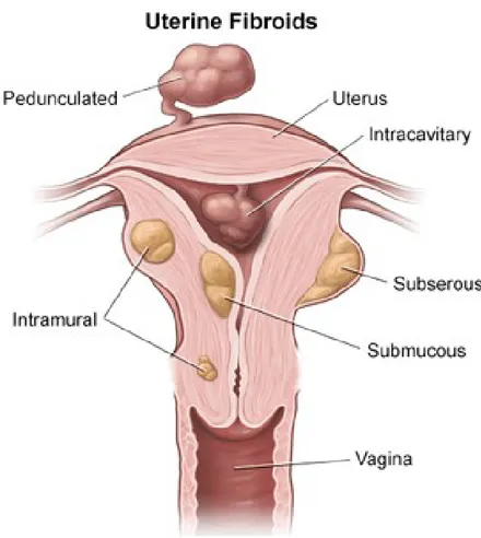
The size of fibroids can vary between a few millimeters up to multiple centimeters. The International Federation of Gynecology and Obstetrics (FIGO) developed a classification system to categorize uterine fibroids by their location, though it is possible to have multiple fibroids in various sites at the same time (Munro, Critchley et al. 2011). Submucosal: these fibroids derive below the endometrium and can grow into the uterine cavity having clinical relevance in regards to the severity of menses, possible cause of infertility and miscarriages as well as indicating outcomes of myomectomies.
Intramural: these fibroids represent the most common type and are located within the uterine wall. Large intramural leiomyomas may distort the uterine cavity and lead to bulk symptoms.
Subserosal: these fibroids extend from the outer layer of the uterus.
Figure 1 – Location of uterine leiomyomas.
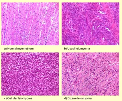
The structure of leiomyomas typically presents with vessels entering at the periphery while showing an avascular center with degeneration and necrosis (Krysiewicz 1992, Murase, Siegelman et al. 1999, Ahmadi, Zafarani et al. 2008). If classified histologically, fibroids can be divided into usual, cellular or bizarre leiomyomas, though other characteristic features, such as epithelioid leiomyomas, clear cell and granular cell leiomyomas, lipoleiomyomas, myxoid leiomyomas, haemorrhagic cellular (apoplectic) leiomyoma, leiomyristioma with haematopoietic elements, benign metastasizing leiomyoma, perinodular hydropic leiomyoma, multinodular hydropic leiomyoma and cotyledonoid dissecting leiomyoma (Toledo and Oliva 2008, Al Hilli and Stewart 2010).
a) Normal myometrium b) Usual leiomyoma
c) Cellular leiomyoma d) Bizarre leiomyoma
Figure 2 – Normal myometrium and different histological types of uterine leiomyoma (Islam, Protic et al. 2013).
1.1.3. Epidemiology and Etiology
There is a gap in assessing the true prevalence and incidence of fibroids as most studies are conducted on symptomatic women with a lack of longitudinal studies (Marshall, Spiegelman et al. 1997, Stewart, Cookson et al. 2017). One of the largest prospective studies conducted over five years that included about 95,000 women showed an incidence of 8.9 per 1000 women years in Caucasian women and 30.6 in African-American women and showed an increase of incidences the older the women were. The impact of ethnicity in the predisposition to leiomyomas has been shown in various studies and it is thought that genetics, environmental factors and lifestyle play an important role (Witherspoon and Butler 1934, Torpin, Pund et al. 1942, Wilcox, Koonin et al. 1994, Marshall, Spiegelman et al. 1997, Amant, Huys et al. 2003). Other risk factors contributing to the development of fibroids are nulliparity, early menarche, alcohol, obesity, diet including vitamin D deficiency (Flake, Andersen et al. 2003, Okolo 2008, Baird, Harmon et al. 2015).
The pathogenesis of leiomyomas is a complex interplay of intrinsic and extrinsic factors such as hormones, genetics, diet and environment. (Walker and Stewart 2005, Islam, Protic et al. 2013). The presence of multiple fibroids is a result of de novo rather than metastatic spread. Recent findings in stem cell research have shown that the highly dependent sex steroid leiomyomas emerge from an estrogen and progesterone receptor deficient precursor, giving rise to the development of new potential therapeutic targets (Ono, Maruyama et al. 2007, Chang, Senaratne et al. 2010, Mas, Cervelló et al. 2012, Ono, Qiang et al. 2012). A side population isolated from healthy human myometrium was identified, showing distinct phenotypical and functional stem cell like character and a leiomyoma-derived side populations presented stem cell characteristics as well, which are required for in vivo leiomyoma xenograft tumours (Ono, Maruyama et al. 2007, Ono, Qiang et al. 2012). Half of all leiomyomas display chromosomal abnormalities, such as karyotypic and cytogenetic, as well as somatic mutations, such as deletions, translocations or duplications (Kiechle-Schwarz, Sreekantaiah et al. 1991, Rein, Friedman et al. 1991, Meloni, Surti et al. 1992). In leiomyomas, multiple chromosomes present genetic alterations, including chromosomes 2,3,6,7,8,11,12,13,14 and 22 and various genes, such as MED12, HMGA2, HMGA1, FH, BHD, TSC2, PCOLCE, ORC5L and LHFPL3, are candidates for leiomyoma development (Ligon, Scott et al. 2002, Sandberg 2005, Walker and Stewart 2005, Ptacek, Song et al. 2007, El-Gharib and Elsobky 2010, Nezhad, Drieschner et al. 2010, Cha, Takahashi et al. 2011, Mäkinen, Heinonen et al. 2011, Mäkinen, Mehine et al. 2011, Velagaleti, Tonk et al. 2011). Out of all these genes, MED12 is present with the highest mutation frequency of about 70% in fibroids (Mäkinen, Heinonen et al. 2011, Mäkinen, Mehine et al. 2011, Je, Kim et al. 2012, Markowski, Bartnitzke et al. 2012, McGuire, Yatsenko et al. 2012, Pérot, Croce et al. 2012). Furthermore, epigenetic modifications, such as microRNA, DNA methylation and histone modification, have been analyzed in leiomyomas (Marsh, Lin et al. 2008, Greathouse, Bredfeldt et al. 2012, Navarro, Yin et al. 2012). The role of hormones, specifically estrogen and progesterone, seem to be the key players in uterine leiomyoma formation by activating receptor
in the nucleus (Maruo, Ohara et al. 2004, Ciarmela, Islam et al. 2011, Kim and Sefton 2011). By targeting genes that enhance cell growth, estrogen and progesterone act as physiologic regulators of gene expression. Though both hormones have decisive roles in the pathogenesis of uterine fibroids progesterone seems to have a superior impact in cell proliferation. Various studies have analyzed the role of cytokines (IL-1, IL-6, IL-11, IL-13, IL-15, TNF-α, GM-CSF, and erythropoietin), chemokine receptors (MIP-1α, MIP-1β, RANTES, Eotaxin, Eotaxin-2, IL-8, CCR1, CCR3, CCR5, CXCR1, CXCR2, and MCP-1) and growth factors (EGF, HB-EGF, PDGF, IGF, TGF-α, TGF-β, VEGF, aFGF, bFGF, activin-A and myostatin) in leiomyoma disease pathogenesis (Sozen, Olive et al. 1998, Hatthachote and Gillespie 1999, Kurachi, Matsuo et al. 2001, Sozen and Arici 2002, Flake, Andersen et al. 2003, Ding, Xu et al. 2004, Luo, Ding et al. 2005, Litovkin, Domenyuk et al. 2007, Ciarmela, Wiater et al. 2008, Syssoev, Kulagina et al. 2008, Ciarmela, Wiater et al. 2009, Chegini 2010, Ciarmela, Bloise et al. 2011, Ciarmela, Islam et al. 2011). The extracellular matrix (ECM) in leiomyoma cells is mostly built of collagen subtypes, proteoglycans and fibronectin. It has been shown that the ECM functions as a reservoir for cytokines, chemokines, growth factors and mediators of angiogenic as well as inflammatory responses (Wolanska, Sobolewski et al. 1998, Arici and Sozen 2000, Walker and Stewart 2005, Norian, Malik et al. 2009, Chegini 2010, Malik, Norian et al. 2010).
1.1.4. Symptoms and Diagnosis
Most cases of leiomyomas are asymptomatic, which can be due to a very small size and a diagnosis in these cases is usually made incidentally. When women become symptomatic, the sign are associated with the size (defined in menstrual weeks as with the gravid uterus), location and number of fibroids (Kashani, Centini et al. 2016). Menorrhagia (heavy and prolonged menstrual bleeding) presents as the most common symptom, though the distinct mechanism is not clear and the severity is dependent on the location of the fibroids, submucosal myomas being the most frequent cause (Fraser, Critchley et al. 2007). Less frequently, larger leiomyomas
can cause pelvic discomfort and bulk symptoms by pressuring neighboring organs (Borah, Nicholson et al. 2013). Depending on the anatomical location this can lead to a urethral obstruction with micturition disturbances such as an alteration urinary frequency up to a complete urinary obstruction. Women may also experience pressure on the rectum presenting with blockage or irritation (Nishino, Togashi et al. 2005). A degeneration of a leiomyoma or torsion of the pedunculated fibroid can lead to acute pain with fever and pelvic tenderness (Laughlin and Stewart 2011). If a large submucosal or intramural leiomyoma deforms the uterine cavity patients may experience difficulties in conceiving and have an increased risk of miscarriage (Pritts, Parker et al. 2009). The main clinical challenges of fibroids are excluding differential diagnosis and comprehending their role in abnormal uterine bleeding, pelvic pain as well as conception and pregnancy problems.
Leiomyomas are diagnosed by pelvic examination and ultrasound as well as by taking the medical history of the patient. A clinical diagnosis is sufficient to confirm the disease. In order to assess submucosal or protruding fibroids hysteroscopy can help visualize the extent of protrusion in the uterine cavity. Additionally, magnetic resonance imaging is the most accurate method for distinguishing between leiomyomas and other conditions arising from the tissues of the uterus, though a high cost and limited availability should be considered (Cicinelli, Romano et al. 1995, Dueholm, Lundorf et al. 2002).
1.1.5. Therapeutic Options
When leiomyomas are asymptomatic, no treatment is required. It is not recommended to prophylactically treat asymptomatic fibroids, though exceptions should be made for women considering pregnancy in order to avoid miscarriage. For symptomatic cases, there are various conservative and surgical options available and decisions should be made based on symptoms, pain level, fibroid size and location, age and comorbidities of the patient as well as reproductive plans (Viswanathan, Hartmann et al. 2007). Hormonal therapies represent the most important
pillar in medical management of leiomyomas. The aim of medical treatment should be to minimize symptoms and reduce bleeding as well as the size of the uterus. Gonadotropin-releasing hormone agonists (GnRHa) cause a downregulation of gonadotropic and gonadal hormones leading to a hormonal state resembling menopause and are the most effective treatment option for leiomyomas. They have been approved by the Food and Drug Administration (FDA) as preoperative treatment to minimize fibroid volume and increase the haematocrit for a better surgical outcome. Side effects include symptoms similar to menopause, such as hot flashes, mood swings, vaginal dryness and sleep disturbance. Long-term use of these drugs causes loss of bone-mass leading to osteoporosis, which represents the most severe adverse event and limits the treatment to a few months. After cessation of treatment there is a rapid rebound-effect resulting in heavy menses and increase of fibroid mass. Therefore, the duration of treatment with GnRH agonists should be limited to a few months in order to attain optimal pre-operative conditions and therapy should be re-evaluated if there is no significant effect on the leiomyomas in order to avoid adverse events (Andreyko, Blumenfeld et al. 1988, Friedman, Rein et al. 1989, Schlaff, Zerhouni et al. 1989, Friedman, Hoffman et al. 1991, Leather, Studd et al. 1993, Stovall, Muneyyirci-Delale et al. 1995). Other treatment options include progesterone receptor modulators, such as ulipristal acetate, which inhibits the progesterone receptor in the hypothalamus, uterus, cervix, and ovaries. It was approved by the European Medicine Agency (EMA) for a short-term treatment course prior to surgery (Talaulikar and Manyonda 2012, Ciarmela, Carrarelli et al. 2014). A trial has shown a superior effect of ulipristal acetate compared to placebo and a similar effect in reduction of fibroid size and bleeding compared to GnRH agonists, with a less frequent occurrence of hot flashes (Donnez, Tomaszewski et al. 2012). The use of antiprogestins has been implicated in treatment of leiomyomas as they target excessive growth of disorganized extracellular matrix and stimulate the growth of smooth muscle cells. Other treatment options include mifepristone, an antiprogestin, asoprisnil, a selective steroid receptor modulator, proellex, a selective
progesterone receptor blocker, aromatase inhibitors and pirfenidone, an antifibrotic and anti-inflammatory agent, which are currently tested in clinical trials (Engman, Granberg et al. 2009, Islam, Protic et al. 2013). The effects of tranilast, an anti-allergic compound, as well as curcumin and vitamin D have shown to inhibit proliferation of fibroids in vitro and represent promising novel target points for future treatment (Islam, Protic et al. 2013).
Surgery is the most frequently applied treatment of leiomyomas, which represent the most common indication for hysterectomy. Hysterectomy is suitable for women who experience severe bleeding unresponsive to medical therapeutics, who do not wish to have any further children or to remain fertile and who are at risk to develop other gynaecological diseases of the uterus in the future (Farquhar and Steiner 2002). The indication for hysterectomy depends on the size, localization and number of leiomyomas, as solitary fibroids may be surgically removed by laparoscopy or hysteroscopy. Another uterus retaining procedure is the myomectomy, suitable for women with menorrhagia though the adverse events include haemorrhage, development of new leiomyomas and adhesion formation hysterectomy (Olufowobi, Sharif et al. 2004). Fibroid treatment via myolysis/cryomyolysis (Phillips, Nathanson et al. 1996, Donnez, Squifflet et al. 2000, Ciavattini, Tsiroglou et al. 2004). Other surgical options include myolisis, uterine artery embolization, uterine artery occlusion and magnetic resonance guided focused ultrasound, though these methods report limited data and are less established than hysterectomy (Phillips, Nathanson et al. 1996, Donnez, Squifflet et al. 2000, Ciavattini, Tsiroglou et al. 2004, Spies, Bruno et al. 2005, Vilos, Vilos et al. 2010, Liu, Li et al. 2011).
1.2. Nutrition and Lipids in Leiomyomas
In regards to the role of nutrition, some scientific reports have concluded the following:
1. A low intake of fruit and vegetables can be associated with an increased risk of developing uterine fibroids.
3. The impact of fish and meat intake needs to be further assessed.
Finally, more studies are necessary to deepen the understanding of the various mechanisms that ultimately lead to the development of fibroids and to analyze the modulating potential of other food classes to identify the correlation to the pathology.
An interesting result showed a correlation between excessive consumption of farmed fish and the growth of leiomyomas. This association is thought to be due to the various components of the diet of these fish. For example, polychlorinated biphenyls (PCB), can be more or less present in fish depending on its provenance (Parazzini, Di Martino et al. 2015).
1.2.1. Therapeutic effect of natural compounds on leiomyomas
Various natural compounds have been analyzed in regards to their therapeutic potential in modulating and preventing myometrial tissue turning into leiomyomas. One of these is epigallocatechin gallate, the ester of epigallocatechin and gallic acid, which can be found in green tea, has shown to induce apoptosis and inhibit the proliferation of leiomyoma cells and a phase II clinical trial using this compound has been completed (Zhang, Al-Hendy et al. 2010). Isoliquiritigenin, which is a calchone flavonoid found in licorice, onions and bean sprouts, can also prevent growth while upregulating cell death in leiomyoma cells (Kim, Ramachandran et al. 2008). Another natural compound, curcumin (diferuloylmethane, a polyphenol originating from the plant Curcuma longa), also known as turmeric, has shown to have significant anti-inflammatory, anti-oxidant and anti-carcinogenic characteristics. Turmeric inhibits mitosis by targeting the apoptotic pathway and leads to a decreased production of fibronectin which is a member of the extracellular matrix (ECM) (Malik, Britten et al. 2007). As mentioned above, vitamin D can also reduce proliferation of leiomyoma cells by down-regulating PCNA, CDK1 and BCL-2 as well as the catechol-O-methyltransferase expression (Sharan, Halder et al. 2011).
1.2.2. Omega-3 fatty acids
Omega-3 fatty acids, also known as ω−3 fatty acids or n−3 fatty acids, are polyunsaturated fats which cannot be synthesized by humans and are therefore ‘essential fatty acids’ that necessarily need to be obtained from marine animals and plant oils (Rose and Connolly 1999). These fatty acids represent an important pillar in the daily diet as they have an important role in maintaining health and preventing diseases (Whelan and Rust 2006). The active components that can be found in fish are eicosapentaenoic acid (EPA) and docosahexaenoic acid (DHA). Omega-3 fatty acids have shown in various in-vitro and in-vivo studies that they possess anti-inflammatory and immunomodulatory effects, highlighting their potential role as therapeutics in inflammatory and autoimmune diseases (Calder 2001). When inflammation occurs in the body, a large amount of cytokines, arachidonic acid-derived eicosanoids (oxidized derivatives such as prostaglandins, thromboxanes and leukotrienes), reactive oxygen species and adhesion molecules are produces. If enough long-chain n-3 polyunsaturated fatty acids are consumed, it has been seen that these can lower the production of inflammatory associated markers (Calder 2001). One of the most important anti-inflammatory effects of omega-3 fatty acids is their ability to interfere with the metabolism of arachidonic acid. An increased presence of omega-3 fatty acids results in a higher expression of adhesion proteins, such as selectins and vascular cell adhesion molecule-1, in the epithelium which consequently interact with the leukocytes. This is achieved by altering the intracellular signaling pathway that controls transcription factors such as nuclear factor-κB and transcription of genes (Deckelbaum, Worgall et al. 2006). Omega-3 fatty acids also present the ability to bind directly to receptors in the nucleus, like retinoid X receptor, operating as transcription factors.
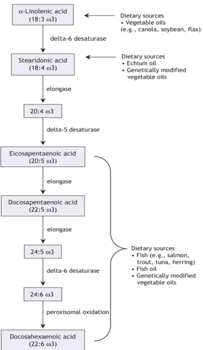
Figure 3 – Chemical structure of eicosapentaenoicacid (EPA) and docosahexaenoic acid (DHA)
Figure 4 – Metabolism and dietary sources of the omega-3 family of polyunsaturated fatty acids. (Whelan and Rust 2006, Surette 2008).
1.2.3. The cell membrane
The cellular membrane is indispensable for existence as it encloses all the metabolic mechanisms occurring in a cell. The membrane does not have a passive role in this, but is actively engaged in the complex operation program which includes nutrient and oxygen diffusion as well as receiving and sending signals responsible for the adaptation to physiological and pathological surroundings in which the cell has to function (Leppert, Jayes et al. 2014).
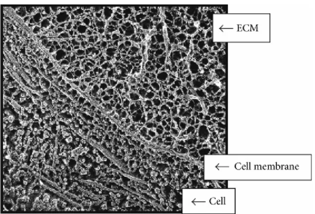
This figure illustrates that mechanical forces can be transmitted across the cell surface and into the cell by means of interconnected structural components (Leppert, Jayes et al. 2014)
Figure 5 – Interconnected structural components of cells and ECM.
Quick freeze, deep etch electron micrograph of a fetal ear cartilage chondrocyte cell to illustrate the integration of structures inside and outside the cell. The ECM (containing a meshwork of proteoglycans, collagen, fibronectin, and laminin), the cell membrane (integrins receptors), and the cell (containing microtubules) are visualized. In the ECM the thinnest fibrils in the meshwork are nm and are presumed to be proteoglycans. The larger thicker fibrils are collagen. Micrograph courtesy of Robert Mecham and John Heuser, Washington University, St. Louis, MO, USA.
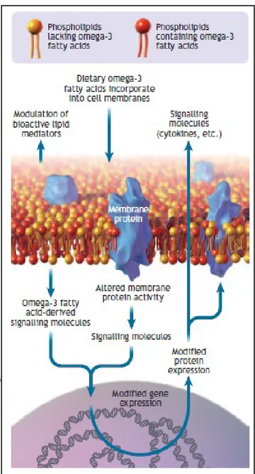
Figure 6 – Cell membrane showing omega-3 fatty acids incorporated into the phospholipid bilayer. Omega-3 fatty acids can modify gene and protein expression, modulate membrane protein activity and act as a reservoir for bioactive molecules (Surette 2008).
1.2.4. Lipidomics
Lipidomics is an uprising field in science, founded less than twenty years ago upon the success of the other “-omics”, the most well-known being genomics. The terminology indicates the innovative interpretation of ‘old-fashioned’ biological molecules that to date are not seen just as cellular elements anymore, but as structures that experience dynamic change in physiological as well as pathological conditions. Lipidomics aims to identify all lipids in the cell, the so called
lipidome and can track changes in the lipids depending on the metabolic conditions. Consequently, it is fundamental to focus research on the cellular membrane, as the most recent findings in biology and medicine have shown that the most important cellular processes are occurring in that site (Bou Khalil, Hou et al. 2010, Vilella, Ramirez et al. 2013).
2. Materials and Methods
Drugs and chemicals
EPA (cis-5,8,11,14,17-Eicosapentaenoic acid) and DHA (cis-4,7,10,13,16,19-Docosahexaenoic acid) were dissolved in absolute ethanol at 30 mM and it was further diluted, with medium to reach 50 µM at the time of cell treatment.
Leiomyoma and myometrial tissue collection and primary cell cultures
Uterine fibroids and myometrial tissues were collected by hysterectomy or laparotomic myomectomy of premenopausal Caucasian women (41-49 years old) with symptomatic fibroids. Samples were collected in Hanks’ Balanced Salt Solution (HBSS) at the time of surgery and rinsed with Dulbecco’s PBS to remove excess blood. Samples were cut into small pieces with 0.1% collagenase type 8 solutions in serum-free Dulbecco's Modified Eagle Medium (DMEM) containing 1% penicillin-streptomycin, 50 µg/L gentamicin, and 1% Amphotericin B. Tissues were then incubated at 37 °C for about 3-5 h in a water bath with manual shaken until complete digestion. Digested cell suspensions were centrifuged at 1200 rpm for 10 min, and washed once with fetal bovine serum (FBS). Finally, the cell pellet was dispersed in complete DMEM media, plated in T25 plastic dishes, and maintained at 37 °C in 95% air-5% CO2. The growth medium
was changed after 48 h or 72 h to remove unattached cells and then subsequently twice a week. The purity of cells was assessed by immunocytochemical staining with specific smooth muscle cells marker, monoclonal mouse anti-α-smooth muscle actin (α-SMA) (Sigma-Aldrich). The lower passage number (up to 5) of cells was used for experiments to avoid changes in phenotype and gene expression.
Transmethylation of cell pellet fatty acids
The direct transmethylation reaction of the cell pellet fatty acids (FA) was performed adapting the method of Harris (Harris, Pottala et al. 2012). Briefly, 30 mg of defrosted cell pellets were suspended in 300 µl of water. The suspension was transferred to a 2ml screwed cap vial and freeze dried. 250 µl of a solution of BF3 (12% in MeOH anhydrous from Sigma Aldrich®) and
250 µl of hexane were added in sequence to the freeze-dried sample. The vial was capped and the suspension was vortexed for 15 seconds. Then, the vial was put in the oven at 100°C for 15 minutes. After cooling, 250 µl of H2O were added to the sample and, after recapping the vial,
the mixture was vortexed for 20 seconds. Two phases were obtained after centrifugation. 150 µl of the upper hexane phase, containing the fatty acids methyl esters (FAME), were transferred in a 300 µl glass insert in a screwed cap vial. The solution was dried under nitrogen flow, and the FAME were dissolved in 50 µl of hexane. Samples were stored at -20°C till the gaschromatographic (GC) analysis.
GC-FID analysis of FAME
GC analysis of the FAME in cell pellets was performed with a Varian® 430 GC. The chromatographic system was equipped with a split/splitless injector, a 100m Varian® capillary column Select™ FAME (0.25 mm, 0.25 µm, #CP7421) and a flame ionization detector (FID). 0.3 µl of sample were injected with a split ratio of 2. The injector and the FID were maintained at 260°C. Helium (He) was employed as carrier gas with a flow rate of 1.6 ml/min. A gradient of temperature was set, with the oven starting at 160°C and reaching 240°C in 20 minutes and keeping for 15 minutes before cooling. Peaks were identified by comparison with known standards (Supelco 37 Component FAME Mix). Fatty acids composition (wt %) were calculated by the corrected peak area normalization method. The chromatographic system and the peak integration were managed by the Galaxie software (Agilent, Santa Clara, CA, USA).
Laurdan fluorescence measurements
Laurdan (6-lauroyl-2-dimethylaminonaphthalene) is a fluorescent probe which locates in the bilayer by its lauric acid tail, with the fluorescent moiety localized at the level of the glycerol backbone (Antollini and Barrantes 1998). It has a very high partition coefficient from aqueous environments to membranes and it is equally distributed between gel and liquid-crystalline (LC) phases; besides it is not affected by pH and by the chemical nature of phospholipid polar heads. Its spectral sensitivity to the membrane phase state can be related to its ability to sense polarity and the dynamics of water molecules (dipolar relaxation of water molecules) in the immediate vicinity of the fluorophore (Parasassi, De Stasio et al. 1991). Laurdan’s spectral features are used to calculate the generalized polarization (GP) equation which provides information about the phospholipid phase of the membrane (Bagatolli and Gratton 1999). Laurdan excitation (Ex) GP spectra were calculated as follows (Parasassi, Loiero et al. 1993). (Parasassi, Loiero et al. 1993): Ex GP = I450 – I490 / I450 + I490 where I450 and I490 are the intensities at each excitation
wavelength, from 320 to 420 nm, obtained using a fixed emission wavelength of 450 and 490 nm, respectively. In the phospholipid gel phase, Laurdan ExGP spectra are wavelength independent; in the LC phase, Ex GP values decrease with increasing excitation wavelength, with two coexisting phases, the GP spectrum has an opposite trend.
Low ExGP values indicate a large water molecular mobility around the probe (thus a fluid membrane), while high ExGP values indicate a rigid membrane with restricted reorientation of water molecules during the fluorescence lifetime.
Laurdan steady-state fluorescence measurements were carried out on a computer-controlled PerkinElmer LS55 spectrofluorimeter. The fluorescence background obtained from cells without Laurdan was always subtracted from the data. Laurdan fluorescence spectra were measured at 37 °C and the temperature was measured in the sample by a digital thermometer. Cells were labeled with Laurdan at a final probe concentration of 1 µM. Each Laurdan spectrum
corresponds to the average of determinations performed on five different samples (n=5). All spectra were normalized by using Perkin Elmer FL WinLab Software (Perkin Elmer Ltd., Buckinghamshire, United Kingdom).
RNA isolation and real-time PCR
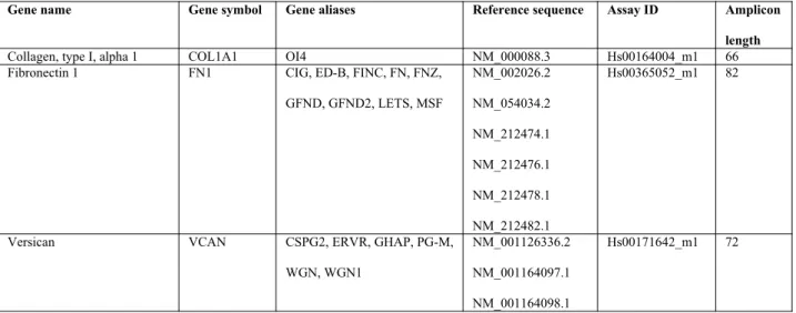
Primary myometrial and leiomyoma cells were treated with EPA or DHA (50 µM), and kept negative control (treated with 0.2 % ethanol as we used to dissolve EPA and DHA) for 48 h, and lysed using TRIzol reagent (Ambion, Life Technologies), and stored at -80 °C. Total RNA (colorless upper aqueous phase) was separated using chloroform according to the manufacturer’s instructions. After that, RNA was purified and concentrated using ReliaPrep™ RNA Cell Miniprep System (Promega Italia, Milan, Italy). The complementary DNA (cDNA) was generated from 1 µg of RNA using high-capacity cDNA reverse transcriptase (RT) kit (Applied Biosystems, Life Technologies), and newly synthesized cDNA was used for real-time PCR. Real-time PCR was performed with 50 ng cDNA in a final volume of 15 µl, containing 1X TaqMan® fast advanced master mix, with the following TaqMan® gene expression assays (Applied Biosystems, Life Technologies) (Table 1). Controls included RNA subjected to RT-PCR without reverse transcriptase.
Table 1. – List of primers.
Gene name Gene symbol Gene aliases Reference sequence Assay ID Amplicon length
Collagen, type I, alpha 1 COL1A1 OI4 NM_000088.3 Hs00164004_m1 66
Fibronectin 1 FN1 CIG, ED-B, FINC, FN, FNZ,
GFND, GFND2, LETS, MSF NM_002026.2 NM_054034.2 NM_212474.1 NM_212476.1 NM_212478.1 NM_212482.1 Hs00365052_m1 82
Versican VCAN CSPG2, ERVR, GHAP, PG-M,
WGN, WGN1
NM_001126336.2 NM_001164097.1 NM_001164098.1
NM_004385.4
Inhibin, beta A INHBA EDF, FRP NM_002192.2 Hs00170103_m1 65
Integrin, beta 1 (fibronectin receptor, beta polypeptide, antigen CD29 includes MDF2, MSK12) ITGB1 CD29, FNRB, GPIIA, MDF2, MSK12, RP11-479G22.2, VLA-BETA, VLAB NM_002211.3 NM_033668.2 NM_133376.2 Hs00559595_m1 75
Protein tyrosine kinase 2 PTK2 FADK, FAK, FAK1, FRNK, PPP1R71, p125FAK, pp125FAK NM_001199649.1 NM_005607.4 NM_153831.3 Hs01056457_m1 76
A kinase (PRKA) anchor protein 13
AKAP13 AKAP-13, AKAP-Lbc,
ARHGEF13, BRX, HA-3, Ht31, LBC, PRKA13, PROTO-LB, PROTO-LBC, c-lbc, p47 NM_001270546.1 NM_006738.5 NM_007200.4 Hs00180747_m1 105
ATP-binding cassette, sub-family G (WHITE), member 1
ABCG1 ABC8, WHITE1 NM_004915.3
NM_016818.2 NM_207174.1 NM_207627.1 NM_207628.1 NM_207629.1 Hs00245154_m1 58
ATP-binding cassette, sub-family A (ABC1), member 1
ABCA1 ABC-1, ABC1, CERP,
HDLDT1, TGD
NM_005502.3 Hs01059118_m1 61
Caveolin 1, caveolae protein, 22kDa CAV1 BSCL3, CGL3, MSTP085, PPH3, VIP21 NM_001172895.1 NM_001172896.1 NM_001172897.1 NM_001753.4 Hs00971716_m1 66
Sterol regulatory element binding transcription factor 2
SREBF2 CTA-250D10.14-005,
SREBP-2, SREBPSREBP-2, bHLHd2 NM_004599.3 NR_103834.1 Hs01081784_m1 91 Cytochrome P450, family 11, subfamily A, polypeptide 1
CYP11A1 CYP11A, CYPXIA1,
P450SCC NM_000781.2 NM_001099773.1 Hs00167984_m1 77 Phosphatidylethanolamine binding protein 1 PEBP1 HCNP, HCNPpp, PBP, PEBP, PEBP-1, RKIP NM_002567.2 Hs00831506_g1 134 Hypoxanthine phosphoribosyltransferase 1 HPRT1 HGPRT, HPRT NM_000194.2 Hs99999909_m1 100
Actin, beta ACT B BRWS1, PS1TP5BP1 NM_001101.3 Hs99999903_m1 171
Data analysis
Statistical analyses were performed using GraphPad Prism software (version 6.01 for Windows) (GraphPad, San Diego, CA). The data were analyzed using non-parametric ‘Kruskal-Wallis’ ANOVA, followed by post hoc ‘Dunn’ test for multiple comparisons. Results are expressed as mean ± SD. Differences were considered significant when *p < 0.05, **p < 0.01, ***p < 0.001. The data obtained from the GC-FID analysis of tissues and cells FA were organized and processed by means of Office Excel software (Microsoft). Principal component analysis (PCA)
of the results was performed by means the Office Excel add-in Multibase package (Numerical Dynamics, Japan).
3. Results and Discussion
In vitro effect of DHA and EPA on fatty acid composition
Myometrial cells.
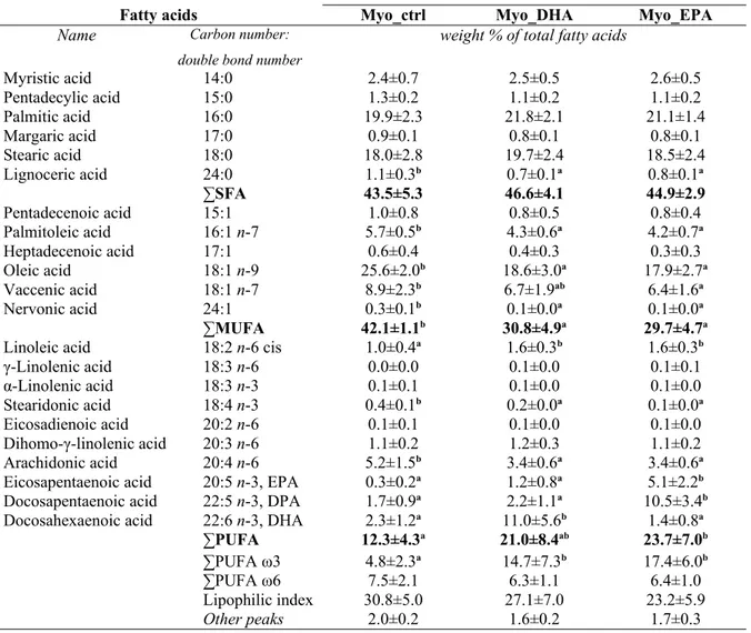
The results regarding the analysis of fatty acid content of cells isolated from myometrial tissue, which were treated in vitro with EPA or DHA are presented in Table 2.
The addition of DHA and EPA to myometrial cells in vitro lead to a change of fatty acid composition of these cells. In both, untreated and treated cells, the SFA were the main parts, while the PUFA were the less frequent. In the untreated group, representing a control samples, the main fatty acid was the oleic acid (25.6±2.0%), followed by palmitic and stearic acids (19.9±2.3% and 18.0±2.8%, respectively). In comparison to the treated cells, palmitic acid had the highest relative content for both myometrial cells treated with EPA and those treated with DHA (21.8±2.1% and 21.1±1.4%, respectively), followed by stearic and oleic acids. Application of DHA on myometrial cells reduced the content of MUFA significantly and led to a not statistically significant increase of the PUFA fraction, compared to the control group. When myometrial cells were treated with EPA, a significant MUFA reduction as well as PUFA increase were measured compared to the untreated cells.
Among the monosaturated types, the addition of DHA to the myometrial cells provided a significant decrease in the relative content of oleic acid (25.6±2.0% vs 18.6±3.0%), palmitoleic acid (5.7±0.5% vs 4.3±0.6%) and nervonic acid (0.3±0.1% vs 0.1±0.0%) while the percentage of vaccenic acid (18:1 ω7) was lower, without statistical significance. Treatment with EPA showed a similar effect on the polyunsaturated fatty acid composition of the myometrial cells, with a reduction of the relative content of oleic acid from the 25.6±2.0% in the control group to the 17.9±2.7% of the treated cells. Also, palmitoleic, nervonic and vaccenic acids significantly decreased after EPA was added to the cell culture.
Omega-3 fatty acids strongly modified the PUFA composition of in vitro treated myometrial cells. The arachidonic acid content decreased from a value of 5.2±1.5% in the untreated cells to values of 3.4±0.6% for cells that received DHA and EPA. Furthermore, linoleic acid levels increased 1.0±0.4% to 1.6±0.3% after addition of DHA and EPA. A small, but statistically significant, decrease of 18:4 ω3 in omega-3 treated myometrial cells was noted. Considering the amounts of EPA, DHA and DPA in cells isolated from the myometrium, the two treatments showed different effects. In the cell group that was treated with DHA, EPA and DPA levels did not vary compared to the control group, though DHA was clearly integrated by the cells as the relative content rose from 2.3±1.2% to 11.0±5.6%. The cells treated with EPA showed a significant increase in the levels of EPA (5.1±2.2% to 10.5±3.4%) and DPA (0.3±0.2% to 1.7±0.9%), while the DHA level was not modified by EPA when added in vitro.
Table 2 - Total fatty acid composition (weight % of total fatty acids) of cells isolated from myometrial tissue, untreated (Myo_ctrl) and treated in vitro with DHA (Myo_DHA) and with EPA (Myo_EPA), with statistical analysis.
Fatty acids Myo_ctrl Myo_DHA Myo_EPA
Name Carbon number: double bond number
weight % of total fatty acids
Myristic acid 14:0 2.4±0.7 2.5±0.5 2.6±0.5 Pentadecylic acid 15:0 1.3±0.2 1.1±0.2 1.1±0.2 Palmitic acid 16:0 19.9±2.3 21.8±2.1 21.1±1.4 Margaric acid 17:0 0.9±0.1 0.8±0.1 0.8±0.1 Stearic acid 18:0 18.0±2.8 19.7±2.4 18.5±2.4 Lignoceric acid 24:0 1.1±0.3b 0.7±0.1a 0.8±0.1a ∑SFA 43.5±5.3 46.6±4.1 44.9±2.9 Pentadecenoic acid 15:1 1.0±0.8 0.8±0.5 0.8±0.4 Palmitoleic acid 16:1 n-7 5.7±0.5b 4.3±0.6a 4.2±0.7a Heptadecenoic acid 17:1 0.6±0.4 0.4±0.3 0.3±0.3 Oleic acid 18:1 n-9 25.6±2.0b 18.6±3.0a 17.9±2.7a Vaccenic acid 18:1 n-7 8.9±2.3b 6.7±1.9ab 6.4±1.6a Nervonic acid 24:1 0.3±0.1b 0.1±0.0a 0.1±0.0a ∑MUFA 42.1±1.1b 30.8±4.9a 29.7±4.7a
Linoleic acid 18:2 n-6 cis 1.0±0.4a 1.6±0.3b 1.6±0.3b
γ-Linolenic acid 18:3 n-6 0.0±0.0 0.1±0.0 0.1±0.1 α-Linolenic acid 18:3 n-3 0.1±0.1 0.1±0.0 0.1±0.0 Stearidonic acid 18:4 n-3 0.4±0.1b 0.2±0.0a 0.1±0.0a Eicosadienoic acid 20:2 n-6 0.1±0.1 0.1±0.0 0.1±0.0 Dihomo-γ-linolenic acid 20:3 n-6 1.1±0.2 1.2±0.3 1.1±0.2 Arachidonic acid 20:4 n-6 5.2±1.5b 3.4±0.6a 3.4±0.6a
Eicosapentaenoic acid 20:5 n-3, EPA 0.3±0.2a 1.2±0.8a 5.1±2.2b
Docosapentaenoic acid 22:5 n-3, DPA 1.7±0.9a 2.2±1.1a 10.5±3.4b
Docosahexaenoic acid 22:6 n-3, DHA 2.3±1.2a 11.0±5.6b 1.4±0.8a
∑PUFA 12.3±4.3a 21.0±8.4ab 23.7±7.0b
∑PUFA ω3 4.8±2.3a 14.7±7.3b 17.4±6.0b
∑PUFA ω6 7.5±2.1 6.3±1.1 6.4±1.0
Lipophilic index 30.8±5.0 27.1±7.0 23.2±5.9
Other peaks 2.0±0.2 1.6±0.2 1.7±0.3
SFA, saturated fatty acid; MUFA, monounsaturated fatty acid; PUFA, polyunsaturated fatty acid. Results represent means ± S.D. (n=8); differences were considered significant for P < 0.05.
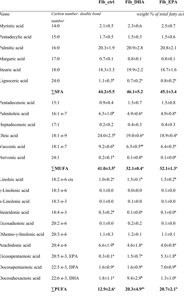
Leiomyoma cells.
Leiomyoma cells responded in a similar way to treatment with EPA or DHA in comparison to myometrial cells. In both, the treated and untreated cells, the SFA represented the highest fatty acids with 45%. The saturated acidic fraction was not affected by the addition of DHA or EPA, except for a minimal decrease of the lignoceric acid in both groups. DHA lead to a reduction of the total MUFA content in leiomyoma cells from 41.0±3.5% of the control group to 32.1±0.4% (P<0.05) in the treated group. It was also noticed that EPA lead to a significant decline of
MUFA content to 32.1±1.3% and also caused a decrease of the relative content of oleic, palmitoleic, vaccenic and nervonic acids. Addition of DHA to leiomyoma cells had a comparable effect on reduction of MUFA subtype frequencies, though the decrease of vaccenic acid level was not statistically significant. The in vitro supplementation of both, EPA and DHA, increased the level of total PUFA in leiomyoma cells, but this was only significant for the cells treated with EPA (P<0.05). Similar to the myometrial cells, treatment with EPA caused a significant decreases of arachidonic acid levels (6.6±1.9% vs 4.0±0.8%) and of C 18:4 ω3 (0.3±0.2% vs 0.1±0.0%) but also a significant increase of EPA, DPA and linoleic acid. The effects of adding DHA to leiomyoma cells were also comparable to its effects on myometrial cells. Apart the significant reduction of the levels of arachidonic and C 18:4 omega-3, the higher level of linoleic acid (P<0.5) was similar to that caused by treatment with EPA.
Different to the effect caused by EPA on leiomyoma cells, DHA led to a significant increase in DHA levels (1.8±1.1% vs 9.4±2.9%), with no noticeable changes on DPA and EPA content. The total PUFA ω3 content of leiomyoma cells changed from 4.1±2.2% in the control group to 12.8±4.2% in DHA treated cells and 13.9±2.0% in EPA treated cells. Addition of DHA and EPA had not effect on the PUFA ω6 fraction.
Table 3 - Total fatty acid composition (weight % of total fatty acids) of cells isolated from leiomyoma
tissue, untreated (Fib_ctrl) and treated in vitro with DHA (Fib_DHA) and with EPA (Fib_EPA), with statistical analysis
Fib_ctrl Fib_DHA Fib_EPA
Name Carbon number: double bond number
weight % of total fatty acids
Myristic acid 14:0 2.1±0.5 2.3±0.6 2.5±0.7 Pentadecylic acid 15:0 1.7±0.5 1.5±0.3 1.5±0.6 Palmitic acid 16:0 20.3±1.9 20.9±2.8 20.8±2.1 Margaric acid 17:0 0.7±0.1 0.8±0.1 0.8±0.1 Stearic acid 18:0 18.3±3.3 19.9±2.2 18.7±1.6 Lignoceric acid 24:0 1.1±0.3b 0.7±0.2a 0.8±0.2a ∑SFA 44.2±5.5 46.1±5.2 45.1±3.4 Pentadecenoic acid 15:1 0.9±0.4 1.3±0.7 1.5±0.8 Palmitoleic acid 16:1 n-7 6.5±1.0b 4.9±0.6a 4.9±0.8a Heptadecenoic acid 17:1 0.2±0.2 0.4±0.3 0.4±0.3 Oleic acid 18:1 n-9 24.0±2.3b 19.0±0.6a 18.9±0.4a Vaccenic acid 18:1 n-7 9.2±0.6b 6.5±0.5ab 6.4±0.5a Nervonic acid 24:1 0.2±0.1b 0.1±0.0a 0.1±0.0a ∑MUFA 41.0±3.5b 32.1±0.4a 32.1±1.3a
Linoleic acid 18:2 n-6 cis 1.0±0.2a 1.5±0.1b 1.5±0.2b
γ-Linolenic acid 18:3 n-6 0.1±0.0 0.0±0.0 0.1±0.0 α-Linolenic acid 18:3 n-3 0.1±0.0 0.1±0.0 0.1±0.0 Stearidonic acid 18:4 n-3 0.3±0.2b 0.1±0.0a 0.1±0.0a Eicosadienoic acid 20:2 n-6 0.1±0.0 0.2±0.2 0.1±0.0 Dihomo-γ-linolenic acid 20:3 n-6 1.1±0.3 1.2±0.1 1.1±0.1 Arachidonic acid 20:4 n-6 6.6±1.9b 4.6±1.8a 4.0±0.8a
Eicosapentaenoic acid 20:5 n-3, EPA 0.3±0.1a 1.5±0.7a 5.3±1.8b
Docosapentaenoic acid 22:5 n-3, DPA 1.6±0.9a 1.6±0.9a 7.0±0.9b
Docosahexaenoic acid 22:6 n-3, DHA 1.8±1.1a 9.4±2.9b 1.3±1.0a
∑PUFA ω3 4.1±2.2a 12.8±4.2b 13.9±2.0b
∑PUFA ω6 8.8±1.9 7.5±1.9 6.8±0.8
Lipophilic index 30.5±4.2 27.2±5.7 25.4±3.1 Other peaks 1.8±0.4 1.5±0.4 1.7±0.2
SFA, saturated fatty acid; MUFA, monounsaturated fatty acid; PUFA, polyunsaturated fatty acid. Results represent means ± S.D. (n=8); differences were considered significant for P < 0.05.
In regards of our data (Table 2 and Table 3), it should be noted that cells isolated from myometrial and leiomyoma tissues had similar responses to treatment with either EPA or DHA. Both treatments provoked a decrease in the MUFA content of the cells and an increase in the PUFA content, though neither affected the fraction of saturated fatty acids. The ω3 fatty acid composition was the only difference between the groups treated with EPA and DHA. The cells treated with DHA showed a higher amount of DHA, with no effects on changes of EPA and DPA levels. On the other hand, treatment of myometrial and leiomyomal cells with EPA caused an increase in the amount of DPA and EPA. Of note, we found that the concentration of DPA in the cells treated with EPA was largely increased in comparison to the level of EPA in the cells. This experiment shows that it is possible for myometrial and leiomyoma cells to incorporate DHA and EPA following treatment with these omega-3 fatty acids. The increase of DPA after EPA treatment can be explained by the activation of the elongation process of EPA.
In both, the treated and untreated cells, the SFA represented the highest fatty acids with 45%. The saturated acidic fraction was not affected by the addition of DHA or EPA, except for a minimal decrease of the lignoceric acid in both groups. DHA lead to a reduction of the total MUFA content in leiomyoma cells from 41.0±3.5% of the control group to 32.1±0.4% (P<0.05) in the treated group. It was also noticed that EPA lead to a significant decline of MUFA content to 32.1±1.3% and also caused a decrease of the relative content of oleic, palmitoleic, vaccenic
and nervonic acids (values in Table 3). Addition of DHA to leiomyoma cells had a comparable effect on reduction of MUFA subtype frequencies, though the decrease of vaccenic acid level was not statistically significant. The in vitro supplementation of both, EPA and DHA, increased the level of total PUFA in leiomyoma cells, but this was only significant for the cells treated with EPA (P<0.05). Similar to the myometrial cells, treatment with EPA caused a significant decreases of arachidonic acid levels (6.6±1.9% vs 4.0±0.8%) and of C 18:4 ω3 (0.3±0.2% vs 0.1±0.0%) but also a significant increase of EPA, DPA and linoleic acid. The effects of adding DHA to leiomyoma cells were also comparable to its effects on myometrial cells. Apart the significant reduction of the levels of arachidonic and C 18:4 omega-3, the higher level of linoleic acid (P<0.5) was similar to that caused by treatment with EPA.
Different to the effect caused by EPA on leiomyoma cells, DHA led to a significant increase in DHA levels (1.8±1.1% vs 9.4±2.9%), with no noticeable changes on DPA and EPA content. The total PUFA ω3 content of leiomyoma cells changed from 4.1±2.2% in the control group to 12.8±4.2% in DHA treated cells and 13.9±2.0% in EPA treated cells. Addition of DHA and EPA had not effect on the PUFA ω6 fraction.
A reduction of the amount of arachidonic acid was noticed in the EPA and DHA treated cells. It is known that omega-3 fatty acids are able to interfere with the arachidonic acid metabolism and represents the core of their anti-inflammatory effects (De Caterina and Libby 1996). Based on these findings we decided to explore which effects omega-3 fatty acids could have in these cells. First, we explored the cell membrane, which plays a key role in mechanisms regarding communication and function of cells.
Effect of EPA and DHA on membrane phase of myometrial and leiomyoma cells
By using Laurdan Ex GP spectra we could identify that the membrane myometrial and leiomyoma cells were in the liquid-crystalline phase. Evaluation of both cell types after
treatment with EPA or DHA showed that the Laurdan Ex GP values were increased compared to the control group, suggesting that the samples were in a more rigid environment with a decreased capacity of mobility due to a lower fluidity level of the membrane. (Figure 7)
These results were unexpected as treatment of myometrial and leiomyoma cells with long-chain PUFA should result in a higher fluidity of the membrane microenvironment, though these findings are comparable with those of Teague and colleagues (Teague, Ross et al. 2013) who showed that DHA, despite its flexible structure, leads to an increase of membrane molecular order in primary B cells, EL4 cells and liposomes of changing composition. Additionally, Kim and colleagues (Kim, Barhoumi et al. 2014) used Laurdan fluorescence polarization microscopy to show that an enrichment of the cell membrane with dietary n-3 PUFA in immunological synapses of CD4+ T cells from DO11.10 T-cell receptor transgenic mice increases membrane molecular order.
The interaction between lipids and proteins in the cell membrane is of high importance as their balance can modulate liquid properties, such as the acyl chain order, the lateral and transmembrane distribution and the mobility (Nyholm 2015). Our results suggest that omega-3 fatty acids have the ability to change the structure of the cell membrane, potentially altering cell signaling. We continued to investigate genes that are characteristic for leiomyomas and that could be modulated in their expression by modifications in the architecture of the plasma membrane. We also analyzed molecules of the extracellular membrane as they are anchored on the exterior to the plasma membrane and therefore participate in mechanical signaling. As myometrial and leiomyoma cells are part of reproductive tissues, we included molecules of mechanisms involved in steroid synthesis and transport of cholesterol.
Figure 7 – (A): Laurdan excitation generalized polarization (Ex GP) spectra in myometrial cells; untreated (—) and treated with EPA ( – · – ) and DHA (— — ). (B): Laurdan excitation generalized polarization (Ex GP) spectra in leiomyomas cells; untreated ( --- ) and treated with EPA ( – · · – ) and DHA (···· ).
Primary myometrial cells
Primary leiomyoma cells
A
Effect of EPA and DHA on mRNA expression of extracellular matrix components and activin A in primary myometrial and leiomyoma cells.
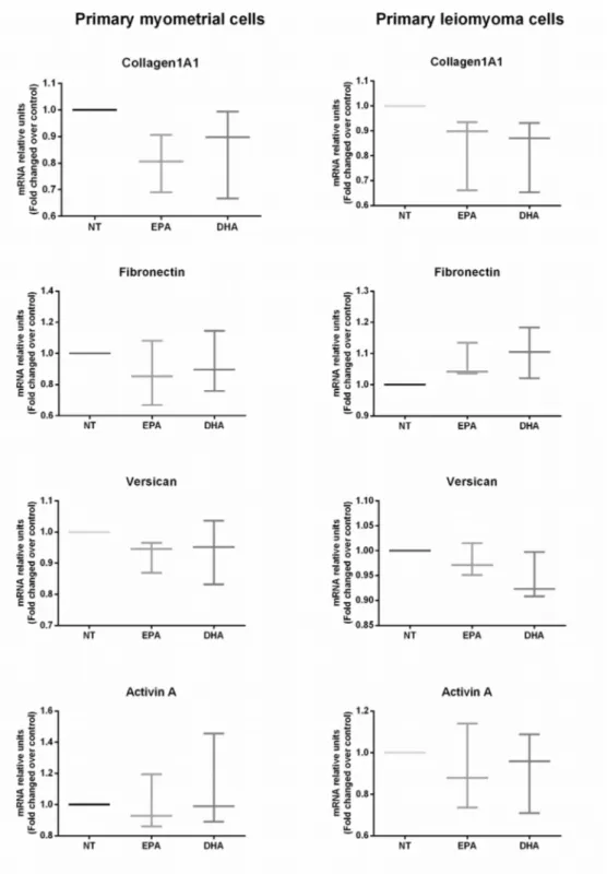
Cells of the myometrium or leiomyomas were treated with EPA or DHA (50 μM for 48 hours) and showed no significant changes of collagen1A1, fibronectin and versican and activin A mRNA expression compared to the untreated sample. (Figure 8)
Figure 8 – Effect of EPA and DHA on extracellular matrix components and activin A mRNA expression in primary myometrial and leiomyoma cells. Data are expressed as mean ± SD (n=3). NT, No treatment; EPA, Eicosapentaenoic acid; DHA, Docosahexaenoic acid.
Effects of EPA and DHA on mRNA expression of mechanical signaling molecules in primary myometrial and leiomyoma cells.
Figure 9 represents the effects of EPA and DHA on expression of mechanical signaling
molecules, such as FAK, AKAP13 and integrin β1 in myometrial and leiomyoma cells. By using the method of real time PCR we could see a significant downregulation of AKAP13 in both cell types treated with the two omega-3 fatty acids EPA and DHA. Furthermore, levels of FAK mRNA were decreased by EPA and DHA only in the cells of the myometrium.
Though no differences in the levels of genes regulating components of the ECM, such as Activin A, could be seen, we found alterations in the expression of molecules involved in mechanical signal transmission such as integrin 1, FAK, AKAP13, despite these changes being limited to myometrial cells. Integrins play an important role in transmitting mechanical signals from the ECM and are characterized as heterodimeric transmembrane receptors. The function of most integrins is to recruit cytoplasmic focal adhesion kinase (FAK) and activate downstream pathways, such as the Rho-dependent signaling (PaszeK, Zahir et al. 2005). A kinase anchor protein 13 (AKAP13) is a RhoA GTPase-specific guanine exchange factor (Rho-GEF) that transforms RhoA from its inactive GDP-bound form to its active GTP-bound form. Interestingly, it has been reported that in leiomyoma cells the expression of integrin β1 (Chen, Lin et al. 2013), AKAP13 (Rogers, Norian et al. 2008) and phosphorylated FAK (Chen, Lin et al. 2013) were higher compared to the myometrium.
Figure 9 – Effect of EPA and DHA on mechanical signaling molecules mRNA expression in primary myometrial and leiomyoma cells. Data are expressed as mean ± SD (n=6). *p < 0.05; **p < 0.01. NT, No treatment; EPA, Eicosapentaenoic acid; DHA, Docosahexaenoic acid.
Effect of EPA and DHA on mRNA expression of sterol regulatory molecules in primary myometrial and leiomyoma cells.
Real time PCR was applied on myometrial and leiomyoma cells that were treated with 50µM EPA or DHA for 48 hours. We could see that the expression levels of ABCG1 and ABCA1 were significantly lower in both cell types treated with the two omega-3 fatty acids. Additionally, SREBF2 was downregulated by addition of DHA in myometrial but not in leiomyoma cells. (Figure 10)
These results show that treatment of cells with omega-3 fatty acids can lead to a decreased expression of genes that play a key role in facilitating lipid accumulation, such as ABCG1 and ABCA1, two proteins responsible for the efflux of cholesterol, a precursor of steroid hormones and the fat-soluble vitamins A, D, E, K. They also are required for the modulation of human oxytocin receptor membrane activity, stability and affinity to oxytocin. It has been shown that the cholesterol content of the myometrium is associated with an abnormal oxytocin-induced contraction of the uterine smooth muscle in rodents. Furthermore, in a mouse model the prevention of accumulation of cholesteryl esters in the myometrium by nuclear receptor liver X receptor- (LXR) occurs via regulation of ABCA1 and ABCG1 (Mouzat, Prod'Homme et al. 2007). The role of cholesterol has been implemented in the pathogenesis of uterine fibroids as higher levels of serum HDL (high-density lipoprotein)-cholesterol were found in women with leiomyomas compared to healthy controls (Sadlonova, Kostal et al. 2008). Also, Borahay et al have published the effect of simvastatin, a drug prescribed to lower cholesterol levels in the blood, to block the growth of fibroids (Borahay, Vincent et al. 2015).
ABCG1 and ABCA1 are part of the evolutionarily conserved family of ATP-binding cassette cholesterol transporters.
ABCG1 is indispensable in cellular cholesterol efflux to HDLs (Wang, Lan et al. 2004) but not to lipid-free apolipoprotein A-I (apoA-I) (Vaughan and Oram 2005). In contrast, the efflux of
cellular cholesterol and phospholipids to apoA-I mediated by ABCA1 converts apoA-I into nascent HDL, which can then act as an efficient acceptor for ABCG1-mediated cholesterol efflux (Smith, Le Goff et al. 2004).
CAV-1 (Caveolin-1) is an element of caveolae, a “cave-like” invagination of the cell membrane. CAV-1 is able to interact with ABCG1 and regulate ABCG1-mediated cholesterol efflux (Gu, Wang et al. 2014). We could not identify an effect of omega-3 fatty acids on CAV-1, but saw that SREBP-2 (sterol-regulatory-element-binding protein-2) levels were decreased in myometrial cells. SREBP-2 plays a central role in cholesterol uptake and synthesis. SREBP-2 can act as a positive regulator of ABCA1 gene expression (Wong, Quinn et al. 2006). The overexpression of ABCG1 increases the processing of SREBP-2 to the transcriptionally active protein, which then leads to an upregulation of SREBP-2 target genes and cholesterol synthesis (Tarr and Edwards 2008).
Figure 10 – Effect of EPA and DHA on sterol regulatory molecules mRNA expression in primary myometrial and leiomyoma cells. Data are expressed as mean ± SD (n=9). *p < 0.05;
**p < 0.01; ***p < 0.001. NT, No treatment; EPA, Eicosapentaenoic acid; DHA, Docosahexaenoic acid.
Effect of EPA and DHA on mRNA expression of mitochondrial enzyme CYP11A1 myometrial and leiomyoma cells.
CYP11A1 plays an important role in the catabolism of steroid hormones and was therefore analyzed in regards to different levels of expressions in myometrial and leiomyoma cells treated with EPA and DHA. Our results show that CYP11A1 mRNA was significantly downregulated in myometrial cells treated with EPA and DHA compared to leiomyoma cells. (Figure 11) CYP11A1 is a mitochondrial enzyme responsible for catalyzing the conversion of cholesterol to pregnenolone, which is a precursor of estrogens, progestogens, mineralocorticoids, glucocorticoids, and androgens, as well as the neuroactive steroids. Estrogen and progesterone have a fundamental part in growth of uterine fibroids. Our results suggest that when leiomyoma cells are treated with omega-3 fatty acids a reduction in the ability to modulate gene expression can be seen in comparison to myometrial cells. However, cells of the myometrium we could see a reduction of CYP11A1 after treatment with EPA or DHA, suggesting that this cellular type differs in the mechanism of regulation of steroidogenesis which could contribute to the pathogenesis of uterine leiomyoma.
Figure 11 – Effect of EPA and DHA on CYP11A1 expression in primary myometrial and leiomyoma cells. Data are expressed as mean ± SD (N= 9). *p < 0.05; **p < 0.01. NT, No treatment; EPA, Eicosapentaenoic acid; DHA, Docosahexaenoic acid.
4. Conclusions
To conclude, omega-3 fatty acids have the ability to modulate the lipid profile, alter the architecture of the cell membrane and downregulate expression of genes relevant for mechanical signal and cellular lipid accumulation in leiomyoma cells. The additional approach of using lipidomics in this study expands knowledge on the pathogenesis of leiomyomas in addition to results gained from genomic, epigenetic and proteomic studies, highlighting new findings on the molecular mechanisms that are responsible for the formation of a leiomyoma. The potential to modulate the lipid content of the cells by applying omega-3 fatty acids as well as knowing which genes are involved, will unfold additional clinical applications in targeted therapies and drug discoveries that can decrease the disease burden of premenopausal women.
5. References
Ahmadi, F., F. Zafarani, M. Niknejadi and A. Vosough (2008). "Uterine Leiomyoma: Hysterosalpingographic Appearances." International Journal of Fertility and Sterility 1(4): 137-144.
Al Hilli, M. M. and E. A. Stewart (2010). "Magnetic resonance-guided focused ultrasound surgery." Semin Reprod Med 28(3): 242-249.
Amant, F., E. Huys, A. Geurts-Moespot, B. G. Lindeque, I. Vergote, F. Sweep and E. F. P. M. Schoenmakers (2003). "Ethnic variations in uterine leiomyoma biology are not caused by differences in myometrial estrogen receptor alpha levels." Journal of the Society for Gynecologic Investigation 10(2): 105-109.
Andreyko, J. L., Z. Blumenfeld, L. A. Marshall, S. E. Monroe, H. Hricak and R. B. Jaffe (1988). "Use of an agonistic analog of gonadotropin-releasing hormone (nafarelin) to treat leiomyomas: assessment by magnetic resonance imaging." American Journal of Obstetrics and Gynecology 158(4): 903-910.
Antollini, S. S. and F. J. Barrantes (1998). "Disclosure of discrete sites for phospholipid and sterols at the protein− lipid interface in native acetylcholine receptor-rich membrane." Biochemistry 37(47): 16653-16662.
Arici, A. and I. Sozen (2000). "Transforming growth factor-beta3 is expressed at high levels in leiomyoma where it stimulates fibronectin expression and cell proliferation." Fertility and Sterility 73(5): 1006-1011.
Bagatolli, L. A. and E. Gratton (1999). "Two-photon fluorescence microscopy observation of shape changes at the phase transition in phospholipid giant unilamellar vesicles." Biophysical Journal 77(4): 2090-2101.
Baird, D. D., Q. E. Harmon, K. Upson, K. R. Moore, C. Barker-Cummings, S. Baker, T. Cooper and G. Wegienka (2015). "A Prospective, Ultrasound-Based Study to Evaluate Risk Factors for Uterine Fibroid Incidence and Growth: Methods and Results of Recruitment." J Womens Health (Larchmt) 24(11): 907-915.
Borah, B. J., W. K. Nicholson, L. Bradley and E. A. Stewart (2013). "The impact of uterine leiomyomas: a national survey of affected women." Am J Obstet Gynecol 209(4): 319.e311-319.e320.
Borahay, M. A., K. Vincent, M. Motamedi, E. Sbrana, G. S. Kilic, A. Al-Hendy and D. Boehning (2015). "Novel effects of simvastatin on uterine fibroid tumors: in vitro and patient-derived xenograft mouse model study." American journal of obstetrics and gynecology 213(2): 196.e191–196.e198.
Bou Khalil, M., W. Hou, H. Zhou, F. Elisma, L. A. Swayne, A. P. Blanchard, Z. Yao, S. A. Bennett and D. Figeys (2010). "Lipidomics era: accomplishments and challenges." Mass Spectrom Rev 29(6): 877-929.
Buttram Jr, V. C. and R. C. Reiter (1981). "Uterine leiomyomata: etiology, symptomatology, and management." Fertility and Sterility 36(4): 433-445.
Calder, P. C. (2001). "Polyunsaturated fatty acids, inflammation, and immunity." Lipids 36(9): 1007-1024.
Cha, P. C., A. Takahashi, N. Hosono, S. K. Low, N. Kamatani, M. Kubo and Y. Nakamura (2011). "A genome-wide association study identifies three loci associated with susceptibility to uterine fibroids." Nature genetics 43(5): 447-450.
Chang, H. L., T. N. Senaratne, L. H. Zhang, P. P. Szotek, E. Stewart, D. Dombkowski, F. Preffer, P. K. Donahoe and J. Teixeira (2010). "Uterine leiomyomas exhibit fewer stem/progenitor cell characteristics when compared with corresponding normal myometrium." Reproductive Sciences 17(2): 158-167.
Chegini, N. (2010). "Proinflammatory and profibrotic mediators: principal effectors of leiomyoma development as a fibrotic disorder." Seminars in Reproductive Medicine 28(3): 180-203.
Chen, H.-M., Y.-H. Lin, Y.-M. Cheng, L.-Y. C. Wing and S.-J. Tsai (2013). "Overexpression of integrin-beta1 in leiomyoma promotes cell spreading and proliferation." Journal of Clinical Endocrinology and Metabolism 98(5): E837-E846.
Ciarmela, P., E. Bloise, P. C. Gray, P. Carrarelli, M. S. Islam, F. De Pascalis, F. M. Severi, W. Vale, M. Castellucci and F. Petraglia (2011). "Activin-A and myostatin response and steroid regulation in human myometrium: disruption of their signalling in uterine fibroid." Journal of Clinical Endocrinology and Metabolism 96(03): 755-765.
Ciarmela, P., P. Carrarelli, M. S. Islam, M. Janjusevic, E. Zupi, C. Tosti, M. Castellucci and F. Petraglia (2014). "Ulipristal acetate modulates the expression and functions of activin A in leiomyoma cells." Reproductive Sciences 21(9): 1120-1125.
Ciarmela, P., M. S. Islam, F. M. Reis, P. C. Gray, E. Bloise, F. Petraglia, W. Vale and M. Castellucci (2011). "Growth factors and myometrium: biological effects in uterine fibroid and possible clinical implications." Human Reproduction Update 17(6): 772-790.
Ciarmela, P., E. Wiater, S. M. Smith and W. Vale (2009). "Presence, actions, and regulation of myostatin in rat uterus and myometrial cells." Endocrinology 150(2): 906-914.
Ciarmela, P., E. Wiater and W. Vale (2008). "Activin-A in myometrium: characterization of the actions on myometrial cells." Endocrinology 149(5): 2506-2516.
Ciavattini, A., D. Tsiroglou, M. Piccioni, F. Lugnani, P. Litta, F. Feliciotti and A. L. Tranquilli (2004). "Laparoscopic cryomyolysis: an alternative to myomectomy in women with symptomatic fibroids." Surgical Endoscopy 18(12): 1785-1788.
Cicinelli, E., F. Romano, P. S. Anastasio, N. Blasi, C. Parisi and P. Galantino (1995). "Transabdominal sonohysterography, transvaginal sonography, and hysteroscopy in the evaluation of submucous myomas." Obstet Gynecol 85(1): 42-47.
Cramer, S. F. and A. Patel (1990). "The frequency of uterine leiomyomas." American Journal of Clinical Pathology 94(4): 435-438.
De Caterina, R. and P. Libby (1996). "Control of endothelial leukocyte adhesion molecules by fatty acids." Lipids 31(1): 57-63.
Deckelbaum, R. J., T. S. Worgall and T. Seo (2006). "n-3 fatty acids and gene expression." Am J Clin Nutr 83(6 Suppl): 1520s-1525s.
Ding, L., J. Xu, X. Luo and N. Chegini (2004). "Gonadotropin releasing hormone and transforming growth factor beta activate mitogen-activated protein kinase/extracellularly regulated kinase and differentially regulate fibronectin, type I collagen, and plasminogen