University of Naples, Federico II
PhD in Chemical science, XXXI cycle
Structural characterisation of
endotoxins from marine and
halophilic bacteria
Clara Barrau
Tutor: Prof. Alba Silipo
Co-tutor: Prof. Antonio Molinaro
Supervisor: Dr. Angela Arciello
If you can't fly, run,
if you can't run, walk,
if you can't walk, crawl,
but by all means, keep moving.
i
Abstract
Gram-negative bacteria cell envelope is a complex structure that is constantly exposed to its environment. It is composed of an
Inner-membrane (IM), a thin peptidoglycan layer and an Outer-Inner-membrane (OM)a.
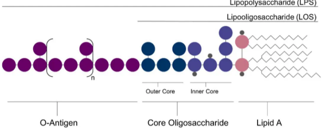
The main component of the OM are Lipopolysaccharides (LPS), also termed as endotoxins. Those molecules are composed of three main parts: a polysaccharide named the O-antigen, a core oligosaccharide and a Lipid
Ab. Endotoxins lacking the polysaccharide are termed Lipooligosaccharides
(LOS).
General structure of LPS and LOS
LPS are known to interact with mammal’s innate immunity through the Toll-like receptor 4 (TLR4) and Myeloid Differentiation factor 2 (MD-2)
receptorial complexc. Depending on their structure, and in particular on their
a Silhavy, T.J.; Kahne, D.; Walker S.; Cold Spring Harb. Perspect. Biol. 2010, 2:a000414
b Raetz, C.R.H.; Whitfield C.; Annu. Rev. Biochem. 2002, 71, 635–700
ii
Lipid A, LPS can either have an agonist or an antagonist activityd.
Discovering new LPS structure is hence necessary in order to develop new therapies, since agonist LPS can be used as vaccines adjuvant and antagonist as drugs against sepsis and septic shock.
In this context, LPS structures from various bacterial sources are currently under study. This project present the characterization of LPS and LOS extracted from marine and halophilic bacteria. As those organisms live in a particular environment, they developed specific strategies to adapt themselves and were hence investigated, as their LPS structure can be
shaped by the adaptation to their environmente. In particular, the study of
LPS from the following strains is here reported.
Pseudoalteromonas sp1A1 is a sponge-pathogen bacterium isolated
from Suberites domunculaf. The full structure of its LPS was resolved using
NMR spectroscopy and Matrix assisted laser desorption (MALDI) Mass spectrometry (MS). Its O-antigen is a branched polysaccharide that have two remarkable features: (i) it possesses a pyruvate linked at 4,6-position of
a Glucosamine and (ii) it possesses a 9-carbon ulosonic acid that is the
3-deoxy-D-glycero-D-galacto-nonulosonic acid(KDN).
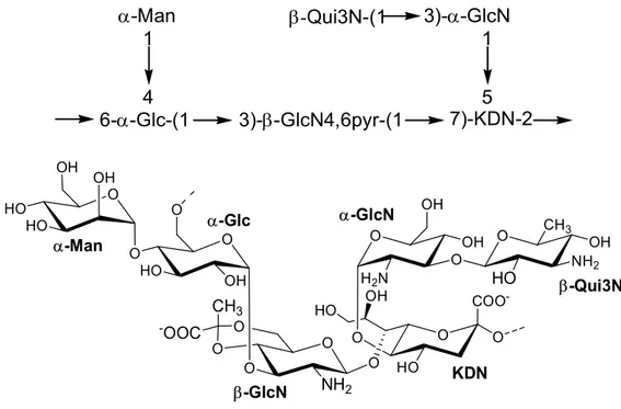
-Man -Qui3N-(1 3)--GlcN 6--Glc-(1 3)--GlcN4,6pyr-(1 7)-KDN-2 1 5 1 4
Structure of the O-antigen from Pseudoalteromonas sp1A1
d Molinaro, A; Holst, O; Di Lorenzo, F; Callaghan, M; Nurisso, A; D’Errico, G; Zamyatina, A; Peri, F; Berisio, R;
Jerala, R; Jimenez-Barbero, J; Silipo, A; Martin-Santamaria, S.; Chem. Eur. J. 2015, 21, 500 – 519
e Di Lorenzo, F.; Billod, J.M.; Martín-Santamaría, S.; Silipo, A.; Molinaro, A. Eur. J. Org. Chem. 2017, 28, 4055–
4073
f Gardères, J.; Bedoux, G.; Koutsouveli, V.; Crequer, S.; Desriac, F.; Le Pennec, G.; Mar. Drugs 2015, 13,
iii
The Lipid A from Pseudoalteromonas sp1A1 was studied by MALDI MS. It is constituted of a mixture of tri- to penta-acylated species, among the penta-acylated Lipid A species, at m/z 1474.6 and 1446.6 were bis-phosphorylated species composed respectively of four C12:0 (3-OH) and one C12:0 and two C12:0 (3-OH), one C10:0 (3-OH), one C11:0 (3-OH) and one C13:0.
Structure Pseudoalteromonas sp1A1 Lipid A species
The structure of the core oligosaccharide from Pseudoalteromonas sp1A1 LPS was also resolved and turned out to be composed of a pentasaccharide containing one Kdo, one heptose, two galactoses and one glucose.
iv Structure of Pseudoalteromonas sp1A1 LOS
Pseudoalteromonas sp1A1 biological activity was assessed using ELISA and Quanti-blue assays. It was found that its LPS does not possess any significant immunostimulant activity on human and murine cell line. The Outer-Membrane properties of Pseudoalteromonas sp1A1 were also studied through Molecular Dynamic (MD) simulation, that showed how the asymmetric repartition of Pseudoalteromonas sp1A1 Lipid A influenced the properties of the phospholipid bilayer, increasing its flexibility. Finally, MD simulation was also performed with Pseudoalteromonas sp1A1 LOS in water and in 0.5 M of NaCl, that is closed to the natural marine environment of the bacterium. Results showed that the presence of salts influenced the conformational behavior of the Kdo-Lipid A region.
O O O NH O O HO O NH O O O O HO P O HO O O 12 12 12 12 12 P O HO OH OH OH O OH O O COOH O HO P O HO OH OH O O O OH OH HO P O O O O OH HO OH OH HO O O HO OH O HO OH HO HO OH
v
The second bacterial strain studied was Spiribacter salinus M19-40T,
a halophile isolated from an intermediate salinity pound of a marine saltern in Spain, the structure of the Lipid A was resolved using MALDI MS and
MS2 experiment. It is a mono-phosphorylated and penta-acylated species
bearing two C10:0 OH), one C12:0, one C14:0 OH) and one C14:0 (3-oxo). This structure possesses two interesting structural features: (i) the 2+3 symmetry that is unusual - as most penta-acylated Lipid A have a 3+3
symmetry and (ii) the occurrence of the C14:0 (3-oxo)g.
Structure of the Lipid A from Spiribacter salinus M19-40T
Halopeptonella vilamensis is a halophilic bacterium that have been
isolated from a saline lagoon in Argentinah whose Lipid A was
characterized using MALDI MS and MS2 experiments. Results showed that
H. vilamensis has a highly heterogeneous mixture of Lipid A species, mono-phosphorylated and hexa-acylated, that differ for the length and saturation of their acyl chains. H. vilamensis main Lipid A species possesses two C10:0 (3-OH), two C12:0 (3-OH), one C12:0 and one C12:1.
g Barrau, C.; Di Lorenzo, F.; Javier Menes R.; Lanzetta R.; Molinaro A.; Silipo A.; Mar. Drugs 2018, 16, 124
vi
Another major species possesses only saturated C12:0. Immunological assays were performed on murine and human cell lines with H. vilamensis LOS and demonstrated its slight immunopotency.
Structure of the Lipid A from Halopeptonella vilamensis
Finally, the characterization of cell envelope components of Halomonas smyrnensis was also attempted. H. smyrnensis is an Exopolysaccharide (EPS) producing halophile isolated from a Turkish salt
lakei. It is known to be a high levan producer and two novel EPS were
isolated. The first one is formed by α-(1→4)-Glc polymer and the second one by α-(1→3)-GlcNAc units.
viii
Abbreviation
BMDM Bone marrow-derived macrophage
COSY Correlation spectroscopy
CPS Capsular polysaccharide
CWA Cell-Wall associated protein
DFQ-COSY Double-quantum filtered COSY
DOC Sodium Deoxycholate
ELISA Enzyme-linked immunosorbent assay
EPS Exopolysaccharide
Gal Galactose
GalNA Galactaminuronic acid
GC-MS Gas chromatography - Mass spectrometry
Glc Glucose
GlcA Glucuronic acid
GlcN Glucosamine
HEK Human Embryonic Kidney
HMBC Heteronuclear multiple-bond correlation
HSQC Heteronuclear single-quantum correlation
IL Interleukin
IM Inner membrane
IMP Inner membrane protein
KDN 3-deoxy-D-glycero-D-galacto-nonulosonic acid
KDO 3-deoxy-D-manno-oct-2-ulosonic acid
LOS Lipooligosaccharide
ix
LTA Lipotechoic acid
MALDI Matrix assisted laser desorption/ionization
Man Mannose
MD-2 Myeloid Differentiation factor 2
MDS Molecular dynamic simulation
MOMs Monocyte-derived macrophages
MS Mass spectrometry
MyD88 Myeloid Differentiation primary response protein 88
NF-κB Nuclear factor-κB
NMR Nuclear magnetic resonance
NOESY Nuclear Overhauser effect spectroscopy
OM Outer membrane
OMP Outer membrane protein
PAGE Polyacrylamide gel electrophoresis
PAMP Pathogen associated molecular pattern
POPE Palmitoyloleoyl Phosphatidylethanolamine
POPG Palmitoyloleoyl Phosphatidylglycerol
PRR Pattern recognition receptor
QuiN Quinovosamine
SDS Sodium dodecyl sulfate
SEAP Secreted alcaline phophate
TH T-Helper cell
TLR4 Toll-like receptor 4
TNF Tumor necrosis factor
TOCSY Total correlation spectroscopy
TOF Time of flight
1
INDEX
SECTION I - INTRODUCTION
5
CHAPTER 1:GRAM-NEGATIVE BACTERIA ... 6
1.1.THE BACTERIAL CELL 7 1.2.BACTERIAL CELL ENVELOPE 8 1.3.LIPOPOLYSACCHARIDES AND LIPOOLIGOSACCHARIDES 10 1.3.1. General structure ... 10
1.3.2. The O-antigen ... 11
1.3.3. The Core Oligosaccharide ... 12
1.3.4. The Lipid A ... 13
1.4.GRAM-NEGATIVE BACTERIA AND INNATE IMMUNITY 15 1.4.1. Innate and adaptive immunity ... 15
1.4.2. TLR4/MD-2 receptorial complex and inflammation ... 16
1.4.3. Structure and function relationship ... 19
1.5.LIPOPOLYSACCHARIDES FROM MARINE AND EXTREME ENVIRONMENT 22 1.5.1. Life at the extreme ... 22
1.5.2. LPS structures from marine bacteria and extremophiles ... 23
CHAPTER 2:CHARACTERIZATION OF LIPOPOLYSACCHARIDES ... 26
2.1.EXTRACTION AND PURIFICATION OF LIPOPOLYSACCHARIDES 27
2.2.CHEMICAL ANALYSIS OF LIPOPOLYSACCHARIDES 28
2.3.ISOLATION OF POLY- AND OLIGO-SACCHARIDE AND LIPID A PORTIONS 30
2.4.NMR OF POLY- AND OLIGO-SACCHARIDES 31
2
SECTION II - PSEUDOALTEROMONAS SP1A1: FROM
ENDOTOXIN TO OUTER-MEMBRANE
36
CHAPTER 3:STRUCTURE AND ACTIVITY OF LIPOPOLYSACCHARIDE OF ... 37
PSEUDOALTEROMONAS SP 1A1 ... 37
3.1.PSEUDOALTEROMONAS SPP. AND THEIR ENVIRONMENT 38
3.2.EXTRACTION AND COMPOSITIONAL ANALYSIS 39
3.3.STRUCTURE OF PSEUDOALTEROMONAS SP 1A1O-ANTIGEN 41
3.4.STRUCTURE OF PSEUDOALTEROMONAS SP1A1LIPID A 45
3.5.STRUCTURE OF PSEUDOALTEROMONAS SP 1A1LOS 48
3.6.IMMUNOLOGICAL ASSAYS 50
3.7.DISCUSSION 51
CHAPTER 4:IN SILICO STUDY OF THE OUTER-MEMBRANE OF PSEUDOALTEROMONAS SP1A1 ... 53
4.1.MOLECULAR DYNAMIC SIMULATION OF GRAM-NEGATIVE BACTERIA OM 54
4.2.MOLECULAR DYNAMIC SIMULATION OF PSEUDOALTEROMONAS SP1A1OM 55
4.3.MOLECULAR DYNAMIC SIMULATION OF PSEUDOALTEROMONAS SP1A1LOS 60
4.4.DISCUSSION 62
SECTION III - STRUCTURE AND ACTIVITY OF LIPID A FROM
HALOPHILES
64
CHAPTER 5:STUCTURE OF THE LIPID A FROM SPIRIBACTER SALINUS M19-40T ... 65
5.1.HALOPHILIC MICRO-ORGANISMS 66
5.2.EXTRACTION, PURIFICATION AND COMPOSITIONAL ANALYSIS 67
5.2.MASS SPECTROMETRY OF THE LIPID A 68
3
CHAPTER 6:STUCTURE AND ACTIVITY OF THE LIPID A FROM HALOPEPTONELLA VILAMENSIS ... 80
6.1.EXTRACTION, PURIFICATION AND COMPOSITIONAL ANALYSIS 81 6.2.MASS SPECTROMETRY OF THE LIPID A 83 6.3.IMMUNOLOGICAL ASSAYS 89 6.4.INVESTIGATION OF HALOPEPTONELLA VILAMENSIS CORE OLIGOSACCHARIDE REGION 93 6.5.DISCUSSION 94
SECTION IV - LOOKING FOR ENDOTOXINS AMONG
EXOPOLYSACCHARIDES: THE CASE OF HALOMONAS
SMYRNENSIS
97
CHAPTER 7:ANALYSIS OF HALOMONAS SMYRNENSIS POLYSACCHARIDES ... 987.1.SWEET AND SALTY: AN EXOPOLYSACCHARIDE PRODUCING HALOPHILE 99 7.2.EXTRACTION, PURIFICATION AND NMR SPECTROSCOPY 100 7.3.DISCUSSION 103
SECTION V - EXPERIMENTAL METHODS
104
CHAPTER 8:MATERIAL AND METHODS ... 1058.1.BACTERIA GROWTH 106 8.1.1. Pseudoalteromonas sp1A1 ... 106
8.1.2. Spiribacter salinus... 106
8.1.3. Halopeptonella vilamensis ... 106
8.1.4. Halomonas smyrnensis ... 107
8.2.EXTRACTION OF LOS AND LPS 108 8.3.CHEMICAL ANALYSIS 109 8.3.1. MGA analysis ... 109
4
8.3.2. AAPM analysis ... 109
8.3.3. Analysis of Fatty acids Methyl-ester derivatives ... 110
8.4.ISOLATION OF LIPID A AND POLYSACCHARIDES MOIETIES 110 8.4.1. Isolation of Pseudoalteromonas sp1A1 O-antigen ... 110
8.4.2. Isolation of Pseudoalteromonas sp1A1 Lipid A ... 111
8.4.3. Isolation of Spiribacter salinus M19-40T Lipid A ... 111
8.4.4. Isolation of Halopeptonella vilamensis polysaccharides ... 112
8.4.5. Isolation of Halopeptonella vilamensis Lipid A ... 112
8.4.6. Isolation of Halomonas smyrnensis polysaccharides ... 112
8.5.MASS SPECTROMETRY 113 8.6.NMR SPECTROSCOPY 114 8.7.BIOLOGICAL ASSAYS 115 8.7.1. ELISA and Quanti-Blue assays with LPS from Pseudoalteromonas sp1A1... 115
8.7.2. Cell culture and ELISA with H. vilamensis LOS ... 116
8.8.MOLECULAR DYNAMIC SIMULATION 117 CONCLUSION ... 119
ANNEX ... 122
PAPERS RELATED TO THIS PHD PROJECT 122 ATTENDED CONGRESS, CONFERENCES, MEETING, AND WORKSHOP 122 ATTENDED INTERNAL SEMINARS 124 ATTENDED COURSES 125 SHORT STAY IN EUROPEAN LABORATORIES 125 ACKNOWLEDGMENT 126 BIBLIOGRAPHY... 127
5
Section I
6
7
1.1. The bacterial cell
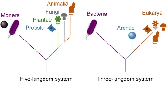
From microorganisms to complex plants and animals, life comprise a remarkable diversity and can be divided into a five-kingdom system that includes Monera, Protista, Plantae, Fungi, and Animalia (Figure 1). Around
30 year ago, Woese et al.1 changed the previous system based on RNA
analysis. They established the three-kingdom system and defined Bacteria as one of the three domains of life, with Archaea and Eukarya (Figure 1). Eukarya are uni- or pluricellular organisms that possess an internal cell compartmentation. Indeed, the presence of organelles within their cytoplasm is characteristic of this domain. Archaea are unicellular prokaryotic organisms devoid of cellular compartmentation. Bacteria are also prokaryotes but they differ from Archaea, inter alia, by the composition of their plasmatic membrane.
Figure 1: The five-kingdom and three-kingdom systems of life
Bacteria include a vast group of microorganisms that grow and reproduce in diverse environments. They comprise an impressive array of
Bacteria Archae Eukarya Monera Protista Plantae Fungi Animalia
8
sizes, shapes and arrangement2. Roughly, Bacteria can be divided on the
basis of their cell morphologies, e.g. rod shape, spherical and curved cells
are respectively called “bacillus”, “cocci” and “spirilli”3. Despite the great
diversity, there are still general features among bacteria (Figure 2). Their cytoplasm comprises most bacteria metabolites and is the place where all transcription and replication of the DNA occurs. Exposed outside the bacteria, flagella and pili are involved in motility. They are anchored in the bacterial cell envelope, which possesses a sophisticated architecture.
Figure 2: Schematic representation of general bacteria composition
1.2. Bacterial cell envelope
Bacterial cell envelope is a complex structure that evolved to adapt to the environment in which they live. Based on their cell external structure, two types of bacteria can be distinguished: positive and Gram-negative bacteria. Historically, those category were termed based on a colorimetric test developed in 1884 by Hans Christian Gram. Nowadays, this determination is still used to describe the bacterial cell envelope
composition4. Flagella Ribosome DNA Pili Cytoplasm Plasma membrane
9 Figure 3: Schematic representation of Gram-positive and Gram-negative bacteria cell envelope. Abbreviations correspond to: Wall techoic acid (WTA), Lipotechoic acid (LTA),
Lipopolysaccharide (LPS), Inner membrane protein (IMP), Cell-wall associated protein (CWA); Outer membrane protein (OMP)
Gram-positive cell envelope is composed of two main parts: the cell membrane and the cell wall (Figure 3). The cell membrane is a phospholipid bilayer. As bacteria do not possess any organelle, all membrane-related biosynthesis occur in the cell membrane, including the
synthesis and transport of molecules composing the cell wall5. This bilayer
also includes inner membrane proteins (IMP). Over this structure, and directly exposed to the environment, there is the cell wall that is composed of a thick layer of a glycopeptide polymer named peptidoglycans. On this
structure are exposed different types of cell-wall glycopolymers6: Wall
techoic acids (WTA) are linked to the peptidoglycan through phosphodiester linkages and lipotechoic acids (LTA) are anchored to the cytoplasmatic membrane through a glycerol phosphate unit. Cell-wall glycopolymer are diverse among bacteria species and strains and possess different ionic charges or number of repeating unit. They are essential for Gram-positive bacteria survival, playing a role in important functions as
Gram-positive Gram-negative Inner membrane Outer membrane Periplasm Inner membrane OMP IMP IMP CWA WTA Cell Wall LTA LPS Peptidoglycan
10
protection, attachment and colonisation. There are also cell-wall associated proteins (CWA) that are exposed at the surface of the cell wall.
Differently from Gram-positive bacteria, Gram-negative cell envelope do not have its peptidoglycan exposed to the environment. It is composed of three parts: the inner membrane, the periplasm and the outer membrane (Figure 3). As for Gram-positive bacteria, the inner membrane is a phospholipid bilayer that plays a key role for the biosynthesis of
Gram-negative other cell wall constituents7. Between the two membranes, the
periplasm is composed of a peptidoglycan layer that is thinner than the one found in Gram-positive bacteria and in turn surrounded by the outer membrane (OM). The OM is a lipid bilayer that acts as a barrier between bacteria and their environment. The OM includes outer membrane proteins (OMP) that are β sheets anchored to the bilayer through hydrophobic residues. Contrary to the inner membrane, the outer membrane is an asymmetric bilayer. Its inner leaflet is still made of phospholipids but its outer leaflet is composed at 75% of Lipopolysaccharides (LPS). LPS are complex molecules that are crucial for the bacteria survival as they are constantly interacting with their environment.
1.3. Lipopolysaccharides and Lipooligosaccharides
1.3.1. General structure
Lipopolysaccharides (LPS) are heat-stable amphiphilic molecules. Initially referred as endotoxins, they are anchored in the bacterial cell membrane and are infamously known for their toxicity. LPS are composed
of three main parts8: a repeating unit of polysaccharide termed the
11
LPS (S-LPS), or Lipopolysaccharides (LPS), are composed of all the three previously mentioned parts. Rough LPS (R-LPS), or Lipooligosaccharides (LOS), do not possess the O-antigen. The Smooth/Rough determination was originally adopted because of the appearance R-LPS and S-LPS give to their bacteria colonies.
Figure 4: General structure of Lipopolysaccharides (LPS) and Lipooligosaccharide (LOS)
Lipopolysaccharides are essential for the bacterial survival. Indeed, they are crucial in many biological roles as adhesion, symbiosis and pathogenicity. In particular, their structure determines the relationships between bacteria and their host. Although LPS share the same broad architecture, each bacterial strain possesses their own variation. Those differences can be seen in the composition of the O-antigen, the core oligosaccharide and the Lipid A.
1.3.2. The O-antigen
The O-antigen is the most variable part of the LPS. It is a hydrophilic polymer that can be composed up to 50 units of from 1 up to 8 monosaccharides. O-antigens can be linear or branched polysaccharides, structural variation of the O-antigen occur in the nature of each
12
monosaccharide, their linkages and the presence of non-carbohydrate constituents. Non-stoichiometric modifications increase the heterogeneity of O-antigen structures. Differences between O-antigen can be observed between each bacterial strain (inter-strain specific) but also within the same bacterial strain (intra-strain specific). For example, E. coli has more than 170 O-serotypes. A single bacterium can synthetize O-antigen with different number of repeating units. The structure of O-antigen play a role in
bacterial pathogenicity and symbiosis9. Its length also influences the
adhesion properties of the LPS10. Interestingly, Gram-negative bacteria can
shorten their O-antigen using a reversible process called phase-variation11,
allowing them to hide from the host immune defence.
1.3.3. The Core Oligosaccharide
The core Oligosaccharide has less chemical diversity than the O-antigen and is composed of two main parts: the Inner and the Outer core (Figure 4). The Outer Core is directly linked to the O-antigen and has the most structural variations. It is often made of negatively charged monosaccharides such as uronic acids. The Inner core is a more conserved
part. It is typically made of 3-deoxy-D-manno-oct-2-ulosonic acid (Kdo) and
L-glycero-D-mannoheptose (L,D-Hep)12 (Figure 5); D-glycero-D
-talo-oct-ulosonic acid (Ko) or D-glycero-D-mannoheptose (D,D-Hep). The Inner core
is usually decorated with phosphates (P).
Figure 5: Structure of (1) 3-deoxy-D-manno-oct-2-ulosonic acid (Kdo) and (2) L-glycero-D -mannoheptose (L,D-Hep)
13
1.3.4. The Lipid A
The lipid A is the hydrophobic part of the LPS that anchors the molecule within the OM. It is composed by a phosphorylated and acylated
β(1’→6) di-Glucosamine backbone (GlcN)13 on which the core
oligosaccharide is attached at C-6’ position. The Lipid A can possess one or two phosphates linked on the C-4’ position of the non-reducing GlcN (GlcN II) and on C-1 position of the reducing GlcN (GlcN I). Lipid As mainly differ on their degree of acylation and the nature of their fatty acids. They are acylated by primary fatty acids on C-2, C-3, C-2’ and C-3’ position through ester and amide bounds. They can also possess ester-linked secondary fatty acids that are attached on the hydroxyl group of primary-linked acyl chains. Finally, Lipid As can also possess substituents as phosphoethanolamine. The first resolved Lipid A structure was from E. coli. It is a bis-phosphorylated and penta-acylated structure with 4+2 symmetry. Many other Lipid As from various bacteria sources pathogen (Figure 6)
were characterized revealing their highly diverse architectures14,15,16,17. The
Lipid A structure is crucial for the survival of the bacteria and its pathogenicity. Indeed, this moiety is known to directly bind with a receptorial complex of mammal’s innate immunity composed of Toll-like receptor 4 (TLR4) and myeloid differentiation factor 2 (MD-2).
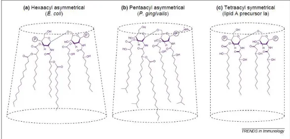
14 Figure 6: Structure of Lipid As from several bacteria, illustrating their structural diversity
among Gram-negative bacteria. (A) Escherichia coli (B) Helicobacter pyroli (C) Porphyromonas gingivalis (D) Salmonella minnesota (E) Rhodobacter sphaeroides
15
1.4. Gram-negative bacteria and innate immunity
1.4.1. Innate and adaptive immunity
Living organisms develop ways to defend against threats in order to survive. Immunity is a complex system that allows the host to detect and supress the intruding pathogens. In 1989, Charles A. Janeway introduced
the concept of innate and adaptive immunity18. The organism’s ability to
prevent infections is broadly based on adaptive immunity. This response is highly specific and is developed after a first encounter with a pathogen. However, around 7 days are needed to recruit the specialised cells during
the first activation19. Therefore, a first line of defence is needed to protect
the organisms against new pathogens. This is the role of innate immunity
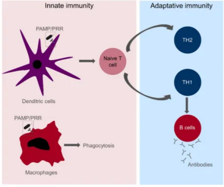
Figure 7: An overview of the innate and adaptive immunity. PAMP: Pathogen-associated Molecular patterns; PRR: Pathogen-Recognition Receptors; TH: T-helper cells
16
Part of the innate immune response is performed by macrophages via phagocytosis (Figure 7). However, dendritic cells (DC) play a key role in
the innate immune recognition and the induction of adaptive immunity20.
Briefly, their activation leads to the secretion of cytokines or direct cell contacts with naïve T cells that triggers the formation of T and B cells (Figure 7). The first step in the activation of the innate immunity is the recognition of the pathogen that is done through Pathogen-Recognition Receptors (PRR) that recognise Pathogen-associated Molecular patterns
(PAMP)21 on the surface of innate immune cells. PAMP are molecules that
are not produced by eukaryotic cells, possess invariant structural features and are necessary for the microbe’s survival. Their recognition by PRR is hence advantageous for the host. There are several PRR that evolved in order to interact with different PAMPs; among them, there is a receptor that is specialized in the recognition of LPS.
1.4.2. TLR4/MD-2 receptorial complex and
inflammation
Firstly discovered in Drosophila, the Toll protein was known to control dorsal-ventral patterning in embryon and inflammatory response in adults. Its analogue was later on discovered in humans as a
transmembrane protein related to NF-κB signaling22. Those proteins are
now referred as Toll-like receptors (TLRs) and known to be part of human’s innate immunity. To better fight infection, there are several types of TLRs that can each recognize a particular PAMP. For example, TLR2 interacts with lipoprotein and TLR5 with flagellin.
17
The first discovered TLR was TLR4 that was shown to specifically
interact with LPS23. Further studies proved that TLR4 does not work alone.
The presence of another protein, the Myeloid Differentiation factor 2 (MD-2)
is necessary to respond to LPS infection24. MD-2 is associated to the
extra-cellular domain of TLR4 and together they form the TLR4/MD-2 receptorial complex. Further studies enhanced the comprehension of TLR4/MD-2 activation and its related signalling pathway. LPS is firstly recruited by LPS Biding proteins (LBP) and is further transferred to the TLR4/MD-2 receptor through CD14. All those interaction spontaneously happen due to the existence of a thermodynamic funnel that prone the assembly of those
molecules25.
Once LPS is linked to TLR4/MD-2, it triggers the dimerization of the receptorial complex and further intra-cellular signalling. In brief, two main supramolecular structures can be formed after the dimerization, triggering
different inflammations pathways26 (Figure 8). When the Toll/interleukin-1
Receptor domain-containing Adaptor Protein (TIRAP) and Myeloid Differentiation primary response protein 88 (MyD88) are linked to the intracellular part of TLR4, an assembly called the Myddosome is formed. Further association with Interleukin-1 Receptor Associated Kinase (IRAK) triggers cytokine production with the activation of transcription factors that are Nuclear Factor-κB (NF-κB) and Activator Protein 1 (AP1). The second supramolecular complex, the Triffosome, is MyD88-independent and involves the assembly of TIR domain-containing adaptor protein inducing IFNβ (TRIF) and TNF receptor-associated factor 3 (TRAF-3). It activates NF-κB, AP1 and also interferon regulatory factor 3 (IRF-3). All the recruited transcription and regulatory factors can then modulate the expression of the
18 Figure 8: General description of TLR4/MD-2 signalling pathway
Inflammation is a physiological phenomenon that is necessary for the organism to protect itself. However, a dysregulation can lead to major deceases. Sepsis and septic shock are disorders that arise from an unbalanced inflammatory response. Those are significant cause of death
among developed countries28. Better understanding TLR4/MD-2
mechanism and finding new regulators is essential to develop therapies. In this context, many bioactive compounds have been tested in order to
modulate the innate immunity29. As natural activators of TLR4/MD-2, LPS
play a key role in the exploration of new modulators. It is now widely known that the interaction between TLR4/MD-2 and LPS is driven by a structure dependent relationship.
19
1.4.3. Structure and function relationship
The idea that LPS structure would influence its endotoxic activity was
already investigated 25 years ago30. The study of Lipid As conformation
from several gram-negative bacteria lead to observation of a correlation between their three-dimensional structures and biological activity. Indeed, the Lipid A toxicity potency is linked to a non-lamellar supramolecular structure. Further researches confirmed the relationship between LPS shape and biological activity. The degree of acylation of the Lipid A and the length of its fatty acids influence its three-dimensional conformation. When the Lipid A has a high degree of acylation, as the hexa-acylated Lipid A from E. coli, it adopts a conical shape. Penta-acylated Lipid A, e.g. from P. gingivalis, have an intermediate shape and hypo-acylated Lipid As, as biosynthetic precursors Lipid Ia and Lipid IVa, adopt a strictly cylindrical
shape (Figure 9)31.
20
It is known that E. coli LPS possesses a strong agonist activity towards TLR4/MD-2 receptorial complex and P. gingivalis LPS activity is
less TLR4-dependent32. Lipid IVa has been identified as a slight agonist in
mice and an antagonist towards human MD-233. All those data confirm that
conical supramolecular structures are correlated to agonist activity and cylindrical shapes to antagonist. Crystal structure of Lipid IVa linked to human MD-2 showed that all fatty acids of the antagonist Lipid A are
inserted in the MD-2 binding pocket34, and that this did not modify the
conformation of MD-2. The interaction between E. coli agonist Lipid A and
TLR4/MD-2 was also investigated by crystallography35. When the
hexa-acylated Lipid A interacts with MD-2, one of its fatty acid does not enter the hydrophobic pocket and remains on the protein surface (Figure 10). The exposed fatty acid can then interact with the hydrophobic region of a second TLR4. As for Lipid IVa, the insertion of E. coli LPS in MD-2 does not change the size of the hydrophobic pocket. However, the di-glucosamine backbone of E. coli LPS adopts a different spatial conformation, compared to Lipid IVa. Such orientation of the Lipid A makes its phosphates groups
interact with positively charged residues of both TLR4. This interaction,
combined to hydrophilic and hydrophobic contacts, are crucial in order to generate the dimerization of TLR4/MD-2. Indeed, the fatty acid of E. coli Lipid A that is at the surface of the hydrophobic pocket can interact with two TLR4s, whereas Lipid IVa does not have fatty acid that mediates the heterodimer formation (Figure 10). As the dimerization of the receptorial complex is necessary for triggering an inflammatory response, those interactions explains why E. coli LPS has a strong agonist activity and Lipid IVa is an antagonist.
21 Figure 10: Representation of the interaction between E. coli Lipid A (A), Lipid IVa (B) and TLR4/MD-2 receptorial complex. Orientation of E. coli Lipid A triggers the dimerization of
the receptor
Finally, Lipid IVa was also studied with murine MD-236. It was shown
that the different amino acids present in the hydrophobic pocket made the Lipid IVa adopt the same overall conformation than E. coli LPS. That explains the slight agonist activity of Lipid IVa in mice. Interactions between endotoxins and TLR4/MD-2 were studied not only by crystallisation but also using other techniques. A more recent investigation of LPS-TLR4 interaction was done by Single Molecule Localisation Microscopy
(SMLM)37. With this technique, it was possible to detect monomeric and
dimeric TLR4 molecules in presence of various LPS. In presence of LPS from E. coli and S. minesotta, most of TLR4 were detected in dimeric forms but in presence of LPS from R. sphaeroides, TLR4 remained in monomeric state.
Thus, the whole structure of the LPS may influence the interaction. Indeed, a lot of work have been dedicated to the Lipid A but other constituents, as the Kdo or heptoses of the core oligosaccharide, contribute
to the biological activity38. In this context, discovering novel LPS structures
is necessary to find new modulators for TLR4/MD-2 receptorial complex. Exploring particular environment where bacteria have specific adaptation strategy is a promising way to detect interesting molecular features.
22
1.5. Lipopolysaccharides from marine and extreme
environment
1.5.1. Life at the extreme
From a human perspective, the optimal conditions for life are at neutral pH, temperature between 20 and 40 °C and at atmospheric pressure. However, life can also exist in conditions that are considered as hostile for many life forms. Organisms living in such conditions are termed
extremophiles and their fascinating properties were studied for decades39.
Extremophiles can be classified in several categories depending on the conditions in which they thrive. For example, psychrophiles can live below 15 °C and thermophiles above 60 °C, barophiles can support up to 80 MPa and halophiles can live in salinity environment with saturated NaCl (Figure 11). Some organisms are able to survive in several extreme conditions, as high pressure and high temperature, and are termed polyextremophile. There is still a lot to discover about extremophiles and current metagenomic analysis lead to the discovery of extremophiles new
genus and species and a better understanding of their metabolism40. The
study of extremophiles is also appealing for biotechnology. Indeed, those organisms can produce metabolites with unique properties that are studied
for industrial applications41. Extremophiles Gram-negative bacteria also
possess LPS on their OM and those molecule can have particular properties.
23 Figure 11: Representation of extremophiles diversity in different environments
1.5.2. LPS structures from marine bacteria and
extremophiles
The ocean is the place where life could be created and it still includes a countless diversity of organisms. Marine microorganism adapted to their environment by creating a various number of metabolites. Their study lead to the discovery of bio-active compounds with large therapeutic
interest, as anti-bacterial, anti-fungal and anti-tumoral molecules42. In
particular, Gram-negative marine bacteria are omnipresent organism whose LPS can possess interesting immunological activity. Indeed, specific
structural features can be observed in these environments43. O-antigen and
core oligosaccharides of marine LPS are usually negatively charged with the presence of uronic acids and non-glycosidic substituents. As LPS are constantly exposed to the marine environment, the anionic residues links to the surrounding cations, strengthening the global membrane structure. This mechanism provides the bacteria higher stability towards its environment.
24
Several Lipid A from marine bacteria were also characterized and common features were observed. Short fatty acids – in comparison to E. coli Lipid A- are often found in marine bacteria with Lipid A containing 10 to 13 carbon acyl moieties. Marine bacteria also possess low acylated (penta and tetra-acylated) and phosphorylated (mono-phosphorylated) Lipid A. Those characteristic are associated with low-agonist and antagonist activity and encourage the exploration of new endotoxin structures in the oceans.
As a consequence of their adaptation, extremophiles also possess
LPS with interesting structures44. As for marine bacteria in general,
extremophiles often possess negatively charged O-antigen and their Lipid A
usually have shorter acyl chains (Figure 12)45,46,47. They are a promising
source of agonist and antagonists towards the TLR4/MD-2 receptorial complex. Characterizing LPS from bacteria that live in marine and extreme conditions is hence necessary in order to understand their adaptation process and to find new modulators of the innate immune system.
25 Figure 12: Structure of Lipid As from some marine and extremophile bacteria: (A) Alteromonas aldita, a marine bacterium (B) Halomonas patelleriensis, an haloalkaliphilic
26
Chapter 2: Characterization of
Lipopolysaccharides
27
2.1. Extraction and purification of
Lipopolysaccharides
Extraction of LPS is the first step towards its structural elucidation. Two main procedures have been designed in order to isolate LPS. The first
one is the PCP extractionin which bacterial cells are exposed to a mixture
of Phenol/Chloroform/Petroleum ether (5:8:2). This methods is more suitable for the extraction of LOS as the extracted material can be found in the phenol phase. The other procedure is the Hot Phenol-Water
extraction48, in which the extraction is more convenient for LPS as they can
be isolated in the aqueous phase. The extracted material is usually impure after this procedure. Indeed, cells contaminants such as proteins and nucleic acids can be also found in the same aqueous phase. In order to remove contaminants and purify the extracted material, the sample is further purified using an enzymatic treatment with several enzymes (Proteases, DNAses and RNAses); the sample then undergoes several Ultra-centrifugation or Size-Exclusion chromatography steps (Figure 13).
Once the extract is lyophilised, electrophoretic methods as Sodium
Dodecyl-Sulfate polyacrylamide gel (SDS-PAGE) and Sodium
Deoxycholate Polyacrylamide gel (DOC-PAGE) are performed. The use of subsequent silver staining procedure allows the detection of the LPS/LOS
by revealing its saccharidic portion49. If the extract is an LOS, a large band
is detected at the bottom of the gel. If it is an LPS, a ladder-like pattern is seen in the middle of the gel, showing the bacteria O-antigen length diversity. Performing electrophoresis with silver staining is a necessary step in the characterization of endotoxin structures. Indeed, typical stains provide a solid validation of the LPS/LOS nature of the extract.
28 Figure 13: Usual procedure used for the extraction and purification of LPS and LOS
2.2. Chemical analysis of Lipopolysaccharides
GC-MS analysis of carbohydrate and fatty acids derivatives provide crucial information about the composition of the extracted and purified LPS. Several methods can be used in order to determine the carbohydrate
composition of the core oligosaccharides and the O-antigen50. The first one
is the Acetylated Methyl glycoside (MGA) procedure. This methods gives information about the nature of the monosaccharides present in the LPS. An aliquot of the extract is methylated at anomeric position in acidic conditions and then per-acetylated. The obtained derivatives are then analysed by GC-MS with a standard that is per-acetylated inositol. The nature of each monosaccharide is given by their retention time on the chromatogram combined with their MS data.
Once the monosaccharide composition of the LPS is assessed, their branching points are determined using the Partially Methylated Alditols Acetates (AAPM) method. In the present procedures, the polysaccharide is methylated at free hydroxyl positions in basic condition with further
treatment by iodomethane (CH3I)51. The linkages are then cleaved with
Trifluoroacetic acid (TFA). A reduction with Sodium Borodeuteride (NaBD4)
DRIED CELLS SOLID PHASE WATER PHASE SUPERNATANT PURE LOS PURE LPS PCP extraction Hot Phenol-Water extraction Dialysis Enzymatic treatment Size exclusion chrommatography Ultracentrifugation Precipitation Washing PHENOL PHASE DOC-PAGE GC-MS Chemical treatment NMR MALDI MS
29
is then performed. This step is crucial because it leads to the ring opening and marks the anomeric position with deuterium atoms. The sample is then acetylated at its remaining free hydroxyls groups (Figure 14). The obtained monosaccharides derivatives are finally injected on GC-MS. It should be noted that with the present procedure, carbohydrates containing carboxylic acids (as uronic or ulosonic acid) cannot be detected. In order to analyse those monosaccharides, it is necessary to first reduce the carboxylic acid group before performing the hydrolysis with TFA (Figure 14). As for MGA, the determination of the branching points is done by analysing their retention time on the GC chromatogram and their fragmentation by MS.
30
Fatty acid content of the LPS can also be analysed by GC-MS52. An
aliquot of the LPS is methylated as described for the MGA procedure and then extracted with hexane. The obtained methyl-ester derivatives are then injected on GC-MS. Each fatty acid has a specific retention time and fragmentation pattern. It is hence possible to determine the composition of the sample by detecting fatty acids with different acyl chain length, hydroxylation and unsaturation.
Compositional analysis give crucial data for the structural analysis of the LPS. However, they are not sufficient to achieve the characterization of the exact structure. Different methods have to be used on the saccharidic part and the Lipid A. Several chemical treatments can be performed in order to separate and isolate those two moieties.
2.3. Isolation of poly- and oligo-saccharide and Lipid
A portions
The Kdo is a particular monosaccharide that is most of the times found between the non-reducing glucosamine of the Lipid A and the core oligosaccharide. Due to the presence of carboxylic acid at anomeric position and the lack of hydroxyl substituent at its C3 position, it is highly labile in acidic position. This is a useful property when the Lipid A needs to be separated from the saccharidic moiety. When the LPS is treated in mild acidic conditions, a cleavage occurs between the non-reducing glucosamine of the Lipid A and the Kdo. The polysaccharide and Lipid A are then easily separated by centrifugation.
Another methods can be used for the isolation of the saccharide
portion. It is the full deacylation of the LPS53. This procedure is typically
31
the sample is treated with hydrazine (N2H4), removing the O-linked acyl
chains. Then a treatment in basic condition (using KOH) cleaves the remaining N-linked acyl chains. After desaltation and purification by column chromatography, the isolated oligosaccharide is ready to be fully characterized.
2.4. NMR of poly- and oligo-saccharides
Nuclear Magnetic Resonance (NMR) is a non-destructive technique that give information on the different nuclei present in a molecule and their atomic environment. There exist many NMR experiments that provide
complementary data for structural characterization of polysaccharides54. 1H
NMR experiment is a 1D spectrum where signals corresponding to each protons of the polysaccharides are recorded. Many information can be assessed with this experiment. Indeed, each region of the spectrum corresponds to a specific kind of proton. Anomeric protons are visible between δ 4.6 and 5.5 ppm, acetyl groups between δ 2.0 and 2.2 ppm and methyl groups between δ 1.0 and 1.3 ppm. Remaining protons are found in the bulk region between δ 2.6 and 4.6 ppm.
1H NMR spectrum can also be used in order to measure anomeric
coupling constant and hence determine each monosaccharide
configuration. Indeed, 3JH1-H2 is around 7-9 Hz for α-configured and 1-4 Hz
for β-configured carbohydrates like glucose and galactose, respectively due to diaxial and equatorial-axial couplings. For D-mannose, different values are measured as its H2 is equatorial. Its β-configuration correspond to a
3JH1-H2 around 1-2 Hz and α-configuration around 0-1 Hz.
To go deeper in the analysis, several 2D spectra must be recorded. Correlation Spectroscopy (COSY), and Total Correlation Spectroscopy
32
(TOCSY) are homonuclear through-bond experiments. Both of the axis in
the spectra correspond to the same kind of atom (1H) and the
magnetization transfer is performed through bounds connections. Those experiments hence measure the correlations of protons among the same monosaccharide unit. All protons from a given unit hence belong to the same spin system. In COSY experiment, observed cross-peaks emerge from magnetization transfer to direct neighbours. This is convenient for the attribution of the precise proton sequences among the same spin system. TOCSY experiment give additional information by transferring the signal through several connections. The two previously described experiments allow the assignment of proton sequence within the same unit but do not give any information about the linkages between each monosaccharides. Nuclear Overhauser Spectroscopy (NOESY) is a homonuclear through-space experiment. Contrary to COSY and TOCSY, the magnetisation is transferred to proton that are spatially closed. With this experiment, it is hence possible to detect a correlation between an anomeric proton and the proton of the other unit to which it is linked (Figure 15).
Other structural data can be assed using heteronuclear 2D experiments. Heteronuclear Single-Quantum Correlation Spectroscopy (HSQC) shows correlations between carbons and protons that are directly linked. As for proton spectra, HSQC can be divided in different region in which the carbon chemical shift is characteristic of its environment. Anomeric carbon are usually found between δ 90-110 ppm, nitrogen-bearing carbons between δ 45-60 ppm, methyl groups around δ 10-20 ppm and sugar ring carbons between δ 60-80 ppm. Heteronuclear Multiple Bond
Correlation (HMBC) gives signals between 13C and 1H that are correlated
33 Figure 15: Representation of atom interactions with different 2D NMR experiments
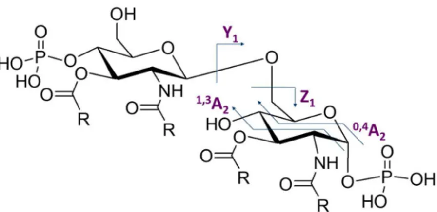
2.5. Mass Spectrometry of the Lipid A
Mass Spectrometry (MS) is a technique that give information about molecular mass. It is a destructive methods that have the advantage to work with very little amount of sample. Several ionisation and fragmentation techniques can be used in MS. For big molecules, as peptides or Lipid A, MALDI ionisation coupled with Time Of Flight (TOF) separation is a suitable technique that is often used. MALDI/TOF MS of Lipid A can be recorded either in positive or negative ion mode.
Negative ion MALDI/TOF MS of Lipid A extracted in acidic conditions contains several clusters of peaks corresponding to species with different degree of acylation. Indeed, under-acylated species can be formed after mild acid hydrolysis and there can also naturally occur a mixture of Lipid A species in intact cells. The mass difference between each clusters usually corresponds to an acyl chain or a phosphate. The composition of the Lipid A, i.e. its phosphorylation, the number and nature of its acyl chains and the presence of other substituents, can be determined with this experiment.
34
However, MALDI/TOF MS analysis alone is not sufficient to obtain a detailed structure. Indeed, it is not possible with this technique to determine the linkage position of each phosphates and acyl chains on the di-glucosamine backbone. MS/MS analysis are then performed on several precursor ions. The fragmentation of the studied Lipid A species are analysed with those experiments. Those fragments can arise from the loss of phosphates and fatty acids but also from sugar ring and linkage cleavage. Those well-known events possess a specific nomenclature that
was determined by Costello et al.55 (Figure 16). The occurrence of those
linkages and sugar ring fragmentation allow the detection of mass corresponding to each glucosamine. This experiment is hence necessary in order to establish the acyl chains repartition among the di-glucosamine backbone.
Figure 16: Main linkages and sugar ring fragmentation that can be observed during MS/MS of the Lipid A
35
Finally, the Lipid A should undergo an ammonium treatment to complete its structural analysis. Indeed, this procedure is necessary in
order to determine if each acyl moieties in ester or amide-linked56. It is
known that with this method secondary ester-linked are more labile than primary ester-linked acyl moieties and amide linked residues. MALDI/TOF MS performed after such extraction procedure hence lead to the detection of species which mass difference correspond to their ester-linked fatty acids. The analysis can be completed with MS/MS of ammonium treated Lipid As. The combination of all previously described MS experiments lead to the precise determination of the Lipid A architecture.
36
Section II
Pseudoalteromonas sp1A1:
From endotoxin to
37
Chapter 3: Structure and activity of
Lipopolysaccharide of
38
3.1. Pseudoalteromonas spp. and their environment
The genus Pseudomonas is part of the ‘gamma-proteobacteria’
group and is composed of gram-negative aerobic and flagellated bacteria57.
Based on 16S rRNA analysis, Gauthier et al. proposed a division of this
group with the new genus Pseudoalteromonas58. Since then,
Pseudoalteromonas spp. is widely studied for its capacity to produce biologically active molecules such as antibiotics and compounds against
fungi59. These properties give Pseudoalteromonas spp. the ability to survive
and coexist in its complex environment60. It can indeed significantly
influence its ecosystem with several mechanisms as the production of
biofilms, anti-fouling and anti-algae agents61.
Pseudoalteromonas spp. encloses several marine species, some known to interact as symbiont or pathogens with eukaryotic organisms
including marine sponges62. Within this ecosystem, sponges developed
molecular strategy to recognize and defend themselves against pathogenic microbes, as the case of Suberites domuncula which developed a
molecular immune response towards bacterial infection63. As gram-negative
bacteria, Pseudoalteromonas spp possess LPS in their OM (See Section 1). The LPS isolated from different Pseudoalteromonas strains were shown to be penta-acylated with low-immunostimulatory and antagonist properties
in murine cells64. For S. domuncula, LPS is recognized by a specific
receptor named S. domuncula LPS interacting protein (SLIP) and then activates the MyD88-dependent pathway of TLR4/MD-2 receptorial
39
The present chapter reports the investigation of the structure and immunological activity of the LPS from Pseudoalteromonas sp1A1. It is a
sponge-pathogen bacterium isolated from S. domuncula66. Further analysis
of the properties of the LPS were made in order to understand Pseudoalteromonas sp1A1 behaviour in its ecosystem and to dissect its immunopotential.
3.2. Extraction and compositional analysis
The LPS was extracted using hot phenol-water procedure and purified by enzymatic treatment. SDS-PAGE confirmed the presence of LPS in the purified water phase with the occurrence of a ladder-like pattern on the gel (Figure 17).
A B
Figure 17: SDS-PAGE 13.5% A. LPS from E. coli standard; B. LPS from Pseudoalteromonas sp1A1, water phase
40
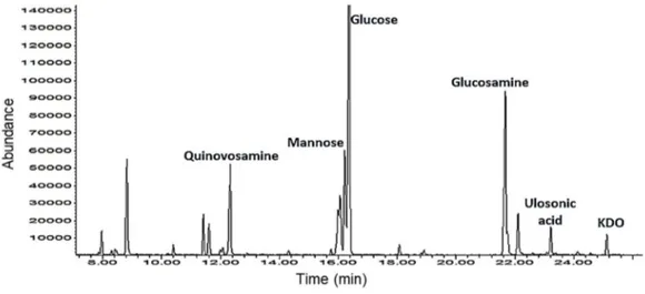
Compositional analysis (MGA) showed the presence of D
-3-deoxy-3-amino-Quinovose (D-QuiN), D-Mannose (D-Man), D-Glucose (D-Glc), D
-Glucosamine (D-GlcN), 3-deoxy-D-manno-oct-2-ulopyranosonic acid (D
-Kdo) an another Ulosonic acid identified as 3-deoxy-D-glycero-D
-galacto-nonulosonic acid (KDN)67 (Figures 18 and 19). Combined with previous
MGA experiment, AAPM analysis showed that Pseudoalteromonas sp1A1 LPS is composed of terminal Qui3N, terminal Man, 4,6-subsituted-Glc, 3-substituted-GlcN and 3,4,6-3-substituted-GlcN.
Figure 18: GC-MS chromatogram of MGA derivative of Pseudoalteromonas sp1A1 LPS
O
COO
-OH
HO
HO
HO
HO
OH
41
Fatty acid analysis revealed the occurrence of (R)-3-hydroxydodecanoic acid [12:0OH)], (R)-3-hydroxydecanoic acid [10:0
(3-OH)], (R)-3-hydroxyundecanoic acid [11:0 (3-OH)],
(R)-3-hydroxytridecanoic acid [13:0 (3-OH)] as primary acyl chains, both in amide and ester linkage, and, as secondary acyl chain, of dodecanoic acid (12:0), decanoic (10:0), undecanoic acid (11:0), tridecanoic acid (13:0), dodecenoic (12:1) and tridecenoic (13:1).
3.3. Structure of Pseudoalteromonas sp 1A1
O-antigen
Pseudoalteromonas sp1A1 LPS was fully de-acylated and its O-antigen was characterised via NMR spectroscopy. A combination of homo- and heteronuclear 2D NMR experiment (DQF-COSY, TOCSY, ROESY,
NOESY, 1H-13C HSQC, 1H-13C HMBC) was executed to assign all the spin
systems and to define the saccharidic sequence. Complete assignment of the spin systems was obtained attributing the proton resonances by DQF-COSY and TOCSY spectra, and subsequently correlating each proton to its related carbon atom through the HSQC spectrum. The anomeric configuration of each monosaccharide unit was assigned on the basis of
3JH-1,H-2 coupling constant obtained by the DFQ-COSY.
A magnitude of 3JH-1,H-2 with values of 7–9 Hz was associated with
the diaxial coupling of a β-configured sugar unit, whereas 2–4 Hz was
indicative of an equatorial-axial coupling of
α-configured residues. Six different units were detected in
42 Figure 20: 1H spectra of deacylated O-antigen from Pseudoalteromonas sp1A1
Unit 1 2 3 4 5 6 7 8 9 A α-GlcN 99.37 5.18 56.21 2.80 80.66 3.54 74.67 3.29 72.57 4.04 3.63-3.51 64.01 -- -- -- B α-Glc 99.18 5.13 72.96 3.36 72.57 3.51 69.60 3.17 72.26 3.61 3.65-3.52 60.47 -- -- -- C α-Man 97.97 5.00 72.10 3.58 71.10 3.47 69.99 3.23 3.365 71.71 3.69-3.52 60.70 -- -- -- D β-GlcN 98.31 4.51 55.87 2.62 3.36 84.0 75.76 3.27 69.54 3.43 3.78-3.82 56.94 -- 1.24 24.37 -- E β-Qui3N 4.43 103.41 72.80 3.10 57.26 2.67 73.89 2.84 69.15 3.35 16.65 1.07 -- -- -- F KDN -- -- 1.72-1.98 34.6 3.99 65.53 3.83 73.5 69.77 4.11 4.03 62.7 85.72 3.40 3.65-3.41 64.01 Table 1: 1H and 13C (italic) chemical shifts (ppm) of de-acylated O-antigen from
Pseudoalteromonas sp1A1
Spin systems A, B, D, E were gluco-configured residues as indicated
by the large 3JH,H ring coupling constant values of their ring protons
43
table 1) were identified as α-GlcN and β-GlcN based on their nitrogen-bearing C-2 respectively at δ 56.21 and δ 55.87ppm. Residue B was recognized as α-Glc (H1 at δ 5.13ppm, table 1). Residue E was identified as β-Qui3N according to the value of its C-3 (δ 57.26 ppm) indicative of a nitrogen-bearing carbon and its downfield H-6/C-6 (δ 1.07/16.65 ppm) corresponding to the methyl group.
Residue C (H1 at δ 5.00ppm, table 1) was attributed as α-mannose.
The manno configuration was established by 3JH-1,H-2 and 3JH-2,H-3 values
(below 3 Hz), the α-configuration was assigned either by the intra-residual NOE contact of H-1 with H-2 and by chemical shift value of its C1 (δ 97.97 ppm, table 1). Finally, residue F was identified as KDN, in accordance with the compositional analysis. Indeed, it possesses characteristic diastereotopic methylene signals at H-3 (δ 1.72/1.98 ppm, table 1) and a 9-carbon sequence.
Moreover, the proton at 1.24 ppm (with corresponding carbon at δ 24.03 ppm) gave NOE contacts with protons H-4 (δ 3.27 ppm) and H-6 (δ 3.78-3.82 ppm) of unit D. It was attributed to a methyl group of a pyruvate that is linked to the β-GlcN.
Figure 21: NOESY (black) and TOCSY (grey) spectra of the anomeric region of the O-antigen of Pseudoalteromonas sp1A1
44
NOESY (Figure 21) and HMBC spectra allowed the determination of the sugar sequence in the polysaccharide and was consistent with previous
compositional analysis. Spin system F was substituted at O-5 by residue A
and O-7 by residue D, as shown by NOE contacts found between H-1 A (δ 5.18 ppm) and 5 F (δ 3.83 ppm) and between 1 D (δ 4.51 ppm) and H-7 F (δ 4.03 ppm). Spin system A was in turn glycosylated by residue E as indicated by NOE contact between H-1 E (δ 4.43 ppm) and H-3 A (δ 3.54 ppm). Spin system D was substituted at O-3 by residue B as shown by NOE correlations between H-1 B (δ 5.13 ppm) and H-3 D (δ 3.36 ppm). Finally, spin system B was glycosylated at O-3 by residue C as proven by
NOE contacts between H-1 C (δ 5.00 ppm) and H-3 B (δ 3.51ppm) (Figure
21). Combined with previous methylation analysis, the full sequence of Pseudoalteromonas sp1A1 O-antigen was elucidated as a branched polysaccharide and its structure is represented in Figure 22.
-Man -Qui3N-(1 3)--GlcN 6--Glc-(1 3)--GlcN4,6pyr-(1 7)-KDN-2 1 5 1 4 O COO -O HO O HO O OH O NH2 O O O -OOC CH3 O OH O HO O O OH OH HO HO O H2N OH O OH O HO CH3 NH2 OH KDN -GlcN -Qui3N -GlcN -Glc -Man
45
3.4. Structure of Pseudoalteromonas sp1A1 Lipid A
Negative-ion MALDI-TOF MS of Pseudoalteromonas sp1A1 was performed on the mild acid hydrolysis product and is shown in Figure 23. The very heterogeneous composition of the Lipid A could be clearly seen, in accordance to previous compositional analysis and other
Pseudoalteromonas spp. LPS Lipid A species68,69,70.. The mass spectrum
showed multiple sets of pseudomolecular ion peaks [M-H]- comprised in the
mass range m/z 1000-1500 Da (Figure 23). Within those clusters, the occurrence of mass differences of 14 amu was attributable to methylene units and thus referred to Lipid A species having different acyl chain lengths.
Figure 23: Negative-ion MALDI-TOF mass spectrum of the isolated lipid A fraction from Pseudoalteromonas sp 1A1 LPS. Assignment of the lipid A species as Tri LA (tri-acylated lipid A species), Tetra LA (tetra-acylated lipid A species), Penta LA (penta-acylated lipid A
species) and the relative groups of ions lacking one phosphate is also indicated
The bis-phosphorylated and penta-acylated species were detected in the cluster around m/z 1474.6 (Figure 23), this last corresponding to a lipid A species carrying four 12:0 (3-OH) primary acyl substituent and one 12:0 as secondary acyl moiety. The species at m/z 1446.6 matched with a lipid A containing two 12:0 (3-OH), one 11:0 (3-OH), one C10:0 (3-OH) and one
894.0 1027.2 1160.4 1293.6 1426.8 1560.0 Mass (m/z) 1541.7 0 10 20 30 40 50 60 70 80 90 100 % In te ns ity In te ns ity 1196.4 1474.6 1276.4 1446.6 1182.4 1262.4 1366.6 1248.3 1390.5 1014.2
Tri-LA Tetra-LA Penta-LA
P 12(3:OH) P 12:0 11(3:OH) 14 14 28
46
C13:0. Its corresponding mono-phosphorylated lipid A could be detected at m/z 1366.6 (Δm/z = 80). Fragments at m/z 1276.4 and 1262.4 matched with tetra-acylated species that have respectively lost C12:0 (3-OH) or C11:0 (3-OH) moieties (Figure 23, Table 2). The most abundant peak, at m/z 1196.4, corresponded to a mono-phosphorylated and tetra-acylated Lipid A species bearing three 12:0 (3-OH) and one 12:0 (Figure 23, Table 2).
Table 2: Proposed interpretation of main peak ions of MALDI-TOF MS (Figure 23)
Pseudoalteromonas sp 1A1 hence possesses the same mixture of Lipid A species that are represented on Figure 24. The bis-phosphorylated and penta-acylated Lipid A species is acylated by four C12:0 (3-OH) in amide linkages and as primary ester-linked fatty acids and one C12:0 as secondary fatty acid. The other penta-acylated Lipid A species is also bis-phosphorylated and penta-acylated and possesses one C10:0 (3-OH), one C11:0 (3-OH) and one C12:0 (3-OH) as primary linked fatty acid and one C13:0 as secondary linked fatty acid. It should be noted that odd numbered fatty acids – C11:0 (3-OH) and C13:0 – likely possesses isopropyl groups
Observed ion
peak (m/z) Substitution Acyl Proposed Fatty acid/phosphate composition 1474.6 Penta-acyl HexN2P2 [C12:0 (3OH)]4[C12:0]
1446.6 Penta-acyl HexN2P2[C12:0 (3OH)]2[C11:0 (3OH)] [C10:0 (3OH)] [C13:0]
1366.6 Penta-acyl HexN2P[C12:0 (3OH)]2[C11:0 (3OH)] [C10:0 (3OH)] [C13:0]
1276.4 Tetra-acyl HexN2P2 [C12:0 (3OH)]3[C12:0]
1262.4 Tetra-acyl HexN2P2 [C12:0 (3OH)]2[C10:0 (3OH)] [C13:0]
1248.4 Tetra-acyl HexN2P2[C12:0 (3OH)][C11:0 (3OH)] [C10:0 (3OH)] [C13:0]
1196.4 Tetra-acyl HexN2P[C12:0 (3OH)]3[C12:0]
1182.4 Tetra-acyl HexN2P[C12:0 (3OH)]2[C10:0 (3OH)] [C13:0]
47
at their extremity. The obtained data are in accordance with the study of Pseudoalteromonas sp 2A, a commensal bacterium also isolated from S.
domoncula71.
Figure 24: Structure of Pseudoalteromonas sp1A1 Lipid A species
Gardères et al.66 previously analysed Pseudoalteromonas sp 1A1
Lipid A and found mono-phosphorylated and penta-acylated Lipid A species, bearing four 12:0 (3-OH) one 12:0 units. However, they did not detected the bis-phosphorylated species and concluded that the secondary fatty acid on the non-reducing glucosamine was linked on the acyloaxyl ester instead of the acyloxyamide. They possibly detected fragments of the lipid A that got degraded.
48
3.5. Structure of Pseudoalteromonas sp 1A1 LOS
The full LOS structure has also been resolved using MALDI-TOF MS (Figure 25). Clusters corresponding to the native LOS could be seen between m/z 2300 and 3000. In source regiospecific cleavage (β-elimination) of the labile linkage involving the Kdo enables both the detection of the core oligosaccharide ions (B-type ions, m/z 1000-1200) and
the Lipid A ions (Y ions, m/z 1400-1500)72. Several species were detected
in the higher mass region, corresponding to the native LOS composed by the Lipid A with four C12:0 (3-OH) and one C12:0 (Figure 24). Peak at m/z 2531.7 was attributed to the LOS with a core oligosaccharide composed of one Kdo, one heptose and three hexoses. The minor peak at m/z 2369.7 corresponded to the LOS that lacks one hexose unit. Another LOS species constituted by four hexoses was detected at m/z 2693.6. The ion at m/z
2487.7 originated from the loss of a neutral CO2 on the Kdo unit. Finally, the
minor peak at m/z 2423.5 was attributed to the native LOS devoid of a phosphate group and constituted by the Lipid A with one C10:0 (3-OH), one C11:0 (3-OH), two C12:0 (3-OH) and one C13:0 (Figure 24).
The main Pseudoalteromonas sp1A1 core oligosaccharide is composed of three hexoses, one heptose, the Kdo and two phosphates. The hexoses comprise one terminal glucose, one terminal galactose and one 4-substituted galactose. The heptose is 4,7-substituted and possesses a phosphate group at its 3-position. Finally, the Kdo is 5-substituted and possesses another phosphate group at its 4-position (Figure 26). Those data match the analysis of another Pseudoalteromonas strain that present
49 Figure 25: MALDI TOF MS analysis of total LOS from Pseudoalteromonas sp1A1; Zoom
on the whole LOS fragmentation is reported in the inset
Figure 26: Structure of Pseudoalteromonas sp1A1 LOS
O O O NH O O HO O NH O O O O HO P O HO O O 12 12 12 12 12 P O HO OH OH OH O OH O O COOH O HO P O HO OH OH O O O OH OH HO P O O O O OH HO OH OH HO O O HO OH O HO OH HO HO OH