SNPs in predicting clinical efficacy and toxicity of chemotherapy:
walking through the quicksand
Raffaele Palmirotta
1, Claudia Carella
1, Erica Silvestris
1, Mauro Cives
1, Stefania
Luigia Stucci
1, Marco Tucci
1, Domenica Lovero
1and Franco Silvestris
11Department of Biomedical Sciences and Human Oncology, Section of Clinical and Molecular Oncology, University of Bari Aldo Moro, 70124 Bari, Italy
Correspondence to: Franco Silvestris, email: [email protected]
Keywords: cancer; targeted therapy; chemotherapy; precision medicine; single nucleotide polymorphisms Received: March 07, 2018 Accepted: April 07, 2018 Published: May 18, 2018
Copyright: Palmirotta et al. This is an open-access article distributed under the terms of the Creative Commons Attribution License
3.0 (CC BY 3.0), which permits unrestricted use, distribution, and reproduction in any medium, provided the original author and source are credited.
ABSTRACT
In the “precision medicine” era, chemotherapy still remains the backbone for the treatment of many cancers, but no affordable predictors of response to the chemodrugs are available in clinical practice. Single nucleotide polymorphisms (SNPs) are gene sequence variations occurring in more than 1% of the full population, and account for approximately 80% of inter-individual genomic heterogeneity. A number of studies have investigated the predictive role of SNPs of genes enrolled in both pharmacodynamics and pharmacokinetics of chemotherapeutics, but the clinical implementation of related results has been modest so far. Among the examined germline polymorphic variants, several SNPs of dihydropyrimidine dehydrogenase (DPYD) and uridine diphosphate glucuronosyltransferases (UGT) have shown a robust role as predictors of toxicity following fluoropyrimidine- and/or irinotecan-based treatments respectively, and a few guidelines are mandatory in their detection before therapy initiation. Contrasting results, however, have been reported on the capability of variants of other genes as MTHFR, TYMS, ERCC1, XRCC1, GSTP1, CYP3A4/3A5 and ABCB1, in predicting either therapy efficacy or toxicity in patients undergoing treatment with pyrimidine antimetabolites, platinum derivatives, irinotecan and taxanes. While formal recommendations for routine testing of these SNPs cannot be drawn at this moment, therapeutic decisions may indeed benefit of germline genomic information, when available. Here, we summarize the clinical impact of germline genomic variants on the efficacy and toxicity of major chemodrugs, with the aim to facilitate the therapeutic expectance of clinicians in the odiern quicksand field of complex molecular biology concepts and controversial trial data interpretation.
INTRODUCTION
The human genome includes 3 billions of
nucleotides, and inter-individual sequence variations are
detected with a frequency of 1/300-1000 nucleotides.
Single nucleotide polymorphisms (SNPs) are germline
sequence variations observed in more than 1% of the
general population, and account for approximately 80%
of the overall genomic heterogeneity [1]. Among the
10 million SNPs identified in the human genome, only
100,000 have a phenotypic and functional impact, since
the majority of them is located in intronic portions of the
DNA [2, 3].
Functional SNPs are key determinants of
inter-individual anthropometric differences, but may also
activate the responses to environmental factors and predict
the individual disease susceptibility [4, 5]. Moreover, a
number of pharmacogenomic studies have demonstrated
that both efficacy and toxicity of drugs are largely
influenced by SNPs [6], and this event appears particularly
www.oncotarget.comOncotarget, 2018, Vol. 9, (No. 38), pp: 25355-25382
relevant in cancer patients receiving chemotherapy since
a definite correlation between chemotherapy efficacy/
tolerability and survival outcomes, cannot be denied.
The optimization of the so-called “patient-therapy
binomial” constitutes one of the main challenges of the
modern oncology [7]. In the “precision medicine” era
it is imperative, indeed, to match the right patient with
the right treatment, and in this context the analysis of
clinically meaningful SNPs may provide better efficacy
outcomes and, at the same time, decreased
treatment-related toxicities. Although a number of studies have
investigated the correlation between specific genotypes
and response to chemotherapy, clinical implementation
of such information has been limited thus far, possibly
as consequence of inconclusive results from unrelated
studies. Thus, while tumor genotyping is currently
routinely used to guide treatment selection in the clinical
arena, patient genotyping is considered very often limited
to cases of cancer predisposition syndromes, in which the
identification of a germline mutation is extremely useful
in defining the therapeutic strategy.
In this review, we aimed at updating clinicians
with the most recent oncogenomic data deriving from the
analysis of selected gene polymorphisms involved in the
metabolism of major chemotherapeutic classes including
fluoropyrimidines, platinum derivatives, irinotecan and
taxanes). In particular, we focused on the biology of
the described genetic variants as well as their potential
impact as predictors of treatment response or toxicity.
Thus, we hope to guide practitioners in: i) requiring the
most appropriate molecular investigations; ii) learning the
results of genetic reports; and iii) tailoring the therapeutic
choices based on the patient genotype, when indicated.
FLUOROPYRIMIDINES
Fluoropyrimidines as fluorouracil, capecitabine
and tegafur, have a prominent role in the treatment of
many tumors, especially of the gastroenteropancreatic
tract, and those of head and neck district [8]. By acting as
pyrimidine analogues, fluoropyrimidines cause defects of
nucleotide synthesis thereby inducing apoptosis in cancer
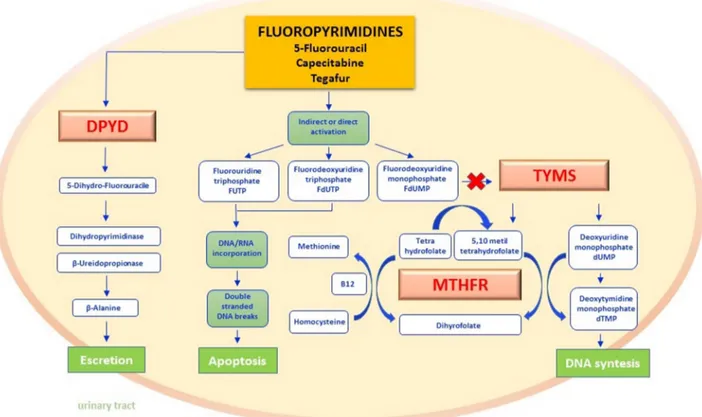
cells [9, 10]. As depicted in Figure 1, active metabolites
of fluoropyrimidines inhibit thymidylate synthetase
(TYMS) and inhibit the folate cycle by interfering
with the methilentetrahydrofolate reductase (MTHFR)
transmetilation reactions [11]. Several enzymes including
dihydropyrimidine dehydrogenase (DPYD) concur in the
metabolism of fluoropyrimidines [12], and their levels of
activity may influence the intracell drug concentration. In
the section below, we synthesize both biologic and clinical
impacts of DPYD, MTHFR and TYMS polymorphisms.
● DPYD - Physiologically, liver DPYD
inactivates the 80-90% of administered fluoropyrimidines,
converting them in 5-fluoro-5,6-dihydrouracil through a
redox reaction exploiting NADPH/NADP
+as a cofactor
[13, 14]. Two further reactions, catalyzed respectively by
dihydropyrimidinase and beta-ureidopropionase, produce
the final metabolites that are ultimately excreted with
urine [15] (Figure 1). DPYD gene extends for 950 kb on
chromosome 1p22 and includes 23 exons [16]. Variations
in gene sequence may cause DPYD deficiency, and are
transmitted as autosomal recessive inheritance. In affected
subjects, the clinical consequences of DPYD deficiency
span from absence of signs/symptoms or mere laboratory
alterations (increased pyrimidine concentration in blood,
urine or liquor) to complex neurological syndro-mes
arising at birth or during childhood (seizures, mental
retardation, microcephaly, muscle hypertonicity, autism
and motor deficits) [17, 18]. Notably, the severity of
clinical presentation is directly related to the extent of
functional enzyme impairment. In patients receiveing
fluoropyrimidine-based chemotherapy, DPYD deficiency
may cause a persistent elevation of the blood drug
concentration, and is therefore associated with an
increased risk of chemotherapy-related toxicities including
neutropenia, nausea, vomiting, diarrhea, stomatitis,
mucositis, hand-foot syndrome and peripheral neuropathy
[19–21].
The individual tolerance to fluoropyrimidine-based
chemotherapy is strictly related to specific polymorphic
variants of the DPYD gene. In fact, within the about 160
known SNPs affecting this enzyme, approximately 15 of
them acquire a clear functional significance [21]. While
for some rare variants (
*3,
*7,
*8,
*9B,
*10,
*11,
*12) the
correlation with DPYD’s reduced activity is very likely,
for others (
*4,
*5,
*6,
*9A) it still remains unclear [22]. As
recently shown in three different metanalysis [23–25],
three SNPs of DPYD (
*2A,
*13 and rs67376798) seem
to be prioritarily associated with side effects in patients
undergoing fluoropyrimidine-based chemotherapy (Table 1).
The IVS14+1G>A variant is characterized by
a single G>A point mutation in the GT splice donor
site IVS14+1, causing the skipping of exon 14 and the
consequent synthesis of a truncated, catalytically inactive
protein [26, 27]. On the other hand, the 1679T>G variant
is characterized by a single aminoacid substitution from
isoleucine to serine at codon 560, encoding for a highly
conserved, functionally important segment of DPYD
[28]. The variant 2846A>T shows a structural alteration
that impairs DPYD function by interfering with cofactor
binding or electron transport [29]. The three variants are
able to decrease DPYD enzyme activity completely or
partially, depending on homozygosity or heterozygosity,
and the severity of fluoropyrimidine-associated toxicities
correlates with the number of functional alleles [30].
Due to the high risk of toxicities, the US Food and Drug
Administratin (FDA; http://www.fda.gov/), the Dutch
Pharmacogenetis Working Group and the European
Medicines Agency (EMA; http://www.ema.europa.eu/ema)
do not recommend the administration of fluoropyrimidines
to subjects carrying IVS14+1G>A, 1679T>G or
2846A>T variants [21]. On the other hand, both the
National Comprehensive Cancer Network (NCCN) and
the American Society of Clinical Oncology (ASCO)
recommend DPYD pharmacogenetic analysis only in
case of suspected toxicities after fluoropyrimidine-based
chemotherapy, while the European Society for Medical
Oncology (ESMO) suggests the pharmacogenetic test in the
pre-therapy setting as an option for selected patients [21,
31–33]. The Clinical Pharmacogenetics Implementation
Consortium (CPIC - https://cpicpgx.org/) has recently
identified wild type subjects as normal metabolizers
and homozygous patients as poor metabolizers, while
heterozygous carriers for any combination of the three
variants were defined as those having enzyme activity
between 30 and 70% compared with the standard
[22, 34, 35]. On this basis, CPIC has contraindicated
fluoropyrimidine-based therapies in patients with mutated
homozygous genotype, while at least a 50% drug dosage
reduction was recommended for heterozygous subjects
[22, 35]. AIOM (Italian Association of Medical Oncology)
and SIF (Italian Society of Pharmacology) suggest the
same dose adjustments proposed by the CPIC guidelines
in the presence of DPYD variants (Table 2) [36].
● MTHFR - The MTHFR gene maps on
chromosome 1 (1p36.3) and encodes for a
homo-dimeric protein that contributes to the folate metabolism
homeostasis as well as to control the turnover of both
nucleic acids and aminoacids [37]. As depicted in Figure
1, MTHFR catalyzes the reduction of 5,10 methylene
tetrahydrofolate (THF) in 5-methyl-THF, which will serve
as methyl group donor in the conversion of homocysteine
in methionine [38].
Figure 1: Fluoropyrimidines pathway.
Fluoropyrimidines (5-fluorouracil and the oral prodrug capecitabine and tegafur) are for the 90% rapidly catabolized in the liver, whereas only 10% is anabolized by forming metabolites responsible for the drug mechanism of action. The rate-limiting step of 5-FU catabolism is catalyzed by dihydropyrimidine dehydrogenase (DPYD) with the synthesis of dihydrofluorouracil (DHFU) and subsequent metabolic reactions lead to the synthesis of inactive compounds excreted by the urinary tract. The main mechanism of action of fluoropyrimidines includes the interaction by either direct or indirect mechanisms, with normal nucleoside biosynthesis. In fact, when active metabolites produced as FUTP, FdUTP, FdUMP are embedded as analogues of pyrimidines in RNA and DNA synthesis, they break the nucleic acid filaments by promoting apoptosis in cancer cells. FdUMP furthermore inhibit the thymidylate synthase (TYMS) enzyme by forming a covalent ternary complex. The inhibition of this reaction not only interrupts the biosynthesis of DNA nucleotides but also interferes with the folate cycle. In this last pathway methylene tetrahydrophilate reductase (MTHFR) is the key enzyme of transmetilation reactions: methyl groups derived from the folate pool in fact permits homocysteine-methionine reconversion by recycling the methyl group bound to Vitamin B12 as a cofactor. DPYD - dihydropyrimidine dehydrogenase; DHFU - dihydrofluorouracil; FUTP - fluorouridine triphosphate; FdUTP - fluorodeoxyuridine triphosphate; FdUMP - fluorouridine monophosphate; TYMS - thymidylate synthase; MTHFR - methylene tetrahydrophilate reductase.Severe MTHFR deficiency is caused by rare
recessive autosomal mutations and is associated with
hyperomocysteinemia and hyperomocysteinuria,
osteoporosis, growth retard, visual defects, and
thrombophilia. Partial enzymatic deficiencies, due to the
presence of common polymorphic variants, can generate
hyperomocysteinemia especially in the presence of folic
acid defect as well as thrombophilia, and increases both
prenatal mortality and coronary heart disease risk [39].
The most studied polymorphic variants of MTHFR
include C677T and A1298C (Table 1). Both SNPs are
characterized by reduced enzymatic activity and their
frequency is greater among Caucasians, especially
in Italians and Hispanics, and lower among Africans
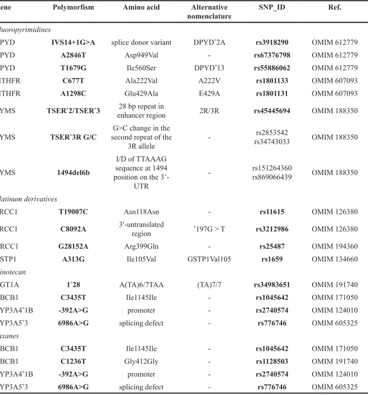
Table 1: Synopses of the major genes variants involved in the metabolism of fluoropyrimidines, platinum derivatives,
irinotecan and taxanes
Gene
Polymorfism
Amino acid
Alternative
nomenclature
SNP_ID
Ref.
Fluoropyrimidines
DPYD
IVS14+1G>A
splice donor variant
DPYD
*2A
rs3918290
OMIM 612779
DPYD
A2846T
Asp949Val
-
rs67376798
OMIM 612779
DPYD
T1679G
Ile560Ser
DPYD
*13
rs55886062
OMIM 612779
MTHFR
C677T
Ala222Val
A222V
rs1801133
OMIM 607093
MTHFR
A1298C
Glu429Ala
E429A
rs1801131
OMIM 607093
TYMS
TSER
*2/TSER
*3
28 bp repeat in
enhancer region
2R/3R
rs45445694
OMIM 188350
TYMS
TSER
*3R G/C
second repeat of the
G>C change in the
3R allele
-rs2853542
rs34743033
OMIM 188350
TYMS
1494del6b
I/D of TTAAAG
sequence at 1494
position on the
3’-UTR
-
rs151264360
rs869066439
OMIM 188350
Platinum derivatives
ERCC1
T19007C
Asn118Asn
-
rs11615
OMIM 126380
ERCC1
C8092A
3′-untranslated
region
*197G > T
rs3212986
OMIM 126380
XRCC1
G28152A
Arg399Gln
-
rs25487
OMIM 194360
GSTP1
A313G
Ile105Val
GSTP1Val105
rs1659
OMIM 134660
Irinotecan
UGT1A
1
*28
A(TA)6/7TAA
(TA)7/7
rs34983651
OMIM 191740
ABCB1
C3435T
Ile1145Ile
-
rs1045642
OMIM 171050
CYP3A4
*1B
-392A>G
promoter
-
rs2740574
OMIM 124010
CYP3A5
*3
6986A>G
splicing defect
-
rs776746
OMIM 605325
Taxanes
ABCB1
C3435T
Ile1145Ile
-
rs1045642
OMIM 171050
ABCB1
C1236T
Gly412Gly
-
rs1128503
OMIM 191740
CYP3A4
*1B
-392A>G
promoter
-
rs2740574
OMIM 124010
CYP3A5
*3
6986A>G
splicing defect
-
rs776746
OMIM 605325
The most relevant polymorphisms with the SNP ID and the OMIM reference (PUBMED database) are reported for each
gene, in keping with the effects on amino acid substitution, and the possible alternative nomenclatures.
[40]. The 5-fluorouracil, a fluoropyrimidine compound
metabolized intracellularly to
5-fluoro-2-deoxyuridine-5-monophosphate (FdUMP) its active form, carries a
cytotoxic effect mediating the formation of a ternary
complexes between 5-10 methylene THF, TYMS and
FdUMP. This complex inhibits the thymidylate and its
intracellular levels decreased with consequent suppression
of DNA synthesis. Due to the catalytic deficit of the
MTHFR, subsequent to its polymorphic variants, the 5-10
methylene THF concentration increased enhancing the
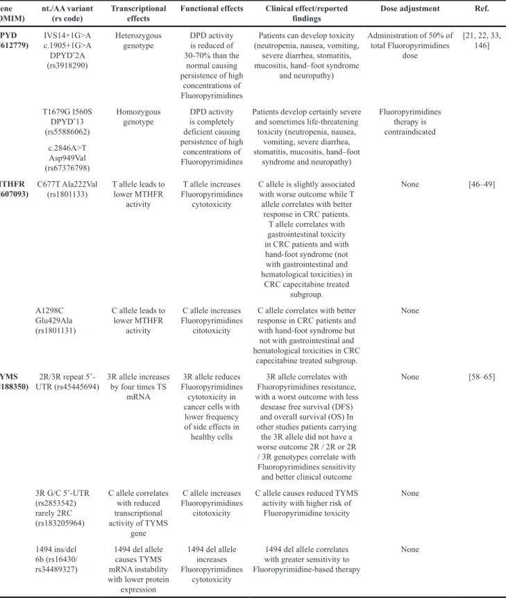
Table 2: Genotype-phenotype correlations and recommended fluoropyrimidines dose adjustments according to
DPYD, MTHFR and TYMS genotypes
Gene
(OMIM) nt./AA variant(rs code) Transcriptional effects Functional effects Clinical effect/reported findings Dose adjustment Ref. DPYD
(#612779) IVS14+1G>A c.1905+1G>A DPYD*2A
(rs3918290)
Heterozygous
genotype DPD activity is reduced of 30-70% than the
normal causing persistence of high
concentrations of Fluoropyrimidines
Patients can develop toxicity (neutropenia, nausea, vomiting,
severe diarrhea, stomatitis, mucositis, hand–foot syndrome
and neuropathy) Administration of 50% of total Fluoropyrimidines dose [21, 22, 33, 146] T1679G I560S DPYD*13 (rs55886062) Homozygous
genotype DPD activity is completely deficient causing persistence of high
concentrations of Fluoropyrimidines
Patients develop certainly severe and sometimes life-threatening
toxicity (neutropenia, nausea, vomiting, severe diarrhea, stomatitis, mucositis, hand–foot
syndrome and neuropathy)
Fluoropyrimidines therapy is contraindicated c.2846A>T Asp949Val (rs67376798) MTHFR
(#607093) C677T Ala222Val (rs1801133) T allele leads to lower MTHFR activity
T allele increases Fluoropyrimidines
cytotoxicity
C allele is slightly associated with worse outcome while T allele correlates with better
response in CRC patients. T allele correlates with gastrointestinal toxicity in CRC patients and with hand-foot syndrome (not with gastrointestinal and hematological toxicities) in CRC capecitabine treated subgroup. None [46–49] A1298C Glu429Ala (rs1801131) C allele leads to lower MTHFR activity C allele increases Fluoropyrimidines citotoxicity
C allele correlates with better response in CRC patients and with hand-foot syndrome but not with gastrointestinal and hematological toxicities in CRC
capecitabine treated subgroup.
None
TYMS
(#188350) UTR (rs45445694)2R/3R repeat 5’- 3R allele increases by four times TS mRNA
3R allele reduces Fluoropyrimidines
cytotoxicity in cancer cells with
lower frequency of side effects in healthy cells
3R allele correlates with Fluoropyrimidines resistance, with a worst outcome with less
desease free survival (DFS) and overall survival (OS) In other studies patients carrying
the 3R allele did not have a worse outcome 2R / 2R or 2R / 3R genotypes correlate with Fluoropyrimidines sensitivity and better clinical outcome
None [58–65] 3R G/C 5’-UTR (rs2853542) rarely 2RC (rs183205964) C allele correlates with reduced transcriptional activity of TYMS gene C allele increases Fluoropyrimidines citotoxicity
C allele causes reduced TYMS activity with higher risk of Fluoropyrimidine toxicity None 1494 ins/del 6b (rs16430/ rs34489327) 1494 del allele causes TYMS mRNA instability with lower protein
expression
1494 del allele increases Fluoropyrimidines
cytotoxicity
1494 del allele correlates with greater sensitivity to Fluoropyrimidine-based therapy
formation and stability of the inhibitory complex, thereby
the cytotoxic potential of fluoropyrimidines. [41].
The C677T polymorphism causes the substitution
of alanine to valine in the aminoacid sequence of exon 4,
reducing the catalytic activity of MTHFR while increasing
its thermolability. At 37 °C, indeed, the enzymatic activity
in subjects with T/T genotype is reduced by approximately
50% with respect to the C/C genotype [42]. However,
the increase in the intracellular stocks of folates has the
potential to stabilize the three-dimensional structure of
MTHFR and improve its enzymatic function [43].
The A1298C SNP is characterized by the substitution
of adenine with cytosine and hence of glutamate with
alanine in exon 7 and results in a decrease of MTHFR
activity [44]. Given the pivotal role of MTHFR in the
metabolism of fluoropyrimidines, its polymorphic variants
have been investigated as possible predictors of response
or toxicity to chemotherapy, but contrasting results have
been reported so far. In a metanalysis of 950 patients
with advanced colorectal cancer treated with a first-line
5-FU-based therapy, no correlation was found between
C667T or A1298C variants and response to therapy [45].
On the other hand, in a metanalysis of 2,402 colorectal
cancer patients treated with 5-FU-based chemotherapy
who alternatively reported clinical benefit outcomes
and/or adverse events, Jennings BA et al. observed a
weak association between MTHFR C677T and dismal
outcomes [46], whereas a positive correlation between
MTHFR SNPs (C677T and A1298C) and response to
fluoropyrimidines was shown in 815 Caucasian patients
with colo-rectal cancer [47]. In terms of toxicity prediction,
a metanalysis of 4,855 colo-rectal cancer patients treated
with 5-FU infusion, demonstrated that MTHFR C677T
inversely correlates with neutropenia (OR: 0.60; 95%
CI: 0.37-0.97) and general toxicity (OR: 0.79; 95% CI:
0.62-1.00) [24]. Similarly, in another study of 450 patients
who underwent 4 cycles of fluoropyrimidine-based
chemotherapy, both C677T and A1298C variants were
not significantly associated with serious hematological
or gastrointestinal side effects. However, in the subgroup
of patients who received capecitabine, a significant
correlation was found between both MTHFR SNPs
and hand-foot syndrome (p=0.0046) [48]. In Wang’s
meta-analysis only in one of three studies applicable for
analyzing the association between MTHFR polymorphism
and toxicity, the association between the 677T allele and
gastrointestinal toxicity was demonstrated (p=0.002) [49,
50]. In a recent study conducted in two cohorts of stage II/
III of CRC patients treated with adjuvant fluoropyrimidine
chemotherapy, the MTHFR 1298CC genotype carriers
underwent worsened disease free survival and overall
survival in both cohorts [51]. Similarly, an even more
recent study of 242 Korean patients with mCRC showed
that the presence of a C677CC genotype was associated
with a good prognosis in multivariate analysis [52]. On the
basis also of the significant ethnic differences in the C677T
and A1298C genotypes frequency, larger studies including
different populations are needed to determine the role of
these polymorphisms in response to fluoropyrimidines
[53]. Therefore, despite the result of such heterogeneous
findings, no formal indications are suggested regarding
the clinical genotyping of MTHFR in patients candidates
to fluoropyrimidine-based treatments although it is likely
that the presence of these polymorphisms in homozygosity
must be taken into account during the therapy planning.
(Table 2) [54, 55].
● TYMS - TYMS is located in the short arm
of chromosome 18 (18p11.32), contains 7 exons and
spans about 30 kb. TYMS is a folate-dependent enzyme
and competes with MTHFR for the availability of
the cofactor 5,10 methylene THF that catalyzes the
reductive methylation of deoxyuridylate (dUMP) to
thymidylate (dTMP), thereby playing a central role in
DNA synthesis and repair [56] (Figure 2). 5-FU efficacy
is directly correlated with TYMS expression levels [57]
and one of the major determinants of TYMS expression
is the presence of three polymorphic variants, namely
TSER
*2R/
*3R (rs45445694), TSER
*3R G/C (rs2853542 –
rs34743033) and 1494del6b (rs151264360 – rs869066439)
[58] (Table 2).
The 5’-UTR of TYMS contains a variable number
(from 2 to 9) of a 28-bp tandem repeat sequence (VNTR)
that acts as enhancer for the promoter by implementing
the gene transcriptional activity (Figure 2). Polymorphic
variants of this region, named thymidylate synthase
enhancer region (TSER), have been described, and
include double (2R) or triple repeats (3R) determining
the genotypes 2R/2R, 2R/3R and 3R/3R [59]. Functional
studies have shown a stepwise increase in TYMS
transcription with an increasing number of tandem repeats
[60], and patients with metastatic colo-rectal cancer with
a 3R/3R genotype show 3.6 times higher mRNA levels
as compared with those homozygous for 2R/2R [61]. The
higher TYMS expression level related to the presence
of 3R/3R genotype accounts for less effective inhibition
of TYMS and worse response to fluoropyrimidines, in
presence of low toxicity. Conversely, the occurrence
of two tandem repeat polymorphism (2R/2R or 2R/3R
genotype) leads to more favorable responses to 5-FU
treatment, [62, 63] but may enhance the 5-FU toxicity
(Table 2) [58].
The TSER
*3R G/C polymorphism consists in a G>C
change in the second repeat of the 3R allele (rs2853542
– rs24743033) (Figure 2). This SNP is associated with
a weaker bond in the promoter region of upstream
transcription factor 1 (USF1) and results in a lower
transcriptional activation of TYMS. It is found in
30%-55% of all TSER 3R alleles and its presence explains
why not all patients with the TYMS 3R allele have poor
outcomes (Table 2) [64, 65].
Besides these variants, an insertion/deletion of
hexanucleotide TTAAAG sequence at 1494 position on
the 3’-UTR of the TYMS gene, has been also described
to be in linkage disequilibrium with the TSER 3R allele
and is associated with worse prognosis in 5-FU treated
patients [66]. The -6 bp deletion results in a 70% decrease
in mRNA levels [67], probably as effect of the accelerated
degradation of the transcript. Thus, by combining the
above mentioned SNPs, it is possible to predict high levels
of TYMS expression in subjects with 2R/3RG, 3RC/3RG,
3RG/3RG; +6bp/+6bp genotypes, and conversely low
levels of the enzyme in those with 2R/2R, 2R/3RC,
3RC/3RC; -6bp/-6bp, -+6bp/-6bp genotypes [58].
Although knockdown or amplification experiments
succeeded in demonstrating the in vitro importance of
TYMS in 5-FU resistance and toxicity, clinical studies
on the predictive role of these polymorphisms have been
controversial so far [68]. For example, no significant
association between TYMS genotype and response rate or
overall survival (OS) has been observed in patients with
gastric cancer treated with platinum/5-FU combinations.
The same study found individuals with a 3R haplotype
to have a significantly lower risk of developing grade 3/4
leukopenia after chemotherapy [49]. In the mentioned
metanalysis performed by Jennings BA et al., on 2,402
patients with colo-rectal cancer, the 2R/2R genotype was
apparently characterized by a significantly higher risk
of toxicities when compared with the 2R/3R and 3R/3R
genotypes [46], whereas the toxicity to capecitabine
appeared globally greater in carriers of 2R/3R and 6bp
insertion polymorphisms [24]. However, no association
between capecitabine efficacy and TYMS polymorphic
variants has been reported so far [64, 69] and, as for
MTHFR, no specific indications have been formulated
thus far for the clinical testing of TYMS SNPs in cancer
patients who may benefit of fluoropyrimidine treatment.
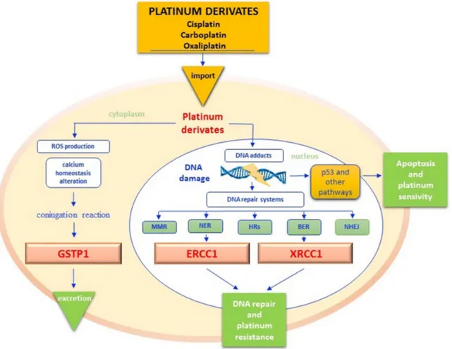
PLATINUM DERIVATIVES
Platinum derivatives are highly efficacious against a
broad spectrum of solid tumors and currently constitute the
backbone for the treatment of pulmonary, head and neck,
gastroentero-pancreatic and genitourinary neoplasms [70].
Resistance to platinum salts is mainly caused by the
hyperactivation of DNA repair systems, with consequent
decrease of pro-apoptogenic DNA adducts. As depicted
in Figure 3, different enzyme groups are enrolled in
DNA repair, including those of the nucleotide excision
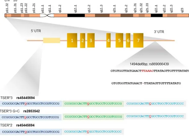
Figure 2: Polymorphisms in the 5’- and 3’-untranslated regions (UTRs) of TYMS gene.
The 5’-UTR of TYMS, named thymidylate synthase enhancer region (TSER), contains a variable number of a 28-bp double (2R) or triple repeats (3R) determining the genotypes 2R/2R, 2R/3R and 3R/3R. TYMS expression level is directly proportional to the number of repeats. The TSER*3R G/Cpolymorphism consists in a G>C change in the second repeat of the 3R allele and results in a lower transcriptional activation of TYMS. The 3’-UTR insertion/deletion of hexanucleotide TTAAAG in position 1494 is in linkage disequilibrium with the TSER 3R allele. The -6 bp deletion results in a 70% decrease in TYMS mRNA levels.
repair (NER) system, base excision repair (BER) system
and mismatch repair (MMR) system [71]. The most
representative enzymes of the NER and BER systems
include the excision repair cross complementation group
1 (ERCC1) and X-ray repair cross-complementing group
1 (XRCC1) respectively. In addition, homologous (HRs)
and non-homologous recombination systems end joining
(NHHRsEJ) are involved in DNA repairing processes
[71] (Figure 3). Cisplatin is characterized by a strong
emetic effect and has a remarkable toxicity profile for
kidney, liver, heart and auditory apparatus, as well as
severe myelo- and neuro-toxicity [72]. Detoxification
of platinum derivatives involves the conjugation with
reduced glutathione (GSH), a reaction catalyzed by
glutathione S-transferase protein 1 (GSTP1) [73]. Given
the key role of enzymes involved in either DNA repair
or drug metabolism in the pharmacodynamics and
pharmacokinetics of platinum salts, several variants of
both ERCC1, XRCC1 and GSTP1 have been investigated
as potential biomarkers of either response or toxicity
(Table 1).
● ERCC1 - ERCC1 is a 297 amino acid protein
encoded by a gene localized on 19q13 chromosome.
After heterodimerization with XP-F, ERCC1 contributes
to elimination of DNA adducts induced by UV light,
ROS, environmental mutagens and especially by cancer
chemodrugs [74] (Figure 3). Moreover, the protein has
a role in the preservation of chromosomal stability and
telomers’ integrity [75]. High levels of ERCC1 have
been associated with platinum resistance, while
ERCC1-Figure 3: Platinum pathway.
Once into cytoplasm, platinum derivatives promote the Reactive Oxygen Species (ROS) synthesis, that cause the alteration of cell membranes permeability, the deregulation of different signal transduction pathways and calcium homeostasis but overall the DNA damage. Glutathione S-transferases protein 1 (GSTP1) catalyzes the conjugation reaction of platinum derivates with reduced glutathione (GSH), in order to increase their hydro-solubility and to facilitate their excretion. When platinum derivatives reach the nucleus, they form intra and interstrand DNA cross-links that block the cell cycle by activating tumor cell apoptosis through different pathways. DNA adducts however may activate sensor proteins and DNA repair systems by avoiding cytotoxicity. Excision repair cross complementation group 1 (ERCC1) is the main endonuclease of DNA NER (Nucleotide Excision Repair) pathway but it also interacts with the BER (Base Excision Repair) function in maintaining chromosomal stability and telomers integrity. X-ray repair cross-complementing group 1 (XRCC1) is another enzyme of BER pathway that repairs DNA bases damaged by X-rays, ROS and mostly alkylating agents (. The efficiency of the GSTP1detoxification reaction and of DNA repairing systems affects the platinum-based treatments response. ROS - reactive oxygen species; GSTP1 - glutathione s-transferases protein 1; ERCC1 - excision repair cross complementation group 1; NER - nucleotide excision repair; BER - base excision repair; XRCC1 - X-ray repair cross-complementing group 1.defective cells appear to be highly sensitive to alkylating
agents [76, 77]. The best carachterized SNPs of ERCC1
include the T19007C (Asn118Asn; rs11615) and the
C8092A (rs321298) variants [78] (Table 1).
The synonym variant T19007C, although not
causing any aminoacid change, results in the low-use
codon AAT, instead of the high-use codon AAC, thus
significantly reducing the efficacy of ERCC1 mRNA [79].
On the other hand, a reduced expression of ERCC1, as
result of the C-allele, has been shown to correlate with
better responses to platinum-based therapies in non small
cell lung cancer (NSCLC) patients, whereas the T-allele
was found to correlate with platinum-resistance in gastric,
ovarian and cervical cancers [80–83] (Table 3). Several
retrospective studies have also demonstrated that NSCLC
patients carrying the T-allele have poor overall survival
when subjected to platinum-based chemotherapy [84,
85] and the association between the T-allele presence
and dismal outcomes has been confirmed in patients with
metastatic colo-rectal cancer treated with platinum. In
fact, the median overall survival (OS) was as low as 15.3
months in subjects with C/C genotype, and 11.1 months in
C/T or T/T carriers [86, 87].
Other studies observed a different
genotype-phenotype correlation between ethnic subgroups, by
showing that the 1907T allele was associated with
unfavorable PFS and OS in Asian, and alternatively,
with favorable prognosis in Caucasians, probably due
to the complexity of interactions between genes and
environment, rather than to polymorphism frequencies in
each group [54, 88].
Also, within the mCRC patients population, another
study on 168 Chinese patients treated with first-line
FOLFOX-4 chemotherapy showed that the CC genotype
was associated with a better response rate and clinical
outcome (ORR: 57.5% vs 36.4%; p = 0.01), as well as PFS
(13 months vs 7 months; P < 0.01), and OS (25 months vs
16 months; P < 0.01) compared to CT ot TT genotypes
[89].
Other studies included a metanalysis conducted
on 1,787 gastric and colon cancer patients treated with
oxaliplatin-based chemotherapy demonstrated the
role of rs11615 T allele as a predictor of low objective
response, shorter PFS and OS in Asian, but not Caucasian
people [90]. The same association was shown in a
subgroup analysis of another metanalysis, reflecting
the strong influence of ethnicity-dependent factors in
pharmacogenetic assay [88]. The combination of several
enzymatic variants involved in fluoropyrimidines and
platinum metabolism has been also reported to correlate
with PFS after first-line chemotherapy in patients
with metastatic colo-rectal cancer. In particular, the
combination of ERCC1-118 T/T, ERCC2-751 A/C, and
ERCC2-751 C/C was independently associated with low
PFS in 166 patients [91] since in post-operative colorectal
cancer evolution of 257 Taiwanese patients, the
ERCC2-751 A/A and ERCC1-118 T/T genotypes predicted higher
incidence of recurrence and worse clinical outcome [92].
Another common variant of ERCC1 is C8092A.
This SNP is located in the 3’UTR of the gene and can
alter polyadenylation, translation efficiency, localization
and stability of mRNA [93]. In particular, the presence
of allele A reduces the stability of the ERCC1 transcript,
thus resulting in lower protein expression and increased
sensitivity to genotoxic chemotherapies [94]. In a recent
metanalysis of 33 studies involving nearly 5,000 patients
with NSCLC treated with platinum-based chemotherapy,
the TT/TC genotypes of the C118T variant and the AA/CA
genotypes of the C8092A SNP were associated with lower
objective response rate (ORR) and OS as compared with
CC genotype. However, this effect was observed only in
the Asian population, but not in Caucasian patients [95].
The role of ERCC1 variants as predictors of
toxicity following platinum therapy has been poorly
investigated. In patients with advanced NSCLC, the
8092A allele, but not the 118T allele, appeared associated
with a significantly increased risk of gastrointestinal
grade 3 or 4 toxicity [96]. In another study of patients
with colorectal cancer treated with platinum-based
adjuvant chemotherapy, the 8092A allele was reported
to predict hematologic toxicity, in particular anemia [97]
(Table 3).
● XRCC1 - The human XRCC1 protein is
encoded by a gene mapping on 19q13.2 chromosome
and plays a pivotal role in the BER pathway, replacing
DNA bases damaged by X-rays, reactive oxygen radicals
and alkylating agents [98–100] (Figure 3). More than
300 SNPs have been shown to affect XRCC1, but only
three of them have been functionally characterized. In
fact, Arg194Trp, Arg280His and Arg399Gln cause amino
acid substitutions in the XRCC1 protein resulting in the
alteration of its function [101]. Such polymorphisms have
been associated to a general increased cancer risk in the
full population as result of impaired capacity of DNA
repair, correlation with greater tumor aggressiveness, and
lower response to platinum derivatives [102, 103].
The G28152A variant, also named Arg399Gln or
rs25487, is the most well-studied SNP of XRCC1 and
maps on the COOH-terminal domain of the gene, coding
for a protein portion devoted to protein-protein interactions
(Table 1) [104, 105]. The 28152A allele is responsible of a
substantial defect of XRCC1 to repair DNA, in particular
after exposure to ionizing radiation [98, 106].
In NSCLC patients, the A/A or G/A genotypes have
been associated with increased risk of all toxicities as
compared with the G/G genotype, and particularly with
a 2.5-fold increased risk of grade 3 or 4 gastrointestinal
toxicities [107]. Also, higher incidence of severe
hematologic adverse effects has been observed in carriers
of the A allele in another study on 487 NSCLC patients
treated with cisplatin, docetaxel and gemcitabine [108].
However, no significant association between XRCC1
399A and chemotherapy-induced toxicities was found
in the TOSCA trial, which evaluated 3,579 patients with
colorectal cancer treated with FOLFOX-4 or XELOX
adjuvant chemotherapy (Table 3) [109].
The G28152A variant has been also investigated
as biomarker of response and survival after
platinum-based chemotherapy. In a study of 112 NSCLC patients,
a progressive increase in average survival times following
platinum treatment has been identified in carriers of the
A/A, A/G and G/G genotypes respectively [102]. By
contrast, a metanalysis of 22 studies investigating
platinum-based chemotherapy in advanced NSCLC demonstrated
the role of A/A + G/A genotypes in predicting objective
responses [110], while worse outcomes were reported for
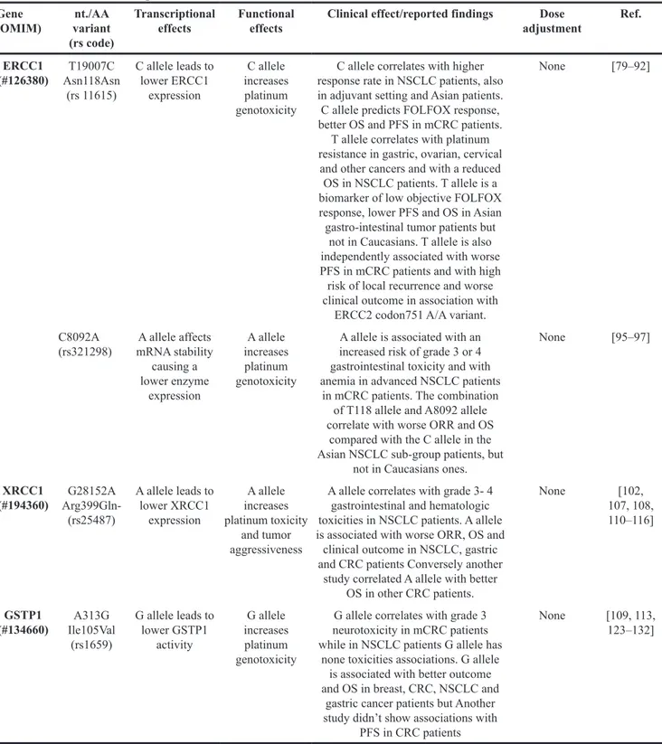
Table 3: ERCC1, XRCC1 and GSTP1 genes variants, potential impact on the enzyme activity and outcome of
platinum derivatives therapies
Gene
(OMIM) variantnt./AA (rs code)
Transcriptional
effects Functional effects Clinical effect/reported findings adjustmentDose Ref. ERCC1 (#126380) Asn118Asn T19007C (rs 11615) C allele leads to lower ERCC1 expression C allele increases platinum genotoxicity
C allele correlates with higher response rate in NSCLC patients, also in adjuvant setting and Asian patients. C allele predicts FOLFOX response, better OS and PFS in mCRC patients.
T allele correlates with platinum resistance in gastric, ovarian, cervical
and other cancers and with a reduced OS in NSCLC patients. T allele is a biomarker of low objective FOLFOX response, lower PFS and OS in Asian gastro-intestinal tumor patients but
not in Caucasians. T allele is also independently associated with worse PFS in mCRC patients and with high risk of local recurrence and worse clinical outcome in association with
ERCC2 codon751 A/A variant.
None [79–92]
C8092A
(rs321298) mRNA stability A allele affects causing a lower enzyme expression A allele increases platinum genotoxicity
A allele is associated with an increased risk of grade 3 or 4 gastrointestinal toxicity and with anemia in advanced NSCLC patients
in mCRC patients. The combination of T118 allele and A8092 allele correlate with worse ORR and OS
compared with the C allele in the Asian NSCLC sub-group patients, but
not in Caucasians ones.
None [95–97] XRCC1 (#194360) Arg399Gln-G28152A (rs25487) A allele leads to lower XRCC1 expression A allele increases platinum toxicity and tumor aggressiveness
A allele correlates with grade 3- 4 gastrointestinal and hematologic toxicities in NSCLC patients. A allele is associated with worse ORR, OS and clinical outcome in NSCLC, gastric and CRC patients Conversely another
study correlated A allele with better OS in other CRC patients. None [102, 107, 108, 110–116] GSTP1 (#134660) Ile105Val A313G (rs1659) G allele leads to lower GSTP1 activity G allele increases platinum genotoxicity
G allele correlates with grade 3 neurotoxicity in mCRC patients while in NSCLC patients G allele has
none toxicities associations. G allele is associated with better outcome and OS in breast, CRC, NSCLC and
gastric cancer patients but Another study didn’t show associations with
PFS in CRC patients
None [109, 113, 123–132]
carriers of the A allele in another study of 235 patients with
NSCLC treated with platinum [108].
Better outcomes following FOLFOX therapy have
been consistently observed in patients with metastatic
colo-rectal cancer carrying a G allele [111, 112]. More recently,
the presence of the A allele has been instead associated
with lower tumor response after oxaliplatin-based
chemotherapy in a cohort of 1,234 patients with colorectal
cancer. Surprisingly, no correlation with PFS was found
[113]. In gastric cancer patients treated with
oxaliplatin-based chemotherapy, the A allele conferred a significant
disadvantage in terms of survival [114]. Contradictory
evidence has been generated on the prognostic role of
XRCC1 399A in patients with colo-rectal cancer [115, 116].
● GSTP1 - The glutathione S-transferases
(GSTs), subdivided in seven enzymatic classes (α, μ, κ,
τ, π, ω and ζ) detoxify mammalian cells by endogenous
and exogenous, hydrophobic and electrophilic toxic
compounds by using reduced glutathione (GSH), thus
avoiding the formation of DNA adducts (Figure 3). The
gene of Pi-class glutathione-S-transferase (GSTP1)
maps on chromosome 11q13.2, extending for about 2.8
Kb [117]. GSTP1 catalyzes the conjugation of platinum
derivatives with reduced glutathione (GSH), in order to
increase their hydro-solubility and excretion [73]. Several
in vitro studies have shown a significant correlation
between resistance to platinum and high levels of
intracellular GSH, as well as between platinum resistance
and elevated GSTP1 expression [118–120].
Seminal studies on the functional polymorphisms
of the GSTP1 identified two variants of the gene (A313G
and C114T), whose combinations result in four functional
haplotypes GSTP1
*A (AC); GSTP1
*B (GC), GSTP1
*C (GT)
and GSTP1
*D (AT) [121]. In this context, the G/G genotype
at nucleotide 313 (A313G; Ile105Val; rs1659)seems to
substantially decrease the enzymatic activity of GSTP1,
as it causes an aminoacid substitution in the active site of
the protein [122]. As consequence, a possible implication
of GSTP1 G-harboring SNPs in the response to platinum
derivatives has been envisaged [73] GSTP1 genotypes G/G
or G/A with lower enzyme activity potentially correlate with
increased response to platinum-based chemotherapy due to
the decreased detoxification activity [73]. The G/G genotype
of GSTP1 has been associated with grade 3 neurotoxicity in
patients with colo-rectal cancer who received FOLFOX [91]
(Table 3). This observation was also confirmed in a study
of n166 Asian patients with metastatic colorectal cancer,
and similar findings were reported for Caucasion patients
with inoperable NSCLC treated with platinum-gemcitabine
[123, 124]. However, not all studies have been consistent in
demonstrating a significant association between platinum
salts, neurotoxicity and GSTP1 variants enriched in alleles
G [125, 126].
While causing increased toxicities, the reduced
detoxifying activity of GSTP1 determined by the G/G
genotype has been also hypothesized to lead to better clinical
outcomes in response to platinum-based protocols. However,
contrasting data have been reported at this regard in several
cancers [91, 127, 128], and a large metanalysis of 13 studies
on colo-rectal cancer patients failed to show any association
between the G allele and PFS [113]. Ethnic differences
among enrolled patients (46% Caucasian, 64% Asian) may
have probably impaired the analysis [54]. Even in NSCLC
patients, different metanalyses and clinical studies have
shown the association between G/G genotype and increased
platinum-based chemotherapy efficacy in terms of both
response rates and OS [129–131]. The positive predictive role
in terms of tumor response, PFS and OS of the G/G genotype
was also confirmed in a recent metanalysis including 8169
cases with gastric cancer subjected to platinum-based
chemotherapy [132].
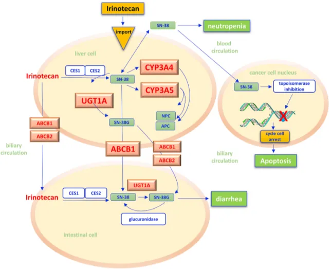
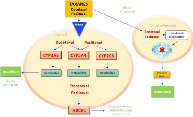
IRINOTECAN
Irinotecan is an camptothecin analogue widely used in
the treatment of gastroenteropancreatic tumors. As a prodrug
irinotecan is intracellularly activated through a hydrolysis
reaction catalyzed by microsomial carboxylesterase within
hepatocytes. This leads to the production of the active
metabolite SN-38 [133], which inhibits the topoisomerase I,
a key enzyme in DNA replication which is 100 times stronger
than the progenitor drug. The SN-38/topoisomerase I/DNA
complex causes major breaks in the DNA replication fork,
with subsequent activation of apoptosis [134] (Figure 4). The
metabolism of SN-38 is mainly mediated by the cytocrome
P450 enzyme CYP3A4 and CYP3A5 isoforms and by the
uridine diphosphate glucuronosyltransferases (UGT), that
catalyze the excretion of the drug into the bile [135, 136].
On the other hand, adenosine triphosphate binding cassettes
(ABCB) allow the transport of irinotecan and its active
metabolites through cell membranes, determining their
distribution between cancer cells, blood and entero-hepatic
circulation [137].
The most serious dose-related toxicity of irinotecan
is diarrhea [138]. This effect seems to correlate with the
varying degree of efficiency of UGT in conjugating SN-38
in its inactive metabolite SN-38G. In fact, in subjects with
low UGT conjugating efficiency, SN-38 is directly reversed
in the intestinal lumen through the bile [135]. On this basis,
it is not surprising that UGT1, CYP3A4, CYP3A5, and
ABCB1 polymorphisms have been explored as appealing
predictors of toxicity and efficacy of irinotecan (Table 1).
● UGT - This superfamily of detoxifying enzymes
includes two subfamilies (UGT1 and UGT2), both having
endogenous and exogenous compounds as substrates.
UGT1A gene is located on chromosome 2q37, and its
promoter sequence (TATA box) contains a polymorphic
syte, with a variable number (from 5 to 9) of dinucleotide
repetitions (TA). An inverse correlation between the number
of dinucleotide repetitions and the UGT1A efficiency has
been demonstrated. The most common genotype in the
general population is 6/6 UGT1A1
*1, characterized by
six homozygous repeat dinucleotides; the presence of 7/7
UGT1A1
*28, typical of patients with Gilbert’s syndrome,
is instead characterized by a glucuronation efficiency as
low as 30-50% than the normal [139]. Patients carrying the
variant 7/7 who undergo irinotecan-based chemotherapy
have a 4-fold increased risk of developing grade 3-4
diarrhea and neutropenia [140, 141] (Figure 4). There is
lack of knowledge about the clinical significance of the
other variants (5/5, 8/8 and 9/9 tandem repeats) [142].
The geographic distribution of allelic variants
frequencies is heterogeneous with maximum value
in African populations (23%), intermediate among
Europeans (11-13%) and minimum among Asians (3%).
Based on observations from previous clinical trials,
a meta-analysis of 9 studies (81 patients with lymphoma
and 740 with advanced colorectal cancer) evaluated
the clinical correlation between severe post-irinotecan
toxicities as grade 3-4 diarrhea and neutropenia, and
occurrence of UGT1A1
*1/UGT1A1
*28 genotypes in both
homozygous and heterozygous carriers. For irinotecan
dosages below 125 mg/m
2, no differences were observed
according to the genotype, whereas higher doses of the
drug were associated with an increased risk of severe
toxicities in subjects homozygous for the UGT1A1
*28
variant [143] (Table 4). Consistently, another metanalysis
of Caucasian patients with metastatic colo-rectal cancer
Figure 4: Irinotecan pathway.
Irinotecan is a prodrug that, after administration, is activated in liver by the hydrolysis reaction catalyzed by carboxylesterases (CES1, CES2) of the microsomal system of hepatocytes, with the release of the more active metabolite SN-38. In cancer cell nuclei, SN-38 acts as inhibitor of the topoisomerase I, a key enzyme in DNA replication. The SN-38/enzyme/DNA complex causes major breaks in the DNA replication fork, with subsequent activation of cancer cells apoptosis. In liver cells, irinotecan and SN-38 may be oxidated by hepatic cytochrome P-450 (CYP 3A4 and 3A5) to form pharmacologically inactive metabolites (NPC, APC). The Uridine diphosphate (UDP) glucuronosyltransferases (UGT) catalyzes the subsequent conjugation reaction of SN-38 with glucuronic acid making its excretion possible through the bile in the intestinal lumen. Adenosine-triphosphate binding cassettes (ABC) transporters (ABCB1/ABCB2) are transmembrane proteins which make possible the absorption of SN-38 from plasma into hepatocytes and hence in interstitial and the excretion of irinotecan and its metabolites by bile into the intestinal lumen. An increased bioavailability of SN-38, i.e. for the reduced efficiency of UGT and CYP 3A4/3A5 reactions, seems to justify the onset of diarrhea and neutropenia as specific side effects of chemotherapy. CES – carboxylesterases; CYP - cytochrome P-450; UGT - uridine diphosphate (UDP) glucuronosyltransferases; ABC - adenosine-triphosphate binding cassettes.who received irinotecan found a 2-to-4-time increased
risk of severe neutropenia and diarrhea in UGT1A1
*1
6/6 carriers as compared with UGT1A1
*28 7/7 carriers.
This effect was more prominent in patients treated with
high doses of irinotecan, or in those receiving the drug in
combination with fluorouracil [144].
While effective in predicting toxicities, the SNPs of
UGT1 appear less useful as biomarkers of efficacy. In fact,
a recent metanalysis performed on 1,898 mCRC patients
treated in first or second line with irinotecan showed no
correlation between UGT1A1
*28 7/7 and response rates
[145].
Based on this body of evidence, in 2005 FDA
recommended a 30% dose reduction for patients
homozygous for UGT1A1
*28 and candidates to irinotecan
therapy at dosages >250mg/m2 [69, 146] (Table 4). No
dose adjustments were indicated in case of UGT1A1
*28
heterozygosity [146]. Similarly, the guidelines published
by the Dutch Pharmacogenomics Working Group
recommend dose reductions in patients with known
UGT1A1
*28 homozygous genotype [146]. In Italy,
assessment of UGT1A variants is indicated in the
pre-therapy setting for those patients where chemopre-therapy
has a high risk/benefit ratio and during therapy in all
cases of grade 3-4 of hematologic and/or gastrointestinal
toxicities or in any case of unexpected ADRs (AIOM-SIF
guidelines) [147] (Table 4).
● CYP3A4/3A5 - The CYP3A4 and CYP3A5
genes contribute to the oxidative metabolism of irinotecan
[148] (Figure 4). CYP enzymatic activity can be largely
influenced by non-genetic factors as diet, ethnicity, or
concomitant therapies, as well as by genetic conditions,
namely the presence of polymorphic variants [149].
Fourty SNPs have been identified in the CYP3A4
gene and among them, the most studied is CYP3A4
*1B
(-392A>G, rs2740574) which is characterized by a
transition from A to G in the regulatory 5’ UTR [150]
(Table 1). This variant is differently distributed among
various ethnic groups (high frequency among Caucasians
and Afro-Americans, very low frequency among Asians)
and may influence the gene transcription, leading to a
substantial increase of the protein levels [151]. As result of
higher CYP3A4 expression, the metabolism of irinotecan
may be accelerated and the intracellular exposure to
SN-38 diminished with consequent decrease of its therapeutic
efficacy [152, 153]. Although limited in its power by a
small sample size, a study of 30 Caucasian patients with
lung or colorectal cancer showed a significant correlation
between CYP3A4 genotype and irinotecan blood clearance
[154]. By contrast, a pharmacokinetic analysis of 177
Japanese individuals with cancer failed to demonstrate
any meaningful impact of CYP3A4 on irinotecan blood
levels [155].
The polymorphism CYP3A5
*3 (6986A>G,
rs776746) is characterized by a transition from A to G
in intron 3 and causes the generation of a splicing site,
leading to the incorporation of an intronic sequence of 131
bp within the transcript. The ultimate consequence of this
mutation is the synthesis of a truncated, non-functional
protein and, indeed, carriers homozygous for CYP3A5
*3
(G/G genotype) have very low levels of the enzyme with
respect to subject with the wild type variant CYP3A5
*1
(A/A genotype) [156] (Table 4). The clinical significance
of the CYP3A5
*3 splice variant has been investigated only
in a sub-analysis of the North American Gastrointestinal
Intergroup N9741 study, that investigated the combination
of irinotecan, 5-fluorouracil and leucovorin in 520 patients
with mCRC. Carriers of the G allele showed a response
rate lower than that observed in patients bearing the A
allele (29% vs 60%, p=0.0074) [157].
On the other hand, hyperfunctional CYP3A4
*1B
and CYP3A5
*1 variants has been associated to protection
from irinotecan-driven toxicities, as consequence of the
accelerated drug metabolism [158] (Table 4). However,
it should be noted that studies on the predictive ability
of CYP polymorphisms are very difficult to interpret as
result of the high inter- and intra-individual variability of
CYP enzymatic activity that may primarily result from
diet-, ethnicity- and therapy-related factors. At present,
CYP3A4/5 genotyping does not have a definite role in
customization of irinotecan-based chemotherapy [159].
ABC transporters - ABC transporters, among
which there is P-glycoprotein (multi-drug resistance
associated resistance protein 1, MDR1 or ABCB1), allow
the absorption of SN-38 from plasma into hepatocytes and
hence into the interstitial space. ABCB1 is a well-known
drug transporter localized in the epithelial cells in the
intestine, liver and kidney, contributes to the absorption
of orally administered drugs, and the excretion of
irinotecan and its metabolites through the bile toward the
intestinal lumen and renal elimination [137]. Indeed ABC
transporters are known for a long time for their ability
to increase efflux of anticancer drugs from cancer cells
leading to the reduction of intracellular chemotherapeutic
agent levels and consequent drug insensitivity [160, 161].
ABCB1 is encoded by a gene on chromosome 7q21.12 that
spans 28 exons [162]. In ABCB1 knockout mice, excretion
of irinotecan and its metabolites appears to be impaired,
resulting in severe alterations of the drug pharmacokinetic
profile [163]. It is therefore not surprising that several
variants of ABCB1 have been studied in their ability to
predicit toxicity following irinotecan-based treatments
(Figure 4).
The ABCB1 SNP 3435C>T (Ile1145Ile, rs1045642)
consists in a silent point mutation that decreases the
mRNA stability and the protein three-dimensional
conformation reducing the enzyme expression [164]
(Table 1). Thus, the presence of this variant determines
a reduction in the excretion of irinotecan and its
metabolites, causing an increased risk of
chemotherapy-associated toxicities [165], (Table 4). In a Korean
study of 107 patients with NSCLC who were treated
with irinotecan-cisplatin, carriers of the 3435T allele
underwent higher incidence of grade 3 diarrhea (p=0.047)
[166]. However, no significant correlation between
ABCB1 genotype and toxicities was found in a French,
phase III, randomized trial of 5-FU and folinic acid with
or without Irinotecan [167].
Similarly to 3435C>T, the ABCB1 SNP C1236T
(Gly412Gly, rs1128503) is characterized by a silent
mutation which alters the transcript stability and reduces
protein expression [168]. In a small study of 65 patients,
the 1236T allele has been associated with prolonged
exposure to irinotecan and its active metabolite SN-38,
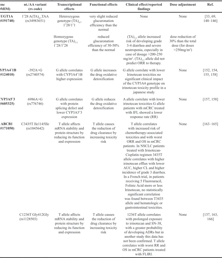
Table 4: Genotype-phenotype correlations and recommended irinotecan dose adjustment according to UGT1A,
CYP3A4
*1B and CYP3A5
*3 genotypes
Gene
(OMIM) nt./AA variant(rs code) Transcriptional effects Functional effects Clinical effect/reported findings Dose adjustment Ref. UGT1A
(#191740) 1
*28 A(TA) 6/7TAA
(rs34983651) genotype (TA)Heterozygous 6/7
1*28/1*1
very slight reduced glucuronation efficiency than the
normal None None [33, 69, 140–146] Homozygous genotype (TA)7/7 1*28/1*28 reduced glucuronation efficiency of 30-50%
than the normal
(TA)7/7 allele increased
risk of developing grade 3-4 diarrhea and severe neutropenia, especially in
case of dosage >200-250 mg/m2. (TA)
7/7 allele did not
predict ORR to therapy.
dose reduction of 30% than the total dose (for doses
>250mg/m2)
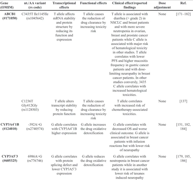
CYP3A4*1B
(#124010) (rs2740574)-392A>G G allele correlates with CYP3A4*1B
higher expression
G allele increases the drug oxidative detoxification
G allele correlate with lower Irinotecan toxicities no significant clinical impact of the CYP3A4 genotype on irinotecan toxicity profile in a
japanese study
None [152, 154, 155, 158]
CYP3A5*3
(#605325) (rs776746)6986A>G G allele correlates with protein splicing defect and lower CYP3A5*3
expression
G allele reduces the drug oxidative
detoxification
A allele correlate with lower irinotecan toxicities G allele patients with mCRC treated with IFL showed a lower
response rate (RR)
None [157, 158]
ABCB1
(#171050) C3435T Ile1145Ile (rs1045642) mRNA stability and T allele affects protein structure by reducing its function
and expression T allele causes the reduction of drug clearance by increasing toxicity risk T allele correlates with increased risk of chemotherapy-associated
toxicities and with worst ORR and OS in mCRC patients. In NSCLC patients
treated with Irinotecan-Cisplatin regimen 3435T allele correlates with higher irinotecan efflux with lower AUC, higher CL and higher incidence of grade 3 diarrhea.
In a French trial, in patients receiving 5 Fluorouracil, Folinic Acid more or less Irinotecan, no statistically significant correlation was found between T3435
allele and hematologic or gastrointestinal toxicities.
None [163–165]
C1236T Gly412Gly
(rs1128503) mRNA stability and T allele affects protein structure by reducing its function
and expression T allele causes the reduction of drug clearance by increasing toxicity risk 1236T allele correlates with prolonged exposure to irinotecan and SN-38, with a greater probability of developing ADRs but in another study this data has not been confirmed. T allele correlates with worst RR and OS in mCRC patients treated
with FLIRI.
None [137, 163, 166]