Table of contents
Riassunto
pg.
i
Abbreviations
pg.
iii
Preface. A bittersweet computational journey
pg.01
Chapter 1. Molecular Models in Glycobiology: an Overview
pg.03
1.1. Introduction
pg.03
1.2. Molecular models techniques applied to study
the glycan-protein interactions
pg.06
1.2.1. Molecular docking
pg.06
1.2.2. Molecular dynamics (MD) simulations
pg.09
1.2.3. Enhanced sampling techniques
pg.14
Chapter 2. Demystifying Heparan Sulfate-Proteins Interactions
pg.17
2.1. Implementation of an incremental docking method to
characterize the heparin/HIV-1 p17 protein interaction
pg.20
2.1.1. Introduction
pg.20
2.1.2. Material and methods
pg.21
2.1.3. Results
pg.22
2.1.4. Conclusions
pg.29
2.2. New insight into the mechanistic effect of heparin and
HSPGs on SARS-CoV-2 S glycoprotein
pg.31
2.2.1. Introduction
pg.31
2.2.2. Material and methods
pg.32
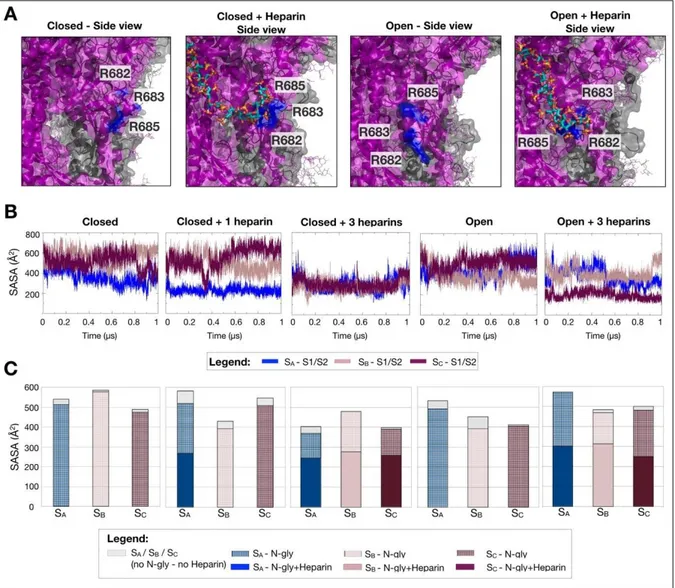
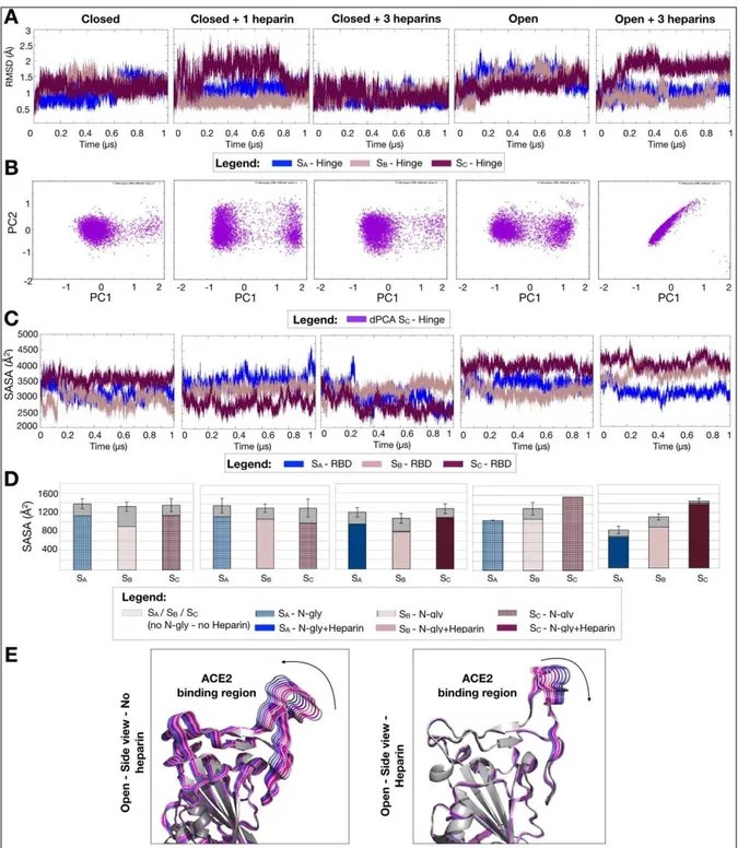
2.2.3. Results
pg.34
2.2.4. Conclusions
pg.42
2.3. New insight into the role of heparin and HSPGs in the
interaction of VEGF and its receptor VEGFR2
pg.44
2.3.1. Introduction
pg.44
2.3.2. Material and methods
pg.46
2.3.3. Results
pg.48
2.3.4. Conclusions
pg.51
Chapter 3. Sialic Acid as a New Target for Drug Discovery
pg.52
3.1. Modelling of the VEGFR2-ST6Gal complex
pg.55
3.1.1. Introduction
pg.55
3.1.2. Material and methods
pg.55
3.1.3. Results
pg.56
3.1.4. Conclusions
pg.69
Concluding Remarks
pg.60
Riassunto
Uno degli obiettivi principali della ricerca medica è lo studio della relazione tra le biomolecole, le loro interazioni e le patologie correlate. A riguardo, sono stati sviluppati numerosi approcci sperimentali volti a ottenere risposte sempre più specifiche, che tuttavia rimangono deficitari riguardo la comprensione dei meccanismi molecolari alla base di queste interazioni. Negli ultimi decenni, grazie al processo tecnologico e sistemi di calcolo sempre più avanzati, gli studi computazionali ed in particolare la simulazione di modelli molecolari sempre più complessi hanno assunto un ormai insostituibile ruolo indirizzamento alla ricerca medica.
Quando si parla di modelli molecolari si fa riferimento alla descrizione numerica di strutture complesse partendo dalle loro caratteristiche (quali geometria, energia, potenziale elettrico, ionizzazione e proprietà spettroscopiche) che permettono la simulazione del loro comportamento e delle loro interazioni a livello atomico basandosi sulle leggi della fisica classica o quantistica.
In questo ambito, è preoccupante il ritardo nello sviluppo dei modelli molecolari per la glicobiologia, nonostante la dimostrata importanza degli zuccheri nei processi fisiologici e patologici. Tale ritardo è principalmente dovuto alla complessità strutturale, flessibilità e lunghezza di queste macromolecole biologiche.
In quest’ottica, lo scopo di questo lavoro è stata l’implementazione di nuove procedure e modelli molecolari computazionali (simulazioni di “docking” e di dinamica molecolare) nel campo della glicobiologia che permettono la migliore comprensione dell’interazione non covalenti degli zuccheri con diverse proteine e le loro conseguenze in molteplici processi fisiologici e patologici.
I modelli molecolari dei complessi proteine-zuccheri sviluppati durante questo lavoro hanno permesso di caratterizzare i meccanismi diretti ed allosterici con i quali queste lunghe catene polisaccaridiche agiscono sulle proteine. L’analisi di questi complessi attraverso dinamiche molecolari ha permesso di dimostrare che questi zuccheri agiscono duplicemente: (i) favorendo l’oligomerizzazione delle proteine legate e inducendo importanti cambi conformazionali alla loro struttura tridimensionale; (ii) esponendo le proteine legate sulla superficie delle cellule favorendo così la loro interazione con recettori specifici.
Oltre che attraverso la loro interazioni non covalenti con distinte proteine, gli zuccheri svolgono un ruolo importante quando covalentemente associati alle proteine (principalmente recettori) a formare le cosiddette “glicoproteine”. A riguardo, in questo lavoro sono riportati modelli molecolari che hanno permesso di dimostrare l’effetto di stabilizzazione e mascheramento dei glicani (soprattutto acido sialico) legati covalentemente ai recettori studiati.
In conclusione, i risultati riportati in questa tesi dimostrano quanto l’applicazione degli studi computazionali alla glicobiologia sia ancora agli albori. La medicina di precisione, che sta assumendo sempre più importanza in ambito biomedico, non può prescindere dall’approfondita conoscenza delle interazioni biomolecolare. A questo riguardo, l’integrazione tra modelli computazionali e risultati sperimentali adottato in questi lavori si è dimostrato estremamente proficuo.
Abbreviations
ACE2 Angiotensin-converting enzyme 2AMBER Assisted model building and energy refinement aMD Accelerated molecular dynamic
CHARMM Chemistry at Harvard macromolecular mechanics Cryo-EM Cryo electron microscopy
dPCA Dihedral principal component analysis ED Essential dynamics
FF Force field
GA Genetic algorithm
GAFF General AMBER force field GAGs Glycosaminoglycans
Glc glucosamine
GlcNAc N-acetyl-D-glucosamine GlcA D-glucuronic acid GPU Graphic processing unit HBD Heparin binding domain
HIV Human immunodeficiency virus HPC High performance computing HS Heparan sulfate
HSPGs Heparan sulfate proteoglycans IdoA Iduronic acid
IFP Interaction fingerprint analysis
LJ Lennard-Jones
MD Molecular dynamics
NEUs sialidases
NMR Nuclear magnetic resonance
NPT Ensembles with constant number of particles, pressure and temperature
NTD N-terminal domain
NVT Ensembles with constant number of particles, volume and temperature OPLS-AA Optimized potentials for ligand simulation-all-atom
PBC Periodic boundary condition PBD Protein data bank
PCA Principal component analysis PME Particle mesh Ewald method
RBm receptor binding motif
REMD Replica exchange molecular dynamic RMSD Root mean square deviation
RMSF Root mean square fluctuation
S Spike glycoprotein
SARS-CoV-2 Severe acute respiratory syndrome coronavirus 2 SASA Solvent accessible surface area
Sia Sialic acid
Ter Terminus
VEGFA Vascular endothelial growth factor
VEGFR Vascular endothelial growth factor receptor VMD Visual molecular dynamic
“Stay hungry. Stay foolish.”
[Stewart Brand. Whole Earth Catalog]
Preface. A bittersweet computational journey
It was in 1902 when Hermann Emil Fischer, german chemistry professor, was awarded for the first time with the Nobel prize in chemistry "in recognition of the extraordinary services he has rendered by his work on sugar synthesis". And this was just the beginning of something revolutionary. Along the years, many other Nobel laureates have been awarded with these prizes in fields related to the history of glycobiology as Karl Landsteiner (Physiology or Medicine, 1930), “for his discovery of human blood groups” and Luis F. Leloir (Chemistry, 1970), “for his discovery of sugar nucleotides and their role in the biosynthesis of carbohydrates”.
Although this long story, there has been relatively little attention paid to the various ways in which proteins are “tweaked” through the attachment of sugars. However, the role of glycans is far from a decorative function. Glycans are not only involved in catalytic reactions but also help determine the three-dimensional structures of proteins, which are inherently linked to their function and their activity. Moreover, in contrast to some of the other chemical tags employed by cells, sugars exhibit a mind-boggling diversity of structures, can confer cell-type specificity, and are crucial components of cell-to-cell signaling. At the same time, glycans make problematic characterizations; they are the most difficult biological molecules to analyze and synthesize.
In my Ph.D. years, I have tried to pinpoint the role of glycans from a computational perspective, studying the interactions between sugars and different - but in some cases mechanistically related - sophisticated biomolecules. Molecular docking, classical molecular dynamics simulations and enhanced sampling have been the “vessels” of this exciting bittersweet computational journey from the role of heparan sulfate proteoglycans and heparin to the sialic acid. Importantly, all the data reported in this work are based on molecular models in line with the experimental data reported in the original paper or already released in literature.
InChapter 1 I introduce the concept of molecular models in glycomics with an overview of the problem related with this field of research. Subsequently, I present the molecular models techniques that I have used in this thesis aimed at reconstructing the ratio of processes.
TheChapter 2 consists of a brief review about structure, role and activity of heparan sulfate proteoglycans and heparin. Sections 2.1, 2.2 and 2.3 contain the results achieved during my work about the mechanistic effects induced by these sugars interacting with p17-HIV protein, SARS-CoV-2 spike glycoprotein and vascular endothelial growth factor, respectively. The results presented in Section 2.1 are published in 2019 in Scientific Reports and consist in the implementation of a computational method to model long sugar interacting with proteins. This project was conducted in collaboration with the National Research Council under the co-supervision of Dott.ssa Pasqualina D’ursi. The results presented in Section 2.2 were conducted thanks to the funding of the PRACE project (partnership for advanced computing in Europe) under the co-supervision of Prof. Rebecca
Wade during my 9 months endorsement at the Heidelberg Institute for Theoretical Studies. The results are collected in a paper submitted to PNAS. In particular, secondary to the COVID-19 pandemia, I have pinpointed the role and the mechanistic effect induced by the sugars mentioned above interacting with spike glycoprotein of SARS-CoV-2. Finally, Section 2.3 gathers the preliminary results of the project that have as specific aim to highlight the structural and dynamical interactions between the vascular endothelial growth factor and the heparan sulphate proteoglycans.
Chapter 3 is focused on the sialic acid as a new target for drug discovery. As in the previous chapter, after an introduction about this sugar, Section 3.1 presents the data obtained in my work. The results offer a pipeline to set up molecular models of ternary complexes simulating the Michaelis complex useful to identify structural frameworks and druggable pockets. Virtual screening of approved small molecules will be subsequently tested to identify compounds able to interfere with the formation of the ternary complex.
Graphical representation on the projects. (1) Characterization of the heparin/HIV-p17 oligomerization (2) New insight into the mechanistic effect of heparin/HSPGs on SARS-CoV-2 spike glycoprotein. (3) New insight into the role of heparin and HSPGs in the interaction of VEGF-A and its receptor VEGFR2. (4) Modelling of the VEGFR2-St6Gal1 complex.
Chapter 1.
Molecular Models in Glycomics: an Overview
1.1. Introduction
One of the main issues of medical research is the study of the relationship between biomolecules and diseases. To this aim, a variety of approaches have been developed over the years, many of which are based on experimental cell-based assays (1), animal models (2) or, less frequently, clinical-based approaches (3). Although reliable and apt to provide the direct connection between biomolecules and diseases, these approaches lack in understanding the molecular mechanisms underlying the pathological processes under study. To fill this gap, always more often a mixed approach is adopted that flanks the experimental models described above to computational studies (4). Among the latter, molecular modelling in particular is gaining a growing interest and wider exploitation.
But, what is molecular modelling?
Imagine a person who for the first time hears about a bicycle and wants to figure out how it works, how to ride it and how to fix the problems when it breaks. Hard to put everything into words, challenging also to explain all with only a picture. Watching a virtual animation or a movie could really help in the aim. The same is for medical research when trying to understand how two biomolecules interact without knowing their structure or dynamicity means facing the same problem. Researchers can identify and characterize biomolecular interactions experimentally but without understanding the mechanisms involved or the mechanistic effects induced by the interactions. Understanding what happens at the molecular and even atomic level during an interaction is extremely helpful since this type of knowledge can give indispensable insight about the functions of biomolecules, and molecular modelling is so far the only way to clarify these mechanisms.
Molecular modelling is the science of taking into account molecular features like geometry, energy, electric and ionization potential and spectroscopic properties and use them to represent molecular structures numerically, simulating their behaviour on the basis of the equations of quantum and classical physics (5). The representation of molecular structures is often combined with a wide range of experimental techniques such as X-ray crystallography, Nuclear Magnetic Resonance (NMR), Cryo Electron Microscopy (Cryo-EM), electron paramagnetic resonance, and Förster Resonance energy transfer, that provide structures of the biomolecules with different degrees of resolution released in free data banks as Protein Data Bank (PBD) ( https://www.rcsb.org/) and are the starting point needed and on which are based the simulations.
tries to predict the interactions between biomolecules starting from their known geometry and the structure of the molecules. The latter allows to study the behaviour of biomolecules at a molecular level through the resolution of the equations of classical physics. In this way, MD simulations can provide dynamic information on a wide range of biological phenomena including conformational changes, protein-ligand and protein-protein interactions, peptide folding as well as predictions of perturbation of the environment as the alteration of pH and salt concentration and mutations (6).
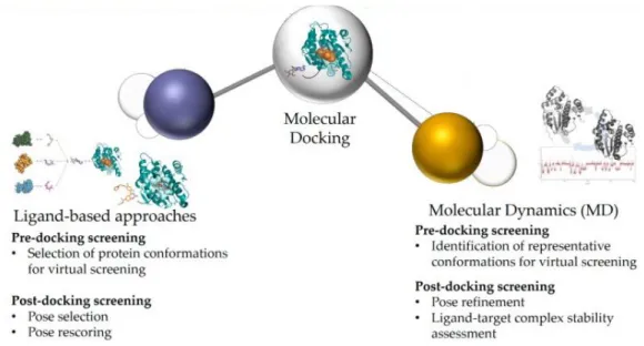
Although molecular docking has been used for a long time as a standalone method for drug design, it is now often integrated into workflows with other computational methods, including MD simulations (Fig. 01). The former is often based on the selection of suitable protein conformations for the docking screening, incorporating structural information and physicochemical properties of the chemical scaffold to select the most suitable poses of the ligand resulting from the molecular docking (Fig. 01). In MD simulation, the flexibility of residues in the binding pocket as well as the potential conformational changes are investigated, greatly improving the results of the virtual studies (Fig. 01) (7).
Fig. 01. Computational methods based on Molecular docking can be divided in ligand-based and
structural-based approaches and in particular the MD simulations is the most suitable example for the former. [Adapted from Pinzi L, et al. (7)].
The so-called omics branch of science is specifically aimed at characterizing and quantifying large pools of biomolecules translating this information into structures, functions and dynamics. Examples of -omics disciplines are genomics, proteomics and glycomics which have as the aim to study the genome, proteins and glycans, respectively. In particular, the latter is the main object of this work. Glycomics, the science which explores the role of carbohydrates in biological processes, is assuming an increasing importance due to the discovery that a large number of biological phenomena are encoded in glycans structures (or glycocode) and interactions (8).
Glycans, also called carbohydrates, consist of a large family of biomolecules which comprises glycosaminoglycans (GAGs), oligosaccharide chains that can exist in a free, soluble form or covalently attached to proteins or lipids (glycoproteins, proteoglycans and glycolipids, respectively). They are widely expressed and involved in a wide range of physiological and pathological processes. Of particular interest are carbohydrate–protein interactions, which regulate biological phenomena such as signal transduction, protein oligomerization, concentration of protein at the cell-surface and viral infection (9). Thus, correct glycosylation patterns are essential for the normal function of organisms while aberrant glycosylation is associated with many human diseases (10).
Clarifying the role of glycans in these processes is a major challenge owing to various factors. First of all, the complexity of glycans biosynthesis. These pathways are not under direct control of genes being rather governed by different enzymes and affected by multiple factors such as the cellular type on which are expressed and the composition of the surrounding environment (11). Again, the structural complexity of glycans is also tricky. These biomolecules can be divided from a structural point of view in non-linear polymers and linear long polyanionic chains, both characterized by distributions of well-defined conformational states rather than robust secondary or tertiary structures, as in case of proteins and nucleic acids. It derives that the main challenge of structural glycoscience is related to the characterization of structural basis and dynamic features of glycans along with the understanding of their behaviour and mechanistic and/or allosteric effect associated with the protein-glycans interactions. Furthermore, some glycans, as in case of the glycoproteins and GAGs, are carriers of information that can be depicted only secondary to their interaction with other biological macromolecules. In this context, it is imperative that spatial and dynamic properties of sugars have to be accurately determined to clarify the relationship between their structure and function.
Unfortunately, experimental structures collected by X-ray crystallography and NMR are limited because of the inherently difficult to crystallize these biomolecules. Indeed, NMR has been used to characterize the dynamics of glycans in solution but with restricted results due to the flexibility of the glycosidic linkage and the limit of molecular weight constraints (12). Also, X-ray crystallography allows to solve only the smaller systems (i.e. the first monosaccharides covalently attached to proteins) due again to the conformational flexibility of glycans that makes their crystallization very difficult. Finally, prediction of glycans sequence and structure based only on computational methods remains a largely unsolved problem.
In conclusion, the progress made in algorithms and computational power allows MD simulations of glycans but the lack in experimental structures of sugar themselves and in interacting with proteins demands strenuous effort to overcome these limits and clarify the biological phenomena.
1.2. Molecular models techniques applied to study the
glycan-protein interactions
1.2.1. Molecular docking
As mentioned above, collecting crystal structures of proteoglycans or protein-glycans complexes is still a challenging task. On one hand, these interactions often occur in the range of microseconds, as in the case of enzymatic reactions, being thus difficult to crystalize the complex in the exact moment in which the reaction occurs; on the other hand, those interactions occur in a specific microenvironment and/or biological settings which are hardly reproducible in x-ray or NMR conditions. It derives that in absence of experimental data, the current molecular docking software, which has not indifferent limitations predicting the protein-glycan interactions, become indispensable to fulfil the tasks proposed in the following works. Hereafter a description of how molecular docking prediction works.
Molecular docking is a computational method that predicts the favoured complex conformation between biomolecules (ligand-receptor) when these are bound to form a stable complex (13). A single biomolecule is described by its dihedral angles, bond distance and bond angles which defines the geometry and its overall structure. Thus, a unique set of these coordinates defines a specific position of each atom within the biomolecule and a subsequent overall of the three-dimensional structure. However, complexes, as in case of protein-glycan systems, are also regulated by additional forces which act both in the catalytic or active site as well as non-specific ones that interact out of the binding site, increasing the complexity of a correct prediction. Another obstacle to this prediction is the flexibility of glycans and of binding sites. The former is related to the already mentioned 1-4 glycosidic linkage between the monosaccharides. The latter is due to possible fitting of the binding site upon its interaction with the sugar. This flexibility, also defined as “conformational change”, is of importance in case of interaction between protein and strong polyanionic chains such as heparin or heparan sulfate (HS) in particular (better detailed in Chapter 2). Finally, although the release of the first docking software specifically dedicated to study glycans interactions such as Vina-Carb (14), HADDOCK (15) or ClusPro (16), improved the quality of the computation of the force field and potential energy by which molecules are described.
To date, the available docking algorithms are basically split into two main groups: the searching algorithms and the scoring algorithms. The formers create an ensemble of possible conformations of the ligand considering the molecule as a dynamic structure and are usually based on a linear combination of many structures or on genetic algorithms (GA) that generate new conformations as they move along the structures or the GA. The latter are based on scoring functions able to evaluate each different conformation of the system and to provide a value that describes the
energyof the system at a given conformation. Low energies indicate better, more stable interactions. Although each docking software is implemented based on the above-mentioned specific algorithms, all of them rank and select the best pose based on its conformation, orientation and translation.
AutoDock software is the docking software used in the following works because implemented with the most suitable force field to compute the glycans flexibility. AutoDock is based on a GA used to find the global minima. This algorithm describes each ligand state as a genotype whereas the corresponding atomic coordinates as a phenotype. Moreover, the fitness is calculated using the energy function and defines the total interaction energy of the complex. Offspring generations of ligand conformations are generated through crossover, by inheritance of genes and additional mutations from either the parents. Obviously, selection of the offspring conformations is based on the fitness value (17).
In a docking scheme, two issues must be considered: the accuracy of the prediction and efficiency of the calculation using reasonable resources and time. AutoDock 4.2 uses two techniques to meet these requirements, namely a rapid grid-based energy evaluation method combined with an efficient search algorithm for torsional degrees of freedom, which gives the specific advantage to predict the glycans conformations (17). In order to evaluate the energies in a rapid way, the affinity potentials for each atom type of the ligand are pre-calculated in AutoDock, where the protein is put in a grid, the size of which is determined by the size of the ligand and can be changed manually according to practical needs. Each grid point stores the affinity potentials for all the atom types of a ligand with the protein. Then, in an AutoDock job, the interaction energy of a particular ligand conformation with the protein target is calculated using the values from the grids.
The search for the ligand conformation related to the torsional degree of freedom is carried out with the search methods implemented in AutoDock. At present, the most efficient search method is the Lamarckian GA, which uses the already described GA in association with the local search algorithm to achieve efficient global phase space coverage and local search optimization. AutoDock 4.2 uses a semi-empirical scoring function to estimate protein-ligand binding free energies that is based on the combination between the classical force fields with empirical data to rapidly rank the candidates (17). In this way, mechanistic approaches are used to compute the enthalpic contributions while empirical approaches are used for solvation free energy and conformational entropy. Finally, the empirical parameters are taken from fitting to known complexes collected through experimental data (17). The protein and ligand molecules start in unbound conformations and form a bound complex after docking, whose binding free energy (∆G) is calculated as follows:
where V stands for the potential energy, P represents the protein and L the ligand, and ∆S conf is the entropic charge upon ligand binding. Each potential energy term includes the van der Waals, hydrogen-bond, electrostatics, and desolvation energies:
The weights (Wvdw, Whbond, Welec, and Wsol) are obtained by fitting experimental binding affinity data. The first term is a classical 12/6 potential for Lennard-Jones interactions. A ij and Bij are parameters for repulsive and dispersion interactions, respectively. The second term is a hydrogen bond term using a 12/10 potential, and Cij and Dij are obtained by fitting to the typical data of hydrogen bonds in the experiment. The term E(t) represents the directionality of the hydrogen bond, and t is the deviation from ideal hydrogen bond geometry. The third term is the electrostatic potential with an apparent dielectric constant ɛ(rij). The last term is an empirical desolvation energy calculated using the volume (V) surrounding a specific atom, a weighted solvation parameter (S), and is a distance-weighting factor σ. Finally, the entropy changes upon ligand binding (∆Sconf) can be estimated from the number of active torsions of the ligand (Ntors) with an empirical parameter Wconf:
Based on all these considerations, AutoDock seems to be really promising in the field of glycomics. Nevertheless, all that glitters are not gold. As already said, AutoDock was designed as an automated docking software for predicting the interactions between flexible ligands and proteins. Thus, the premises are really good but the concept of flexible ligand is underestimated if we consider glycans structure. This is because the algorithm is set to allow at most 32 free torsions, which are good in case of small molecules but if we consider linear tetrameric structures of glycans, they cover 28 of the 32 total free torsions, giving the idea of the limitations of the method and the exigence to implement new methods to overcome the limitations.
1.2.2. Molecular Dynamics (MD) simulations
The history of MD simulations started more than 60 years ago, when in 1957 Ander and Wainwright carried out the first simulation in the context of phase transition in a system of hard spheres but we need to wait until 1977 for the first MD of protein (18). And, slowly, the groundwork that allows to carry out the simulations resulted in the achievement recognized by the 2013 Nobel Prize in Chemistry (19). From there on, MD simulations have become more popular and visible. This
remarkable improvement is in part due to the increased number of available structures in the Protein Data Bank in association with the implementation of computer technologies, such as the high performance computing (HPC), the improvement of supercomputer and the introduction of Graphic Processing Units (GPUs) that allow an easier handling of the techniques and decrease the computing time for the simulations.
MD simulation method consists in solving coupled equations of motion numerically for a system in which the molecules move at a constant velocity. The solution results in a trajectory of the biomolecule, from which thermodynamic and dynamic properties of the system can be extracted by statistical mechanical relationship. Importantly, the reliability of the method for predicting the behaviour of a system depends on the assumptions used to describe the interactions of the system.
In this context, the interest in applying MD simulations to the glycans is increasing and associated with the peculiar features of these biomolecules. The exigence of applying MD simulations is due to the lack of crystal structures as well due to the possibility of pinpoint the dynamic behaviour of molecules which are characterized by a cloud of conformations more than a single secondary or tertiary structure. At this regard, Franck and co-workers highlighted the importance of MD simulations in this field carrying out a simulation of Human Immunodeficiency Virus (HIV) -gp120 glycoprotein fully glycosylated, showing for the first time the cloud trend assumed by the N-glycans covalently attached to the protein and demonstrating the shielding capability of the sugars as a mechanisms adopted by viruses to escape the immunity defences (20). More recently, MD simulations have been instrumental in demonstrating the same shielding effect by Casalino and co-workers for the Severe Acute Respiratory Syndrome Coronavirus 2 (SARS-CoV2) virus. MD simulations results have also suggested promising molecular targets to design specific vaccines and small molecules (21).
Hereafter are described the main features on which MD simulations are based to describe systems as realistic as possible.
Describing the systems with molecular dynamics. Considering a system of N particles, this is characterized by a set of atomic positions (R={R1,R2...RN} and relative momenta (P={P1,P2...PN}). Each atom can be seen and represented as a single point in a 6N multidimensional space, called phase space (Γ). Thus, a single point in the phase space represents a microscopic state of the system, while a collection of points in the Γ defines an ensemble. Using MD simulations we can generate a time sequence of points in the phase space, i.e. a sequence of different positions and momenta of the system belonging to the same ensemble. For each microscopic state of the system in the phase space, it is possible to estimate the observable value of a certain property A as a function of Γ, A(Γ), as the ensemble average or thermodynamic average:
where ρΓ is the probability distribution function of collection of points Γ, and δΓ = dR1...dRNdP1...dPN.
The thermodynamic state of the system is defined by parameters as the number of particles N, volume V, temperature T and pressure P. Based on these parameters, the probability distribution function has the form of the Boltzmann distribution function:
where H(Γ) is the classical Hamiltonian of the system defined as:
where R1, P 1and M1are the position, momentum and mass of the particle I, U is the potential energy, KB is the Boltzmann constant and Z is the canonical partition function.
On these basis, through the integration of the Newton’s equation of motion, we can estimate the evolution of the system as a function of time starting from its microstate at time 0 until its microstate at time τ. Along the MD simulations, a set of microstates of the system, i.e. a trajectory of points in the phase space Γ(t) is collected. According to the “ergodic hypothesis” and starting from a trajectory, we can calculate and connect the time average value of an observable ‹A›τ to‹A›obs(22). Basically, if the system will be able to evolve for an infinite time, it would be it would be able to sample all the
possible states and conformations and thus its behaviour averaged over time and over the phase space
become the same:
So, the longer is the simulation the better is the sampling of the states. Thus, the average behaviour of molecular biological systems can be predicted by estimating macroscopic observables using MD simulations. In this way, the Lagrangian equation of motion can be used to write the Newton’s equations of motion taking in consideration the variation of time:
where qk are the generalized coordinates and q ̣̇ k are their time derivatives. In addition, the Lagrangian function ℒ(q,q ̣̇ ,t) can be defined in terms of kinetic K(qq ̣̇ ,t) and potential Γ(qq ̣̇ ,t) energies:
For a system of atoms with Cartesian coordinates Ri the equation of motion described below yields:
where miand a i are the mass and acceleration of particle iand F iis the force of that particle. Thus, starting from a set of initial coordinates, as PDB crystal structures, NMR data, cryo-EM or homology modelling coordinates, and velocities randomly generated from the Maxwell-Boltzmann probability distribution at a given temperature T.
Force Fields in MD simulations . How atoms and molecules interact with each other is a central part for MD simulations. The potential energy of molecules is described as an empirical force field (FF) parametrized to reproduce the experimental data, which represent our knowledge of the potential energy surface of the system and are used to calculate the forces for propagating the dynamical systems. In classical MD simulations, the electronic degrees of freedom are not taken into account and atoms are considered as point particles simply approximated by using parametric functions of nuclear coordinates, the force field. Many force fields are available in the literature, such as CHARMM (Chemistry at Harvard Macromolecular Mechanics), OPLS-AA (Optimized Potentials for Liquid Simulations, All-Atom), AMBER (Assisted Model Building and Energy Refinement), and GAFF (General AMBER Force Field). Notably, in all the projects proposed this dissertation, I have employed AMBER force fields. Each of these force fields is a package that includes multiple parametric functions specifically set up for different biomolecules: protein, ions, water, nucleic acids, sugars. These force fields have similar basic functional form, representing bond stretching, angle bending, dihedral torsion, van der Waals, and Coulombic interactions for each molecule, and are described as:
where Utot is the total energy; k l, kθ and kχ are the bond, angle, and dihedral angle force constants, respectively; leq, θeq, and χ the bond length, bond angle, and dihedral angle, respectively, and the subscript eq represents the equilibrium values for the individual terms. Coulomb and Lennard-Jones (LJ) terms contribute to non-bonded interactions where εij is the LJ well depth and σij is the finite distance at which the inter-particle potential is zero, qi is the partial atomic charge, and rij is the
distance between atoms i and j . Due to this fact, LJ potential is considered a short-range potential and calculated using a cut-off of 10 Å (23).
MD simulations in explicit solvent consider the solvent explicitly, that is characterized by atoms connections between the individual water molecules. The water molecules are described by appropriate force fields. Actually, the most used family of water-models are the so-called fixed charge force fields, in which the fixed charges are assigned on the four fixed sites of the molecule: two charges are positive and simulate the hydrogen atoms, whilst the other two are negative and simulate the lone pairs. The four charges are arranged tetrahedrally about the oxygen (24). Within this family there are some of the most popular FF, for example TIP3P and TIP4P (25), which are widely used in the simulations of biological systems. In conclusion, if the essential physical interactions of molecules and/or particles are represented faithfully, explicit methods can reliably predict properties.
Glycans Force Field in MD simulations . Being the glycans the subject of my work, it is important to describe the force fields specifically developed for sugars, that are the main features characterizing glycans in MD simulations. Here a description of the GLYCAM_06 force field (26), used in this dissertation. Before the release of the GLYCAM_06 force field, the performance of several parameter sets, which included the first versions of GLYCAM (27), has been quantitatively evaluated against data from quantum mechanical calculations as well as on a relative basis. The conclusion was that all the parameters and force field evaluated perform similar results and no one was top ranked compared to the others. On a low note, the early version of GLYCAM used to simulate sugars in explicit solvent worked poorly in reproducing diffusion rate and differ substantially compared to the other force fields in predicting the putative radial pair distribution function (RPD) between hydroxyl groups and water model (TIP3P) (26).
The low results compared to the other parameters prompted the optimization and release of the GLYCAM_06 force field. At first, the structures of the monosaccharides were collected using experimental neutron diffraction with particular attention at the main features of the glycans such as the conformation or glycosidic linkage. In addition, the new force field package comprises new parameters such as bond and valence angle deformation force constants, dihedral angle rotational barriers, and electrostatic properties, which are collected with quantum mechanical (QM) calculations because of the difficulty or impossibility to calculate them with experimental data. Finally, the partial atomic charges were derived by fitting the QM molecular electrostatic potentials. In particular, this method starts from the general practice of assigning partial atomic charges to every atom in the molecule adopted (26) by not fitting partial charges to aliphatic hydrogen atoms. In addition the special treatment for the 1–4 non-bonded interaction was removed because it was redundant (26).
Periodic boundary condition. Consider a molecule arranged in a 10 × 10 × 10 water box. Nearly half the water molecules are on the outer faces of the box and would lie at the edge with the vacuum, generating artifacts due to finite-size effects of the system, and these will have a large effect on the measured properties. To avoid this problem, periodic boundary condition (PBC) can be applied. This method allows to ideally surround the central cube, the only one that will be explicitly treated, with infinite water boxes with the result of simulating an infinite solution system around the box (28). Finally, it’s important to bear in mind the periodicity of the system when considering properties which are influenced by long-range correlations. Fortunately, many algorithms have been implemented to overcome this problem and among them particle mesh Ewald method (PME) is the one adopted in the MD simulations carried out in this dissertation (29). The basic idea behind this technique is to split the relevant part of the potential into a short-range part, ordinarily treated within a cut-off, and a long-range term, in which the remaining interactions are Fourier transformed (30).
Temperature and Pressure. To carry out MD simulations as realistic as possible, the systems are described as microcanonical ensembles in which the number of particles (N), volume (V) and total energy (E) are kept constant. However, to obtain more accurate information that can be related to the experiments, MD simulations can be coupled with thermostat and barostat. The former allows to simulate canonical ensembles (NVT) in which the volume (V) and temperature (T) are kept constant. The latter maintains pressure (P) and temperature (T) stable (NPT ensemble). During this time, many algorithms have been implemented but I will briefly describe the two used for the simulations presented in this dissertation (30).
Langevin thermostat. In the Langevin thermostat the equation of motion is modified to maintain a stable temperature as following:
where Γ is a friction coefficient and Wiis a random force unrelated to time and across particles. The result is that the smaller particles create a damping force to the momenta when the large particles push the smallest out of the way. The smaller (thermal) particles also move with kinetic energy and give random kicks to the large particles, resulting in a random force that acts on all the particles and decreases their velocity using a constant friction. The Langevin thermostat is mostly used in the NVT-MD simulations carried out in my work (http://www.math.ucsd.edu/).
Berendsen thermostat. In the NPT ensemble, the pressure is controlled by coupling the system with a barostat. One of the main problems of the velocity-rescaling method is that it does not allow temperature fluctuations which are present in the canonical ensemble. To overcome this problem, Berendsen and co-workers introduce a first-order coupling barostat in conjunction with the
temperature control method. With this algorithm, the pressure of the simulated system is relaxed using a time constant toward the reference pressure (31). One advantage of the Berendsen thermostat is that it allows the temperature fluctuations, thereby not fixing it to a constant value. The motivation for the Berendsen thermostat is the minimization of local disturbances of a stochastic thermostat while keeping the global effects unchanged (http://www.math.ucsd.edu/).
1.2.3. Enhanced sampling techniques
Classical MD simulations have been shown as powerful tools to understand and predict the evolution of complex biological systems, but this method also has limitations. Using this technique is hard to sample conformational variations of small peptides or complex systems because it is hard to overcome high-energy-barriers to jump between the states with a reasonable amount of replicas and simulation time. Thus, enhanced sampling methods have been developed in recent years to accelerate rare events with notable success. These methods allow us to overcome the local-minimum states by enhancing the sampling as a function of one or a few predefined collective variables relevant for the event, increasing the probability of these events. Among these methods, replica exchange MD (REMD) simulation and the accelerate MD (aMD) were used in my work and are briefly presented hereafter.
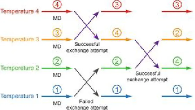
REMD. This technique allows to overcome high energy-barrier and to sample the conformation of peptides and proteins by combining MD simulations with the Monte Carlo algorithm (32). With this algorithm, several replicas of the starting system are simulated in parallel at different temperatures or at the same temperature but using different Hamiltonians at which the replica can
easily surmount the energy barriers. Periodically, the configurations of neighbouring replicas are
exchanged and this exchange is accepted by a Metropolis acceptance criterion that guarantees the
detailed balance (Fig. 02).
Fig 02. Illustration of replica exchange molecular dynamics (REMD) method.
The prerequisite for an efficient sampling is an adequate overlap of potential energy distributions between replicas. The overlap is large enough for satisfying acceptance ratios (10-50 %)
in case of systems with a small number of degrees of freedom and small temperature differences between the replicas. In this way, REMD is able to overcome the local minimum and successfully sample the conformational space through the exploration of the free energy landscape (33). REMD simulations are therefore restricted to systems comprising not more than a few thousand atoms and/or small peptides, typically in the context of peptide folding.
aMD. Hamelberg and co-workers proposed an aMD algorithm which is based on a bias potential function that can be used to overcome the local minimum without knowing other information about the potential energy neither the saddle point (28). With this algorithm, it is possible to sample the potential energy landscape using a bias potential added to the pre-existing one which allows to accelerate the time scale of the simulation. The representation of the potential energy landscape on the modified surface derives and is an expression of the bias potential, permitting it to successfully sample the potential energy minima. Using this bias potential it is possible to sample the conformational space more exhaustively than in the case of classical MD simulations, thus reaching the convergence of the system (34). aMD simulations demonstrate to be useful also for the sampling of systems composed of small peptides up to complete protein structures.
Based on all the considerations and information collected in this introductory chapter, we can conclude that glycomics studies are still in infancy beside the huge potential of the molecular model to sustain the experimental research. For this reason, the overarching goal of the studies reported in this work is the development of proper bioinformatics tools to allow the computational characterization of sugar-protein interaction and the prediction of their biological consequences, with the specific aim of merge the predicted results with experimental data to fulfil the specific objectives of the glycobiology.
Chapter
2.
Demystifying Heparan Sulfate-Proteins interactions
Proteoglycans are the most abundant and heterogeneous family of co-receptors expressed on the cell surface and in the extracellular matrix. Characterized by one or more polysaccharide chains (GAGs) covalently attached to the core protein, proteoglycans are involved in a variety of physiological and pathological processes, so that the comprehension of their mechanistic effects at cellular levels are required for the comprehension of many body functions and diseases.Heparan sulfate proteoglycans (HSPGs) is a subset of proteoglycans composed of a core protein on which are covalently attached long linear heparan sulfate (HS) polyanionic chains. Whether the structure of the core protein can affect the function of the HSPGs is not clear but is known that the core can directly interact with both intra- and extra-cellular protein. HSPGs are classified based on their localization and structure of the core protein. The main families are represented by HSPGs whose core is a trans-membrane protein with a short intracellular tail (Syndecans) or are anchored to the membrane through a glycophosphatidylinositol residue (Glypicans). Other HSPGs are instead secreted by cells and deposited in the extracellular matrix (collagen XVII, agrin and perlecan). Finally, serglycin are HSPGs found in the cytoplasmic secretory granules of endothelial, endocrine, and hematopoietic cells.
The long linear HS are composed by repetitive disaccharide units of β1–4-linked D-glucuronic acid (GlcA) or and α1–4-linked N-acetyl-D-glucosamine (GlcNAc) that undergo different modifications including the sulfation at specific positions to confer highly negative charge and binding-specificity. In particular, during the biosynthesis, HS can be subjected to a series of reactions processes: (i) deacetylation or sulfation of the GlcNAc (the most frequent at position C2 and C6 to GlcNS6S); (ii) epimerization of adjacent C5-GlcA to L-iduronic acid (IdoA); (iii) ester-linked sulfate groups at C2-IdoA (IdoA2S) or at C6-GlcA or more rarely at C3-GlcNAc. Interestingly, these modifications do not occur along all the HS at the same rate giving a rise of specific domain based on the sulfation: sections with variably sulfated domains (so-called NS domains), other with no modifications (the so-called NAc domains), other with a mix of NAc/NS domain located in the transition zone. Finally, at variance with proteins, HS are not synthesized under the direct control of genes, thus the synthesis of HS is not template driven and can greatly change among the different species and even cell types. What dictates the size and composition of HS in different cells at different times in development remains one of the great enigmas in modern cell glycobiology (9).
Relevantly, the terms HS and heparin are often used interchangeably but this is a simplification that needs an explanation. HS are synthesized by almost all eukaryotic cells, covalently attached to proteoglycans and their length can span from 50 to 250 disaccharides units (20-100kDa). Differently,
sequences (Fig. 03). Heparin can be found in two main states: unfractionated heparin (spanning from 12 to 14 kDa, ≃30 disaccharides) and low molecular weight heparin (spanning from 4 to 6 kDa, ≃15 disaccharides). Interestingly, the latter are extensively used as anticoagulant because the high pattern of sulfation also comprises the rare sulfation at 3-O-GlcNAc which specifically acts inhibiting the antithrombin III (35). Finally, due to the high degree of heterogeneity that characterizes the HS structure depending on their source, in both experimental and computational studies very often heparin is used as a “simplification” of HS.
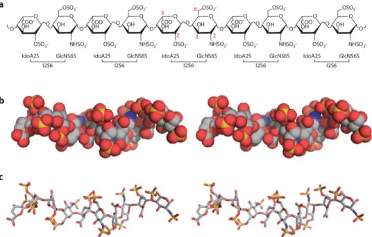
Fig. 03. (a) Heparin chemical structure. I2S6 is the most abundant disaccharide unit identified in heparin and is characterized by the sulfation at C2-IdoA and at N- and C6-GlcNac. The red numbers associated with the carbon atoms highlight the possible substitutions. ( b and c) Heparin crystal structures (PDBid 1E0O) are
represented in spheres and sticks, respectively. Both the structures are colored by elements: carbon, oxygen, nitrogen and sulfate atoms are colored in grey, red, blue and yellow, respectively. In both the structures it is possible to notice the helical nature of the heparin structure. Abbreviations: GlcNS6S, N-sulfo 6-sulfo Glc; IdoA2S, 2-sulfo L-iduronic acid. [Adapted from Xu D et al (9)].
HSPGs are involved in multiple biological phenomena through their capability to interact with a wide range of protein-partners, the so-called HS- or heparin-binding proteins. These proteins are characterized by the presence of basic domains, corresponding to sequences of basic residues that primarily interact with the negatively charged sulfated groups of HS/heparin. These basic motifs were identified for the first time by Cardin and Weintraub in 1989 and for this reason called Cardin-Weintraub motifs. The Cardin-Weintraub motifs, also known as heparin-binding domains (HBDs) because of the similarity between HSPGs and heparin, consist of specific sequences ‘XBBXBX’ and ‘XBBBXXBX’, where X represents an hydropathic residue and B a basic residue (R or K), which are responsible for the interactions with the sulfate groups of the HS (36). Through the
interactions with the heparin-binding proteins, HSPGs can regulate a plethora of biological processes including protein oligomerization (37), ligand/receptor interaction (38) and protein internalization (39) that can affect cell proliferation, adhesion and migration (40), angiogenesis (41) and viral infection (42).
Finally, HSPGs and heparin are able to induce conformational drifts secondary to the interaction with the heparin-binding proteins. For this reason, they carry information that can be depicted only pinpointing and characterizing the systems at molecular level. Unfortunately, a few number of crystal structures of short heparin/HSPGs and no crystal structure of long heparin/HS in complex with protein have ever been released. In addition, publications in which the dynamic behaviour of these systems is characterized are so far limited despite the importance of issue.
Thus, the detailed understanding of the HS and heparin-dependent processes is required to clarify both physiological and pathological processes at molecular level. For this reason, the first aim of my research, described here below in Sections from 2.1 to 2.3, has been directed to the implementation of proper bioinformatics tools to allow the computational characterization of protein-HSPGs/heparin molecular models and the characterization of the mechanistic effects related to their biological consequences.
2.1. Implementation of an incremental docking method to
characterize the heparin/HIV-1 p17 protein interaction
The results from this work has been collected in a published paper: Bugatti A*, Paiardi G*, Urbinati C, Chiodelli P, Orro A, Uggeri M, Milanesi L, Caruso A, Caccuri F, D’Ursi P and Rusnati M. Heparin and heparan sulfate proteoglycans promote HIV-1 p17 matrix protein oligomerization: computational, biochemical and biological implications. Scientific Reports. *Co-authorship. (2019) 9:15768. Supplementary information is available for this paper at https://doi.org/10.1038/s41598-019-52201-w.
2.1.2.
Introduction
Matrix protein p17 contributes to structural integrity of HIV virions and regulates viral replication (43). Also, p17 is released by HIV-infected cells, being detectable in the nanomolar range in plasma, brain and lymph nodes of patients treated with HAART (44). In its extracellular form, p17 deregulates the functions of B-lymphocytes (45). Importantly, p17 also induces endothelial cell activation and angiogenesis (46). With these multiple effects, p17 contributes to AIDS progression and to the pathogenesis of AIDS-associated diseases (47). HSPGs consist of a core protein and of heparin-like GAG composed of repeated disaccharides units of 2-O-sulfated L-Iduronic acid (IdoA) and N, 6-O-sulfated D-glucosamine (Glc) occasionally interrupted by non-sulfated uronic acids and under-sulfated hexosamines (48). At the surface of leukocytes, they act as receptors for extracellular p17. In the extracellular environment, free heparin released by mast cells sequesters p17, modulating its bioavailability (49). Besides p17, HSPGs act as receptors also for HIV-1 gp120 (50) and Tat (51) while free heparin promotes Tat oligomerization and biological activity (52). HSPGs act as co-receptor also for many other viral proteins and cytokines, promoting their oligomerization required for receptors clustering and activation (53).
p17 spontaneously oligomerizes forming trimers and even hexamers (54). Two different domains have been identified in p17 that specifically mediate its self-assembling: the C-terminus (ter) that self-interacts with the same region of other p17 molecules (55) and amino acids E 42 -N47, Q59 , Q63, that interact with the Q69-E74 region of another p17 molecule (56). On the other hand, due to the presence of “coiled coil” sequences, p17 tends to misfold, behaving as an “amyloidogenic” protein that forms toxic assemblies in the brain that have been demonstrated to contribute to AIDS-associated neurodegeneration (44).
Taken together, the capacity of p17 to oligomerize and to bind to heparin/HSPGs, along with the involvement of the GAG in the process of oligomerization of many cytokines prompted us to study the effect of heparin/HSPGs on p17 oligomerization and biological consequences by adopting a multidisciplinary approach including bioinformatics, biochemical and cell-based models.
analogue of HSPGs) and MD simulations have been limited only to short (di-, tetra- or hexa-) oligosaccharides, mainly due to heparin conformational flexibility and high charge density, the weak surface complementarity of heparin/ protein interactions, the absence of well-defined binding pockets, the difficulty to define the impact of solvation/desolvation, the large electrostatic interactions involved and the large number of torsional angles between glycosidic bonds (57). Only in selected cases, 14-mer heparin were used with molecular docking to study FGF, VEGF and CXCL8 dimerization (58-60). Relevant to this point, longer heparins or HS are found in nature that are responsible for cytokine oligomerization. With these premises, to corroborate our experimental data, we have here modelled heparin up to 24-mer and performed docking and MD simulations with p17 dimer.
2.1.2. Material and methods
Models: p17 and the heparin tetrasaccharide (4-mer heparin) were modelled as described (54). A 4-mer heparin probe with the deletion of the H atom of the hydroxyl groups at position 4 in the first IdoA and of the hydroxyl group at position 1 of the Glc (4-mer heparin modified probe) was prepared and used in docking simulations to promote the 1→4 glycosidic linkage. Two p17 mutants were modelled by the Alanine Scanning Method in which the lysine residues of both the N- and C-ter basic domains were replaced with alanine (N-ter K→A p17 and NC-ter K→A p17).
Heparin path identification:Blind docking simulations were performed by ClusPro web-server (16) using 4-mer heparin and N-ter K→A or NC-ter K→A p17 dimers to identify HBDs on p17 other than those in the N- and C-ter basic domains. The identified heparin probe sites were filtered by best score, cluster size, visual inspection and positioned in a dimer of wild type (wt) p17 to finally achieve an alignment and hence a traced heparin path.
Incremental docking and heparin modelling: The 4-mer heparin modified probe was used in local docking simulation along the traced heparin path in the wt p17 dimer by AutoDock 4.2 (17). The “sliding window method” was set up to create a sequence of overlapping sliding grids, each covering a whole 4-mer heparin and the last saccharide unit of the previous one. Local docking poses were filtered for free energy of binding, clusters size and correct orientation. The aligned 4-mer heparin modified probes were joined by 1→4 glycosidic linkages using Pymol. Gasteiger-Hückel charges were assigned to the sugar and then minimized using steepest descendent and conjugate gradient methods by Chimera (61), obtaining heparins of increasing length.
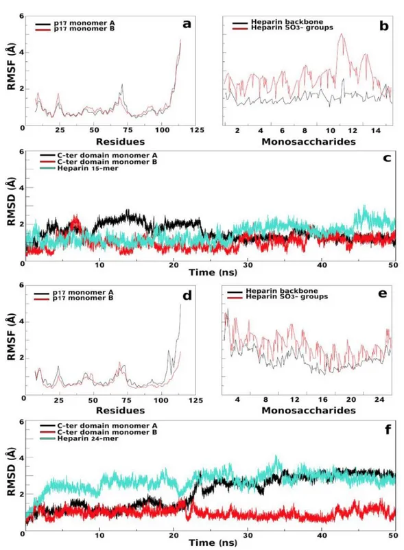
MD simulations:Amber14 package (62)was used for MD simulations of p17 dimer in complex with 15- and 24-mer heparins. MD simulations were carried out using f99SB force field parameters for protein and GLYCAM_06 for heparins. Each complex was neutralized by adding Ca 2+restrained
away from the protein (63)and solvated with TIP3P water model. Energy minimization was carried out with the non-bonded cut-off of 8Å through the following steps: (i) p17/heparin complexes and
counter ions were restrained by a harmonic potential of 5 kcal/mol×Å 2, while water molecules were
relaxed using 2,500 cycles of steepest descent and conjugate gradient methods; (ii) counter ions and hydrogens were relaxed using 5,000 cycles of steepest descent and conjugate gradient methods and restrained by a harmonic potentials of 3 kcal/mol×Å 2and then of 1 kcal/mol×Å 2; (iii) the system was
relaxed using 5,000 cycles of steepest descent and conjugate gradient methods without any restraint; iv) the system was heated from 0.1K to 100.0K in NVT (constant volume) and from 100.0 to 300.0K in NPT (1.0atm constant pressure). The two generated complexes were simulated in periodic boundary conditions using the Langevin algorithm at 300.0K. During heating and simulations, the Ca2+ ions were restrained at 500 kcal/mol×Å 2. Equilibration (3 ns) and simulation were validated
using the physical observables parameters of the system confirming that the complexes obeyed the NPT ensemble. Electrostatic interactions were calculated using the Particle Mesh Ewald method. A cut-off of 8.0Å was applied to van der Waals forces. Integration time step was set to 1.0 fs during equilibration and 2.0 fs during simulation. MD simulations were performed over 50ns using the pmemd CUDA program of the Amber14 package and a server Tesla K20 GPU.
2.1.3. Results
As already mentioned, computational docking of heparin (mostly used as a structural analogue of HSGPs) to its binding-protein still represents a challenge. Accordingly, the p17/heparin complex has been resolved only with a protein monomer and a 6-mer heparin, prompting us to conceive a new computational approach to study the interaction of longer heparins with p17 dimers (Fig. 04). The first step of this workflow consists in a blind docking simulation using ClusPro web server. The results of the simulations with 4-mer heparin probes and wt p17 dimer show that the tetra-saccharides always take position in the N-ter HBDs of the two monomers. On the other hand, using a N-ter K→A p17 dimer, the 4-mer heparin probes dock mainly in the C-ter basic domain.
Fig.04. Workflow of computational studies.
Thus, we used a NC-ter K→A p17 dimer to predict alternative heparin-binding pose, allowing the definition of two alternative shorter and longer heparin paths. In the shorter path, the succession of 4-mer heparin probes starts from the N-ter HBD of p17 monomer A and goes directly to the N-ter HBD of p17 monomer B (Fig. 05A). Also in the longer path the succession of 4-mer heparin probes starts from the N-ter HBD of monomer A but it makes then contact with H4, C-ter basic domain and H3 of monomer A and with H3, C-ter basic domain, H4 and finally N-ter HBD of monomer B (Fig. 05B). Subsequently, the sliding window and incremental docking methods were used to identify and join the best binding poses of each 4-mer heparin in the traced paths (Fig. 05C), predicting two heparin models: the 15-mer heparin, modelled on the shorter path, and the 24-mer heparin modelled on the longer path (Fig. 06). In the complexes formed by p17 dimer with the 15- and 24-mer heparins, H-bonds are mainly formed with SO3− or COO−. In the 15-mer heparin complex H-bonds occur
mainly within the N-ter HBD domain and α-helix 2 of both monomers (Fig. 06A and Tab. 01), while in the 24-mer heparin complex they occur in all the helices of both monomers and in the C-HBD (Fig. 06B and Tab. 02).
Fig.05. Shorter (a) and longer (b) heparin path identification by ClusPro web server. Position of the 4-mer heparin probes (in sticks) obtained by blind docking on the wt p17 dimer are in green, those obtained on N-ter K→A or NC-ter K→A p17 dimers are in magenta and yellow. p17 monomers are represented in grey cartoons. (c) Schematic representation of the sliding window method. Different regions composing the p17 protein are shown (H, α-helices; L, loops). The grid box of the first docked probe is in green, the second and third boxes, covering a whole 4-mer heparin and the last monomer of the precedent probe are in yellow and magenta, according to the heparin probes color. p17 monomers are represented as cartoons. For each heparin probe, in sticks, the atoms involved in 1→4 glycosidic linkages are represented as spheres.
Fig.06.Electrostatic interactions. Docking prediction (lef) and complex stability after MD simulations (right) of 15-mer (a) or 24-mer (b) heparin (yellow and green sticks for 15- and 24-mer) in complex with p17 dimer (represented as electrostatic surface). Zoom insets: interactions before and after MD simulations are reported. P17 amino acids and monosaccharides involved in the networks are in sticks, blue and yellow.
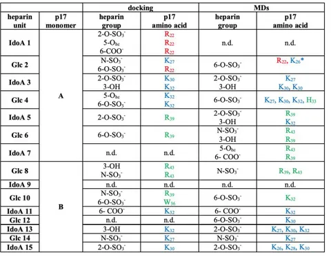
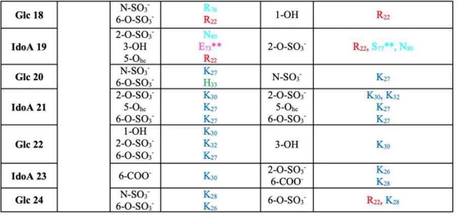
Tab. 01. H-bond analysis in the 15-mer heparin/p17 dimer complex from docking or MD simulations. The 15-mer heparin makes stable contact with amino-lysine and guanidine-arginine groups of the residues R 22
(87%), K26 (99%), K27 (99%), K32 (95%), R39 (97%), R43 (93%) of p17 monomer A and with K 26(98%), K27
(98%), K28(87%), K30 (86%), K32 (92%) of p17 monomer B. In brackets are the average persistency of the
different H-bonds. P17 amino acids in loop 1 are in red, those in the N-ter HBD in blue and those in the -helix 2 in green. IdoA: 2-O-sulfated L-iduronic acid. Glc: N, 6-O-sulfateD-Glc. Hc: heterocyclic. n.d.: not defined. *H-bond with heparin is mediated by the N atom of the peptide bond. In all the other cases, N atoms of the side chain are instead involved.