The copy number variation landscape of congenital
anomalies of the kidney and urinary tract
Miguel Verbitsky
1,51, Rik Westland
1,2,51, Alejandra Perez
1, Krzysztof Kiryluk
1, Qingxue Liu
1,
Priya Krithivasan
1, Adele Mitrotti
1, David A. Fasel
1, Ekaterina Batourina
3, Matthew G. Sampson
4,
Monica Bodria
5, Max Werth
1, Charlly Kao
6, Jeremiah Martino
1, Valentina P. Capone
1, Asaf Vivante
7,8,
Shirlee Shril
7, Byum Hee Kil
1, Maddalena Marasà
1, Jun Y. Zhang
1, Young-Ji Na
1, Tze Y. Lim
1,
Dina Ahram
1, Patricia L. Weng
9, Erin L. Heinzen
10, Alba Carrea
5, Giorgio Piaggio
5, Loreto Gesualdo
11,
Valeria Manca
12, Giuseppe Masnata
12, Maddalena Gigante
11, Daniele Cusi
13, Claudia Izzi
14,
Francesco Scolari
15, Joanna A. E. van Wijk
2, Marijan Saraga
16,17, Domenico Santoro
18, Giovanni Conti
19,
Pasquale Zamboli
20, Hope White
1, Dorota Drozdz
21, Katarzyna Zachwieja
21, Monika Miklaszewska
22,
Marcin Tkaczyk
23, Daria Tomczyk
23, Anna Krakowska
23, Przemyslaw Sikora
24, Tomasz Jarmoliński
25,
Maria K. Borszewska-Kornacka
26, Robert Pawluch
26, Maria Szczepanska
26, Piotr Adamczyk
26,
Malgorzata Mizerska-Wasiak
27, Grazyna Krzemien
27, Agnieszka Szmigielska
27, Marcin Zaniew
28,
Mark G. Dobson
29,30, John M. Darlow
29,30, Prem Puri
30,31, David E. Barton
29,32, Susan L. Furth
33,
Bradley A. Warady
34, Zoran Gucev
35, Vladimir J. Lozanovski
35,36, Velibor Tasic
35, Isabella Pisani
37,
Landino Allegri
37, Lida M. Rodas
38, Josep M. Campistol
38, Cécile Jeanpierre
39, Shumyle Alam
40,
Pasquale Casale
40,41, Craig S. Wong
42, Fangming Lin
43, Débora M. Miranda
44, Eduardo A. Oliveira
44,
Ana Cristina Simões-e-Silva
44, Jonathan M. Barasch
1, Brynn Levy
45, Nan Wu
46,47,
Friedhelm Hildebrandt
7, Gian Marco Ghiggeri
5, Anna Latos-Bielenska
48, Anna Materna-Kiryluk
48,
Feng Zhang
49, Hakon Hakonarson
6, Virginia E. Papaioannou
50*, Cathy L. Mendelsohn
3*,
Ali G. Gharavi
1* and Simone Sanna-Cherchi
1*
Congenital anomalies of the kidney and urinary tract (CAKUT) are a major cause of pediatric kidney failure. We performed a genome-wide analysis of copy number variants (CNVs) in 2,824 cases and 21,498 controls. Affected individuals carried a sig-nificant burden of rare exonic (that is, affecting coding regions) CNVs and were enriched for known genomic disorders (GD). Kidney anomaly (KA) cases were most enriched for exonic CNVs, encompassing GD-CNVs and novel deletions; obstructive uropathy (OU) had a lower CNV burden and an intermediate prevalence of GD-CNVs; and vesicoureteral reflux (VUR) had the fewest GD-CNVs but was enriched for novel exonic CNVs, particularly duplications. Six loci (1q21, 4p16.1-p16.3, 16p11.2, 16p13.11, 17q12 and 22q11.2) accounted for 65% of patients with GD-CNVs. Deletions at 17q12, 4p16.1-p16.3 and 22q11.2 were specific for KA; the 16p11.2 locus showed extensive pleiotropy. Using a multidisciplinary approach, we identified TBX6 as a driver for the CAKUT subphenotypes in the 16p11.2 microdeletion syndrome.
C
AKUT has a devastating impact on childhood renal sur-vival1–4. A better understanding of the pathogenesis ofCAKUT is imperative to improve the prognosis of affected children5. CAKUT encompasses a broad spectrum of phenotypes,
which can result from early disruptions in transcription factors and signaling molecules, such as PAX2, EYA1, RET, BMP4 (refs. 5,6) and
others, or are directly related to spatiotemporal interactions of the outgrowing ureteric bud and metanephric mesenchyme7–9. Early
disturbance of these interactions leads to renal agenesis, hypopla-sia or hypodysplahypopla-sia, whereas later perturbations in the outgrowth
A full list of affiliations appears at the end of the paper.
of the ureteric bud result in obstructive uropathy (OU), vesicoure-teral reflux (VUR) or ectopic or horseshoe kidney (EK-HK)6,10–13.
Maldevelopment of the lower urinary tract can result in epispa-dias or hypospaepispa-dias (LUTM) or posterior urethral valves (PUV)14.
Genetic manipulation in mice has indicated that disruption of the same cellular pathways can lead to multiple different genitouri-nary phenotypes15–19. Similarly, mutations in genes associated with
Mendelian forms of CAKUT can lead to different subphenotypes in individuals from the same families18,20,21, thus suggesting that a
single genetic lesion can have pleiotropic manifestations across the
0.10 0.25 0.50 1.00 2.50 5.00 10.00 25.00 50.00 0.0 2.5 5.0 7.5 10.0 12.5 15.0 17.5 20.0
Largest CNV per genome (Mb)
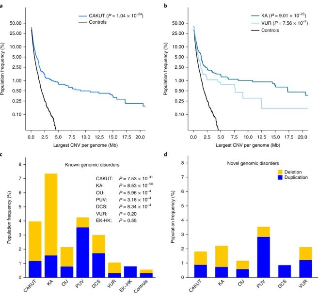
P opulation frequency (%) CAKUT (P = 1.04 × 10⁻²⁴) CAKUT: P = 7.53 × 10–41 KA: P = 8.53 × 10–50 OU: P = 5.96 × 10⁻⁴ PUV: P = 3.16 × 10⁻⁴ DCS: P = 8.34 × 10⁻⁴ VUR: P = 0.20 EK-HK: P = 0.55 Controls a 0.10 0.25 0.50 1.00 2.50 5.00 10.00 25.00 50.00 0.0 2.5 5.0 7.5 10.0 12.5 15.0 17.5 20.0
Largest CNV per genome (Mb)
P opulation frequency (%) KA (P = 9.01 × 10⁻²⁵) VUR (P = 7.56 × 10⁻⁷) Controls b
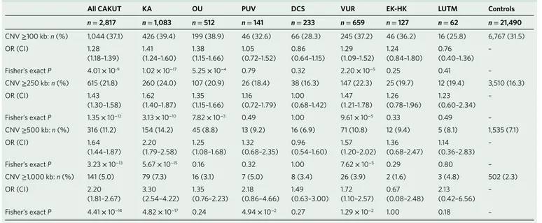
Known genomic disorders
0 1 2 3 4 5 6 7 8 CAKUT KA OU PUV DCS VUR EK−HK Contr ols P opulation frequency (% ) c
Novel genomic disorders
0 1 2 3 4 5 6 7 8 CAKUT KA OU PUV DCS VUR P opulation frequency (% ) Deletion Duplication d
Fig. 1 | Burden of rare copy number variants in CAKuT cases compared with controls. a,b, Burden of large, rare, exonic CNVs in all CAKUT cases and controls (a) and in KA and VUR cases and controls (b). c,d, Prevalence of known genomic disorders (c) and novel likely pathogenic CNVs (d) in CAKUT cases and controls. Deletions are marked in yellow; duplications are marked in blue. KA, OU, PUV and DCS were significantly enriched for genomic disorders. Deletions predominated in KA, whereas duplications were more frequent in PUV and DCS. P values are shown for comparison between cases and controls.
spectrum of CAKUT. Conversely, differences in the prevalence and severity of structural malformations point toward a distinct molecular basis and genetic architecture22,23. To date, there are
more than 50 single-gene disorders known to underlie isolated and nonisolated (that is, syndromic) CAKUT5,24,25. Furthermore,
a substantial number of CAKUT patients carry copy number variants (CNV) that were previously associated with a syndrome diagnosis or are large and extremely rare in the general popula-tion26–28. Nevertheless, a molecular diagnosis can be established
in less than 20% of affected individuals15–17,29–32, thus
emphasiz-ing that large studies across the entire phenotypic spectrum of CAKUT are indispensable to identify genes and allelic variants that either are specific to subcategories of disease or have pleio-tropic effects across the entire genitourinary tract, and to discover novel cellular pathways that are implicated in kidney and urinary development.
Here, we show the presence of a distinct genetic architecture as well as pleiotropic mutations for the different subphenotypes of CAKUT. Our CNV analysis of nearly 3,000 cases across the pheno-typic spectrum of CAKUT sheds light on the genomic architecture
of disease and implicates TBX6 as a main driver for the various CAKUT phenotypes in the 16p11.2 microdeletion syndrome.
Results
Burden of rare CNVs is high in CAKUT. We conducted a study in 2,824 CAKUT cases and 21,498 population controls (Supplementary Tables 1 and 2 and Supplementary Fig. 1) to compare the prevalence of rare CNVs that intersect genes. The case cohort represented common CAKUT subcategories: kid-ney anomalies (KA; including renal agenesis, hypoplasia, dys-plasia and multicystic dysdys-plasia), vesicoureteral reflux (VUR), obstructive uropathy (OU; including congenital hydronephro-sis, ureteropelvic junction obstruction, ureterovesical junction obstruction and congenital megaureter), duplicated collecting system (DCS; including duplications of the ureter or kidney, par-tial and complete), posterior urethral valves (PUV), ectopic kid-ney or horseshoe kidkid-ney (EK-HK), and other lower urinary tract malformations (LUTM; including anomalies of the bladder and anterior urethra). Our analysis focused on large (≥ 100-kb) CNVs that are present in fewer than 1:1,000 population controls across
Further annotation identified 54 large, rare, exonic CNVs in an additional 47 CAKUT cases (1.7%) that fulfilled ACMG criteria35,36
as likely pathogenic imbalances (Methods and Supplementary Table 11). Among these, the CNVs classified as pathogenic were an atypical deletion at the 16p11.2 locus (described below), a 300-kb deletion involving PAX2, a 100-kb duplication containing TBX18, a 571-kb deletion spanning PBX1 and a 6.8-Mb duplication includ-ing BMP4 (Supplementary Fig. 8a–d). We also identified overlap-ping duplications at the 15p11.2 locus in five cases with ureteric defects (VUR, OU) and PUV, as well as two KA cases with deletions proximal to the 16p11.2 microdeletion syndrome. In line with the burden test results, the distribution of deletions and duplications at known and novel GD-CNV loci (Fig. 1c,d) was significantly dif-ferent among CAKUT categories (6 × 2 Fisher’s exact test P = 8.93 × 10⁻⁵). In particular, subjects with KA and OU were enriched for
deletion syndromes, contrary to PUV and DCS, which showed an excess of duplications at the same genomic loci.
When we examined associations with disease severity, cases with a known GD-CNV were more likely to have multiple sites of the urinary tract affected (OR 1.60, 95% CI 1.05–2.42; P = 0.02) and more frequently harbored extrarenal malformations (OR 4.79, 95% CI 3.21–7.17; P = 6.00 × 10⁻¹⁵) than did cases without a known
GD-CNV. However, in agreement with the burden tests conducted above, analysis of simplex isolated cases still showed a greater burden than that of controls (OR 3.12, 95% CI 2.06–4.61; P = 1.86 x 10⁻⁷),
thus strongly implicating the importance of GD-CNVs in milder forms of CAKUT. To test more complex genetic models for modes of disease determination, we examined cases and controls for second-site CNVs. Among the 159 cases with GD-CNVs (112 known, 47 new/likely pathogenic, altogether called ‘diagnos-tic CNVs’ (DCNVs)), 11 (6.9%) carried more than one DCNV (Supplementary Table 12). In cases, the presence of zero, one or more than one DCNV increased the likelihood of extrarenal malformations (chi-square test for 3 × 2 table: P = 6.94 x 10⁻⁷;
Supplementary Fig. 9).
KA, OU and VUR show distinct genomic characteristics. A com-parison of CNV landscapes between the largest CAKUT subcat-egories revealed both commonalities and differences between KA, OU and VUR (Table 2). We found significant CNV enrichment for all three phenotypes, which was most uniformly shown by a larger different ancestries, as estimated by principal component analysis
(Supplementary Fig. 2).
This analysis revealed a marked enrichment for large, rare CNVs in CAKUT compared with controls (P = 1.04 × 10⁻²⁴; Fig. 1a), which
was consistent across virtually all metrics examined, including the number of individuals with large, rare CNVs, the median size and total CNV span per genome, and the fraction of GD-CNVs, thus indicating an important role for gene-disrupting CNVs across the entire CAKUT spectrum (Table 1 and Supplementary Table 3). This signal was driven by cases with and without extrarenal manifesta-tions, because the burden difference was still highly significant in analysis of cases without extrarenal defects separately (P = 3.21 × 10⁻⁸; Fig. 1a,b and Supplementary Figs. 3 and 4). Even when only
simplex isolated CAKUT cases (that is, cases without extrarenal manifestation and with no additional CAKUT phenotypes other than the primary one) were considered, there was still an excess burden of large, rare CNVs compared with controls (P = 1.15 × 10⁻⁸;
Supplementary Fig. 5). Comparison of burden metrics for cases and controls indicated a population attributable risk of 4.1% for large, rare CNVs ≥ 500 kb in CAKUT (odds ratio (OR) 1.64, 95% confi-dence interval (CI) 1.44–1.87; P = 3.23 × 10⁻¹³). This excess burden
is predominantly attributable to exonic deletions, most prominently in the KA cases (Supplementary Tables 4 and 5). Interestingly, we observed an enrichment for rare duplications compared with dele-tions in VUR and PUV cases compared with controls (P = 3.33 × 10⁻² and P = 8.67 × 10⁻⁴, respectively; Supplementary Fig. 6).
Secondary analysis also showed enrichment of the number of genes per individual genome that were affected by rare CNVs for nearly all CAKUT subcategories (Supplementary Table 6 and Supplementary Fig. 7).
Genomic disorders inform about the genetic architecture of CAKUT. We cross-annotated all rare CNVs with a curated list of known GDs33,34 (Supplementary Table 7) and identified 45
distinct known GDs in 112 (4.0%) independent cases (Fig. 1c
and Supplementary Table 8). Five cases carried more than one known GD-CNV, resulting in a total number of 117 known GD-CNVs in our cohort (Supplementary Table 9); in compari-son, known GD-CNVs were found in only 134/21,498 (0.6%) population controls (OR 6.58, 95% CI 5.05–8.55; P = 7.53 × 10−41;
Supplementary Table 10).
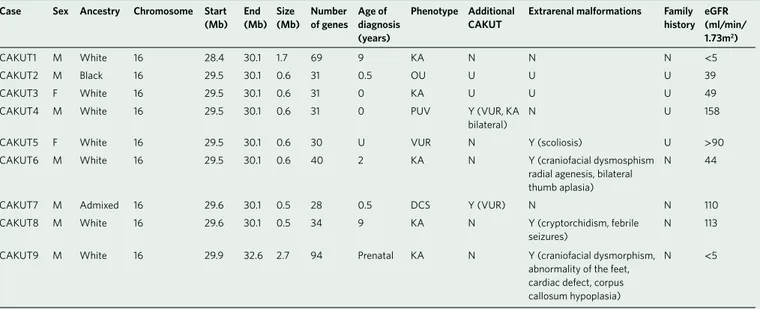
Table 1 | Distribution of largest, rare, exonic CNV per individual across different size thresholds
All CAKuT KA Ou PuV DCS VuR EK-HK LuTM Controls
n = 2,817 n = 1,083 n = 512 n = 141 n = 233 n = 659 n = 127 n = 62 n = 21,490 CNV ≥ 100 kb: n (%) 1,044 (37.1) 426 (39.4) 199 (38.9) 46 (32.6) 66 (28.3) 245 (37.2) 46 (36.2) 16 (25.8) 6,767 (31.5) OR (CI) 1.28 (1.18–1.39) 1.41 (1.24–1.60) 1.38 (1.15–1.66) 1.05 (0.72–1.52) 0.86 (0.64–1.15) 1.29 (1.09–1.52) 1.24 (0.84–1.80) 0.76 (0.40–1.36) – Fisher’s exact P 4.01 × 10⁻⁹ 1.02 × 10⁻¹⁷ 5.25 × 10⁻⁴ 0.79 0.32 2.20 × 10⁻⁵ 0.25 0.41 – CNV ≥ 250 kb: n (%) 615 (21.8) 260 (24.0) 107 (20.9) 26 (18.4) 38 (16.3) 147 (22.3) 25 (19.7) 12 (19.4) 3,510 (16.3) OR (CI) 1.43 (1.30–1.58) 1.62 (1.40–1.87) 1.35 (1.15–1.66) 1.16 (0.72–1.79) 1.00 (0.68–1.42) 1.47 (1.21–1.78) 1.26 (0.78–1.96) 1.23 (0.60–2.34) – Fisher’s exact P 1.35 × 10⁻¹² 3.13 × 10⁻¹⁰ 7.82 × 10⁻³ 0.49 1.00 9.61 × 10⁻⁵ 0.33 0.49 – CNV ≥ 500 kb: n (%) 316 (11.2) 154 (14.2) 45 (8.8) 13 (9.2) 16 (6.9) 71 (10.8) 12 (9.4) 5 (8.1) 1,535 (7.1) OR (CI) 1.64 (1.44–1.87) 2.20 (1.79–2.58) 1.25 (1.08–1.68) 1.32 (0.68–2.35) 0.96 (0.54–1.60) 1.57 (1.20–2.02) 1.36 (0.68–2.47) 1.14 (0.36–2.83) – Fisher’s exact P 3.23 × 10⁻¹³ 5.67 × 10⁻¹⁵ 0.16 0.32 1.00 7.62 × 10⁻⁵ 0.29 0.80 – CNV ≥ 1,000 kb: n (%) 141 (5.0) 79 (7.3) 16 (3.1) 7 (5.0) 8 (3.4) 26 (3.9) 2 (1.6) 3 (4.8) 502 (2.3) OR (CI) 2.20 (1.81–2.67) 3.30 (2.54–4.22) 1.35 (0.76–2.23) 2.18 (0.86–4.66) 1.49 (0.63–3.00) 1.72 (1.10–2.57) 0.67 (0.08–2.48) 2.13 (0.42–6.56) – Fisher’s exact P 4.41 × 10⁻¹⁴ 4.82 × 10⁻¹⁷ 0.24 4.94 × 10⁻² 0.27 1.29 × 10⁻² 1.00 0.18 – Proportion of individuals with their largest rare CNV at least as large as the indicated size threshold, comparing cases to controls.
indicating that novel syndromes account for the molecular basis of this disorder (Table 2, Fig. 1d and Supplementary Figs. 3 and 10). Finally, OU cases fell in an intermediate category, with CNVs that were not statistically larger than those of controls, yet the excess CNV burden (P = 0.01; Supplementary Fig. 3) was reflected by a greater proportion of individuals with large imbalances that were also more likely to be classified as pathogenic GD-CNVs (11 cases with GD-CNVs, OR 3.50, 95% CI 1.70–6.52; P = 5.96 × 10⁻⁴; Table 3
and Supplementary Fig. 3). These data suggest a distinct genomic architecture among CAKUT subcategories, with enrichment of GD-CNVs in KA and enrichment for novel, large- or intermediate-sized imbalances in VUR and OU, respectively.
Six GD loci account for 65% of CAKUT cases with known GD-CNVs. We conducted a literature search, including a survey of proportion of cases carrying exonic CNVs ≥ 100 kb than controls.
KA cases had the highest CNV burden (P = 9.01 x 10⁻²⁵; Fig. 1b), as
evidenced by median CNV size and total span, as well as the pro-portion of individuals with large imbalances that could be classified as pathogenic GD-CNVs (80 cases with GD-CNVs, OR 12.65, 95% CI 9.40–16.94; P = 8.53 × 10−50; Table 2 and Fig. 1c). After removal
of individuals with a known GD-CNV, the strength of the excess CNV burden in KA was still detectable but markedly attenuated (P = 1.27 × 10⁻³), in agreement with the major role for GD-CNVs in
the pathogenesis of KA (Supplementary Fig. 10). In contrast, VUR cases were also affected by a high CNV burden (P = 7.56 × 10⁻⁷),
but these CNVs predominantly involved duplications and were less likely to be classified as GD-CNVs (seven cases with GD-CNVs, OR 1.71; 95% CI 0.67–3.64; P = 0.20; Fig. 1c). The large rare CNVs in VUR were mostly classified as likely pathogenic, thus potentially
Table 2 | Comparison of CNV landscapes across major CAKuT subcategories
CAKuT (n = 2,824) KA (n = 1,088) Ou (n = 512) VuR (n = 660) Controls (n = 21,498)
Median CNV size (kb) (IQR)a 245 (315) 258 (394) 223 (244) 248 (273) 223 (248)
P (Wilcoxon) 3.4 × 10⁻⁶ 3.6 × 10⁻⁶ 0.87 1.9 × 10⁻² –
Median total CNV span (kb) per genome (IQR)a
350 (598) 414 (815) 306 (451) 353 (503) 300 (437)
P (Wilcoxon) 1.4 × 10⁻⁷ 1.9 × 10⁻⁸ 0.61 1.7 × 10⁻² –
% individuals with at least one large, rare CNVsa
37.1 39.3 38.9 37.2 31.5
P (Fisher’s exact) 4.0 × 10⁻⁹ 1.0 × 10⁻⁷ 5.3 × 10⁻⁴ 2.2 × 10⁻³ – % individuals with at least two large,
rare CNVsa
10.5 12.6 10.4 9.7 8.1
P (Fisher’s exact) 1.6 × 10⁻⁵ 1.0 × 10⁻⁶ 7.1 × 10⁻² 0.12 –
% individuals with known GDs 4.1 7.4 2.1 1.1 0.6
P (Fisher’s exact) 7.5 × 10−41 8.5 × 10−50 6.0 × 10⁻⁴ 0.20 – Ratio of number of large, rare
deletions: duplicationsa
0.90 1.02 1.05 0.74 0.94
P (Fisher’s exact) 0.34 0.35 0.37 3.3 × 10⁻² –
CNVs here refer to autosomal, exonic CNVs ≥ 100 kb with a frequency of < 1:1,000 in controls. aMetrics derived from burden analysis. CAKUT cases were compared with controls. P values were derived
from the indicated tests.IQR, interquartile range.
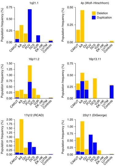
Table 3 | Clinical characteristics of CAKuT cases affected by the heterozygous 16p11.2 microdeletion
Case Sex Ancestry Chromo some Start
(Mb) End (Mb) Size (Mb) Number of genes Age of diagnosis (years)
Phenotype Additional
CAKuT Extrarenal malformations Family history eGFR (ml/min/ 1.73m2)
CAKUT1 M White 16 28.4 30.1 1.7 69 9 KA N N N < 5
CAKUT2 M Black 16 29.5 30.1 0.6 31 0.5 OU U U U 39
CAKUT3 F White 16 29.5 30.1 0.6 31 0 KA U U U 49
CAKUT4 M White 16 29.5 30.1 0.6 31 0 PUV Y (VUR, KA
bilateral)
N U 158
CAKUT5 F White 16 29.5 30.1 0.6 30 U VUR N Y (scoliosis) U > 90
CAKUT6 M White 16 29.5 30.1 0.6 40 2 KA N Y (craniofacial dysmosphism
radial agenesis, bilateral thumb aplasia)
N 44
CAKUT7 M Admixed 16 29.6 30.1 0.5 28 0.5 DCS Y (VUR) N N 110
CAKUT8 M White 16 29.6 30.1 0.5 34 9 KA N Y (cryptorchidism, febrile
seizures)
N 113
CAKUT9 M White 16 29.9 32.6 2.7 94 Prenatal KA N Y (craniofacial dysmorphism, abnormality of the feet, cardiac defect, corpus callosum hypoplasia)
N < 5
lesions result in specific CAKUT subphenotypes, whereas other genetic lesions, such as the 16p11.2 microdeletion syndrome, have high pleiotropic effects across the genitourinary tract. These loci provide a list of regions that are likely to encompass critical regula-tors of kidney and urinary tract development in humans, offering a unique opportunity for gene discovery.
Exome sequencing indicates haploinsufficiency underlying CNV deletions. To test the mechanism through which pathogenic CNVs confer risk for CAKUT, we conducted whole-exome sequencing (WES) in 23 patients with pathogenic microdeletions at 14 indepen-dent loci (Supplementary Table 13). On the basis of recessive loss-of-function (LOF) inheritance, WES would uncover a hemizygous LOF mutation on the nondeleted allele (unmasking effect). WES was performed as previously described18,23,54. We retrieved all
iden-tified candidate hemizygous LOF variants located within the case-specific deletion loci. Overall, we identified only one LOF variant in
EFCAB12 (p.Q437*; observed once in heterozygosity in the Exome
Aggregation Consortium database (ExAC) database55,56) in a patient
affected by unilateral KA and multiple extrarenal manifestations, including neurodevelopmental delay, epilepsy, corpus callosum agenesis, left radial bone agenesis and a patent ductus arteriosus, who had a 14.9-Mb deletion at chromosome 3q13.22–13.1 (ref. 57)
(Supplementary Fig. 11). The EFCAB12 variant was inherited from a heterozygous unaffected mother, whereas the CNV occurred de novo57 (Supplementary Fig.11). A query of 15,469 control
indi-vidual exomes at the Institute of Genomic Medicine at Columbia University did not reveal any homozygous or compound heterozy-gous truncating variants, thus suggesting that biallelic truncating mutations are probably not tolerated in humans. This finding fur-ther points toward recessive mutations in EFCAB12 as contributors to the developmental syndrome of this individual. Additionally, we performed clinical annotation of genes known to be implicated in Mendelian forms of CAKUT by querying WES data for an in-house gene list as previously described58. We did not find any pathogenic
variants in known CAKUT genes for any of the 23 deletion car-rier patients. Hence, our WES studies suggest haploinsufficiency as the main pathogenetic mechanism for the CAKUT-associated deletion CNVs.
CAKUT is a common phenotype in the 16p11.2 microdeletion syndrome. We identified nine CAKUT cases and five controls with overlapping 16p11.2 microdeletions (0.32% versus 0.02%; OR 13.7, 95% CI 4.1–52.2; P = 4.39 × 10⁻⁶; Fig. 2, Table 3 and Supplementary
Tables 9 and 10), thus implicating CAKUT as an important feature of this syndrome. To better estimate the prevalence of genitouri-nary malformations in individuals with this GD-CNV, we first analyzed the clinical reports from 186 cases with 16p11.2 microde-letion syndrome in the DECIPHER database (Supplementary Table 14). The most prevalent associated conditions were abnormalities of the nervous system (92.5%), abnormalities of the head or neck (26.9%), growth defects (23.7%) and abnormalities of the limbs (14.5%). Abnormalities of the genitourinary system (including KA, hydronephrosis and VUR) were reported in only ten cases (5.4%), but these estimates probably reflect the standard clinical indication for ordering a DNA microarray for diagnosis of a syndromic dis-ease, that is, neurodevelopmental delay and dysmorphic features. Abnormalities of the skeletal system, which are a hallmark of the 16p11.2 microdeletion syndrome59,60, were reported in only 19
cases (10.2%), because, similarly to individuals with CAKUT, these patients are rarely referred for genetic testing. To obtain a more unbiased estimate of prevalence of genitourinary defects in patients with 16p11.2 microdeletion, we queried the data warehouse of the Center for Applied Genomics (CAG) at the Children’s Hospital of Philadelphia (CHOP). We analyzed the medical records of 42 chil-dren with the 16p11.2 microdeletion syndrome (Supplementary databases of genomic variants such as DECIPHER37,38 and ISCA35
and found a known link to CAKUT5,23,26,39–46 or case series or reports
in which CAKUT was part of the clinical phenotype47–53 in 43 out
of 45 GD-CNVs. This finding underlines that these GD-CNVs are causally related to CAKUT.
Although most GD-CNVs affected unique or few cases (Supplementary Table 8), six loci explained 73 of the 112 (65.2%) subjects who carried a known GD-CNV (Fig. 2). These com-mon GD loci included chromosome 1q21.1 (seven (6.3%) cases with a GD-CNV; five deletions, two duplications), chromosome 4p16.1-p16.3 (Wolf–Hirschhorn syndrome; five (4.5%) cases with a GD-CNV; all deletions), chromosome 16p11.2 (nine (8.0%) cases with a GD-CNV; eight deletions, one duplication), chromo-some 16p13.11 (nine (8.0%) cases with a GD-CNV; four deletions, five duplications), chromosome 17q12 (26 (23.2%) cases with a GD-CNV; 23 deletions, three duplications) and chromosome 22q11.2 (17 (15.2%) cases with a GD-CNV; 14 deletions, three duplications) loci. Genotype–phenotype correlations indicated that microdeletions but not duplications were enriched in upper urinary tract defects, especially KA (1q21.1, 4p16.1-p16.3, 17q12, 22q11.2), whereas the 16p11.2 microdeletion syndrome was identified in all CAKUT subcategories, thus suggesting a high pleiotropic effect. These observations provide further support that some genetic
0.00 0.25 0.50 0.75 Population frequency (%) 1q21.1 0.00 0.25 0.50 Population frequency (%) Deletion Duplication 4p (Wolf−Hirschhorn) 0.00 0.25 0.50 0.75 1.00 1.25 1.50 Population frequency (%) 16p11.2 0.00 0.25 0.50 0.75 Population frequency (%) 16p13.11 0.00 0.25 0.50 0.75 1.00 1.25 1.50 1.75 2.00 Population frequency (% ) 17q12 (RCAD) 0.00 0.25 0.50 0.75 1.00 1.25 Population frequency (% ) 22q11 (DiGeorge)
CAKUTKA OUPUVDCSVUREK−HKLUTMControls CAKUTKA OUPUVDCSVUREK−HKLUTMControls
CAKUTKA OUPUVDCSVUREK−HKLUTMControls CAKUTKA OUPUVDCSVUREK−HKLUTMControls
CAKUTKA OUPUVDCSVUREK−HKLUTMControls CAKUTKA OUPUVDCSVUREK−HKLUTMControls
Fig. 2 | Common genomic disorders loci in CAKuT cases and their prevalence in controls. Deletions are marked in yellow; duplications are marked in blue. Among these common genomic loci, the chromosome 16p11.2 deletion showed high pleiotropy, whereas the Wolf–Hirschhorn, 17q12 and 22q11.2 deletions were mostly identified in KA cases. RCAD, renal cysts and diabetes.
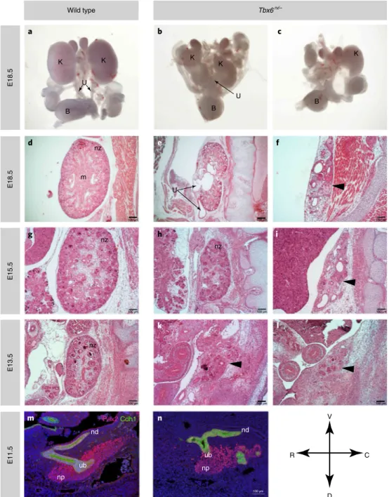
renal hypoplasia and dysplasia, and obstructive uropathy character-ized by hydronephrosis and hydroureter (Table 4 and Fig. 3a–l). Microscopic analysis of renal tissue from Tbx6rv/– embryos at stages
from E13.5 to E18.5 showed variable severity of kidney anomalies (Fig. 3d–l). Severe phenotypes included unilateral or bilateral renal agenesis, rudimentary kidneys and undeveloped renal parenchyma embedded in the paraspinal musculature (Fig. 3f,i,l). Milder pheno-types included unilateral and bilateral renal hypoplasia with hydro-ureter, tubule dilation and hydronephrosis (Fig. 3e,h,k). These data strongly implicate TBX6 as a major driver of CAKUT phenotypes. Comparative analysis was performed on wild-type and Tbx6rv/–
embryos at E11.5 after staining with E-cadherin (Cdh1), an epithe-lial marker, which labels the common nephric duct and the ureteric bud, which gives rise to the collecting duct system, and Pax2, which prevalently labels mesenchymal progenitors that produce neph-rons72–74. This analysis revealed that the events leading to CAKUT
(renal parenchyma abnormalities and obstruction) are present at early stages during development (Fig. 3m,n). In wild-type E11.5 embryos, the ureteric bud had invaded the metanephric blastema and had undergone a round of branching (Fig. 3n). In the Tbx6rv/–
mutant (Fig. 3n), the ureteric bud had not fully invaded the meta-nephric blastema and had not branched, a defect predicted to lead to KA. Additional Cdh1-positive cells in Fig. 3n might represent persistent mesonephros or ectopic ureteric buds; in that possibil-ity, ectopic buds would explain the duplication of kidney and ureter phenotype, as observed in Fig. 4.
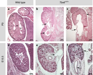
Because more subtle CAKUT phenotypes such as OU or DCS may require a minimum glomerular filtration rate or might be masked in more severe models, we examined a milder model that might be closer to the genetic architecture of humans with 16p11.2 microdeletion syndrome. We therefore analyzed embryos homo-zygous for the Tbx6 hypomorphic allele (Tbx6rv/rv). We generated
25 homozygous Tbx6rv/rv animals at E15.5 (n = 10), E18.5 (n = 10)
and postnatal day (P) 0 (n = 5) and observed incomplete penetrance and variable expressivity of multiple phenotypes that are typical of human CAKUT, including mild unilateral or bilateral hypoplasia with asymmetric kidneys, bifid ureter or DCS, and OU with pro-found hydronephrosis (Table 4 and Fig. 4). This pleiotropy of CAKUT phenotypes, ranging from renal parenchyma defects (KA) to hydronephrosis (OU) or duplication of ureters (DCS), is highly reminiscent of the pleiotropic manifestations of the 16p11.2 micro-deletion in humans, thus suggesting that TBX6 gene dosage is a major determinant of the pathogenesis of CAKUT and the observed variable expressivity of phenotypes in this syndrome.
Discussion
CAKUT has a profound effect on child health and alone accounts for about 50% of kidney failure requiring dialysis and transplantation Table 15). Neurodevelopmental defects and brain anomalies and
spine defects were the most prevalent conditions, recorded in 22 (52.4%) and 6 (14.2%) cases, respectively. Imaging studies and clinical data assessing the kidney and urinary tract were available for 15 out of 42 cases, and genitourinary anomalies were present in 9 of 15 cases (40.0%), thus highlighting that CAKUT, similarly to scoliosis, is a common feature in this syndrome and is charac-terized by incomplete penetrance. In agreement with our obser-vation of a high pleiotropic effect for the 16p11.2 microdeletion, the phenotype was also variable in this cohort: two patients with OU, two with VUR, one with PUV and one with LUTM. These data implicate CAKUT as a major feature of the chromosome 16p11.2 microdeletion syndrome and provide an opportunity to identify novel key regulators of kidney and urinary tract development.
TBX6 is a driver of CAKUT in the 16p11.2 microdeletion
syn-drome. We have recently demonstrated that the combinatorial use of genomic analyses with functional modeling in vertebrates can lead to the identification of key drivers of CAKUT phenotypes from microdeletion syndromes23. Analysis of breakpoints in nine
CAKUT cases with typical or atypical 16p11.2 microdeletions iden-tified a ~175-kb minimal region of overlap predicted to harbor the genetic driver(s) for CAKUT (Supplementary Fig. 12). Among the 19 genes included in the minimal region of overlap, TBX6 appeared as a strong candidate: it is included in the list of essential genes from the International Mouse Phenotyping Consortium61; it is depleted
from LOF mutations in subjects from the ExAC database (aggregate prevalence of LOF < 0.001); it has a role in paraxial and intermedi-ate mesoderm development62,63; and its inactivation in humans and
rodents leads to spine defects, including congenital scoliosis and spondylocostal dysostosis59,62,64–66. The connection of TBX6
muta-tions with vertebral anomalies is of particular interest because of the historically well-known clinical association between congenital scoliosis and CAKUT67–69.
To provide a functional link between the inactivation of TBX6 and CAKUT, we generated an allelic series by cross-breeding mice with two different alleles, a Tbx6-null allele (Tbx6tm2PA)70
and a Tbx6 hypomorphic allele, the spontaneous mutant Tbx6rv
(hereafter referred to as Tbx6– and Tbx6rv, respectively), which has
been studied for vertebral development and spine defects71. Because
the homozygous null mutation is lethal at embryonic day (E) 9.5, thus precluding analysis of the developing urinary tract, we first studied compound heterozygous embryos (Tbx6rv/–), which retain
sufficient residual expression of Tbx6 for survival past E9.5. We studied 19 Tbx6rv/– embryos at E17.5–E18.5 and observed full
pen-etrance of CAKUT with variable expressivity of phenotypes charac-terized by unilateral or bilateral renal agenesis, unilateral or bilateral
Table 4 | Tbx6 dosage-dependent kidney and urinary tract phenotypes
Phenotype Tbx6rv/− Tbx6rv/rv Tbx6rv/rv Tbx6rv/rv Tbx6+/– Tbx6rv/+ Tbx6+/+
E17.5− E18.5a E15.5 E18.5 P0− P1 E18.5a E18.5a E18.5
Bilateral renal agenesis 4/19 0/8 0/5 0/3 0/4 0/6 0/12
Unilateral renal agenesis 6/19 0/8 0/5 0/3 0/4 0/6 0/12
Bilateral renal hypoplasia/dysplasia 8/19 3/8 0/5 0/3 0/4 0/6 0/12
Unilateral renal hypoplasia/dysplasia 3/19 3/8 4/5 1/3 0/4 0/6 0/12
Hydronephrosis/ hydroureter 5/19 1/8 1/5 3/3 0/4 0/6 0/12
Duplex kidney/ureter 1/19 0/8 4/5 0/3 0/4 0/6 0/12
Total embryos with defects (%) 19/19 (100) 6/8 (75) 4/5 (80) 3/3 (100) 0/4 (0) 0/6 (0) 0/12 (0)
aBased on gross morphology only. We analyzed four Tbx6+/– and six Tbx6+/rv E18.5 embryos according to gross morphology, thus hampering ability to assess milder phenotypes and incomplete penetrance
kidney disease and to mitigate the associated neurocognitive and cardiac disease. In this respect, it is relevant to address two features of CAKUT: incomplete penetrance and variable expres-sivity of disease. Whereas prediction of a targeted treatment that prevents or reverses the disease is still limited by current knowl-edge, understanding the intrinsic interindividual mechanisms of in children1,3,75–78. Moreover, CAKUT is often accompanied by
extrarenal comorbidities, such as neurodevelopmental and car-diovascular diseases, thus contributing further to the disease burden in affected children5,26,27,79. Understanding the genetic
architecture of CAKUT has important implications for the devel-opment of therapeutic tools that aim to slow the progression of
100 µm 100 µm 100 µm 100 µm 100 µm 100 µm 100 µm 100 µm 100 µm 100 µm Wild type Tbx6rv/– E18.5 E18.5 E15.5 E13. 5 E11. 5 Pax2 Cdh1 K a b c d e f g h i j k l n m K K K K U U B B B nz m U nz nz nz nd ub np nd ub np V R D C
Fig. 3 | Analysis of urinary tract phenotypes in Tbx6rv/– mutants. a–c, Whole mounts of urogenital tracts. isolated from E18.5 wild type (a), and Tbx6rv/– mutants showing severe bilateral renal hypoplasia (b) and unilateral renal agenesis with contralateral renal hypoplasia (c); K, kidney; U, ureter; B, bladder. d–f, Hematoxylin and eosin (H&E)-stained sagittal sections from E18.5 wild type (d) and Tbx6rv/– mutants (e,f). The normal developing nephrogenic zone (nz) and kidney medulla (m) are clearly discernible in the wild type but not the mutant mice. The arrows in e point to the dilated renal pelvis (upper arrow) and ureter (lower arrow), which are indicative of hydronephrosis and hydroureter, respectively. In e, the kidney parenchyma also appears severely hypoplastic. The arrowhead in F points to the rudimentary kidney, which is embedded in paraspinal musculature. Few dilated tubule and microcysts are present. g–i, H&E stained kidney from an E15.5 wild type embryo (g) and E15.5 Tbx6rv/– mutant embryos (h,i). The mutants show moderate to severe hypoplasia with reduction of nephrogenic zone (nz) (h); the arrowhead indicated severely underdeveloped kidney tissue with tubule dilation and microcysts (i). j–l, H&E histological analysis of kidneys from E13.5 wild type embryos (j) and Tbx6rv/– mutants (k,l). The arrowheads point to the rudimentary kidneys that are embedded in the body wall. m,n, Immunostaining of E11.5 wild type and Tbx6rv/– mutant embryos stained with Pax2 (red) and Cdh1 (green) showing the ureteric bud (ub) nephron progenitors (np) and nephric duct (nd). In the wild type embryo, the ureteric bud has invaded the metanephric mesenchyme and branched, whereas in the mutant, the ureteric bud has not fully invaded the metanephric mesenchyme.
16p11.2, in which patients with deletions show macrocephaly, and patients with duplications are microcephalic90,91. Larger human
cohorts and experimental data will be required to investigate this hypothesis. A closer look at common GD loci assigns specificity to CAKUT subcategories, such as deletions, but not duplications, at 4p (Wolf–Hirschhorn syndrome)92, 17q12 (renal cysts and
diabe-tes syndrome)40 and 22q11.2 (DiGeorge syndrome)93, which were
nearly exclusively identified in cases with upper urinary tract malformations (mainly KA). On the other end, microdeletions at 16p11.2 were observed across the entire phenotypic spectrum of CAKUT, thus underlining the highly pleiotropic effect of this genomic region on human kidney and urinary tract development. The finding that pleiotropic urogenital defects are an important feature of this syndrome was replicated in two independent series from DECIPHER and CHOP.
We previously reported on the potential of large-scale genetic studies coupled with functional modeling in vertebrates in the identification of genetic drivers for kidney phenotypes of micro-deletion syndromes23. Here, we focused on the 16p11.2
micro-deletion syndrome, because its pleiotropic effect and incomplete penetrance provide an ideal scenario to identify genetic factors that might aid in devising therapeutic intervention. Deletion mapping and prioritization analyses pointed to TBX6 as the main genetic driver for CAKUT in cases with this syndrome. Tbx6 mouse allelic series showed that compound heterozygous embryos for a null (Tbx6tm2Pa) and a hypomorphic (Tbx6rv) allele displayed
fully penetrant CAKUT with variable expressivity of phenotypes, from bilateral renal agenesis to hypodysplasia and obstructive uropathy. Analysis of E11.5 embryonic tissue stained for renal progenitors and epithelial markers suggested that the initiating events for the CAKUT phenotypes are likely to occur very early in development in these mice. These data are in line with results from recent fate-mapping studies showing that Tbx6 is expressed in renal progenitor cells before commitment to ureteric bud or meta-nephric mesenchyme lineages94. When we analyzed embryos and
mice with milder Tbx6 inactivation (Tbx6rv/rv) as a closer model
to the human 16p11.2 microdeletion, we observed incomplete penetrance of multiple kidney and urinary tract malformations. Interestingly, similarly to the observations in our 16p11.2 micro-deletion cases, we observed that the mutant mice showed pheno-types across the phenotypic spectrum of CAKUT, including KA, OU and DCS. Overall, our mouse data at least partly suggest that the variable expressivity and incomplete penetrance of CAKUT in cases with the 16p11.2 microdeletion are attributable to a fine regulation of TBX6 gene expression during organogenesis.
In summary, with this study on children and young adults with kidney and urinary tract malformations, we provide substantial insight into the genomic landscape of human CAKUT. We iden-tify several susceptibility genetic loci and genes, and we highlight
TBX6 as a main genetic driver for CAKUT in subjects with the
chromosome 16p11.2 microdeletion syndrome, with notable implications for understanding, and potentially modifying, dis-ease penetrance.
URLs. Annovar, http://annovar.openbioinformatics.org/en/latest/; ATAV, https://redmine.igm.cumc.columbia.edu/projects/atav/wiki; DECIPHER, https://decipher.sanger.ac.uk/; Exome Aggregation Consortium, http://exac.broadinstitute.org/; ISCA, https://www. iscaconsortium.org/; Python Software Foundation, https://www. python.org/; lifelines, https://github.com/CamDavidsonPilon/ lifelines/; pandas, https://pandas.pydata.org/; R Foundation for Statistical Computing, https://www.R-project.org/; dplyr pack-age, https://CRAN.R-project.org/package= dplyr; survival package:
https://CRAN.R-project.org/package= survival; Seattle seq, http:// snp.gs.washington.edu/SeattleSeqAnnotation138/; UCSC Genome Browser, https://genome.ucsc.edu/.
compensation or amplification of disease are critical for devising treatments that might slow or halt the progression of kidney dis-ease and associated extrarenal malformations.
The study of CNVs has provided enormous insight into the genetic architecture of many developmental traits80–89. We
con-ducted a large study on rare CNVs in nearly 3,000 CAKUT cases and over 21,000 controls. By studying the burden of rare CNVs, we identified that KA, OU and VUR are particularly enriched for large and rare structural variants that affect coding regions of the genome. When examining known GD-CNVs, we found particu-lar enrichment in CAKUT cases with KA, OU, DCS and PUV compared with controls. Overall, we identified 45 distinct GDs at 37 independent genomic loci in 4% of the CAKUT cases, thus indicating substantial genetic heterogeneity. We identified novel GD-CNVs in an additional ~2% of CAKUT cases, providing multiple novel susceptibility loci to kidney disease. With respect to mechanism, WES in cases with deletions was consistent with haploinsufficiency as the most frequent pathogenic mechanism in individuals affected by GD-associated deletions. Moreover, increased burden of second-site CNVs was associated with KA and increased prevalence of extrarenal malformations. These data indicate a role for background genomic burden in variable penetrance and expressivity of disease. CNVs at six of the known 37 loci accounted for ~65% of the cases with a known GD, thus identifying major susceptibility CNVs for CAKUT. At these six loci, deletions were associated with KA, whereas duplications were enriched in cases with ureteric and lower tract defects, such as DCS and PUV. This preliminary observation gives rise to the hypothesis that CAKUT subcategories such as ureteric and lower urinary tract defects (DCS, PUV) may represent, at a molecular level, mirror traits of conditions affecting the upper urinary tract (KA, OU). This ‘mirroring phenomenon’ has been recognized in autism spectrum disorder caused by structural variation at the
P0 Tbx6rv/rv Wild type E15. 5 rc m p a b c d e f 200 μm 100 μm 100 μm 100 μm 200 μm 200 μm
Fig. 4 | Analysis of urinary tract phenotypes in Tbx6rv/rv mutants. a–c, H&E
stained sagittal sections from a wild type P0 pup (a) and P0 Tbx6rv/rv pups (b,c). The renal cortex (rc), which originates from the nephrogenic zone, is clearly distinguishable in the wild-type but not the mutant mice. The pelvis (p) and the medulla (m) are indicated. The arrows in b point to the duplicated kidneys. The arrows in c point to the hypoplastic kidney, dilated renal pelvis and proximal ureter. d–f, H&E-stained sections from an E15.5 wild type embryo (d) and Tbx6rv/rv mutant embryos (e,f). The arrow in e points to the dilated renal pelvis and proximal ureter. The arrows in f points to the hypoplastic kidney and dilated ureter.
25. Vivante, A., Kohl, S., Hwang, D. Y., Dworschak, G. C. & Hildebrandt, F. Single-gene causes of congenital anomalies of the kidney and urinary tract (CAKUT) in humans. Pediatr. Nephrol. 29, 695–704 (2014).
26. Sanna-Cherchi, S. et al. Copy-number disorders are a common cause of congenital kidney malformations. Am. J. Hum. Genet. 91, 987–997 (2012). 27. Verbitsky, M. et al. Genomic imbalances in pediatric patients with chronic
kidney disease. J. Clin. Invest. 125, 2171–2178 (2015).
28. Westland, R. et al. Copy number variation analysis identifies novel CAKUT candidate genes in children with a solitary functioning kidney. Kidney Int.
88, 1402–1410 (2015).
29. Ulinski, T. et al. Renal phenotypes related to hepatocyte nuclear factor-1beta (TCF2) mutations in a pediatric cohort. J. Am. Soc. Nephrol. 17, 497–503 (2006).
30. Madariaga, L. et al. Severe prenatal renal anomalies associated with mutations in HNF1B or PAX2 genes. Clin. J. Am. Soc. Nephrol. 8, 1179–1187 (2013).
31. Hoskins, B. E. et al. Missense mutations in EYA1 and TCF2 are a rare cause of urinary tract malformations. Nephrol. Dial. Transplant. 23,
777–779 (2008).
32. Heidet, L. et al. Spectrum of HNF1B mutations in a large cohort of patients who harbor renal diseases. Clin. J. Am. Soc. Nephrol. 5, 1079–1090 (2010). 33. Carvalho, C. M. & Lupski, J. R. Mechanisms underlying structural variant
formation in genomic disorders. Nat. Rev. Genet. 17, 224–238 (2016). 34. Lupski, J. R. Genomic disorders: structural features of the genome can lead
to DNA rearrangements and human disease traits. Trends Genet. 14, 417–422 (1998).
35. Miller, D. T. et al. Consensus statement: chromosomal microarray is a first-tier clinical diagnostic test for individuals with developmental disabilities or congenital anomalies. Am. J. Hum. Genet. 86, 749–764 (2010).
36. South, S. T. et al. ACMG Standards and Guidelines for constitutional cytogenomic microarray analysis, including postnatal and prenatal applications: revision 2013. Genet. Med. 15, 901–909 (2013).
37. Swaminathan, G. J. et al. DECIPHER: web-based, community resource for clinical interpretation of rare variants in developmental disorders. Hum. Mol. Genet. 21, R37–R44 (2012).
38. Firth, H. V. et al. DECIPHER: database of chromosomal imbalance and phenotype in humans using Ensembl resources. Am. J. Hum. Genet. 84, 524–533 (2009).
39. Grisaru, S., Ramage, I. J. & Rosenblum, N. D. Vesicoureteric reflux associated with renal dysplasia in the Wolf-Hirschhorn syndrome. Pediatr. Nephrol. 14, 146–148 (2000).
40. Mefford, H. C. et al. Recurrent reciprocal genomic rearrangements of 17q12 are associated with renal disease, diabetes, and epilepsy. Am. J. Hum. Genet.
81, 1057–1069 (2007).
41. Portnoi, M. F. Microduplication 22q11.2: a new chromosomal syndrome. Eur. J. Med. Genet. 52, 88–93 (2009).
42. Kobrynski, L. J. & Sullivan, K. E. Velocardiofacial syndrome, DiGeorge syndrome: the chromosome 22q11.2 deletion syndromes. Lancet 370, 1443–1452 (2007).
43. Weber, S. et al. Mapping candidate regions and genes for congenital anomalies of the kidneys and urinary tract (CAKUT) by array-based comparative genomic hybridization. Nephrol. Dial. Transplant. 26, 136–143 (2011).
44. Hildebrandt, F. et al. A novel gene encoding an SH3 domain protein is mutated in nephronophthisis type 1. Nat. Genet. 17, 149–153 (1997). 45. Willatt, L. et al. 3q29 microdeletion syndrome: clinical and molecular
characterization of a new syndrome. Am. J. Hum. Genet. 77, 154–160 (2005).
46. Mattina, T., Perrotta, C. S. & Grossfeld, P. Jacobsen syndrome. Orphanet. J. Rare. Dis. 4, 9 (2009).
47. Sampson, M. G. et al. Evidence for a recurrent microdeletion at chromosome 16p11.2 associated with congenital anomalies of the kidney and urinary tract (CAKUT) and Hirschsprung disease. Am. J. Med. Genet. A 152A, 2618–2622 (2010).
48. Fontes, M. I. et al. Genotype-phenotype correlation of 16p13.3 terminal duplication and 22q13.33 deletion: Natural history of a patient and review of the literature. Am. J. Med. Genet. A 170, 766–772 (2016).
49. Goh, E. S. et al. Definition of a critical genetic interval related to kidney abnormalities in the Potocki-Lupski syndrome. Am. J. Med. Genet. A 158A, 1579–1588 (2012).
50. Yamamoto, T. et al. A large interstitial deletion of 17p13.1p11.2 involving the Smith-Magenis chromosome region in a girl with multiple congenital anomalies. Am. J. Med. Genet. A 140, 88–91 (2006).
51. Kim, Y. M. et al. Phelan-McDermid syndrome presenting with developmental delays and facial dysmorphisms. Korean J. Pediatr. 59, S25–S28 (2016).
52. Ozgun, M. T. et al. Prenatal diagnosis of a fetus with partial trisomy 7p. Fetal. Diagn. Ther. 22, 229–232 (2007).
Online content
Any methods, additional references, Nature Research reporting summaries, source data, statements of data availability and asso-ciated accession codes are available at https://doi.org/10.1038/ s41588-018-0281-y.
Received: 12 October 2017; Accepted: 18 October 2018; Published online: 21 December 2018
References
1. Wuhl, E. et al. Timing and outcome of renal replacement therapy in patients with congenital malformations of the kidney and urinary tract. Clin. J. Am. Soc. Nephrol. 8, 67–74 (2013).
2. Chesnaye, N. C. et al. Mortality risk disparities in children receiving chronic renal replacement therapy for the treatment of end-stage renal disease across Europe: an ESPN-ERA/EDTA registry analysis. Lancet 389, 2128–2137 (2017).
3. Sanna-Cherchi, S. et al. Renal outcome in patients with congenital anomalies of the kidney and urinary tract. Kidney Int. 76, 528–533 (2009).
4. Goodyer, P. R. Renal dysplasia/hypoplasia. in Pediatric Nephrology 5th edn. (eds. Avner, E. D., Harmon, W. E. & Niaudet, P.) 83–91 (Lippincott Williams & Wilkins, Philadelphia, 2004).
5. Sanna-Cherchi, S., Westland, R., Ghiggeri, G. M. & Gharavi, A. G. Genetic basis of human congenital anomalies of the kidney and urinary tract. J. Clin. Invest. 128, 4–15 (2018).
6. Schedl, A. Renal abnormalities and their developmental origin. Nat. Rev.
Genet. 8, 791–802 (2007).
7. Costantini, F. & Kopan, R. Patterning a complex organ: branching morphogenesis and nephron segmentation in kidney development. Dev. Cell. 18, 698–712 (2010).
8. Short, K. M. & Smyth, I. M. The contribution of branching morphogenesis to kidney development and disease. Nat. Rev. Nephrol. 12,
754–767 (2016).
9. dos Santos Junior, A. C., de Miranda, D. M. & Simoes e Silva, A. C. Congenital anomalies of the kidney and urinary tract: an embryogenetic review. Birth Defects Res. C. Embryo Today 102, 374–381 (2014). 10. Chen, F. Genetic and developmental basis for urinary tract obstruction.
Pediatr. Nephrol. 24, 1621–1632 (2009).
11. Vainio, S. & Lin, Y. Coordinating early kidney development: lessons from gene targeting. Nat. Rev. Genet. 3, 533–543 (2002).
12. Sanna-Cherchi, S. et al. Genetic approaches to human renal agenesis/ hypoplasia and dysplasia. Pediatr. Nephrol. 22, 1675–1684 (2007). 13. Nicolaou, N., Renkema, K. Y., Bongers, E. M., Giles, R. H. & Knoers, N. V.
Genetic, environmental, and epigenetic factors involved in CAKUT. Nat. Rev. Nephrol. 11, 720–731 (2015).
14. Georgas, K. M. et al. An illustrated anatomical ontology of the developing mouse lower urogenital tract. Development 142, 1893–1908 (2015). 15. Thomas, R. et al. HNF1B and PAX2 mutations are a common cause
of renal hypodysplasia in the CKiD cohort. Pediatr. Nephrol. 26, 897–903 (2011).
16. Weber, S. et al. Prevalence of mutations in renal developmental genes in children with renal hypodysplasia: results of the ESCAPE study. J. Am. Soc. Nephrol. 17, 2864–2870 (2006).
17. Nicolaou, N. et al. Prioritization and burden analysis of rare variants in 208 candidate genes suggest they do not play a major role in CAKUT. Kidney Int. 89, 476–486 (2016).
18. Sanna-Cherchi, S. et al. Mutations in DSTYK and dominant urinary tract malformations. N. Engl. J. Med. 369, 621–629 (2013).
19. Vivante, A. et al. Mutations in TBX18 cause dominant urinary tract malformations via transcriptional dysregulation of ureter development. Am. J. Hum. Genet. 97, 291–301 (2015).
20. Schimmenti, L. A. et al. Further delineation of renal-coloboma syndrome in patients with extreme variability of phenotype and identical PAX2 mutations. Am. J. Hum. Genet. 60, 869–878 (1997).
21. Bekheirnia, M. R. et al. Whole-exome sequencing in the molecular diagnosis of individuals with congenital anomalies of the kidney and urinary tract and identification of a new causative gene. Genet. Med. 19, 412–420 (2017).
22. Ichikawa, I., Kuwayama, F., Pope, J. Ct, Stephens, F. D. & Miyazaki, Y. Paradigm shift from classic anatomic theories to contemporary cell biological views of CAKUT. Kidney Int. 61, 889–898 (2002).
23. Lopez-Rivera, E. et al. Genetic drivers of kidney defects in the DiGeorge syndrome. N. Engl. J. Med. 376, 742–754 (2017).
24. Hwang, D. Y. et al. Mutations in 12 known dominant disease-causing genes clarify many congenital anomalies of the kidney and urinary tract. Kidney Int. 85, 1429–1433 (2014).
81. Greenway, S. C. et al. De novo copy number variants identify new genes and loci in isolated sporadic tetralogy of Fallot. Nat. Genet. 41, 931–935 (2009).
82. Osoegawa, K. et al. Identification of novel candidate genes associated with cleft lip and palate using array comparative genomic hybridisation. J. Med. Genet. 45, 81–86 (2008).
83. Serra-Juhe, C. et al. Contribution of rare copy number variants to isolated human malformations. PLoS ONE. 7, e45530 (2012).
84. Brunetti-Pierri, N. et al. Recurrent reciprocal 1q21.1 deletions and duplications associated with microcephaly or macrocephaly and developmental and behavioral abnormalities. Nat. Genet. 40, 1466–1471 (2008).
85. Sanders, S. J. et al. Multiple recurrent de novo CNVs, including duplications of the 7q11.23 Williams syndrome region, are strongly associated with autism. Neuron 70, 863–885 (2011).
86. Sebat, J. et al. Strong association of de novo copy number mutations with autism. Science 316, 445–449 (2007).
87. Stefansson, H. et al. Large recurrent microdeletions associated with schizophrenia. Nature 455, 232–236 (2008).
88. Yu, L. et al. De novo copy number variants are associated with congenital diaphragmatic hernia. J. Med. Genet. 49, 650–659 (2012).
89. Mannik, K. et al. Copy number variations and cognitive phenotypes in unselected populations. JAMA 313, 2044–2054 (2015).
90. Golzio, C. & Katsanis, N. Genetic architecture of reciprocal CNVs. Curr. Opin. Genet. Dev. 23, 240–248 (2013).
91. Golzio, C. et al. KCTD13 is a major driver of mirrored neuroanatomical phenotypes of the 16p11.2 copy number variant. Nature 485, 363–367 (2012).
92. Gandelman, K. Y., Gibson, L., Meyn, M. S. & Yang-Feng, T. L. Molecular definition of the smallest region of deletion overlap in the Wolf-Hirschhorn syndrome. Am. J. Hum. Genet. 51, 571–578 (1992).
93. Driscoll, D. A., Budarf, M. L. & Emanuel, B. S. A genetic etiology for DiGeorge syndrome: consistent deletions and microdeletions of 22q11. Am. J. Hum. Genet. 50, 924–933 (1992).
94. Concepcion, D. et al. Cell lineage of timed cohorts of Tbx6-expressing cells in wild-type and Tbx6 mutant embryos. Biol. Open 6,
1065–1073 (2017).
Acknowledgements
We thank all patients and family members for participating in this study. We thank J. R. Lupski for critical review of this manuscript. This work was supported by grants (1R01DK103184, 1R21DK098531 and UL1 TR000040, to S.S.-C.; 2R01DK080099, to A.G.G.; 3U54DK104309, to A.G.G., C.L.M. and J.M.B.; R37HD033082, to V.E.P.; and 1R01DK105124 to K.K.) from the National Institutes of Health (NIH); a grant-in-aid (13GRNT14680075, to S.S.-C.) from the American Heart Association; a grant (RF-2010–2307403, to S.S.-C. and G.M.G.) from the Joint Italian Ministry of Health and NIH Young Investigators Finalized Research; a grant (to G.M.G.) from the Fondazione Malattie Renali nel Bambino; grants to D.E.B. and P.P. from the National Children’s Research Centre and the Irish Health Research Board (HRA-POR-2014–693); a grant (AAE07007KSA, to C.J.) from the GIS-Institut des Maladies Rares; by the Polish Ministry of Health (to A.M.K. and A.L.-B.); by the Polish Kidney Genetics Network (POLYGENES), the Polish Registry of Congenital Malformations (PRCM) and the NZOZ Center for Medical Genetics (GENESIS); by grants (to the Chronic Kidney Disease in Children Study) from the National Institute of Diabetes and Digestive and Kidney Diseases and the Eunice Kennedy Shriver National Institute of Child Health and Human Development; by grants (U01DK66143, U01DK66174, U01DK082194, U01DK66116 and RO1DK082394) from the National Heart, Lung, and Blood Institute; and by the Paul Marks Scholar Award (to S.S.-C.); CNPq grant 460334/2014-0 and FAPEMIG grant PPM-005555-15 (to D.M.M., E.A.O. and A.C.S.-e.-S.); and a Kolff Postdoc Fellowship Abroad grant (15OKK95, to R.W.) from the Dutch Kidney Foundation. S.S.-C. is supported as the Florence Irving Assistant Professor of Medicine at Columbia University. We thank D.B. Goldstein for providing infrastructure for whole-exome sequencing at the Institute for Genomic Medicine (IGM) at Columbia University, and for critical review of the manuscript. Acknowledgments to the investigators who contributed of whole-exome sequencing data for 15,469 controls from the IGM warehouse can be found in the Supplementary Note.
Author contributions
S.S.-C. directed the project. V.E.P., C.L.M., A.G.G. and S.S.-C. designed the project. M.V., R.W., A.P., Q.L., P.K., D.A.F., E.B., M.W., J.M., V.P.C., Y.-J.N., T.Y.L., D.A. and H.W. performed the experiments and/or data generation. M.G.S., M.G.D., J.M.D., P.P., D.E.B., S.L.F., B.A.W., C.J., D.M.M., E.A.O., A.C.S.-e.-S., F.H. and H.H. contributed array genotype data for CNV analyses. M.V., R.W., P.K., A.P., E.B., A.M., V.E.P., C.L.M. and S.S.-C. analyzed the data. K.K., J.M.B. and B.L. provided critical intellectual content for the design of the study. All other authors (A.M., M.B., C.K., A.V., S.S., B.H.K., M.M., J.Y.Z., P.L.W., E.L.H., A.C., G.P., L.G., V.M., G.M., M.G., 53. Trachoo, O., Assanatham, M., Jinawath, N. & Nongnuch, A. Chromosome
20p inverted duplication deletion identified in a Thai female adult with mental retardation, obesity, chronic kidney disease and characteristic facial features. Eur. J. Med. Genet. 56, 319–324 (2013).
54. Westland, R. et al. Phenotypic expansion of DGKE-associated diseases. J. Am. Soc. Nephrol. 25, 1408–1414 (2014).
55. Karczewski, K. J. et al. The ExAC browser: displaying reference data information from over 60,000 exomes. Nucleic Acids Res. 45, D840–D845 (2017).
56. Lek, M. et al. Analysis of protein-coding genetic variation in 60,706 humans. Nature 536, 285–291 (2016).
57. Materna-Kiryluk, A. et al. The emerging role of genomics in the diagnosis and workup of congenital urinary tract defects: a novel deletion syndrome on chromosome 3q13.31-22.1. Pediatr. Nephrol. 29, 257–267 (2014). 58. Lata, S. et al. Whole-exome sequencing in adults with chronic kidney
disease: A pilot study. Ann. Intern. Med. 168, 100–109 (2018). 59. Wu, N. et al. TBX6 null variants and a common hypomorphic allele in
congenital scoliosis. N. Engl. J. Med. 372, 341–350 (2015). 60. Al-Kateb, H. et al. Scoliosis and vertebral anomalies: additional
abnormal phenotypes associated with chromosome 16p11.2 rearrangement. Am. J. Med. Genet. A 164A, 1118–1126 (2014).
61. Dickinson, M. E. et al. High-throughput discovery of novel developmental phenotypes. Nature 537, 508–514 (2016).
62. Chapman, D. L. & Papaioannou, V. E. Three neural tubes in mouse embryos with mutations in the T-box gene Tbx6. Nature 391, 695–697 (1998).
63. Chapman, D. L., Agulnik, I., Hancock, S., Silver, L. M. & Papaioannou, V. E. Tbx6, a mouse T-Box gene implicated in paraxial mesoderm formation at gastrulation. Dev. Biol. 180, 534–542 (1996).
64. Abe, K. et al. Novel ENU-induced mutation in Tbx6 causes dominant spondylocostal dysostosis-like vertebral malformations in the rat. PLoS ONE. 10, e0130231 (2015).
65. Lefebvre, M. et al. Autosomal recessive variations of TBX6, from congenital scoliosis to spondylocostal dysostosis. Clin. Genet. 91, 908–912 (2017).
66. Sparrow, D. B. et al. Autosomal dominant spondylocostal dysostosis is caused by mutation in TBX6. Hum. Mol. Genet. 22,
1625–1631 (2013).
67. MacEwen, G. D., Winter, R. B., Hardy, J. H. & Sherk, H. H. Evaluation of kidney anomalies in congenital scoliosis. 1972. Clin. Orthop. Relat. Res. 434, 4–7 (2005).
68. Cowell, H. R., MacEwen, G. D. & Hubben, C. Incidence of abnormalities of the kidney and ureter in congenital scoliosis. Birth Defects. Orig. Artic. Ser.
10, 142–145 (1974).
69. MacEwen, G. D., Winter, R. B. & Hardy, J. H. Evaluation of kidney anomalies in congenital scoliosis. J. Bone Joint Surg. Am. 54, 1451–1454 (1972).
70. Hadjantonakis, A. K., Pisano, E. & Papaioannou, V. E. Tbx6 regulates left/ right patterning in mouse embryos through effects on nodal cilia and perinodal signaling. PLoS ONE. 3, e2511 (2008).
71. Watabe-Rudolph, M., Schlautmann, N., Papaioannou, V. E. & Gossler, A. The mouse rib-vertebrae mutation is a hypomorphic Tbx6 allele. Mech. Dev.
119, 251–256 (2002).
72. Batourina, E. et al. Distal ureter morphogenesis depends on epithelial cell remodeling mediated by vitamin A and Ret. Nat. Genet. 32, 109–115 (2002).
73. Batourina, E. et al. Vitamin A controls epithelial/mesenchymal interactions through Ret expression. Nat. Genet. 27, 74–78 (2001).
74. Batourina, E. et al. Apoptosis induced by vitamin A signaling is crucial for connecting the ureters to the bladder. Nat. Genet. 37, 1082–1089 (2005).
75. Harambat, J., van Stralen, K. J., Kim, J. J. & Tizard, E. J. Epidemiology of chronic kidney disease in children. Pediatr. Nephrol. 27,
363–373 (2012).
76. Westland, R., Kurvers, R. A., van Wijk, J. A. & Schreuder, M. F. Risk factors for renal injury in children with a solitary functioning kidney. Pediatrics
131, e478–e485 (2013).
77. Westland, R., Schreuder, M. F., Bokenkamp, A., Spreeuwenberg, M. D. & van Wijk, J. A. Renal injury in children with a solitary functioning kidney: the KIMONO study. Nephrol. Dial. Transplant. 26, 1533–1541 (2011).
78. Westland, R., Schreuder, M. F., van Goudoever, J. B., Sanna-Cherchi, S. & van Wijk, J. A. Clinical implications of the solitary functioning kidney. Clin. J. Am. Soc. Nephrol. 9, 978–986 (2014).
79. Verbitsky, M. et al. Genomic disorders and neurocognitive impairment in pediatric CKD. J. Am. Soc. Nephrol. 28, 2303–2309 (2017).
80. Cooper, G. M. et al. A copy number variation morbidity map of developmental delay. Nat. Genet. 43, 838–846 (2011).