Journal of Physics: Conference Series
PAPER • OPEN ACCESS
SEM tomography for the investigation of hybrid
structures
To cite this article: M Ferroni et al 2017 J. Phys.: Conf. Ser. 902 012031
View the article online for updates and enhancements.
Related content
Morphological Investigation of Disturbed Ionosphere during Intense Geomagnetic Storms
Bhupendra Malvi, Prateek S. Srivastav, Azad A. Mansoori et al.
-3D Nano-characterisation of materials by FIB-SEI/EDS tomography
F A Lasagni, A F Lasagni, I Huertas-Olivares et al.
-Structural and Morphological Investigation for Water-Processed Graphene Oxide/Single-Walled Carbon Nanotubes Hybrids
M R Muda, M M Ramli, S S Mat Isa et al.
1
Content from this work may be used under the terms of theCreative Commons Attribution 3.0 licence. Any further distribution of this work must maintain attribution to the author(s) and the title of the work, journal citation and DOI.
Published under licence by IOP Publishing Ltd
1234567890
Electron Microscopy and Analysis Group Conference 2017 (EMAG2017) IOP Publishing IOP Conf. Series: Journal of Physics: Conf. Series 902 (2017) 012031 doi :10.1088/1742-6596/902/1/012031
SEM tomography for the investigation of hybrid structures
M Ferroni1, 2, M Donarelli2, V Morandi1, A Migliori1, L Ortolani1A Signoroni2, C R Chandraiahgari3,4, G De Bellis3,4 and M S Sarto3,4
1 CNR – IMM National Research Council - Microelectronic and Microsystems Institute, via Gobetti 101, 40129 Bologna, Italy
2 Department of Information Engineering, University of Brescia, Via Branze 38, 25123 Brescia, Italy
3 Department of Astronautical, Electrical and Energy Engineering, Sapienza University of Rome, via Eudossiana 18, Rome 00184, Italy.
4 Research Center for Nanotechnology Applied to Engineering of Sapienza (CNIS), SNNLab, Sapienza University of Rome, Piazzale Aldo Moro, 5, Rome 00185, Italy E-mail: [email protected]
Abstract. The morphological investigation at the micrometric scale of a graphene - ZnO
nanorods hybrid structure is performed by scanning electron microscopy. When operated in the scanning-transmission imaging mode, the detection strategy allows implementation of a tomographic approach to recover the three dimensional spatial arrangement of the sample constituents. This tomographic approach complements the serial-sectioning imaging methods and is suitable for thin, self-standing specimens.
1. Introduction
Recent developments in material fabrication exploit the realization of hybrid structures combining different growth techniques and pursuing a positive tailoring of the functional properties.
As an example, graphene combined with ZnO crystalline nanorods (NRs) is presently under investigation because of the useful photocatalytic properties of this n-type semiconducting metal oxide and the electrical properties of graphene. In the form of crystalline NRs, ZnO has been used for a number of applications, ranging from gas sensors to solar cells [1-4]. A graphene-NRs system can be obtained by dispersing the NRs through the multilayered graphene flakes or promoting the nucleation of NRs directly over graphene [5-8]. In the present study, graphene flakes have been produced by solvo-thermal exfoliation method, and ZnO NRs have been grown in-situ through aqueous hydrothermal method [9]. A significant advantage of this approach is the compatibility with flexible and transparent polymeric substrates [10].
2. Characterization by scanning electron microscopy
The understanding and modelling of hybrid structures calls for an investigation of the spatial arrangement of the constituents at the micrometric scale. This intermediate magnification range could be easily covered by scanning electron microscopy (SEM); however, the interpretation of sample
2
1234567890
Electron Microscopy and Analysis Group Conference 2017 (EMAG2017) IOP Publishing IOP Conf. Series: Journal of Physics: Conf. Series 902 (2017) 012031 doi :10.1088/1742-6596/902/1/012031
morphology from the conventional secondary-electron (SE) images should be carried out with caution, and quantitative three dimensional information is beyond the limits of the technique.
2.1. Conventional imaging with secondary electrons
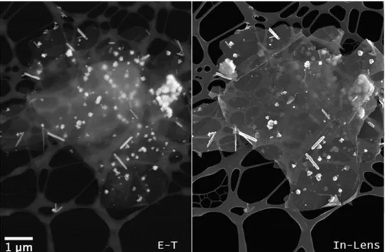
Figure 1 (left and right) compares two low-magnification SEM images obtained with SE of a flake of graphene in-situ decorated with ZnO NRs and nanoparticles.
For the observation, the specimen was drop cast from the aqueous suspension over a carbon grid, according to a standard procedure for transmission electron microscopy (TEM) samples. When implemented in the SEM, the suspension of the thin sample over a lacy film prevents the saturation of the SE/BSE signals from the bulk substrate and, also, allows the exploitation of the transmitted-electrons imaging mode.
The two images in Figure 1, obtained with the standard Everhard-Torley (E-T) detector (left) and the In-lens detector (right) integrated in the Gemini column of the SEM (ZEISS-LEO 1530 FEG SEM), are quite different because of the limited difference in SE emission between the carbon-based material and nanorods dispersed at the top surface. The resulting images feature low contrast and a significant compositional contribution: in particular, the sensitivity of the E-T detector to the SE2 component of the SE signal, together with the +300 V bias of the collector grid, allows a significant compositional contribution related to the back-scattered electrons and promotes the visibility of most of the ZnO nanostructures underlying the graphene layer present at the centre of the selected area.
Figure 1: SEM images of a graphene – ZnO nanorods hybrid structure by secondary-electron imaging: Everhart-Thorley (left) and In Lens detector (right).
2.2. Imaging with transmitted electrons
The scanning-transmission imaging mode in the SEM (STEM in SEM or T-SEM) is a standard implementation for the SEM platform, which has gained interest for the observation of light-element and nanostructured materials as a low-energy counterpart of TEM.
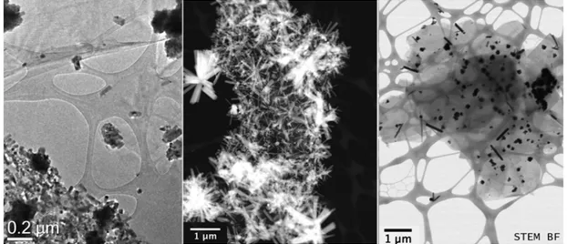
Figure 2 compares the TEM images (FEI Tecnai F20 microscope) of the graphene-ZnO sample obtained with conventional Bright-Field (left) and STEM-HAADF (center) with the one obtained by the STEM in SEM (right). Figure 2-right shows the transmitted-electron image of the very same area shown
3
1234567890
Electron Microscopy and Analysis Group Conference 2017 (EMAG2017) IOP Publishing IOP Conf. Series: Journal of Physics: Conf. Series 902 (2017) 012031 doi :10.1088/1742-6596/902/1/012031
in Figure 1, obtained in the STEM-in-SEM, operated in a Bright-Field condition. In this image, the ZnO NRs and nanoparticles are clearly recognizable from graphene and the supporting lacy carbon film of the TEM grid.
The image contrast is monotonically governed by the sample density and projected thickness, while the resolution is limited by the size of the focused electron beam. This imaging mode meets the requirements for a tomographic approach to the three dimensional reconstruction of the mass distribution of the sample.
Figure 2: TEM and SEM images of the graphene – ZnO nanorods structure by transmitted-electron imaging. Conventional TEM Bright-Field (left) and TEM STEM-HAADF (center).
(right) SEM image in STEM Bright-Field condition.
3. Electron tomography in the SEM
The SEM platform is already used in the 3D visualization of samples in both biological and physical science through operation of the microscope in a slice and view mode, where a section of the bulk sample is progressively obtained by a focused ion beam and visualized with the electron beam. This approach performs well for uniform and compact specimens, as the section cutting is required to proceed evenly throughout the entire reconstructed volume. Limitations may also arise when the sample features a significant degree of porosity and voids. In the case of the graphene-ZnO NRs sample, such structure is not suitable for serial sectioning as the sample is extremely light, self-standing and lacks rigid support from an embedding media.
As a complementary approach to three dimensional imaging, the scanning-transmission imaging was recently integrated with a rotating sample holder to demonstrate a technically viable realization of electron tomography in the SEM [11-12]. The tomographic reconstruction algorithm introduces the additional requirement for the detection strategy: the STEM signal is based on the intensity of the electrons forward-scattered by the thin specimen, and the mass-thickness contrast should vary monotonically when the sample is rotated and projected from different directions.
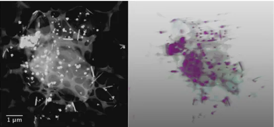
For the graphene-ZnO NRs sample, the optimal imaging condition corresponds to the Dark-Field (DF) mode, owing to the difference in forward scattering between the thin graphene and the nanometric ZnO crystals. The image presented in Figure 3-left has been acquired in DF condition, so the highest signal-to-background ratio can be measured, the brightest areas being related to the ZnO nanorods. The light-grey area is the graphene flake, which is rippling over the carbon grid. The beam acceleration voltage was 27 kV, and the detection strategy was adapted to maintain a complete collection of the transmitted signal upon tilting the sample for the series of projections.
4
1234567890
Electron Microscopy and Analysis Group Conference 2017 (EMAG2017) IOP Publishing IOP Conf. Series: Journal of Physics: Conf. Series 902 (2017) 012031 doi :10.1088/1742-6596/902/1/012031
Figure 3: (left) STEM image of the graphene-NRs in dark-field condition. (right) A colored rendering of the reconstructed tomogram.
The series of the sample DF projection images has been acquired by tilting the specimen from -60° to +50°, at 2° steps. The projection images have been aligned each other with a cross-correlation algorithm, and the result of the reconstruction of the 56 projection images is reported in Figure 3-right. It turns out that the tomogram, obtained by standard retroprojection and followed by iterative refining, retrieves the correct spatial disposition of the hybrid system.
4. Discussion and concluding remarks
SEM is a valuable tool for the preliminary characterization and for the optimization of the preparation process of hybrid structures. In addition, the proposed approach to electron tomography in SEM is suitable for the assessment of the dispersion of ZnO NRs in graphene flakes. This method is potentially applicable to a large category of partially electron-transparent samples, and volumes up to 1 μm3 could be reconstructed. The detection strategy could also take advantage of recent algorithmic implementations, such as discrete tomography or compressed sensing, to optimize the projection scheme and to reduce the number of projections required to retrieve the structure of the sample with nanometric resolution.
Acknowledgments
Ferroni M and Donarelli M acknowledge the financial support from the Ministero degli Affari Esteri e della Cooperazione Internazionale within the ITA-EGYPT CYAMOXSOLAR project.
References
[1]! Wang H T, Kang B S and Ren F 2005 Appl. Phys. Lett. 86 243503 [2]! Jiaqiang X et al 2006 Sens. and Act. B 113 526
[3]! Gao T and Wang T H 2005 Appl. Phys. A 80 1451 [4]! Hsu Y F et al 2008 Appl. Phys. Lett. 92 133507 [5]! Zou R et al 2013 J. Mater. Chem. A 1 8445 [6]! Chandraiahgari C R et al 2015 RSC Adv. 5 49861 [7]! Ding J et al 2015 RSC Adv. 5 22935
[8]! Chung R et al 2013 Nanoscale Res. Lett. 8 350 [9]! Chandraiahgari C R et al 2016 RSC Adv. 6 83217
[10]! Casiraghi C and Whiters F 2016 in 2D Materials for Nanoelectronics (Houssa M, Dimoulas A and Molle A, eds.) p 434
[11]! Ferroni M et al 2016 Scientific Reports 6 33354 [12]! Ferroni M et al 2015 J. Phys.: Conf. Ser. 644 012012
Journal of Physics: Conference Series
PAPER • OPEN ACCESS
Peer review statement
To cite this article: 2017 J. Phys.: Conf. Ser. 902 011002
View the article online for updates and enhancements.
Related content
Peer review statement
-Peer review statement
-Peer review statement
1
Content from this work may be used under the terms of theCreative Commons Attribution 3.0 licence. Any further distribution of this work must maintain attribution to the author(s) and the title of the work, journal citation and DOI.
Published under licence by IOP Publishing Ltd
1234567890
Electron Microscopy and Analysis Group Conference 2017 (EMAG2017) IOP Publishing
IOP Conf. Series: Journal of Physics: Conf. Series 902 (2017) 011002 doi :10.1088/1742-6596/902/1/011002
Peer review statement
All papers published in this volume of Journal of Physics: Conference Series have been peer reviewed through processes administered by the proceedings Editors. Reviews were conducted by expert referees to the professional and scientific standards expected of a proceedings journal published by IOP Publishing.