UNIVERSITA’ DELLA CALABRIA
Dipartimento di Chimica e Tecnologie Chimiche – CTC
Rende, Italy 2017
Dottorato di Ricerca in
Medicina Traslazionale
CICLO (XXIX)
TRANSITION METAL-BASED COMPLEXES AS
CHEMOTHERAPEUTIC AGENTS. THEORETICAL INVESTIGATION
OF MoA, INTERACTION WITH BIOLOGICAL MOLECULES AND
Abstract
Molti metalli e, conseguentemente, molti complessi metallici svolgono ruoli importanti all’interno di sistemi biologici e biochimici. Oggigiorno, è risaputo che essi rappresentano ingredienti molto importanti per la vita altrettanto quanto i composti organici. Per esempio, i complessi di ferro svolgono un ruolo fondamentale nel trasporto di O2 nel sangue, i complessi di calcio sono alla base delle ossa, lo zinco è presente nell’ insulina che regola la quantità di zucchero nel nostro corpo. Questo è possibile perché i metalli possiedono particolari proprietà chimiche. Infatti, tendono facilmente a perdere elettroni, diventando specie elettron-deficienti più reattive nei confronti di diverse molecole biologiche e più solubili in soluzioni acquose. I metalli non solo sono elementi vitali in molti fenomeni biologici, ma possono anche essere sfruttati per il trattamento di diverse malattie.
L'esempio più importante di questa categoria di composti è il complesso organometallico cisplatino [Pt(Cl)2(NH3)2], il chemioterapico più potente disponibile sul mercato. Scoperto nel 1969 da Rosenberg, esso svolge un ruolo chiave nell’ inibizione della divisione cellulare, causando la morte delle cellule tumorali.
Dopo la scoperta della sua attività citotossica, l'applicazione di farmaci metallici nelle varie terapie ha registrato una crescita enorme e la continua ricerca dell'uso di metalli in medicina, in particolare per la cura del cancro, è diventata una disciplina, indicata come Medicinal Inorganic Chemistry.
Dopo la scoperta del cisplatino, altri due farmaci antitumorali a base di platino, derivati del cisplatino, sono stati scoperti: carboplatino e ossaliplatino. Questi complessi esercitano la loro azione citotossica coordinando il DNA e bloccando la divisione cellulare. Tuttavia, anche se questi complessi, di formula generale [Pt(X)2(L)2], sono ancora tra i farmaci più frequentemente utilizzati, risultano essere tossici a causa della loro reattività e instabilità.
In questi anni, per superare i problemi relativi all’uso di complessi di Pt(II), l’ attenzione è stata concentrata sullo sviluppo di nuovi complessi, generando due diverse categorie di farmaci antitumorali: i complessi di Pt(IV), considerati "profarmaci" e ottenuti per ossidazione dai complessi di Pt(II), e i "farmaci che non contengono platino", come i complessi di iridio, rodio, osmio o rutenio. Le caratteristiche di questi farmaci dovrebbero renderli più inerti, quindi più efficaci dei complessi di Pt(II), e provocare una differenziazione nel meccanismo di azione.
Lo studio teorico dei sistemi biologici ha ormai raggiunto la maturità necessaria per fornire informazioni complementari rispetto a quelle ottenibili sperimentalmente nello studio di molte proprietà e fenomeni. Infatti, l’impressionante sviluppo sia delle metodologie teoriche, sia della tecnologia informatica ha consentito di fornire un importante supporto di studi teorici alle bioscienze. Inoltre, l’uso della teoria del funzionale della densità (DFT), che rappresenta un ottimo compromesso tra accuratezza e costi computazionali, è particolarmente adatto allo studio dei sistemi biologici come dimostrato dal fatto che negli ultimi anni sono stati completati con successo molti studi DFT e sono state affrontate questioni fondamentali nelle simulazioni biologiche. Scopo di questa tesi è lo studio teorico di farmaci antitumorali contenenti metalli, in particolare complessi di Pt(IV) e Ir(III), delle principali reazioni del farmaco che avvengono dal momento della sua iniezione o somministrazione orale al raggiungimento del suo bersaglio biologico e investigazione del meccanismo di azione che può essere sia quello classico, già proposto per il cisplatino e, quindi, associato all’accumulo cellulare e al legame con il DNA, sia un meccanismo di diversa natura. In alcuni casi, la teoria del funzionale della densità è stata utilizzata come approccio computazionale supportato da calcoli eseguiti a livello Coupled Cluster post Hartree-Fock nella sua versione CCSD(T).
i
INTRODUCTION
Many metals, and, consequently, many metal complexes play important roles in biological and biochemical systems. It is known, today, that they are very important ingredients in life as well organic compounds. For example, the iron complexes have a fundamental role in the O2 transport in the blood, the calcium complexes are the basis of the bones, the zinc is present in the insulin, which regulates the amount of sugar in our body. This is possible because metals have particular chemical properties. Indeed, they easily tend to lose electrons, becoming electron deficient species, reactive towards various biological molecules and soluble in water solutions. Metals not only are vital elements in many biological phenomena, but they can be also exploited in therapeutics for a variety of diseases.
The most prominent example of such class of compounds is the organometallic complex cisplatin [Pt(Cl)2(NH3)2], the most potent chemotherapeutic available on the market. Discovered in 1969 by Rosenberg, it plays a key role in the inhibition of cell division, killing the cancer cells. Since its discovery the application of metal drugs for therapeutics has experienced a tremendous and continuous growth and the investigation of the use of metals in medicine, particularly for the treatment of
ii
cancer, has become a discipline that is the Medicinal Inorganic Chemistry.
After the discovery of cisplatin, other two Pt-anticancer drugs, derivatives of cisplatin, have been discovered: carboplatin and oxaliplatin. These complexes exert their cytotoxic action by coordinating to DNA and stopping cell division. However, although these complexes, with general structure [Pt(X)2(L)2], are still among the most frequently used drugs, they appear to be toxic, because of their chemical reactivity and instability.
In these years, to overcome Pt(II) complexes disadvantages, the research has turned its attention to the development of new complexes, producing two different anticancer drugs classes: Pt(IV) complexes, which are considered “prodrugs” and are obtained by oxidation of the Pt(II) complexes, and “no-platinum containing drugs”, such as iridium, rhodium, osmium or ruthenium complexes. The characteristics of such drugs should both make them more inert and, consequently, more effective than Pt(II) complexes and cause a differentiation in the mechanism of action.
The theoretical investigation of biological systems has now achieved the needed maturity to complement the experimental approaches for the study of a large variety of properties and phenomena. The impressive development accomplished by both theoretical approaches and computer technology provides an
iii
important support to biosciences in many fields. Density Functional Theory (DFT) use represents an optimal compromise between accuracy and computer resources, particularly suitable for the investigation of biological systems. Myriad of DFT studies, indeed, have been successfully completed in recent years and crucial issues in biological simulations have been addressed.
Aim of this PhD thesis is the theoretical study for metal-containing anticancer drugs, particularly Pt(IV) and Ir(III) complexes, of the main reactions in the complicated route of the drug from its injection or oral administration to its biological target and the elucidation of the mechanism of action that could be either the classical one already proposed for cisplatin and thus associated to both cellular accumulation and DNA binding, or could also follow different patterns. The Density Functional Theory is used as computational approach supported, in some cases, by calculations carried out at post Hartree-Fock Coupled Cluster level in its CCSD(T) version.
This PhD thesis is organized in five chapters:
Chapter I: Cancer and Anticancer Drugs: General Principles;
Chapter II: Theoretical Concepts and Computational Methods;
iv
Chapter III: Carnosine Role in Inhibition of Platinum II Drugs;
Chapter IV: Platinum Complexes Hydrolysis;
Chapter V: A New Class of Anticancer Drugs: Organoiridium(III) Complexes.
vi
List of Papers included in this thesis
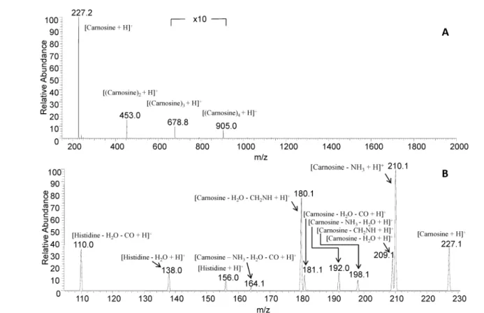
1. Collisionally-Induced Dissociation Products of the Protonate Dipeptide Carnosine: Structural elucidation, Fragmentation Pathways and Potential energy surface analysis.
Eslam Moustafa, Ida Ritacco, Emilia Sicilia, Nino Russo, Tamer Shoeib, Phys. Chem. Chem. Phys., 17, 12673-12682, 2015.
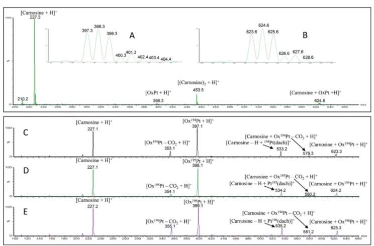
2. Fragmentation Pathways Analysis for the gas Phase Dissociation of Protonated Carnosine-Oxaliplatin Complexes.
Ida Ritacco, Eslam Moustafa, Emilia Sicilia, Nino Russo, Tamer Shoeib, Dalton Trans., 44, 4455-4467, 2014.
3. Mass spectrometric and computational investigation of the
protonated carnosine-carboplatin complex fragmentation.
Ida Ritacco, Mohamed Korany, Tamer Shoeib, Nino Russo, Emilia Sicilia, Inorg. Chem., 54, 7885-7897, 2015.
4. Investigation of the inertness to hydrolysis of Platinum(IV) produgs
Ida Ritacco, Gloria Mazzone, Nino Russo, Emilia Sicilia, Inorg. Chem., 2016, 55 (4), 1580-1586
5. DFT Investigation of the Mechanism of Action of Organoiridium(III)Complexes As Anticancer Agents
Ida Ritacco, Nino Russo, Emilia Sicilia, Inorg. Chem., 2015, 54 (22), 10801-10810
6. Hydrolysis in acidic environment and degradation of satraplatin: a joint experimental and theoretical investigation
vii 7. Hydrolysis of Platinum Compounds: Insights into Reactivity
from Accurate Ab Initio Calculations.
Ida Ritacco, Zhen Liu, Jeremy Harvey, Emilia Sicilia, Manuscript in preparation.
viii
Contents
Introduction……….i
List of paper included in this thesis………vi
1
I Cancer and Anticancer Drugs: General Principles ….. 1 Introduction………...3
4
1.1 Creation of a “cell tumor”………...4 4
1.1.1 Cancer cells vs healthy cells………...….4 4
1.2 Cancer therapies………..5 6
1.2.1 Surgery………..5 7
1.2.2 Radiotherapy……….5 7
1.2.3 Targeted therapies……….…6 7
1.2.4 PDT: Photodynamic therapy……….…6 1.2.5 Third cancer therapies: Chemotherapy…………...8 1.2.5.1 Chemotherapeutic drugs: classification and action mechanism ……….8 1.2.5.2 Chemotherapeutic drugs administration
Modes………10 1.2.5.3 Chemotherapeutic drugs resistance…………...10
7
1.3 Platinum(II) anticancer drugs………...11 9
1.3.1 Cisplatin: chemical properties………....12 1.3.2 Cell death: Cisplatin action mechanism………….13
ix
1.3.3 Cisplatin disadvantages………16 1.3.3.1 Cancer cell resistance to cisplatin………...…..17 1.3.4 Second and third generation drugs………...18 1.3.4.1 Second generation platinum drugs:
Carboplatin………..…...19 1.3.4.2 Third generation platinum drugs:
Oxaliplatin………...21
12
1.4 Platinum(IV) prodrugs. General aspects………....22 1.4.1 Platinum(IV) complexes with
no-bioactive axial ligands………...25 1.4.2 Platinum(IV) complexes with
bioactive axial ligands………...…26 1.4.3 Photoactivatable platinum(IV) ………...27 1.4.4 Platinum(IV) complexes reduction………...29 1.4.4.1 Reduction by glutathione: mechanistic hypothesis……….…31 1.4.4.2 Reduction by ascorbate: mechanistic hypothesis……….…33 1.4.4.3 Basolo reduction………...34 14
1.5 Non-platinum anticancer drugs………..…35 15
1.5.1 Ruthenium complexes as anticancer drugs……...36 1.5.2 Gold complexes as anticancer drugs……….37 1.5.3 Copper complexes as anticancer drugs………….38 1.5.4 Iridium complexes as anticancer drugs………….38
x References Chapter I………...40
II Theoretical Concepts and
Computational Methods...45
Introduction………...47
25
2.1 The Slater determinant ………..…49 25
2.2 The Hartree-Fock approximation………..…50 27
2.3 Density functional theory (DFT)………....51 29
2.3.1 The Hohenberg-Kohn theorems………...…52 2.3.2 The Kohn-Sham equations………...54
36 2.4 Coupled cluster approximation………60
References Chapter II………...62
43
III Carnosine Role in the Inhibition of
Platinum(II) Drugs………65
Introduction ...67
48
3.1 Aim of the study……….…69 48
3.2 Collisionally-Induced Dissociation Products of the
Protonated Dipeptide Carnosine: Structural elucidation, Fragmentation Pathways and
Potential energy surface analysis (Paper I)…………....70
51
3.3 Fragmentation Pathways Analysis for the Gas Phase Dissociation of Protonated
xi
3.4 Mass Spectometric and Computational Investigation of the Protonated Carnosine-Carboplatin Complex
Fragmentation (Paper III)...74
References Chapter III………..76
Paper I………..79
Paper II………...91
Paper III……….107
IV. Platinum Complexes Hydrolysis ………...122
Introduction……….124
4.1 Aim of the study………...127
4.2 Hydrolysis of Platinum Compounds: Insights into Reactivity from Accurate ab Initio Calculations (Manuscript in preparation)………128
4.3 Investigation of the inertness to hydrolysis of platinum(IV) prodrugs (Paper IV)……….136
4.4 Hydrolysis in acidic environment and degradation of satraplatin: a joint experimental and theoretical investigation (Paper V)……….138
References Chapter IV...141
Paper IV………..145
xii V. A New Class of Anticancer Drugs: Organoiridium(III)
Complexes………...192
Introduction………..194 5.1 Aim of the study………...195 5.2 DFT investigation of the mechanism of action
of organoiridium(III) complexes as
anticancer agents (Paper V)………...196 References Chapter V………..199
CHAPTER I
__________________
______
Cancer and Anticancer
Drugs: General Principles
3 Introduction
Cancer is a disease caused by abnormal cells present in our body, which multiply uncontrollably. No one knows the causes that originate this phenomenon, but, certainly, environmental, hereditary and related to lifestyle factors can contribute to the tumor onset.
There are two types of cancer: solid tumor, formed by a mass of diseased cells, and liquid tumor, generated by diseased cells present in liquid matrices, such as blood [1]. Furthermore, these tumors can be classified into “malignant and benign tumors”, which difference lies in the ability of malignant tumors to generate “metastasis”, that is other diseased cells within the body [2]. Cancer is the main cause of death in developed countries, and, in the last years, some types of cancer, such as breast, lung, colon and prostate cancer, have become increasingly common [2].
Currently, cancer is treated by three approaches: surgery, radiation therapy, in which radiations are used to kill the diseased cells, and chemotherapy, in which particular drugs are swallowed either intravenously or orally [3], being the former the most used. So, in this chapter, we will try to explain and understand, in general, how a tumor is generated, what are the drugs most commonly used in chemotherapy, advantages, disadvantages and recent developments of anticancer drugs.
4 1.1 Creation of a “cell tumor”
A cancer cell is born when the DNA of a healthy cell undergoes mutations [4].
To understand a cancer cell emergence, it is necessary to understand the cell cycle, in which the basis is called phosphorylation, and its phases. The phosphorylation consists of phosphate groups transfer from particular high-energy molecules to various substrates. The enzymes that catalyze this transfer are called "kinases". So, in our body cells, the four phases of the cell cycle are regulated by a set of protein kinases, called CDK (Cyclin-dependent Kinases). The CDK allow the passage from one phase to another and, if they are damaged, an abnormal cell cycle and, consequently, an abnormal cell division will occur. The result is the appearance of an abnormal cell, defined tumor cell [5]. The abnormal cells are eliminated by apoptosis. In the case of tumor cells, the apoptosis phenomenon doesn’t occur, so they continue to multiply uncontrollably.
1.1.1 Cancer cells vs healthy cells
Cancer cells differ from healthy cells for four reasons [2]: 1. uncontrolled proliferation;
2. loss of functionality: healthy cells, once they are formed, acquire a specific function that they will carry out for all
5
their life cycle. In contrast, cancer cells, particularly malignant ones, are all the same and, for that reason, they haven’t a specific role. The main consequence is that they lose functionality;
3. invasiveness: The tumor cells, in contrast to healthy ones, are located outside of the tissues or organs of origin;
4. ability to form metastases, which are small tumors located in body areas different from tumor origin site.
1.2 Cancer therapies
There are several methods for the treatment of cancer. To understand what is the best, it is needed to know and to interpret some information, for example the type and location of the tumor or the patient's health status [6].
1.2.1 Surgery
This treatment is used for tumors that have well-defined characteristics. In fact, they must be solid, small and, in particular, must be localized in the areas of the body easily reached by classical surgery [7].
1.2.2 Radiotherapy
This treatment consists of local irradiation of X-rays (external radiotherapy) or in ingestion of drugs, source of gamma rays
6
(internal radiotherapy). These rays damage the DNA of diseased cells, which cannot multiply and, so, they die.
Unfortunately, this treatment is not selective. In fact, it damages both cancer cells and healthy cells [7].
1.2.3 Targeted therapies
The use of targeted therapies is based on knowledge of the molecular mechanisms that lead to the development, growth and spread of cancer. They act in a selective way on some of these cellular processes and, for this reason, targeted therapies are defined “smart drugs”. Smart drugs are able to specifically coordinate molecular targets that are in cancer cells. In this way the therapy action appears to be selective and healthy cells are not damaged. Basically, the targeted therapies involve the correction of genetic mutations that are the basis of onset tumor, through inhibition or stimulation of the molecular target altered in the diseased cell. So, each genetic mutation underlying the disease has a specific therapy [8].
1.2.4 PDT: Photodynamic therapy
Photodynamic therapy is a non-invasive technique that uses a photosensitizing substance as a drug. The photosensitizer is activated by light at a particular wavelength.
7
The first step of PDT is the injection of the photosensitizer in the bloodstream. So, the drug enters in the metabolism of cancer cells, which are exposed to light.
At this point, the photodynamic therapy may act in three different ways:
1. ROS Production: The photosensitizer, activated by light, releases ROS, that is singlet oxygen, superoxide anion and hydrogen peroxide, which determine the diseased cell apoptosis;
2. Lack of nutrients: The photosensitizer can damage the blood vessels of cancer cells, preventing the tumor to receive the necessary nourishment;
3. Immune system activation: PDT can activate the immune system to attack cancer cells.
PDT can be used for the treatment of lungs and esophagus cancers. In this case, the light is provided by optical fiber cables inserted into endoscopes. For superficial tumors, such as skin cancers, light-emitting diodes (LEDs) are used.
The main disadvantage of PDT is that the light which is used to activate the photosensitizer cannot pass through more than 1 centimeter of tissue. So, this therapy can be used effectively only for the treatment of skin cancers and small tumors. It cannot be used for the treatment of metastases.
8
The main advantage of PDT is that healthy tissues are not damaged because diseased tissues are treated in a limited way. Furthermore, PDT may be used with other therapies, such as surgery, radiation therapy, or chemotherapy [9].
1.2.5 Third cancer therapies: Chemotherapy
One of the main advantages of chemotherapy is that it can be used for the treatment of solid tumors, such as testicular cancer, breast, colon, and fluids, such as leukemia, through the use of particular drugs, called "chemotherapeutic drugs". These drugs act on the cells that grow very quickly, i.e. the cancer cells, going to damage some steps of cell division. In this way, the cell replication is hampered and, as a result, the diseased cells die [7,10].
These various therapies can be used separately or can be used in mixed treatments. In fact, in the case of very large tumors, one can think to use radiotherapy or chemotherapy to reduce the tumor, and then to remove it surgically.
1.2.5.1Chemotherapeutic drugs: classification and Mechanism of Action
Chemotherapy drugs are classified according to their mechanism of action (MoA). In fact, they are divided into [1]:
9
1. Antimetabolites. The mechanism of action consists in the block or change the DNA biosynthetic pathways of cancer cells;
2. DNA interactive agents. They are divided into:
-Alkylating agents, which act by forming covalent bonds with the DNA of diseased cells. In this way, cell replication is blocked;
-Cross-linking agents, which coordinate the DNA on the same filament (intra-strand cross-linking) or between two different strands (inter-strand cross-linking). Platinum complexes belong to this subclass;
-Intercalating agents. The intercalation is a type of non-covalent interaction, in which planar molecules are inserted between pairs of the DNA bases;
-Kinases inhibitors, that is drugs that inhibit kinases; -DNA-cleaving agents. They, coordinating the DNA, cause the resolution of the filament to which they bind. From this division free radicals are formed.
3. Antitubulin agents. They interfere with the synthesis of microtubules, blocking the nuclear division and causing the death of the diseased cell.
10 1.2.5.2 Chemotherapeutic drugs administration modes
The chemotherapy consists of "treatment cycles", which can vary from three to eight, depending on the type of cancer and of the drugs used. On these last two factors also depend administration modes of anticancer drugs. In fact, they can be administered intravenously, to date the most widespread way, and by intramuscular or subcutaneous injection. In these cases, the drugs reach the diseased cells by means of transport in the blood. These types of administration have limitations and disadvantages, such as long times of administration, high costs for healthcare and, mainly, possible interaction of drugs with species present in the blood, with the consequent loss of their efficacy. For these reasons, in the last years, it has become always more important the oral administration, consisting in the use of particular drugs [11].
1.2.5.3 Chemotherapeutic drugs resistance
All anticancer drugs listed above, appear to be quite effective in cancer diseases. Unfortunately, in some patients there is the problem of the "resistance", that is the cancer cells survive, despite administration of chemotherapy drug, by adaptations toward the antitumor agent [7]. The resistance phenomenon could increase with the succession of chemotherapy cycles.
11
Among the many anticancer drugs, some cross-linking agents, in particular platinum complexes, appear to be very effective. So, in the following paragraphs we will focus on the detailed description of platinum complexes, comparing them with complexes having different metals than platinum, but equally effective in the fight against cancer.
1.3 Platinum(II) anticancer drugs
Over the years, the metal-containing compounds have had a fundamental role in the pharmaceutical field, due to their chemical-physical properties. The metals most commonly used in the chemical-pharmaceutical industry are the "transition metals". In fact, they have partially filled d orbitals, which are exploitable in coordination with various ligands due to their interesting electronic properties. Transition metals can be used in the design of new anticancer drugs [12].
The transition metal most commonly used in the anticancer drugs design is Platinum, which, by coordinating various ligands, generates anticancer platinum complexes. The large use of platinum complexes in the treatment of cancer is due to Barnett Rosenberg who, in 1969, discovered, casually, the importance of cisplatin [cis-diamminedichloridoplatinum (II)] in the inhibition of cell division. In fact, during an experiment to analyze the effects of the electric field on the bacterial growth,
12
he observed that the bacteria proliferation on platinum electrode had ceased. On this electrode, Rosenberg identified the formation of an electrolysis product, identified as Cisplatin (cis-[Pt(NH3)2(Cl)2]), considered to be responsible for cellular anti-proliferation. Assuming that the platinum complexes could be used in cancer treatment, Rosenberg and co-workers performed experiments with Sarcoma 180 and the L1210 leukemia. Due to the attained results, in 1971, cisplatin went in the phase I clinical trials and, in 1978, it was approved for use in testicula and ovarian cancer by the US Food and Drug Administration [13,14].
1.3.1 Cisplatin: chemical properties
In cisplatin, the metal ion, that is platinum, has a state of oxidation equal to +2, it has a d8 electronic configuration, has a coordination number equal to four and cisplatin has a planar-square structure.
The Pt(II) can coordinate the four ligands, two amines and two chlorides, as well as in cis position, also in trans position, generating, in this way, the transplatin trans-[Pt(NH3)2(Cl)2] (Fig. 1) [15].
13
Fig. 1: Chemical structure of a) cisplatin and b) transplatin Cisplatin, conversely transplatin, can coordinate the DNA of cells. For this reason, it was considered to be an active complex for the inhibition of cell division.
1.3.2 Cell death: Cisplatin action mechanism
Experimental and theoretical studies carried out over the years have provided many information concerning the mechanism of action of cisplatin. Cisplatin, and generally the platinum (II) complexes, induce the death of diseased cells through four steps: 1. Accumulation inside the cancer cell. The cisplatin comes into contact with the blood stream of patients intravenously. In plasma, it finds a high concentration of chloride ions (100 mM), which limits the replacement of its chloride ligands with water molecules (hydrolysis of cisplatin). In this way, the cisplatin preserves its neutrality, necessary requirement for its transport into the cell membranes and, consequently, for the accumulation in the diseased cell. Cisplatin can be transported into the cell membrane by passive diffusion or active transport.
14
2. Activation of the drug by hydrolysis. In the cytoplasm, the concentration of chloride ions decreases (4 mM). Consequently, the cisplatin chloride ligands are replaced by two molecules of water through an associative substitution reaction (SN2) (Scheme 1). The loss of chlorides, via hydrolysis, is the step that determines the activation of the drug. In fact, the reaction product, positively charged, is highly reactive towards nucleophiles present inside the cancer cell (Chapter IV).
Scheme 1: Hydrolysis reaction of cisplatin
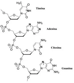
3. Interaction with DNA. The literature confirms that the hydrolysis products coordinate the DNA nucleobases of diseased cells, producing structural distortions. Consequently, the cancer cells are no longer able to reproduce themselves, because the DNA transcription is blocked, and, therefore, they die. The main DNA coordination sites are (Fig. 2):
the N1 or N7 nitrogens of adenine; The N3 nitrogen of cytosine;
15
The N7 nitrogen of guanine.
Theoretical and crystallographic studies have shown that the cisplatin prefers to coordinate the nitrogen N7 of guanine [16] and that it can coordinate the DNA in three different modes (Fig. 3):
1,2-intrastrand. 1,3-intrastrand; interstrand.
The 1,2-adduct intrastrand is the adduct more present in the cancer cell (65%) and more reactive, because it induces a significant distortion in the DNA double helix. The 1,3-intrastrand (10%) and the interstrand (3%) modes are significantly less important [17].
4. Cell Apoptosis. Generally, the platinum complex coordination inhibits the DNA replication, causing the death of the cancer cell. This phenomenon is called as "cell apoptosis" and is directly proportional to the cytotoxicity of cisplatin and, in general, to the cytotoxicity of platinum (II) complexes.
16
Fig. 2: Cisplatin binding sites in DNA
Fig. 3: DNA-Cisplatin adducts. a) 1,2-intrastrand cross-link, b) 1,3-intrastrand cross-link and c) interstrand cross-link.
1.3.3 Cisplatin disadvantages
Although cisplatin appears to be very cytotoxic to certain cancers, in vivo and in vitro studies have demonstrated that this
17
drug has many disadvantages. For example, nephro-, oto- and neuro-toxicity, nausea and/or vomiting may occur in patients subjected to this type of chemotherapy. In addition, the cisplatin is not very soluble in aqueous solutions, therefore, the intravenous administration is a limit, and it can be easily complexed from peptides that are in the extra- and intra-cellular environment. Anyway, the main disadvantage of the cisplatin treatment is the resistance of the cancer cell to the drug [17].
1.3.3.1 Cancer cell resistance to cisplatin
Many tumors appear to be resistant to cisplatin, and in general to anti-cancer drugs. In the particular case of cisplatin, the resistance is of two types:
intrinsic resistance, that means that the tumor cells do not respond to the drug;
acquired resistance. In this case, the cancer cell becomes resistant after exposure to the drug. For example, the ovarian cancer cells, initially, respond to the action of cisplatin but, in time, acquire resistance to the drug. The factors that modulate the resistance are three:
1. Drug changes during its accumulation in the intracellular
18
2. Presence of thiols in the intracellular environment. A cellular response to the accumulation of cisplatin in the cancer cell is an intracellular increase of thiols concentration. The thiol mainly present in the cells is the glutathione (GSH), which may coordinate the drug, decreasing its cytotoxicity. So, a result of the complexation phenomenon, which will be discussed specifically in Chapter III, is the platinum(II) complex inactivation;
3. Ability of the cancer cell to repair the DNA structural
modifications. In some cases, the diseased cells are able
to survive to DNA damage. It is not clear how this mechanism of cellular resistance develops, but some studies have shown that it happens [18].
Ultimately, cellular resistance to cisplatin is a complicated and multifactorial phenomenon.
1.3.4 Second and third generation drugs
Over the years, research has focused the attention on the design of new platinum (II) complexes, hoping to improve the clinical performance and to overcome the limits of cisplatin. Cleare and Hoeschele, affirmed in their articles that cisplatin derivatives can show antitumor activity only if the structural criteria listed below are fulfilled [19,20]:
19
presence of two amine groups in cis, because trans geometry is not active;
presence of two leaving groups in cis relative to one another;
the leaving groups must be removed easily;
the compounds must be neutral at time of administration; compounds with alkyl substituents and, at least, one
hydrogen atom on the amine ligands are more cytotoxic. So, the general structure for the new platinum (II) complexes is:
where X are the leaving groups, for example two chlorides or bidentate malonate, while R are hydrogen atoms or alkyl substituents. However, the “rules of thumb” are no longer generally followed because today are known numerous active Pt(II) complexes possessing trans geometry or positive charge.
1.3.4.1 Second generation platinum drugs: Carboplatin
Carboplatin [cis-diammine(1-1-cyclobutanedicarboxylato) platinum(II)] is called second-generation drug and it has been developed to overcome the toxicity limits of cisplatin (Fig. 4).
20
Fig. 4: Chemical structure of Carboplatin
The carboplatin MoA is very similar to that of cisplatin. However, carboplatin undergoes hydrolysis more slowly than cisplatin for the presence of the bidentate ligand cyclobutanedicarboxylate [21]. Therefore, being the carboplatin less reactive than cisplatin, it is also less toxic. Indeed, neuro- and oto-toxicity appear to be less evident after carboplatin treatment. This allows using higher doses of the drug during chemotherapy cycles. Currently, carboplatin is the drug most widely used for the treatment of ovarian cancer [22]. It appears to be not very effective against bladder, head and testicles cancer. In these cases, the drug most used is cisplatin.
The main disadvantage of carboplatin is the cell resistance [23]. Furthermore, the use of high doses of this drug can generate phenomena of myelosuppression (decreased production of cells in the blood by the bone marrow) and thrombocytopenia (decreased production of platelet). Another carboplatin problem is the possible complexation of the drug in the extra- or intra-cellular environment (Paper III).
21 1.3.4.2 Third generation platinum drugs: Oxaliplatin
Oxaliplatin [trans-(1R,2R-Diaminocyclohexane)oxalato platinum(II)] was developed to overcome cellular resistance limitations of cisplatin and carboplatin. Oxaliplatin is a cisplatin derivative having two bidentate ligands, that is 1R, 2R-diaminocyclohexano ligand (DACH) and the oxalate group as a leaving group (Fig. 5).
Fig. 5: Chemical structure of Oxaliplatin
The presence of the oxalate reduces the drug reactivity, limiting its toxicity, while the presence of the DACH increases the oxaliplatin lipophilicity, favoring its accumulation in the cytoplasm by passive absorption.
Oxaliplatin, as carboplatin, is hydrolyzed more slowly than cisplatin [24] and the hydrolysis product interacts with the DNA of the cancer cell to form the DNA-oxaliplatin adduct. This adduct is much more efficient in the inhibition of cell replication compared to cisplatin-DNA and DNA-carboplatin adducts, because the DACH ligand has a higher lipophilicity and steric hindrance [18]. To date, the oxaliplatin is used for the treatment
22
of breast, stomach, pancreas and, in combination with other drugs, to the colon cancers. Similarly to cisplatin and carboplatin, the main problem of oxaliplatin is its complexation by means of peptides that are in the extra- or intra- cellular environment (Paper II).
Cis-, Carbo- and Oxaliplatin play a key role in the fight against various cancers. However, their clinical use is limited due to their toxicity and cellular resistance. In addition, the research of new and more effective platinum(II) drugs did not produce satisfactory results, except in rare cases [25,14].
The requirement to develop platinum drugs strongly cytotoxic, has encouraged researchers to a clinical investigation of platinum(IV) complexes as anticancer prodrugs.
1.4 Platinum(IV) prodrugs. General aspects
The platinum(IV) complexes are anticancer prodrugs of structure [PtA2L2X2], that show a low-spin d6 octahedral geometry. These compounds are obtained by adding the axial ligands (X) to the platinum(II) complexes through oxidative addition. The axial ligands can be chloro, hydroxo or carboxylato groups and they regulate the pharmacological properties of platinum(IV) complexes, such as lipophilicity, reduction potentials, charge, selectivity and accumulation in the
23
cancer cell. Instead, the equatorial ligands, that is amine ligands (A) and leaving groups (L), determine the prodrugs cytotoxicity. The platinum(IV) prodrugs are more inert than platinum(II) drugs, therefore, more stable and, consequently, less reactive. This all translates into a lower tendency to hydrolysis or substitution of the ligands in the blood, after the intravenous administration and intra-cellular environment. Due to this low reactivity, the platinum(IV) complexes may be taken orally and accumulate in the cytosol in higher quantities than platinum(II) complexes.
In cancer cells, platinum(IV) prodrugs are activated by reduction, which consists in the release of the two axial ligands and the real drug, that is the platinum(II) complex. Then, this complex coordinates the final target, i.e. the DNA (Scheme 2) [26].
Scheme 2: Synthesis and reduction of platinum(IV) complexes According to various studies, in the cytoplasm, the reduction of the platinum(IV) complexes occurs due to glutathione (GSH) and ascorbate as reducing agents. The reduction is considered
24
the activation step of the platinum(II) drugs, because it favors the transition from the platinum(IV) complexes, kinetically inert and not very reactive, to platinum(II) complexes, kinetically labile, and, therefore, more cytotoxic of platinum(IV) prodrugs [27].
The intracellular reduction rate of the platinum(IV) complexes depends on the type of cell and on the ligands coordinated to the metal, which define the coordination sphere of the complex. Generally, the platinum(IV) prodrugs have many advantages compared to the platinum(II) complexes [13]: due to their stability they could be assumed orally, they have fewer side effects than the platinum(II) complexes and the axial ligands can be modified to improve and increase the pharmacological properties of the complex.
Platinum(IV) anticancer complexes can be divided into three categories based on the type of axial ligands:
Complexes with non-bioactive axial ligands; Complexes with bioactive axial ligands; Photoactivatable complexes.
25
1.4.1 Platinum(IV) complexes with non-bioactive axial ligands
The main platinum(IV) complexes that do not exhibit bioactive axial ligands are Tetraplatin, iproplatin and satraplatin, formally called JM-216 (Fig. 6).
Fig. 6: Chemical structure of a) tetraplatin, b) iproplatin and c) satraplatin
Clinically, tetraplatin and iproplatin have been abandoned because the former exhibited a strong neurotoxicity, while the latter was less active than cisplatin and carboplatin.
Satraplatin, instead, has been the first platinum(IV) complex to be administered orally, because being inert and, therefore, stable, accumulates in large quantities in the cancer cell and do not exhibit cellular resistance.
From satraplatin, by reduction, a highly active platinum(II) complex is formed, i.e. the amminedichlorido (cyclohexylamine) platinum(II) (JM-118). As it happens for the cis-, carbo- and
26
oxaliplatin, inside the cancer cell JM118 undergoes hydrolysis and, subsequently, interacts with DNA, generating the DNA- JM118 adduct and inhibiting cell division (Scheme 3).
Schema 3: Satraplatin reduction
Although satraplatin is responsible for the regression of various cancers, such as prostate cancer, it is not used as a chemotherapeutic drug because it does not guarantee patient survival. The satraplatin can be used in combination with other drugs for the treatment of prostate cancer, lungs cancer and small solid tumors [13].
1.4.2 Platinum(IV) complexes with bioactive axial ligands
The selectivity of platinum(IV) complexes towards the cancerous tissues or intracellular targets depends heavily on the axial ligands [13]. An example of a bioactive axial ligand is estradiol. The platinum(IV) complex, shown in Figure 7, which has estradiol in axial position, turns out to be highly cytotoxic towards the breast and ovarian cancer cells, because the estrogen
27
released after the intracellular reduction of the platinum(IV) complex encourages HMGB1 protein expression [28].
Another complex belonging to this category is the platinum(IV) prodrugs with ethacrynic acid in the axial position (Fig. 7), developed to overcome the limitations related to cellular resistance. In fact, the ethacrynic acid is an inhibitor of glutathione-S-transferase [29].
Subsequently, Lippard and co-workes proposed as platinum(IV) prodrug with bioactive axial ligands the Mitaplatin, a platinum(II) complex which has dichloroacetates in the axial position (Fig. 7). The dichloroacetates, once released into the cancer cells by reduction, change the mitochondrial membrane potential, causing apoptosis of cancer cell [30].
Fig. 7: Chemical structure of Platinum(IV) complexes with bioactive axial ligands
1.4.3 Photoactivatable platinum(IV)
Platinum(IV) complexes activated directly into the tumor tissue by irradiation are defined photoactivable complexes. These
28
complexes are inert in the dark and do not show interaction with the glutathione that is in the cancer cell. Furthermore, in the active and cytotoxic form, they have azides and amines in trans between them and the hydroxyl groups in axial position (Fig. 8). Amines, that are in the equatorial position, may be replaced by pyridine, which makes the complexes more cytotoxic and more resistant to irradiation [31].
The MoA differs from that of platinum(IV) and platinum(II) complexes. In this case, a drug photodecomposition takes place through the transfer of one electron from each azido ligand to platinum, generating N2 and platinum(II) complex (Scheme 4). The latter, then, by interacting with the DNA of the cancer cell, generates the Pt-DNA adduct [13].
Fig. 8: Chemical structure of Photoactivatable Platinum(IV) complexes.
29
Scheme 4: Possible mechanism for the photoreaction of a generic photoactivatable platinum(IV) upon irradiation with UVA.
1.4.4 Platinum(IV) complexes reduction
Since many years, the literature affirms that the antitumour activity of platinum(IV) complexes is directly proportional to their ability to reduce themselves in the cancer cell. So, to understand the biological activity of these prodrugs it is necessary to understand what are the factors that influence their reduction. Generally, the reduction of the platinum(IV) complexes involves the loss of the two axial ligands and the release of the platinum(II) drug, which, by binding to DNA, stops cell replication and induces cellular apoptosis.
The reduction depends on the structure of the prodrug, therefore on the nature of axial and equatorial ligands, and on the reducing agents involved in the process. A measure of the platinum(IV) complexes reduction can be obtained through cyclic voltammetry experiments. Indeed, Choi et al. explained that the reduction kinetics strongly depends on the reduction potentials of the complexes submitted to voltammetric analysis [32].
30
Furthermore, it is worth mentioning that the reduction Pt(IV)-Pt(II) is irreversible, therefore, to define the reduction potential of platinum(IV) complexes all the rules of the irreversible cyclic voltammetry must be observed [33,34].
Cyclic voltammetry studies have confirmed the relationship between the nature of the ligands and the reduction degree. According to these studies, the axial ligands have a greater influence on the reduction kinetics than the equatorial ligands. For example, platinum(IV) complexes, which have chlorides in axial position and have reduction potential of about E=-4mV, are reduced more easily than complexes that in axial position have carboxylate groups (-250≤ E ≥ -350 ) or hydroxides (E=-664mV). Then, higher is the negativity of reduction potential, lower is the reduction kinetic of the prodrugs. The variation of the equatorial ligands has an influence next to nothing on the reduction kinetics.
The main reducing agents, inside the cancer cell, are glutathione and ascorbic acid, although the reduction mechanism of the platinum(IV) to platinum(II) complexes is not clear. It is thought that the reduction of the platinum(IV) complexes using glutathione and/or ascorbic acid can occur with inner- or outer-sphere electron-transfer mechanisms [35-37].
The inner-sphere electron-transfer mechanism proceeds through the formation of a strong covalent bond between the oxidant and
31
reductant species. This covalent bond is seen as an ideal bridge, due to which the reductant species can transmit electrons to the oxidant species. In a chemical reaction, the "bridge structure" can be a very stable minimum, a reaction intermediate or a transition state.
The outer-sphere electron-transfer mechanism does not require for the formation of bridges between oxidizing and reductant species, but consists in an electron transfer from the reaction environment to the oxidizing species.
The reaction can also be catalyzed by Pt(II) [38].
1.4.4.1 Reduction by glutathione: mechanistic hypothesis
The reduction mechanism of the platinum(IV) complexes by glutathione should proceed with the formation of a covalent bond between the axial ligand of the complex and the glutathione sulfur, favoring an electron transfer from the reductant species to the axial ligand. In this way, the electronic density of the axial ligand is shifted to the metal center, making the Pt-axial ligand bond weaker and the release of the axial ligand easier. For example, Ranford et al. suggested that the reduction of the complex [PtCl2(OCOCH3)2(NH3)2] by glutathione, occurs via the formation of a bridge between the sulfur of the reductant species and the acetate oxygen coordinated to platinum (Fig. 9).
32
After bridge formation, there is an electrical charge transfer from glutathione, which is considered as a nucleophile, to the axial ligand and, subsequently, to the platinum. This weakens the acetate-Pt bond and facilitates the axial ligand release [39,27].
Fig. 9: Transition state form in the case of the interaction [PtCl2(OCOCH3)2(NH3)2] and Glutathione
Starting from the transition state, shown in Figure 9, two hypothesis of mechanism have been proposed.
The first hypothesis proposes the formation of an intermediate, following the release of the first axial ligand, in which the platinum is pentacoordinated and it has an oxidation number equal to +3. Then, from this minimum, by interaction with another glutathione and release of the second axial ligand, the platinum(II) drug would be released (Scheme 5a).
The second hypothesis proposes a concerted mechanism, that is the release of the first axial ligand, due to glutathione, and, simultaneously, the release of the second axial ligand to form the corresponding platinum(II) complex (Scheme 5b).
33
Scheme 5a: First hypothesis of platinum(IV) complexes reduction
Scheme 5b: Second hypothesis of platinum(IV) complexes reduction
1.4.4.2 Reduction by ascorbate: mechanistic hypothesis
In the case of reduction by ascorbate, the hypothesis of mechanism proposes an outer sphere electron transfer as result of the interaction between the platinum(IV) complex and the reductant agent. It is not clear how the electron transfer occurs, but it has been hypothesized that the reductant species releases two electrons to the reaction environment, and, subsequently, these electrons are transferred from reaction environment to the
34
platinum(IV) complex, causing the release of the axial ligands together with the drug (Scheme 6) [27,37].
Scheme 6: Platinum(IV) complexes reduction by ascorbate
1.4.4.3 Basolo reduction
Basolo studied the reduction of some platinum(IV) complexes, such as [Pt(en)3]4+ (en = ethylenediamine), by [Cr(bipy)3]2+ (bipy = 2,2′-bipyridine). Basolo affirmed that this reduction occurs through an outer-sphere mechanism, in which there is an electron transfer to generate a Pt(III) intermediate, without, however, direct interaction with the reductant species. The Pt(III) could react rapidly with another [Cr(bipy)3]2+ to yield Pt(II) complex. When the platinum(IV) complex has chloride ligands in the axial position, such as [Pt(NH3)5Cl]3+, it is suggested an inner sphere mechanism where one axial chloride is coordinated to the complex of Cr(II), forming a bridge. He also suggested a two electron reduction mechanism to yield Pt(II) and Cr(IV) complexes with the latter that is reduced directly to Cr(II) [38].
35
The Basolo reduction mechanism can also be applied when the reductant species is a platinum(II) complex.
To date, only the Basolo reduction mechanism is supported by experimental and theoretical data (Paper IV). Unfortunately, this mechanism has limitations, because it can be used only when the platinum(IV) complexes has certain types of axial ligands.
As regards the reduction mechanisms by glutathione and ascorbate only few experimental data are available that are not supported by theoretical data. For this reason, future studies should be devoted to the design of prodrugs inert to hydrolysis (Paper IV), the interaction of this complexes with proteins that are in the blood and cancer cells and the understanding of their reduction mechanism.
1.5 Non-platinum anticancer drugs
Currently, only the platinum-based drugs have been approved for clinical practices. However, in the last years, research has turned its attention to the study of new metal-containing complexes. These non-platinum complexes have low toxicity and high activity against tumors that appear to be resistant to cisplatin. Inside the cancer cells, these drugs, which contain
36
main group or transition metals, can act on different targets, such as DNA, enzymatic functions, and/or the formation of ROS. The metals most frequently used for this purpose are ruthenium(II) and ruthenium(III), gold(I) and gold(III), copper(II), and iridium(III) [40,41]. The iridium (III) seems to be an excellent candidate for the development of new anticancer drugs, due to its peculiar characteristics (Paper VI).
1.5.1 Ruthenium complexes as anticancer drugs
Ruthenium(II) and Ruthenium(III) exhibit antitumor activity. The ruthenium(II) forms penta- and hexa-coordinated complex, while the ruthenium(III) forms only hexa-coordinated complexes. Furthermore, ruthenium(III) is considered a prodrug, because, as confirmed by in vivo experiments, before reaching the final target, it is always reduced to ruthenium(II).
The most studied ruthenium(II) complexes, due to their high cytotoxic activity, are those having arene ligands. The cytotoxic activity of ruthenium complexes should be based on their ability to interact with the DNA of cancer cells or result from other interaction types. The ruthenium(II) complexes exhibit cytotoxic activity similar to that of carboplatin and lower than that of cisplatin.
In most cases, the ruthenium(II) complexes have, in addition to arene ligands, a chloride ligand. It seems that the hydrolysis is
37
the fundamental step for the activation of these complexes. In fact, they may undergo rapid hydrolysis leading to the loss of the chloride ligand and to the rapid interaction with aminoacids and various nucleobases.
The more cytotoxic ruthenium(III) complexes, are those that have indazole ligands, able to interact with the DNA of cancer cells and to induce apoptosis via the intrinsic mitochondrial pathways [40,41].
1.5.2 Gold complexes as anticancer drugs
Gold(I) complexes which have phosphine ligands are used as anticancer drugs, because they are inhibitors of the enzyme thioredoxin reductase (TR). This enzyme is present in high concentrations in cancer cells and promotes the diseased cells proliferation. The inhibition of TR enzyme by gold(I) complexes causes cell apoptosis.
Gold(III) complexes have a chemical behavior very similar to that of the platinum(II) complexes. In fact, gold(III) complexes are isoelectronic with platinum(II) complexes, although gold(III) complexes in the physiological environment undergo rapid hydrolysis and subsequent reduction to gold(I). For this reason, over the years, gold(III) complexes more inert to hydrolysis and, then, to reduction have been designed [40,41].
38 1.5.3 Copper complexes as anticancer drugs
Many copper(II) complexes, containing N-, S-, and/or O-ligands, have a good cytotoxic activity. The action mechanism of copper(II) complexes is not yet clear, but it is assumed that these complexes cause cellular apoptosis by interacting with the DNA or by inhibiting the enzymes that are, in high amounts, inside the cancer cell.
The use of copper(I) complexes as anticancer drugs is limited by their low stability and by their tendency to be oxidized [40,41].
1.5.4 Iridium complexes as anticancer drugs
In the last years, a great deal of efforts has been devoted to the study of new anticancer drugs containing iridium as a metal center. In particular, iridium(III) complexes seem to be very inert and less reactive. While ruthenium complexes are stabilized by the presence of arene ligands, iridium(III) complexes are stabilized by the presence of the pentamethylcyclopentadienyl ligand (Cp*), which is negatively charged. Furthermore, half-sandwich organoiridium(III) cyclopentadienyl complexes of general formula [(η5 -Cpx )Ir(III)- (X∧Y)Z]0/+, where Cpx=Cp*, Cpxph (tetramethyl-(phenyl)-cyclopentadienyl) or Cpxbiph (tetramethyl(biphenyl)-cyclopentadienyl), X∧Y=bidentate ligand with nitrogen, oxygen,
39
and/or carbon donor atoms, and Z=Cl, H2O, or pyridine (py), have high cytotoxicity.
These complexes can undergo hydrolysis, losing the Z ligand, and, subsequently, can interact with the DNA of the cancer cell, or to produce ROS, by interacting with other species that are in the cancer cell, increasing the intracellular oxidative stress. In both cases cellular apoptosis occurs (Paper VI).
40 REFERENCES CHAPTER I
[1] Nussbaumer, S., Bonnabry, P., Veuthey, J. L., Fleury- Souverain, S., Talanta 2011, 85, 2265-2289.
[2] Rapporti Associazione Italiana Registri Tumori.
[3] Shewach, D. S., Kuchta, R. D., Chem. Rev. 2009, 109, 2859-2861.
[4] Evan, G. I., Vousden, K. H., Nature 2001, 411, 342-348. [5] Collins, K., Jacks, T., Pavletich N. P., PNAS USA 1997, 94,
2776-2778.
[6] World Health Organization (WHO), Geneva: World Health
Organization 1979, 48.
[7] Smith, H. J., Introduction to the Principles of Drug Design
and action-Harwood academic publishers, III edition, 1998,
1-568.
[8] Urruticoechea, A., Alemany, R., Balart, J., Villanueva, A., Viñals, F., Capellá, G., Current Pharmaceutical Design,
2010, 16, 3-10.
[9]Wilson, B. C., Canadian Journal of Gastroenterology, 2002, 16(6), 393–396.
[10] Johnstone, R. W., Ruefli, A. A., Lowe S. W., Cell 2002, 108, 153-164.
41
[12] Baile, M. B., Kolhe, N. S., Deotarse, P. P., Jain, A. S., Kulkarni, A. A., International Journal of Pharma Research
& Review 2015, 4 (8), 59-66.
[13] Dilruba, S., Kalayda, G. V., Cancer Chemother Pharmacol
2016, 77 (6), 1103-1124.
[14] Wong, E., Giandomenico, C. M., Chem. Rev. 1999, 99, 2451-2466.
[15] Alderden, R. A., Hall, M. D., Hambley, T. W., Journal of
Chemical Education 2006, 83, 728-734.
[16] Alberto, M. E., Butera, V., Russo, N., Inorg. Chem. 2011, 50 (15), 6965-6971.
[17] Jamieson, E. R., Lippard, S. J., Chem. Rev. 1999, 99, 2467-2498.
[18] Perez, R. P., Eur. J. Cancer 1998, 34, 1535-1542.
[19] Cleare, M. J., Hoeshcele, J.D., Plat. Met. Rev. 1973, 17, 2-13.
[20] Cleare, M. J., Hoeshcele, J.D., Bioinorg. Chem. 1973, 2, 187-210.
[21] Pavelka, M., Lucas, Maria Fatima A., Russo, N., Chem.
Eur. J. 2007, 13, 10108-10116.
[22] Ozols R. F., Bundy B. N., Greer B. E., Fowler J. M., Clarke-Pearson D., Burger R. A., Mannel R. S., DeGeest K., Hartenbach E. M., Baergen R., J. Clin. Oncol. 2003, 21, 3194-3200.
42
[23] Stewart, D. J., Crit. Rev. Oncol. Hematol. 2007, 63, 12-31. [24] Lucas, Maria Fatima A., Pavelka, M., Alberto, M., Russo,
N., J. Phys. Chem. 2009, 113, 831-838.
[25] Galanski, M., Jakupec, M. A., Keppler, B. K., Curr. Med.
2005, 12, 2075-2094.
[26] Wexselblatt, E., Yavin, E., Gibson D., Angew. Chem. Int.
Ed. 2013, 52, 6059-6062.
[27] Wexselblatt, E., Gibson D., J. Inorg. Biochem. 2012, 117, 220-229.
[28] He, Q., Liang, C. H., Lippard, S. J., Proc. Natl. Acad. Sci.
USA 2000, 97, 5768-5772.
[29] Ang, W. H., Khalaila, I., Allardyce, C. S., Juillerat-Jeanneret, L., Dyson, P. J., J. Am. Chem. Soc. 2005, 127, 1382-1383.
[30] Dhar, S., Lippard, S. J., Proc. Natl. Acad. Sci. USA 2009, 106, 2219-2220.
[31] Butler, J. S., Sadler, P. J., Curr. Opin. Chem. Bio.2013,
17, 175-188.
[32] Choi, S., Filotto, C., Bisanzo, M., Delaney, S., Lagasee, D., Whitworth, J. L., Jusko, A., Li, C., Wood, N. A., Willingham, J., Schwenker, A., Spaulding, K., Inorg. Chem.
1998, 37, 2500.
43
[34] Evans, D. H., O’Connel, K. M., Petersen, R. A., Kelly, M. J., J. Chem. Educ. 1983, 60 (4), 290.
[35] Hall, M. D., Hambley, T. W., Coordination Chemistry
Reviews 2002, 232, 49-67.
[36] Johnstone, T. C., Suntharalingam, K., Lippard, S. J., Chem.
Rev. 2016, 116 (5), 3436-3486.
[37] Ejehi, Z., Ariafard, A., Chem. Commun., 2017, DOI: 10.1039/ C6CC07834F.
[38] Basolo., Chem. Rev. 2016, 116 (5), 3436-3486.
[39] Chen, L., Lee, P. F., Ranford, J. D., Vittal, J. J., Wong, S. Y., J. Chem. Soc. Dalton 1999, 1209-1212.
[40] Trudu, F., Amato, F., Vanhara, P., Pivetta, T., Pena-Mèndez, E. M., Havel, J., Journal of applied biomedicine
2015, 13, 79-103.
[41] Ott, I., Gust, R., Arch. Pharm. Chem. Life Sci. 2007, 340, 117-126.
CHAPTER II
__________________
______
Theoretical Concepts and
Computational Methods
47
Introduction
The main goal of theoretical chemistry is to explore the electronic structure and properties of more or less complex systems.
Quantum mechanics emerged in the beginning of the twentieth century as a new discipline because of the need to describe phenomena, which could not be explained using Newtonian mechanics or classical electromagnetic theory. These phenomena include the photoelectric effect, blackbody radiation and the rather complex radiation from an excited hydrogen gas. These and other experimental observations led to the concepts of quantization of light into photons, the particle-wave duality, the de Broglie wavelength and the fundamental equation describing quantum mechanics, namely the Schrödinger equation.
Particles defined as "quantum systems” are described by a wave function (𝜓) [1], that is a mathematical object that contains all the information of the system under examination. A quantum system can be described by the non-relativistic time-independent
Schrödinger equation[2]:
𝐻̂𝜓 = 𝐸𝜓 (2.1)
where 𝐻̂ is the Hamiltonian operator for a system of nuclei and electrons described by position vectors RA and ri, respectively.
48 In atomic units, the Hamiltonian for N electrons and M nuclei is
𝐻̂ = − ∑ 1 2𝛻𝑖 2 𝑁 𝑖=1 − ∑ 1 2𝑀𝐴𝛻𝐴 2 𝑀 𝐴=1 − ∑ ∑ 𝑍𝐴 𝑟𝑖𝐴 𝑀 𝐴=1 𝑁 𝑖=1 + ∑ ∑ 1 𝑟𝑖𝑗 𝑁 𝐽>1 𝑁 𝑖=1 + ∑ ∑ 𝑍𝐴𝑍𝐵 𝑅𝐴𝐵 𝑀 𝐵>𝐴 𝑀 𝐴=1 (2.2)
In Eq. (2.2) the first term is the operator for the kinetic energy of the electron; the second term is the operator for the kinetic energy of nuclei; the third term is the coulomb attraction between electrons and nuclei; the fourth and fifth terms are the repulsion between electrons and nuclei, respectively.
Due to the Born-Oppenheimer approximation [3], in equation (2.2), the second term can be neglected and the last term can be considered constant. The remaining terms in (2.2) compose the electronic Hamiltonian, that is the Hamiltonian describing the motion of N electrons in the field of M point charges.
𝐻̂ 𝑒𝑙𝑒𝑐 = − ∑ 1 2𝛻𝑖 2 𝑁 𝑖=1 − ∑ ∑ 𝑍𝐴 𝑟𝑖𝐴 𝑀 𝐴=1 𝑁 𝑖=1 + ∑ ∑ 1 𝑟𝑖𝑗 𝑁 𝐽>1 𝑁 𝑖=1 (2.3)
Quantum chemistry tries to find approximate solutions to the electronic Schrödinger equation, because it can be solved exactly only for one- electron systems, such as hydrogen atom or H2+ molecule.
49
2.1 The Slater determinant
A single electron is described by the wave function 𝜓(r,s), spin
orbital, that define both its spatial distribution,𝜓(r), and its spin,
α(s) or β(s).
𝜓(𝑟, 𝑠) = {𝜓(𝑟)𝛼(𝑠)𝑜𝑟 𝜓(𝑟)𝛽(𝑠)
(2.4)
For a system of N electrons, the total electronic wave function is equal to the product of the wave functions of each electron and it is known as Hartree Product. The Hartree product doesn’t respect the antisymmetry principle (a general statement of the
Pauli exclusion principle) that requires that electronic wave
functions be antisymmetric with respect to the interchange of the space and spin coordinates of any two electrons [4]. So, the
Slater determinant has been introduced as the simplest
antisymmetric wave function which can describe the ground state of an N-electron system:
𝜓𝑁𝑆𝐷= 1 √𝑁!|| 𝜓𝑖(𝑟1, 𝑠1) 𝜓𝑗(𝑟1, 𝑠1) … 𝜓𝑗(𝑟1, 𝑠1) 𝜓𝑖(𝑟1, 𝑠1) 𝜓𝑗(𝑟2, 𝑠2) … 𝜓𝑘(𝑟2, 𝑠2) ⋮ 𝜓𝑖(𝑟𝑁, 𝑠𝑁) ⋮ 𝜓𝑗(𝑟𝑁, 𝑠𝑁) … ⋮ 𝜓𝑘(𝑟𝑁, 𝑠𝑁) || (2.5)
The Slater determinant incorporated both exchange effects, arising from the requirement that |𝜓|2 be invariant to the
![Fig. 2 Energy-resolved collision induced dissociation curves of [carnosine + H] + as obtained on the Aquity TQ tandem mass spectrometer employing argon as a collision gas as described in the Instrumental section](https://thumb-eu.123doks.com/thumbv2/123dokorg/2867287.9108/100.892.73.427.71.516/collision-dissociation-carnosine-spectrometer-employing-collision-described-instrumental.webp)
![Fig. 4 Proposed mechanism for the collision induced fragmentation of [carnosine + H] + to produce the fragments observed at m/z 198 and 180.](https://thumb-eu.123doks.com/thumbv2/123dokorg/2867287.9108/101.892.204.695.75.335/proposed-mechanism-collision-induced-fragmentation-carnosine-fragments-observed.webp)
![Fig. 6 Proposed mechanism for the collision induced fragmentation of [carnosine + H] + to produce the fragments observed at m/z 210, 192 and 164.](https://thumb-eu.123doks.com/thumbv2/123dokorg/2867287.9108/102.892.208.695.66.641/proposed-mechanism-collision-induced-fragmentation-carnosine-fragments-observed.webp)
![Fig. 9 Proposed mechanism for the collision induced fragmentation of [carnosine + H] + to produce the fragment ion observed at m/z 156, 138 and 110.](https://thumb-eu.123doks.com/thumbv2/123dokorg/2867287.9108/104.892.207.695.69.674/proposed-mechanism-collision-induced-fragmentation-carnosine-fragment-observed.webp)
![Fig. 2 Energy-resolved collision induced dissociation curves of [Carnosine + OxPt + H] +](https://thumb-eu.123doks.com/thumbv2/123dokorg/2867287.9108/113.892.140.740.269.1022/energy-resolved-collision-induced-dissociation-curves-carnosine-oxpt.webp)


