whale (Kogia sima). In spite of their low extant
di-versity, physeteroids are also known by several fossil species, which suggest an Oligocene origin (Mchedlidze 1970) and, especially, a significant ra-diation during the subsequent Miocene (Bianucci & Landini 2006; Kimura & Hasegawa 2006; Lambert 2008; Lambert et al. 2010, 2016, in press; Boers-ma & Pyenson 2015; Collareta et al. 2017, in press; Benites-Palomino et al. 2020; Paolucci et al. 2020). The Miocene sperm whales display an impressive variety of cranial morphologies and reconstructed
A NEW RECORD OF PHYSETEROIDEA FROM THE UPPER MIOCENE OF THE PIETRA LECCESE (SOUTHERN ITALY): SYSTEMATICS, PALEOECOLOGY AND TAPHONOMY OF A FOSSIL MACRORAPTORIAL SPERM WHALE
EMANUELE PERI1,2,*, ALBERTO COLLARETA2 & GIOVANNI BIANUCCI2
1*Corresponding author. Dottorato Regionale Pegaso in Scienze della Terra, Università di Pisa, via Santa Maria 53, 56126 Pisa, Italy. E-mail: [email protected]
2Dipartimento Scienze della Terra, Università degli Studi di Pisa, Via S. Maria 53 56126 Pisa, Italy. E-mail: [email protected]; [email protected]; [email protected]
To cite this article: Peri E., Collareta A. & Bianucci G. (2020) - A new record of Physeteroidea from the Upper Miocene of the Pietra leccese (southern Italy): systematics, paleoecology and taphonomy of a fossil macroraptorial sperm whale. Riv. It. Paleontol. Strat., 126(3): 751-769.
Abstract. We report on a partial skeleton of sperm whale (Cetacea, Odontoceti, Physeteroidea) from the Pietra
leccese, a Miocene limestone widely exposed in the Salento Peninsula (southern Italy). This specimen was found in Tortonian strata cropping out at the Cisterna quarry, not far from the holotype of the stem physeteroid Zygophyster varolai. The presence of a deep and rectilinear groove medial to the tympanosquamosal recess of the squamosal, the
bowed mandibles, and some dental features suggest that this specimen belongs to a still undescribed new genus and species of macroraptorial sperm whale that displays some affinities with the late Miocene Acrophyseter from Peru.
Nevertheless, due to the incompleteness and poor preservation state of the skull, we abstain from creating a new taxon. The teeth exhibit both apical wear and deep occlusal facets, and three teeth even lost their crowns. These dental modifications suggest that the studied specimen used a raptorial feeding strategy for preying upon food items such as large-sized bony fishes or diminutive marine mammals. The bones are mostly disarticulated and broken, and some of them preserve traces hinting at the action of macro-scavengers, possibly including both sharks and bony fishes. Furthermore, the skull is pervasively encrusted by oysters, which suggests that it laid on the seafloor for a long time before being buried. This find provides new clues about the composition of the Miocene vertebrate assemblage of the Pietra leccese and indicates that various macroraptorial sperm whale species inhabited the Mediterranean Basin during the Tortonian.
Received: April 9, 2020; accepted: August 29, 2020
Keywords: Cetacea; Odontoceti; teeth; biostratinomy; scavenging; Mediterranean Sea; Salento Peninsula; Tortonian.
I
ntroductIonNowadays, the superfamily Physeteroidea represents a small group of odontocete cetaceans consisting of only three living species: the great sperm whale (Physeter macrocephalus), the pygmy
G
eoloGIcalandpaleontoloGIcal frameworkThe Pietra leccese is a Miocene calcareous formation exposed in the Salento Peninsula, i.e., the southern portion of the Apulia region of southea-stern Italy (Fig. 1a). It consists of yellowish, weakly stratified biomicrites and biosparites that are rich in planktic foraminifera and calcareous nannofossils (Bossio et al. 2005; Margiotta 2015). The Pietra lec-cese overlies the Aquitanian Lecce Formation and, at the top, it gradually passes into the Messinian Calcareniti di Andrano Formation, which closes the Miocene sedimentary cycle of Salento (Mazzei et al. 2009). According to micropaleontological studies, the Pietra leccese spans between the early Burdiga-lian and the late Tortonian, and its depositional en-vironment was the outer shelf (Bossio et al. 2005; Mazzei et al. 2009). The deposition of the Pietra leccese was repeatedly interrupted, resulting in si-gnificant hiatuses that are often marked by accumu-lations of glauconite (Bossio et al. 2005; Margiotta 2006, 2015; Mazzei et al. 2009). These interruptions in deposition might explain why the thickness of this formation is so reduced (i.e., a few tens of me-ters) even though its sedimentation lasted for about 11 million years (Margiotta 2006; Mazzei et al. 2009). body sizes. Furthermore, differing from modern
Physeteroidea that feed upon deep-water cephalo-pods and fish by means of suction, several Miocene sperm whales were raptorial predators that used their powerful bites to grab, break or tear apart their prey items (Lambert et al. 2010, 2014, 2016; Lam-bert & Bianucci 2019). Unfortunately, a significant part of the rich fossil record of physeteroids con-sists of fragmentary and poorly diagnostic remains (mostly isolated teeth) (Hampe 2006; Vélez-Juarbe et al. 2015; Lambert et al. 2016). For this reason, the real extent of the past ecological diversity of this odontocete clade is still far from being completely understood. Here we report on a new partial skel-eton of Physeteroidea from the Tortonian strata of a calcareous formation of southern Italy, informally named Pietra leccese. This new specimen, kept at the Museo di Storia Naturale, Università di Pisa, consists of several fragmentary bones pervasively encrusted by oysters. Hereinafter, we provide a de-tailed description of this specimen and briefly dis-cuss its systematic affinities, functional morphology, paleoecology and taphonomic history.
Institutional abbreviations: IRSNB: Institut Royal des
Sciences Naturelles de Belgique, Bruxelles, Belgium; MAS, Museo Arqueologico de Salango, Salango, Ecuador; MAUS, Museo dell’Am-biente, Università del Salento, Lecce, Italy; MGPUF, Museo di Storia Naturale, Sezione di Geologia e Paleontologia, Università degli Studi di Firenze, Firenze, Italy; MLP, Museo de La Plata, La Plata, Argen-tina; MNHN, Muséum National d’Histoire Naturelle, Paris, France; MSNC, Museo Civico di Storia Naturale di Comiso, Comiso, Italy; MSNUP, Museo di Storia Naturale, Università di Pisa, Calci, Italy; MUSM, Museo de Historia Natural de la Universidad Nacional Ma-yor de San Marcos, Lima, Peru; MUSNAF, Museo di Storia Naturale dell’Accademia dei Fisiocritici, Siena, Italy; USNM, National Museum of Natural History, Smithsonian Institution, Washington DC, USA.
List of specimens directly examined for comparison:
Acrophyseter deinodon (MNHN SAS 1626); Acrophyseter robustus
(MNHN PI 239); Acrophyseter sp. (MUSM 2182); Albicetus oxymycterus (USNM 10923); Aprixokogia kelloggi (USNM 187015); Diaphorocetus poucheti (MLP 5-6); Eudelphis mortzelensis (IRSNB M.523); Kogia breviceps (MAS 4000; MNHN 1976-37; USNM 283625); Kogia pusilla
(MGPUF 1540V); Kogia sima (MSNC 3450; MUSNAF Mam410); Koristocetus pescei (MUSM 888); Livyatan melvillei (MUSM 1676); Orcinus orca (MSNUP 298; MSNUP 301); Orycterocetus crocodilinus (USNM
22926); Orycterocetus sp. (MAUS 29/1); Physeter macrocephalus (MNHN
1831; MSNUP 265, 266, 267); Physeterula dubusi (IRSNB M.527); Pliokogia apenninica (MSNUP I-17603); Scaldicetus caretti (IRSNB
M.512); Scaphokogia cochlearis (MNHN PPI 229, MUSM 971, MUSM
1998); Scaphokogia totajpe (MUSM 973); Scaphokogia sp. (MUSM
972); Scaphokogiinae sp. (MUSM 3291; MUSM 3405); Thalassocetus antwerpiensis (IRSNB M.525); Zygophyseter varolai (MAUS 229/1).
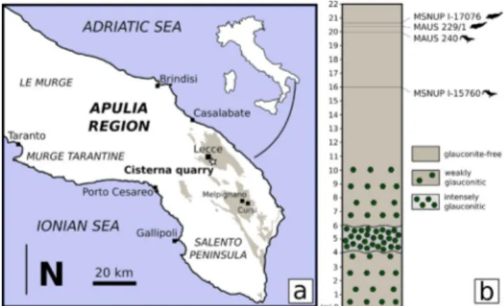
Fig. 1 - Geographic locations of the finding site and stratigraphic position of the studied specimen. (a) Location of the site
where the fossil cetacean specimen MSNUP I-17076 was found (Cisterna quarry, black star) on a schematic regional geological map. Sand brown-shaded areas indicate the expo-sures of the Pietra leccese, the Miocene calcarenite limestone from which the fossil originates. Redrawn and modified after Calia et al. (2013). (b) Section of the Cisterna quarry with the
stratigraphical position of the cetacean specimens MSNUP I-17076 (the physeteroid specimen studied herein), MAUS 229/1 (holotype of the physeteroid Zygophyseter varolai),
MAUS 240 (holotype of the ziphiid Messapicetus longirostris),
and MSNUP I-15760 (referred specimen of Messapicetus lon-girostris). Redrawn and modified after Mazzei et al. (2009).
The Pietra leccese is widely known for its abundant fossil record of marine vertebrates that includes cetaceans, sirenians, turtles, and bony and cartilaginous fishes. Cetaceans are represented by both Odontoceti (toothed whales) and Mysticeti (baleen whales) (Bianucci & Varola 2014). Odon-tocetes include the holotypes of “Eurhinodelphis” salentinus (Eurhinodelphinidae), Hesperoinia dalpiazi
(Inioidea?), Messapicetus longirostris (Ziphiidae), Ru-dicetus squalodontoides (Kentriodontidae), Zygophyseter varolai (Physeteroidea), and fragmentary remains of
Inticetidae and Squalodontidae (Moncharmont Zei 1950, 1956; Bianucci et al. 1992, 1994a, b, 2016; Bia-nucci & Varola 2014; BiaBia-nucci & Landini 2006; Peri et al. 2019). Mysticetes include the holotype of Ar-chaeschrichtius ruggieroi (Eschrichtiidae) and
fragmen-tary remains of Balaenidae and Cetotheriidae (Bi-sconti & Varola 2000, 2006; pers. obs.). Sirenians are represented by the dugongid Metaxytherium medium
(Bianucci et al. 2003). Sea turtle remains belong to
Psephophorus polygonus (Dermochelyidae), Trachyaspis lardyi (Cheloniidae) and indeterminate cheloniids
(Chesi et al. 2007). Bony fishes (Osteichthyes) inclu-de members of the families Istiophoridae (Makaira),
Scombridae (Acanthocybium and Scomberomorus),
Fi-stularidae (Fistularia), Serranidae and Holocentridae
(Carnevale et al. 2002). Cartilaginous fishes (Chon-drichtyes) belong to Lamniformes, Carcharinifor-mes and MyliobatiforCarcharinifor-mes (Menesini 1969; Sorce 2009). Lastly, marine vertebrate Digestichnia (inclu-ding one coprolite and putative gastroliths) have also been reported (Tavani 1973; Collareta et al. 2019b).
S
yStematIcpaleontoloGyCETACEA Brisson, 1762 OdOntOceti Flower, 1867
Physeteroidea Gray, 1821
Physeteroidea gen. et sp. indet.
Figs. 2-13
Referred material: MSNUP I-17076, a partial skeleton
consisting of the fragmentary cranium and mandibles, 20 detached teeth (some of which are incomplete), the incomplete atlas, the left radius and the incomplete ulna, six fragmentary ribs, and other frag-ments of postcranial bones. All these remains were enclosed in six blocks of limestone cut during the quarrying activities. Unfortuna-tely, only some of the blocks containing the partial skeleton were recovered. Most of the preserved bones are broken and/or cut in several portions due both to taphonomic processes and cutting of the limestone blocks while still in the quarry.
Horizon and locality: MSNUP I-17076 was found in 1987
by Angelo Varola at the Cisterna quarry (indicative geographic coor-dinates: 40°19’35” N; 18°11’40” E), near Cavallino (Salento Peninsu-la, southern Italy) (Fig. 1a). In this quarry, a 22-m-thick stratigraphic section of Pietra leccese was measured (Fig. 1b) and dated by means of foraminiferal biostratigraphy to an age interval spanning from the Langhian to the middle Tortonian (Mazzei et al. 2009). MSNUP I-17076 was collected ca. 1.3 m below the top of the section, i.e., ca. 25 cm above the holotype of Zygophyseter varolai and ca. 80 cm
abo-ve the holotype of Messapicetus longirostris (Bianucci & Landini 2006;
Bianucci et al. 1992, 2016). The middle and upper portions of the
Cisterna section, including MSNUP I-17076 and the holotypes of Z. varolai and M. longirostris (Fig. 1b), was dated to the Neogloboquadrina acostaensis zone of Iaccarino & Salvatorini (1982), sensu Foresi et al.
(1998), which ranges between ca. 10.5 and 8.14 Ma (middle Torto-nian) (Mazzei et. al. 2009; Bianucci et al. 2016).
Description. Except when explicitly stated
otherwise, the anatomical terminology utilized in the present paper follows Mead & Fordyce (2009).
Total body length estimate and ontogeny. In MSNUP
I-17076, the distance between the proximal limit of the left mandibular fossa and the sagittal plane of the cranium (275 mm) is slightly lower than in Zygo-physeter varolai (305 mm), whose total body length
was estimated at about 6.5–7 m (Bianucci & Lan-dini 2006). Based on these data, it is reasonable to hypothesize that MSNUP I-17076 was likely slightly smaller than Z. varolai (total body length between
5.5 and 6.5 m). The closed pulp cavity of the teeth and the pronounced degree of dental wear suggest that MSNUP I-17076 was an adult individual.
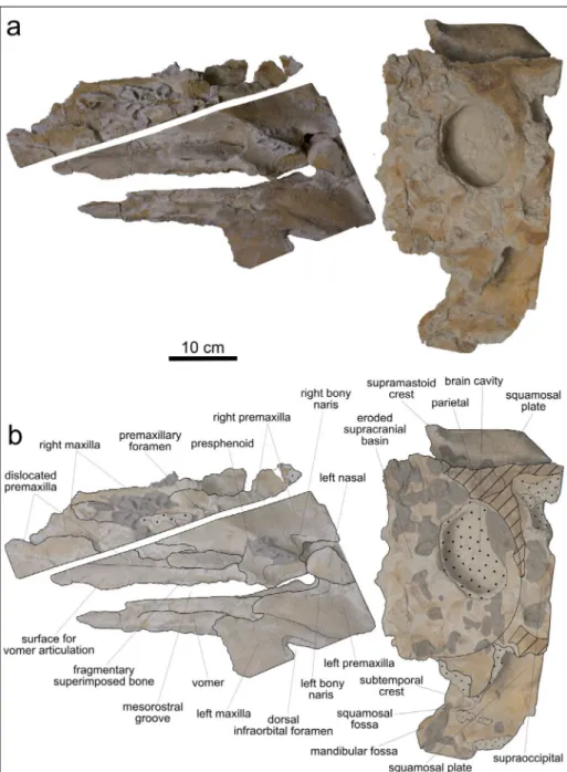
Cranium. The cranium was extracted from
th-ree limestone blocks: one of these blocks contained most of the neurocranium, excluding its anterior portion, whereas the other two blocks contained the fragmentary rostrum and the anterior portion of the neurocranium. The preserved portion of the rostrum includes the incomplete vomer, the frag-mentary premaxillae and maxillae, the left nasal, and the presphenoid.
In dorsal view (Fig. 2), the bones exposed along the rostrum (i.e., the maxillae, premaxillae and vomer) are more or less displaced laterally from their original (anatomical) position. Consequently, it is difficult to reconstruct the original geometry of these bones. The left maxilla exhibits a large, ellipti-cal dorsal infraorbital foramen. The maxilla/prema-xilla suture is not clearly discernible; it is probably represented by a light groove running near to the dorsal infraorbital foramen. The right premaxilla exhibits a longitudinal depression that extends la-teral to the mesorostral groove. This depression
re-presents the sutural surface for the dislocated vomer. At about half of its length, the preserved portion of the right premaxilla hosts a small, elliptical prema-xillary foramen (transverse width: 1.4 cm; anteropo-sterior length: 2.0 cm). The left premaxilla widens transversely, reaching a maximum width of 9.5 cm behind the left bony naris, at level of the left nasal. The vomer is dorsally exposed along the left half of the rostrum; it is V-shaped in cross section and rea-ches a maximum transverse width of 25 mm. Due to post-mortem breakage, the presphenoid is displa-ced with respect to its original anatomical position. It is transversely concave and borders the right half of the left bony naris. The right bony naris is signi-ficantly smaller than the left one (transverse width:
right naris, 5.4 cm; left naris, 1.7 cm). The left naris is aligned to the mesorostral groove. In this respect, MSNUP I-17076 apparently differs from Acrophyse-ter deinodon, OrycAcrophyse-terocetus crocodilinus and ZygophyseAcrophyse-ter va-rolai, all having the right bony naris aligned with the
mesorostral groove (Bianucci & Landini 2006; Lam-bert et al. 2016). However, the pronounced
displace-ment of the bones that comprise the preserved part of the rostrum of MSNUP I-17076 suggests con-sidering this character with caution. The preserved left nasal displays a sub-rectangular shape and is su-perimposed to the posteromedial preserved portion of the left premaxilla. There are no hints suggesting that a right nasal was originally present, this bone being lacking in extant Physeter and Kogia and never Fig. 2 - Cranium, in dorsal view, of MSNUP I-17076, Physe-teroidea indet. from the upper Miocene of the Pie-tra leccese (southern Italy). Photograph (a) and expla-natory line drawing (b). The grey-shaded areas denote the presence of encrusting oysters. The dotted pattern indicates skull areas covered by hard sediment. The obli-que black stripes correspond to the areas damaged by the quarrying activities. The stippled lines indicate the re-constructed margins.
detected in any fossil physeteroid except Acrophyseter.
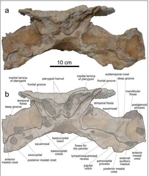
The supracranial basin is largely hidden by encru-sting oysters and patches of hardened sediment that, coupled with the overall poor state of preservation of the fossil, prevent us to identify the bone sutures on most of the neurocranium. Moreover, based on the preserved material, it is not clear whether the su-pracranial basin extended onto the rostrum. The po-sterodorsal portion of the neurocranium is broken and a large hole exposes the brain cavity. Posteriorly to this cavity, the incomplete supraoccipital and part of the right parietal are preserved. Lateral to the su-pracranial basin, the preserved glenoid process of the squamosal forms an almost right angle with the sagittal plane. The dorsal surface of the squamosal
is posteromedially expanded to form a broad squa-mosal plate. Delimiting anterodorsally the extent of the squamosal plate, the subtemporal crest is steep and robust, and appears as completely preserved on the left side only. Posterodorsal to the subtemporal crest, the squamosal plate exhibits a wide and deep squamosal fossa.
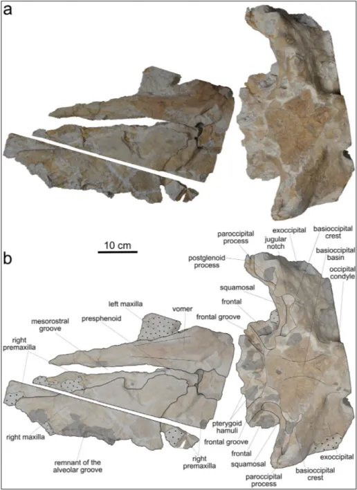
In ventral view (Fig. 3), the right maxilla is broadly extended on the rostrum, whereas the right premaxilla displays a narrow exposure medial to the right maxilla. The ventral portion of the left maxilla is missing or covered by hardened sediment, whe-reas only a small eroded piece of the left premaxil-la is visible ventrally. The preserved portion of the right maxilla exhibits a roughly triangular shape. On
Fig. 3 - Cranium, in ventral view, of MSNUP I-17076, Physete-roidea indet. from the upper Miocene of the Pietra lec-cese (southern Italy). Pho-tograph (a) and explanatory line drawing (b). The grey-shaded areas denote the pre-sence of encrusting oysters. The dotted pattern indicates skull areas covered by hard sediment. The stippled lines indicate the reconstructed margins.
the medial side, the right maxilla displays a large and deep notch. The lateral margin of the right maxilla is mostly eroded, except for a small portion along its anterior half. At this level, the maxilla displays a shal-low groove covered by encrusting oysters. We inter-pret this groove as a remnant of the upper alveolar groove. The laterally shifted vomer is longitudinally exposed on the ventral surface of the rostrum; this ventral exposure is triangular and tapers anteriorly. The basioccipital displays a roughly trapezoidal sha-pe and is wider than long. The basioccipital basin is deep, but the angle formed by the basioccipital crests cannot be ascertained due to the poor preservation state of these structures. The exoccipital exhibits a wide ventral exposure posterior to the squamosal.
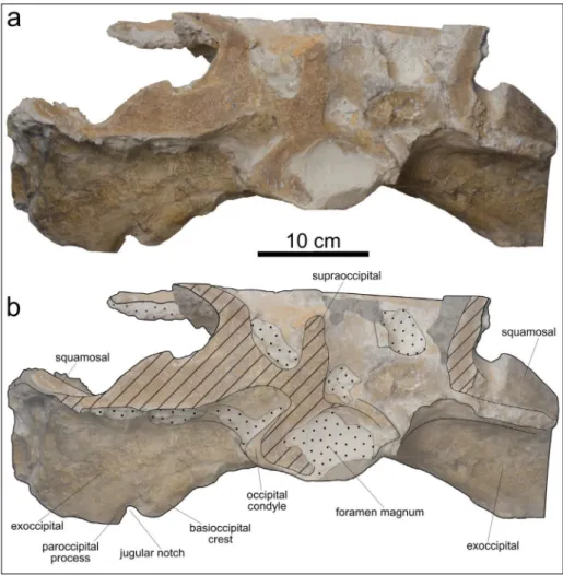
In posterior view (Fig. 4), a wide jugular notch is visible between the ventromedial margin (paroc-cipital process) of the exoc(paroc-cipital and the posterior end of the basioccipital crest. The jugular notch se-emingly differs from that of Acrophyster spp. in not
being ventrally closed (Lambert et al. 2016); howe-ver, the poor preservation state of MSNUP I-17076 prevents from drawing definitive conclusions in this respect.
In anterior view (Fig. 5), the preserved por-tion of the neurocranium displays right and left ven-tral bulges that are best observable on the broken transverse surface. These bulges represent the col-lapsed remains of the pterygoid hamuli. Ventrolate-ral to the pterygoid hamuli, the frontal grooves, the displaced and poorly preserved medial laminae of the pterygoids, and the elongated and robust squa-mosals are discernible. In frontal view, both the pre-served proximal portions of the squamosals form an angle of about 20° with the horizontal plane of the cranium. Medial to the partially preserved left mandibular fossa, the triangular tympanosquamosal recess of the squamosal is laterally delimited by a deep, elongated, and rectilinear groove that recalls a similar feature observed in Acrophyseter spp.
(Lam-bert et al. 2008; Lam(Lam-bert et al. 2016) Posterior to the tympanosquamosal recess and the mandibular fossa, the external auditory meatus is limited by the anterior and posterior meatal crests. Between the ju-gular notch and the ventral border of the squamosal, a deep fossa for the insertion of the periotic is di-scernible. Only the left fossa is visible, the right one being covered by a large oyster.
Fig. 4 - Cranium, in posterior view, of MSNUP I-17076, Physe-teroidea indet. from the upper Miocene of the Pie-tra leccese (southern Italy). Photograph (a) and expla-natory line drawing (b). The grey-shaded areas denote the presence of encrusting oysters. The dotted pattern indicates skull areas covered by hard sediment. The obli-que black stripes correspond to the areas damaged by the quarrying activities. The stippled lines indicate the re-constructed margins.
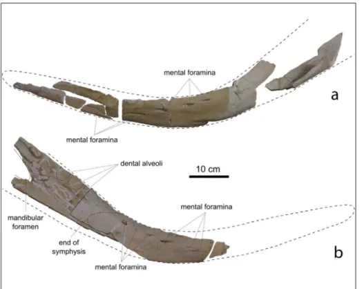
Mandibles. The mandibles of MSNUP I-17076
are badly damaged and fragmented (Fig. 6). They exhibit a bowed shape, having a convex ventral mar-gin and a concave dorsal marmar-gin in lateral view. Such a peculiar shape of the mandible is also observed in
Acrophyseter spp. and, less pronounced, in Zygophyse-ter varolai and PhyseZygophyse-terula dubusi. On the inZygophyse-ternal wall
of a posterior fragment of the right mandible, three shallow depressions, representing remains of the po-steriormost dental alveoli, are discernible. As in Acro-physter deinodon, these posterior alveoli are roughly
vertical and closely spaced. On the same mandibu-lar fragment, the anterior portion of the mandibumandibu-lar foramen is preserved, although it is not possible to reconstruct in detail its shape due to the poor pre-servation state. The anterior edge of the mandibular foramen seems to be located slightly posterior to the posterior margin of the posteriormost dental alveo-lus, a condition similar to Z. varolai (where the
ante-rior edge of the mandibular foramen is located about 5 cm posterior to the posterior margin of the po-steriormost alveolus) and different from A. deinodon
(where the anterior edge of the mandibular foramen is located at the level of the anterior margin of the penultimate mandibular tooth). On the lateral surfa-ce of the right mandible, seven mental foramina are discernible; they are placed inside longitudinal groo-ves and are roughly aligned along an oblique line that rises posteriorly. Seven mental foramina, displaying a similar alignment, are also observed on the right mandible of A. deinodon, whereas in Z. varolai only
three foramina aligned along a horizontal line are observed. A fragment, excavated by grooves, of the medial surface of the right mandible represents the posterior portion of the symphyseal surface, sugge-sting that the dentaries were not fused to each other, differing from the living sperm whale Physeter macro-cephalus that exhibits fused dentaries (Werth 2004). Fig. 5 - Cranium, in anterior view, of
MSNUP I-17076, Physete-roidea indet. from the upper Miocene of the Pietra lec-cese (southern Italy). Pho-tograph (a) and explanatory line drawing (b). The grey-shaded areas denote the pre-sence of encrusting oysters. The dotted pattern indicates skull areas covered by hard sediment. The stippled lines indicate the reconstructed margins.
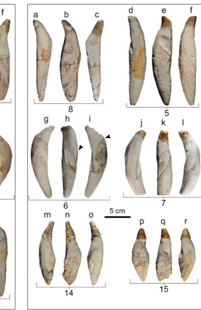
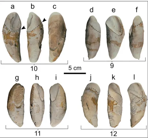
Teeth. All the 20 preserved teeth are detached
and more or less complete; they are moderately slender, elongated, and straight to weakly curved. Their shape varies from fusiform to cylindrical (Figs. 7, 8). Measurements of the 16 best preserved teeth are reported in Table 1 (note that the tooth numbering used both here, in figure 7 and 8, and in Table 1 does not reflect the anatomical position of the teeth). The enamelled crown is conical, with a roughly circular transverse section. The ratio betwe-en the crown lbetwe-ength (including the estimated api-cal portion of the crown missing, due to wear) and the total length of the tooth ranges from 0.22 to 0.23, inside the wider range of variation of Acro-physeter sp. MUSM 2182 (0.22-0.27). A smaller ratio
is observed in Livyatan melvillei (0.10-0.11) and
Zygo-physeter varolai (0.18). The enamel layer is
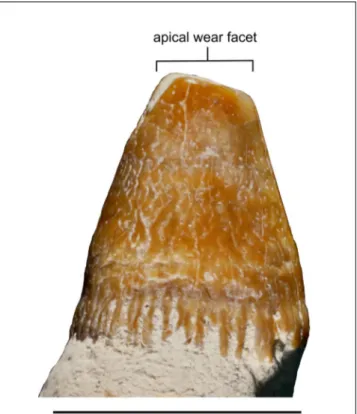
modera-tely thick (ca. 1 mm) and its surface is sculptured by longitudinal, shallow, sinuous and anastomo-sing grooves; a 5-8 mm high crown-root transition area is characterized by interdigitating portions of enamel covering the proximal portion of the root (Fig. 9). A similar sculpture and interdigitation of the enamel surface, although less pronounced, was also observed in Z. varolai and in some teeth of the
holotype of Scaldicetus caretti (Lambert & Bianucci
2019). The total length of the complete root
ran-ges from 126 to 141 mm, these values being inter-mediate between those of Acrophyster sp. MUSM
2182 (59-90 mm) and Z. varolai (140-177 mm). The
root is moderately slender (ratio between maximum transverse diameter and total length of the root
ran-1 Table Table 1 1 2 3 4 5 6 7 8 9* 10* 11* 12* 13 14 15 16 Total length 167.0 163.1 144.5 129.8 158.8 +131.2 +118.6 +148.4 108.5 111.8 97.4 +98.5 +131.2 +120 +98.4 +124.2 Root length 136.9 141.4 129.4 111,2 142.5 126.1 +109.1 129.6 108.5 111.8 97.4 +98.5 +131.2 +92.9 +80.4 +124.2 Crown length 28.1 22.6 15.9 21.1 15.9 - 13.8 23.7 - - - 26.2 19.3 -
Maximum labiolingual diameter of root 38.5 39.3 30.6 32.6 37.4 29.6 27.5 38.9 38.3 31.5 32.4 37.4 38.0 - - 39.9
Maximum mesiodistal diameter of root 30.4 33.1 32.9 32.5 31.9 29.4 30.0 - 29.6 - 37.8 31.4 26.8 - - 32.7
Transverse diameter at crown base 20.3 18.8 17.1 - 19.0 20.5 17.6 18.3 - - - 16.6 17.8 -
Mesiodistal diameter at crown base - 17.9 - 17.0 17.9 - 18.4 18.2 - - - 17.3 18.0 -
Tab. 1 - Measurements of detached teeth of Physeteroidea indet. (MSNUP I-17076) from the Tortonian of the Pietra leccese (southern Italy). Note that the tooth numbering used here does not reflect the anatomical position of the teeth. All measurements are reported in millimeters. *, crown completely lost due to in vivo wearing; +, incomplete.
Fig. 6 - Left (a) and right (b) man-dibular rami of MSNUP I-17076, Physeteroidea in-det. from the upper Miocene of the Pietra leccese (sou-thern Italy), in lateral view. The stippled lines indicate the hypothesized outline of the mandibles and the poste-rior alveoli.
ging between 0.23 and 0.30) when compared with
Acrophyster sp. MUSM 2182 (0.27-0.38) and Z. varolai
(0.31-0.35). All roots are fusiform and display their greatest transverse diameter roughly at mid-length, with the exception of tooth 7 that has a cylindrical shape. With respect to this character, the teeth of MSNUP I-17076 differ from those of Acrophyseter
spp. and Z. varolai, in which the maximum
diame-ter of the root is located closer to the crown. The transverse section of the root varies from elliptical to circular. As in Z. varolai, the proximal extremity
of the root tapers, and the pulp cavity is fully closed
in all teeth, suggesting that MSNUP I-17076 was an adult specimen. This condition is different from that observed in other physeteroids such as Physeter macrocephalus and the holotype of Scaldicetus caretti, in
which the pulp cavity remains open also in physi-cally mature specimens (Boschma 1938; Lambert et al. 2016; Lambert & Bianucci 2019). The external root is partially covered by a layer of dark material (Fig. 7d-f, j, l-r; Fig. 8g, i, p, q; Fig. 10a, b, d, f, j-l), also observed in other fossil teeth of physeteroids, named ‘gingival collar’ and interpreted as the area of connection between the gum and the teeth by
Fig. 7 - Six detached teeth of MSNUP I-17076, Physeteroidea in-det. from the upper Miocene of the Pietra leccese (southern Italy) in lingual/labial (a, c, d, f, g, i, j, l, m, o, p, r) and distal (b, e, h, k, n, q) views. The stippled lines indicate the apical limit of the gingival collar. The dotted lines delimit the oc-clusal facets. The arrows indicate the lowering of the apical limit of the gingival collar. The numbers indicate the corre-sponding series of measurements in Table 1.
Fig. 8 - Six detached teeth of MSNUP I-17076, Physeteroidea in-det. from the upper Miocene of the Pietra leccese (southern Italy) in lingual/labial (a, c, d, f, g, i, j, l, m, o, p, r) and distal (b, e, h, k, n, q) views. The stippled lines indicate the apical limit of the gingival collar. The dotted lines delimit the oc-clusal facets. The arrows indicate the lowering of the apical limit of the gingival collar. The numbers indicate the corre-sponding series of measurements in Table 1.
Bianucci & Landini (2006). The gingival collar may represent fossil evidence of the calculus (dental plate) observed in extant odontocetes (Loch et al. 2011; Lambert & Bianucci 2019). The gingival col-lar is rather proximoapically long in the teeth of MSNUP I-17076, as in Acrophyseter deinodon,
where-as in Z. varolai it forms a narrower band. The
sha-pe and orientation of the gingival collar provide information on the original inclination of the to-oth with respect to the horizontal plane (Bianucci & Landini 2006). Based on these observations, all the roots appear to have been significantly incli-ned within their alveoli. Moreover, two opposite sides of the root show an obliquely oriented gin-gival collar (Fig. 7d, j, l, p-r; Fig. 8g, I; Fig. 10d, f, j, l), and as such, they are here interpreted as the labial/lingual surfaces of the tooth, whereas the other opposite sides where the gingival collar turns roughly orthogonal to the main axis of the root (Fig. 7e, n; Fig. 8q) are here interpreted as the mesial/distal surfaces of the tooth. In this way, it is possible to reconstruct that most of the teeth
exhibit a mesiodistal compression of the root, a feature that is also observed in the closely spaced posterior upper teeth of A. deinodon, as well in the
posterior teeth of the extant delphinid Orcinus orca
(pers. obs. on MSNUP 298 and MSNUP 301). The degree of apical wear varies significantly among the teeth, with teeth 1 and 14 having only a few millimeters of their apexes missing, whereas teeth 5 and 7 lack a significant portion of crown. Most of the teeth display occlusal facets caused by the opposing teeth and affecting the upper portion of root not covered by the gum. In some teeth, the occlusal facet is deep and occupies a significant fraction of the root surface (up to 50%). Three teeth display two occlusal facets: 1) a short, almost vertical occlusal surface affecting the crown (e.g., tooth 3) and in one case extending also over the upper portion of the root (e.g., tooth 4); and 2) a longer and deeper occlusal surface that obliquely crosses the distal surface of the root. Four teeth completely lack the crown: their preserved apical portion is rounded and grooved by a deep, wide, obliquely oriented occlusal facet (Fig. 10). Such a tooth shape, also observed in one tooth of Z. varo-lai (Bianucci & Landini 2006: fig. 8D) and in several
physeteroid teeth from the Miocene of Germany (Hampe 2006: figs. 3-5), could be a consequence of the breakage of the distal portion of the teeth, probably due to the progressive wear at the inter-face between opposing teeth. In teeth with most developed occlusal facets, there is also an abrupt lowering of the gingival collar that recalls the con-dition observed in teeth of Z. varolai and S. caretti.
This suggests the presence of a gum cavity hosting the opposing tooth during jaw adduction.
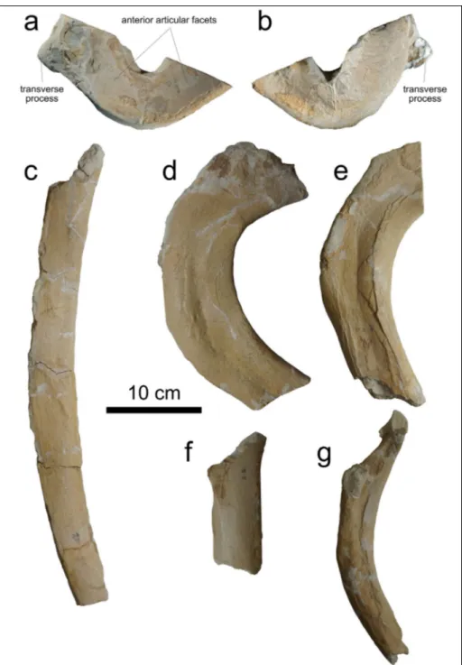
Vertebrae. The incomplete atlas and a single
thoracic vertebra are the only preserved elements of the vertebral column of MSNUP I-17076.
As a consequence of quarrying activities, the atlas was saw-cut at an angle of about 23° with the horizontal plane, and its dorsal half was con-sequently lost (Fig. 11a, b). Among fossil physete-roids, the atlas was described only in Acrophyseter robustus, Placoziphius duboisi, Zygophyseter varolai and,
based on fragmentary material, in Physeterula dubusi
and Ferecetotherium kelloggi (Mchedlidze 1970;
Bia-nucci & Landini 2006; Lambert 2008; Lambert et al. 2016). Therefore, our comparison was made with these fossil species and with the extant Ko-gia spp. and Physeter macrocephalus. The fragmentary Fig. 9 - Close-up of a tooth crown of MSNUP I-17076,
Physete-roidea indet. from the upper Miocene of the Pietra leccese (southern Italy), showing the ornamentation pattern of the enamel and the crown-root transition area.
atlas of MSNUP I-17076 is a robust and massi-ve bone having an anteroposterior massi-ventral length of 63 mm and an estimated transverse width of about 200 mm. In anterior view, the ventrolateral margin is semi-circular and regularly convex, thus differing from the almost rectilinear margin obser-ved in P. macrocephalus. The poorly preserved right
transverse process appears prominent and pointed as in A. robustus, P. dubusi and Z. varolai, whereas
in P. macrocephalus the transverse process is short
and dorsoventrally developed. We observed an intermediate condition (transverse process short and moderately developed dorsoventrally) in Kogia
spp. and P. duboisi. The articular facet is moderately
concave, as in the other physeteroids, and the tran-sverse width across the lateral margins of the arti-cular facets could be estimated at 150 mm, slightly smaller than in Z. varolai (175 mm).
The single thoracic vertebra lacks the neural arch and the transverse processes. The preserved centrum, which is fused to both the epiphyses, is deformed by diagenetic compression and its ventral surface is partially covered by encrusting oysters. The anterior and posterior profile are di-stinctly heart-shaped. Judging by the broken bases of the pedicles, which are located at the
dorsola-teral corners of the centrum, the neural arch was originally wide. The anteroposterior length of the centrum is 57 mm, whereas its transverse width is 83 mm.
Forelimb. The left radius and the
incomple-te left ulna are the only forelimb bones preserved in MSNUP I-17076 (Fig. 12). Comparisons were made with the extant Kogia spp. and Physeter macro-cephalus, as well as with the only fossil physeteroids
having these bones preserved (i.e., Brygomophyse-ter shigensis and Ferecetotherium kelloggi; Mchedlidze
1970; Hirota & Barnes 1994).
The almost complete left radius (Fig. 12a, d) is an elongated and lateromedially compressed bone. The total length of the radius is 204 mm, whereas its anteroposterior width at mid-length is 63 mm. The ratio between these two measure-ments is 0.31, a value greater than in B. shigensis and F. kelloggi (0.26) but significantly smaller than in P. macrocephalus (0.43-0.48) and Kogia breviceps
(0.47-0.59), both having a proportionally shorter radius than MSNUP I-17076. In lateral view, the anterior margin of the radius is straight, whereas the po-sterior margin is weakly concave. On the proximal epiphysis, the articular surface with the humerus is oriented obliquely with respect to the main axis
Fig. 10 - Four detached teeth of MSNUP I-17076, Physete-roidea indet. from the up-per Miocene of the Pietra leccese (southern Italy) in lingual/labial (a, c, d, f, g, i, j, l) and posterior (b, e, h, k) views. Note how the tooth crowns are completely mis-sing due to in vivo wear. The stippled lines indicate the gingival collar. The dotted lines delimit the occlusal fa-cets. The arrows indicate the lowering of the apical limit of the gingival collar. The numbers indicate the corre-sponding series of measure-ments in Table 1.
of the radius. The anteroposterior length of this articular surface is about 67 mm, whereas its ma-ximum lateromedial width is 33 mm. The distal epiphysis is more lateromedially compressed than the proximal epiphysis and, in lateral view, it exhi-bits a distal margin that is more convex than in P. macrocephalus. The radius of MSNUP I-17076 lacks
the peculiar posterodistal projection that characte-rizes the radius of B. shigensis (Hirota & Barnes
1994).
The ulna of MSNUP I-17076 (Fig. 12b, c) is incomplete, lacking its distal epiphysis. Compa-red with the radius, the ulna shows a similar
late-romedial compression; however, in lateral view, it is significantly narrower than the radius, reaching a minimum anteroposterior width of 41 mm. Considering its pronounced anteroposterior nar-rowing, the ulna of MSNUP I-17076 appears as more slender than in other physeteroids having this bone preserved. The partially preserved ole-cranon process appears well developed, forming a semi-circular notch with the posterior margin of the diaphysis. A large oleocranon is also observed in other physeteroids, with the exception of Kogia
spp. in which this process is significantly reduced.
Ribs. MSNUP I-17076 includes several rib Fig. 11 - Postcranial bones of
MSNUP I-17076, Physete-roidea indet. from the up-per Miocene of the Pietra leccese (southern Italy). (a-b) Atlas in anterior (a) and posterior (b) view. (c-g) Five fragmentary ribs in lateral view.
fragments. It was not possible to reconstruct the original position of these bones along the rib cage due to their incompleteness. The only exceptions are two large proximal portions belonging respec-tively to a right and a left rib (Fig. 11c-g). They are flat and remarkably wide, their maximum tran-sverse width approaching 80 mm, and they exhibit a pronounced curvature, all features that compare favorably with the peculiar first ribs of cetaceans.
t
aphonomIc featureSDiffering from the holotype of Zygophyster varolai, whose skeletal elements were found only
slightly displaced from their anatomical position (Bianucci & Landini 2006), the fossil bones of MSNUP I-17076 are scattered and lay far from their original position. In addition, many bones display deeply eroded surfaces, pervasive brea-kages and an overall poor state of preservation. Based on the irregular broken surface that deli-mits anteriorly the preserved portion of the neu-rocranium, the latter was not separated from the rostrum as a consequence of quarrying opera-tions, but rather as a consequence of pre-burial biostratinomic processes. Moreover, the roof of the neurocranium is largely lost, thus exposing the brain cavity. The preserved rostral bones are displaced from their original position and display several breakages. The mandibles are highly frag-mented, and all the preserved teeth are no longer hosted within the dental alveoli. The few preser-ved rib fragments exhibit some irregular broken surfaces indicating burial breakages. The pre-servation of forelimb bones is remarkable from a taphonomic point of view; indeed, elements of the forelimb are only rarely preserved in fossil Physeteroidea. Unfortunately, detailed informa-tion about the disposiinforma-tion and orientainforma-tion of the bones was not collected during the fossil prepara-tion and the removal of the entombing sediment.
A noteworthy taphonomic peculiarity of MSNUP I-17076 lies in the presence of numerous incisions on the external surface of the bones (Fig. 13). These traces suggest intense scavenging upon the bones before the definitive burial of the skeleton. Based on their morphology, these traces can be divided into three groups. The first inci-sion type (Fig. 13a), the most commonly obser-ved, consists of numerous sub-rectilinear sulci with variable length (from a few millimeters up to more than 30 mm) and depth. In most cases, these scars appear randomly organized, showing no preferential orientation. In a few cases, for example on a fragment of mandible (Fig. 13a-b) and on the left radius, the observed sulci are shal-low, relatively short (i.e., not exceeding 135 mm in length), and arranged in a subparallel, closely spaced fashion. The second incision type (Fig. 13c-d) has been observed on a mandible
frag-Fig. 12 - Left radius (a, d) and ulna (b, c) of MSNUP I-17076, Physe-teroidea indet. from the upper Miocene of the Pietra leccese (southern Italy), in lateral (a, b) and medial (c, d) view.
ment only. It consists of a 5-mm-wide, 9-mm-long series of parallel and undulated striae. The third incision type (Fig. 13e-f) consists of a few marks arranged in half-circles; it is found only on an indeterminate bone fragment that was supe-rimposed onto the neurocranium. These marks have an uneven depth and usually display one or two deeper points. The diameter of the half-cir-cles ranges between 14 and 19 mm.
Another impressive taphonomic feature of MSNUP I-17076 is represented by the pervasive colonization of the bone surfaces by encrusting oysters (Figs. 2-5). The main concentration of oysters is observed on the cranium. In particular, clusters of oysters partially cover the supracra-nial basin, the pterygoid hamuli, the left squa-mosal, the upper part of the medial wall of the temporal fossa, the dorsal surface of the right premaxilla, and the ventral surface of the right maxilla. Their maximum length varies between 10 and 30 mm, suggesting the co-occurence of individuals at different ontogenetic stages.
d
IScuSSIonTaxonomic identification and affinities
As mentioned above, the bones of MSNUP I-17076 are largely disarticulated, severely fractured, and often dislocated due to biostratinomic and dia-genetic processes, as well as to the quarrying activi-ties. This frustrates any attempt to reconstruct the general outline of the skull of MSNUP I-17076. Furthermore, the bones of the cranium are inten-sively eroded and partially covered by cemented oysters that obliterate most of the potentially di-agnostic characters. Nevertheless, some didi-agnostic features of the skull and teeth are preserved. The presence of a supracranial basin and the asymmetry of the bony nares (the left naris being significantly larger than the right one) allow us to refer MSNUP I-17076 to the superfamily Physeteroidea (Kellogg 1965; Bianucci & Landini 2006; Lambert 2008; Lambert et al. 2008, 2016; Vélez-Juarbe et al. 2015; Collareta et al. 2019a; Paolucci et al. 2020). The pres-ence of a crown with thick and ornamented enamel
Fig. 13 - Close-up of some skele-tal elements of MSNUP I-17076, Physeteroidea in-det. from the upper Miocene of the Pietra leccese (sou-thern Italy), exhibiting bite marks by macro-scavengers. (a, b) Mandible fragment displaying incisions of the first type attributed to the gnawing action of a bony or cartilaginous fish. (c, d) Mandible fragment display-ing incisions of the second type attributed to a scrape by a serrated shark tooth. (e, f) Indeterminate bone frag-ment displaying incisions of the third type attributed to the bite of a bony fish with
Sparus-like anterior teeth.
is a plesiomorphic character absent in most of the crown Physeteroidea (Physeteridae and Kogiidae); as such, it suggests a stem position for MSNUP I-17076 within the Physeteroidea. Furthermore, the elongated, deep, and rectilinear groove that is found medial to the tympanosquamosal recess of the squa-mosal, the bowed shape of mandible in lateral view, the anteroposterior compression of the root of some teeth (probably the upper posterior teeth), and the broad gingival collar observed on the teeth are all characters shared by MSNUP I-17076 and Acrophyse-ter spp. Nevertheless, the larger size of the cranium
and some peculiar features of the teeth (e.g. the pres-ence of enamel ornamented by longitudinal, shallow, sinuous, and anastomosing grooves and the enamel interdigitations extending over the upper portion of the root) suggest referral of MSNUP I-17076 to a new genus possibly related to (but distinct from) Ac-rophyseter. Considering the poor state of preservation
and the lack of some highly diagnostic skeletal ele-ments (e.g., the ear bones), MSNUP I-17076 likely belongs to and underscribed taxon of sperm whale within the stem group of Physeteroidea, pending the discovery of more complete specimens.
Paleoecology
The deep occlusal facets observed on the te-eth of MSNUP I-17076 suggest a strong degree of occlusion and a great use of the biting action. Si-milar dental features have been observed in other fossil physeteroids proposed to have been macro-raptorial predators (Bianucci & Landini 2006; Ham-pe 2006; Lambert et al. 2008, 2010, 2014, 2016). Repetitive mandible adduction probably led to the formation of deep occlusal facets; this process may have resulted, as an extreme consequence, in the breakage of some tooth crowns. It is worth mentio-ning the absence of vertical fractures on the roots, which differs from the condition observed in the teeth of the holotype of Scaldicetus caretti (Lambert
& Bianucci 2019). This is probably due to the com-pletely filled pulp cavity of MSNUP I-17076, which provided the teeth with a certain degree of robu-stness and prevented vertical fractures. The gene-ral morphology of the teeth, the presence of deep occlusal facets, whose deepening led in some cases to the breakage of the crowns, and the presence of flat apical wear surfaces in several tooth crowns lead us to hypothesize that MSNUP I-17076 could have been a macroraptorial sperm whale. The prey items
of MSNUP I-17076 could have included large bony fishes as well as small marine mammals. We exclu-de sharks from the usual diet of MSNUP I-17076: indeed, a shark-rich diet would have likely reflected in a stronger apical wear caused by frequent friction with the highly abrasive skin of these cartilaginous fishes. This kind of wearing, leading to the flatte-ning of the teeth down to the gum level, has been observed in living adult offshore killer whales ( Orci-nus orca) by Foote et al. (2009) and Ford et al. (2011);
interestingly, offshore killer whales are known to include a large proportion of sharks in their diet (Dahlheim & Heyning 2008).
MSNUP I-17076 represents a specimen of prime importance for reconstructing the paleoeco-logical framework of the late Miocene Mediterra-nean Sea. MSNUP I-17076 likely shared its habitat with the contemporaneous Zygophyseter varolai,
ano-ther macroraptorial sperm whale whose holotype skeleton was found near and roughly in the same layer as MSNUP I-17076. Moreover, at least ano-ther raptorial physeteroid lived in the Mediterrane-an Sea during the early late Miocene, as indicated by nine isolated teeth from the Tortonian of Ces-saniti, southwestern Italy (Marra et al. 2016). The Cessaniti physeteroid teeth clearly differ from both
Z. varolai and MSNUP I-17076 by the absence of
enamel interdigitations at the base of the crown and by displaying the greatest transverse diameter of the root closer to the crown base; moreover, the teeth of both the Cessaniti physeteroid and Z. varolai
dif-fer from those of MSNUP I-17076 in being signifi-cantly less slender. Interestingly, the Tortonian Me-diterranean Sea was populated by large-sized sharks such as Anotodus agassizi, Carcharocles megalodon, Co-smopolitodus hastalis, Galeocerdo aduncus and Parotodus benedeni (Menesini 1969; Sorce 2009). Such a load
of top predators strongly contrasts with the pre-sent-day scenario, which sees Carcharodon carcharias
and the relatively rare Orcinus orca as the sole apex
predators of the Mediterranean vertebrate trophic chains (Notarbartolo di Sciara et al. 1993; Morey et al. 2003; Abdulla 2004; Notarbartolo di Sciara 2016). It is thus tempting to hypothesize that the central Mediterranean was more productive during the Tortonian than it is today. Some clues about this come from the Serravallian-Tortonian phosphate-rich sediments, usually associated with glauconite, that were found within the Pietra leccese (Föllmi et al. 2015; Vescogni et al. 2018). These deposits have
been interpreted as the result of nutrient-rich current system that flowed in the central Mediterranean from the east, thus setting an upwelling regime in this area (Vescogni et al. 2018). These phosphatic sediments have been observed not only in the Salento Peninsu-la (Föllmi et al. 2015; Vescogni et al. 2018), but also in the Maltese Islands and Sicily (Catanzariti & Gatt 2014; Föllmi et al. 2008). Interestingly, along the stra-tigraphic section of the Cisterna quarry, at least three glauconitic levels are discernible (Fig. 1).
MSNUP I-17076 sheds new light on this pa-leoenvironment, which was able to sustain several taxa of large-sized predators, and as such, a complex network of trophic interactions. The co-occurrence of several cetacean apex predators could be explai-ned by hypothesizing a certain degree of ecological segregation, bound to different diet regimes. The differences observed between MSNUP I-17076, Z. varolai and the Cessaniti physeteroid in terms of
den-tition could indeed reflect different target preys, and as such, different trophic niches.
Taphonomic history
The aforementioned taphonomic observations allow us to propose a reconstruction of the complex taphonomic history of MSNUP I-17076, which is here summarized.
After the death of the cetacean, we cannot exclude a floating phase before deposition of the car-cass on the seafloor; however, such a putative phase of floating was certainly not prolonged. Indeed, in case of a prolonged floatation, the head and the fo-relimbs would have likely separated from the body, as observed in extant dolphins by Schäfer (1972) and suggested for Miocene fossil odontocetes of the East Pisco Basin by Bianucci et al. (2010, 2018).
Before and/or after the deposition of the car-cass on the seafloor, the defleshing action of macro-scavengers would have occurred, as testified by the abundant incisions found on several bones. The ma-jority of these traces, and especially the chaotically ar-ranged scars referred to the first incision type, cannot be attributed to a specific tracemaker. Judging from their arrangement, the series of subparallel incisions of the first type can instead be attributed to gnawing of the bone by large-sized bony or cartilaginous fi-shes provided with numerous, sharply pointed teeth (Bianucci et al. 2018). In the light of the morpho-logical-genetic classification scheme proposed by Ci-gala Fulgosi (1990) and then emended by Bianucci
et al. (2010) and Collareta et al. (2017), the parallel and undulated striae that are found on the mandi-bular fragment and referred to the second incision type likely originated as the serrated cutting edge of a shark tooth scraped the bone, with an undulatory movement, perpendicular to the dental axis (Cigala Fulgosi 1990). Shark species from the upper Mioce-ne portion of the Pietra leccese that might account for the production of this mark include Carcharocles megalodon and Galeocerdo aduncus (Menesini 1969; Sorce
2009). The half-circular marks of the third type are very similar to those found on recent sea urchin tests by Sievers & Nebelsick (2017: fig. 4 c-d). These bite marks were attributed to two species of bony fishes belonging to the family Sparidae, which are provided by incisiform anterior teeth that are moderately bent outwards. Therefore, we tentatively refer the semicir-cular marks found on bones of the studied physete-roid to the biting action of a bony fish with a
Sparus-like morphology of anterior teeth.
The abundant presence of encrusting oysters at different ontogenetic stages suggests a prolonged exposure of the bones on the seafloor before burial. The highest accumulation of oysters has been found on the skull (Figs. 2-5). This observation seemingly indicates that the skull lied on the seafloor in a higher position above the sediment-water interface with re-spect to the other bones. In addition, the mostly dor-sal collocation of the oyster clusters suggests that the neurocranium mostly rested dorsal side-up and only underwent limited extent of rolling before burial.
c
oncluSIonS andperSpectIveSWe reported on MSNUP I-17076, a new par-tial skeleton of sperm whale from the upper Mio-cene of southern Italy. The presence of a relatively thick enamel layer coating the tooth crown apices supports a placement of this specimen within the stem-Physeteroidea. Despite the poor preservation state of MSNUP I-17076, and the lack of important diagnostic features, the fossil sperm whale presented here displays several morphological affinities with the roughly coeval Peruvian genus Acrophyseter, but also
some specific features suggesting the referral of this skeleton to an as yet undescribed genus possibly rela-ted to (but distinct from) Acrophyseter.
The morphology of the preserved teeth and the presence of deep occlusal facets led us to
hypo-thesise that MSNUP I-17076 was a macroraptorial sperm whale that probably fed upon large bony fi-shes or diminutive marine mammals.
Several scars have been observed incising the external surfaces of various bones; they testify to the defleshing action of macro-scavengers, possi-bly including both cartilaginous and bony fishes. The abundant presence of oysters encrusting the bones indicates a long period of exposure of the preserved parts of the skeleton on the seafloor.
This new find increments our current knowledge on the diversity and ecological structure of the Tortonian Mediterranean, which appears to have been rich in large-sized macroraptorial pre-dators, both among cetaceans (Zygophyseter varolai
and two different species of indeterminate physe-teroids from Cessaniti and the Cisterna quarry, respectively) and among selachians (e.g., Anotodus agassizi, Carcharocles megalodon, Cosmopolitodus hastalis, Galeocerdo aduncus, Parotodus benedeni and Physogaleus contortus). All these taxa formed a peculiar guild of
high-trophic level predators that presents no equi-valent in the present-day Mediterranean; similar trophic positions are occupied by only two species, namely, the killer whale and the great white shark. Such an assemblage of large predators is extremely intriguing for and should be further investigated in future studies about the Pietra leccese fossil assem-blage, studies which will hopefully contribute to unravel the trophic relationships within the Pietra leccese vertebrate ocryctocoenosis as well as the feeding strategies and trophic niches of its high trophic level predators.
Acknowledgments: We are grateful to Fabrizio Cancelli, Letizia
Del Favero, Gianni Insacco, Simone Farina, Mariagabriella Forna-siero, Olivier Lambert, Walter Landini, Christine Lefèvre, Giuseppe Manganelli, James G. Mead, Christian de Muizon, Charles W. Potter, Nicholas D. Pyenson, Luca Ragaini, Daniel Robineau, Rodolfo Sa-las-Gismondi and Chiara Sorbini, for providing access to specimens under their care for fruitful discussions about several issues dealt with in the present paper. We also thank Olivier Lambert and an anonymous reviewer, who greatly contributed to improve the qua-lity of this paper with their useful advice. Finally, we are grateful to Lorenzo Rook for his expert editorial support.
This work is dedicated to the memory of the late Ange-lo Varola, prominent paleontoAnge-logist who discovered and prepared most of the fossil vertebrates of the Pietra leccese, including the specimen herein described from Cisterna quarry. Angelo Varola’s prime discoveries have earned us one of the reference assemblages worldwide for reconstructing the Miocene marine vertebrate faunas and Mediterranean ecosystems, as well as an outstanding training ground for various generations of paleontologists. We remember him with affection and gratitude.
RefeRences
Abdulla A. (2004) - Status and conservation of sharks in the Mediterranean Sea. IUCN Technical paper, 144, 7 pp.
Benites-Palomino A., Vélez-Juarbe J., Salas-Gismondi R. & Urbina M. (2020) - Scaphokogia totajpe, sp. nov., a new
bulky-faced pygmy sperm whale (Kogiidae) from the late Miocene of Peru. Journal of Vertebrate Paleontology,
e1728538.
Bianucci G. & Landini W. (2006) - Killer sperm whale: a new basal physeteroid (Mammalia, Cetacea) from the Late Miocene of Italy. Zoological Journal of the Linnean Society,
148: 103-131.
Bianucci G. & Varola A. (2014) - I cetacei fossili della Pietra leccese nei musei del Salento. Museologia Scientifica, 13:
114-119.
Bianucci G. (2001) - A new genus of kentriodontid (Cetacea: Odontoceti) from the Miocene of South Italy. Journal of Vertebrate Paleontology, 21: 573-577.
Bianucci G., Collareta A., Bosio G., Landini W., Gariboldi K., Gioncada A., Lambert O., Malinverno E., Muizon C. de, Varas-Malca R., Villa I.M., Coletti G., Urbina M. & Di Celma C. (2018) - Taphonomy and palaeoecology of the lower Miocene marine vertebrate assemblage of Ullu-jaya (Chilcatay Formation, East Pisco Basin, southern Peru). Palaeogeography, Palaeoclimatology, Palaeoecology, 511:
256-279.
Bianucci G., Collareta A., Post K., Varola A. & Lambert O. (2016) - A new record of Messapicetus from the Pietra
leccese (Late Miocene, Southern Italy): antitropical dis-tribution in a fossil beaked whale (Cetacea, Ziphiidae).
Rivista Italiana di Paleontologia e Stratigrafia, 122: 63-74.
Bianucci G., Landini W. & Varola A. (1992) - Messapicetus lon-girostris, a new genus and species of Ziphiidae (Cetacea)
from the Late Miocene of “Pietra leccese” (Apulia,
Ita-ly). Bollettino della Società Paleontologica Italiana, 31: 261-264.
Bianucci G., Landini W. & Varola A. (1994) - New remains of Cetacea Odontoceti from the “Pietra leccese” (Apulia, Italy). Bollettino della Società Paleontologica Italiana, 33:
215-230.
Bianucci G., Landini W. & Varola A. (1994) - Relationships of Messapicetus longirostris (Cetacea, Ziphiidae) from the
Miocene of South Italy. Bollettino della Società Paleontologica Italiana, 33: 231-241.
Bianucci G., Landini W. & Varola A. (2003) - New records of Metaxytherium (Mammalia: Sirenia) from the late
Mio-cene of Cisterna quarry (Apulia, southern Italy). Bollet-tino della Società Paleontologica Italiana, 42: 59-64.
Bianucci G., Sorce B., Storai T. & Landini W. (2010) - Killing in the Pliocene: shark attack on a dolphin from Italy.
Palaeontology, 53: 457-470.
Bisconti M. & Varola A. (2000) - Functional hypothesis on an unusual mysticete dentary with double coronoid process from the Miocene of Apulia and its systematic and be-havioural implications. Palaeontographia Italica, 87: 19-35.
Bisconti M. & Varola A. (2006) - The oldest eschrichtiid mys-ticete and a new morphological diagnosis of
Eschrich-tiidae (gray whales). Rivista Italiana di Paleontologia e Strati-grafia, 112: 447-457.
Boersma A.T. & Pyenson N.D. (2015) - Albicetus oxymycterus, a
new generic name and redescription of a basal physe-teroid (Mammalia, Cetacea) from the Miocene of Cali-fornia, and the evolution of body size in sperm whales.
PloS one, 10: e0135551.
Boschma H. (1938) - On the teeth and some other particulars of the sperm whale (Physeter macrocephalus L.). Temminck-ia, 3: 151-278.
Bossio A., Mazzei R., Monteforti B. & Salvatorini G. (2005) - Stratigrafia del Neogene e Quaternario del Salento sud-orientale (con rilevamento geologico alla scala 1: 25.000). Geologica Romana, 38: 31-60.
Brisson M.-J. (1762) - Regnum Animale in classes IX distribu-tum, sine synopsis methodical. Theodorum Haak, Paris. Calia A., Laurenzi Tabasso M., Maria Mecchi A. & Quarta G.
(2013) - The study of stone for conservation purposes: Lecce stone (southern Italy). Geological Society, London, Special Publications, 391: 139-156.
Carnevale G., Sorbini C., Landini W. & Varola A. (2002) -
Makaira cf. M. nigricans Lacépède, 1802 (Teleostei:
Per-ciformes: Istiophoridae) from the Pietra leccese, Late Miocene, Apulia, Southern Italy. Palaeontographia Italica,
88: 63-75.
Catanzariti R. & Gatt M. (2014) - Calcareous nannofossil bio-stratigraphy from the middle/late Miocene of Malta and Gozo (Central Mediterranean). Stratigraphy, 11: 303-336.
Chesi F., Delfino M., Varola A. & Rook L. (2007) - Fossil sea turtles (Chelonii, Dermochelyidae and Cheloniidae) from the Miocene of Pietra Leccese (late Burdigalian-early Messinian), Southern Italy. Geodiversitas, 29:
321-333.
Cigala Fulgosi F. (1990) - Predation (or possible scavenging) by a great white shark on an extinct species of bottle-nosed dolphin in the Italian Pliocene. Tertiary Research,
12: 17-36.
Collareta A., Cigala Fulgosi F. & Bianucci G. (2019a) - A new kogiid sperm whale from northern Italy supports psy-chrospheric conditions in the early Pliocene Mediterra-nean Sea. Acta Palaeontologica Polonica, 64: 609-626.
Collareta A., Gemelli M., Varola A. & Bianucci G. (2019b) - Trace fossils on a trace fossil: a vertebrate-bitten verte-brate coprolite from the Miocene of Italy. Neues Jahrbuch für Geologie und Paläontologie-Abhandlungen, 293: 117-126.
Collareta A., Lambert O., Landini W., Di Celma C., Malinver-no E., Varas-Malca R., Urbina M. & Bianucci, G. (2017) - Did the giant extinct shark Carcharocles megalodon target
small prey? Bite marks on marine mammal remains from the late Miocene of Peru. Palaeogeography, Palaeoclimatology, Palaeoecology, 469: 84-91.
Collareta A., Lambert O., Muizon C. de, Benites-Palomino A.M., Urbina M. & Bianucci G. (in press) - A new physe-teroid from the late Miocene of Peru expands the diver-sity of extinct dwarf and pygmy sperm whales (Cetacea: Odontoceti: Kogiidae). Comptes Rendus Palevol, in press.
Collareta A., Lambert O., Muizon C. de, Urbina M. & Bianucci G. (2017) - Koristocetus pescei gen. et sp. nov., a diminutive
sperm whale (Cetacea: Odontoceti: Kogiidae) from the late Miocene of Peru. Fossil Record, 20: 259-278.
Dahlheim M.E. & Balcomb K.C. (2008) - Eastern temperate North Pacific offshore killer whales (Orcinus orca). In:
Ridgway S.H. & Harrison R.S. (Eds.) - Handbook of Ma-rine Mammals: 719-728. Academic Press, San Diego. Flower W.H. (1867) - Description of the skeleton of Inia
geof-frensis and the skull of Pontoporia blainvillii, with remarks
on the systematic position of these animals in the Order Cetacea. Transactions of the Zoological Society of London, 6:
87-116.
Föllmi K.B., Gertsch B., Renevey J.P., De Kaenel E. & Stille P. (2008) - Stratigraphy and sedimentology of phosphate-rich sediments in Malta and south-eastern Sicily (latest Oligocene to early Late Miocene). Sedimentology, 55:
1029-1051.
Föllmi K.B., Hofmann H., Chiaradia M., de Kaenel E., Frijia G. & Parente M. (2015) - Miocene phosphate-rich sediments in Salento (southern Italy). Sedimentary Geology, 327: 55-71.
Foote A.D., Newton J., Piertney S.B., Willerslev E. & Gilbert M.T.P. (2009) - Ecological, morphological and genetic di-vergence of sympatric North Atlantic killer whale popu-lations. Molecular Ecology, 18: 5207-5217.
Ford J.K.B., Ellis G.M., Matkin C.O., Wetklo M.H., Barrett-Len-nard L.G. & Withler R.E. (2011) - Shark predation and tooth wear in a population of northeastern Pacific killer whales. Aquatic Biology, 11: 213-224.
Fordyce R.E. & Muizon C. de (2001) - Evolutionary history of cetaceans: a review. In: Mazin J-M., de Buffrénil V. (Eds) - Secondary adaptation of tetrapods to life in water: 169-233. Verlag Dr. Friedrich Pfeil, München.
Foresi L.M., Iaccarino S., Mazzai R. & Salvatorini G. (1998) - New data on calcareous plankton stratigraphy of the Middle to Late Miocene (Serravallian/Tortonian) of the Mediterranean area. Rivista Italiana di Paleontologia e Strati-grafia, 104: 95-114.
Hampe O. (2006) - Middle/late Miocene hoplocetine sperm whale remains (Odontoceti: Physeteridae) of North Ger-many with an emended classification of the Hoploceti-nae. Fossil Record, 9: 61-86.
Hirota K. & Barnes L.G. (1994) - A new species of Middle Mio-cene sperm whale of the genus Scaldicetus (Cetacea; Phy-seteridae) from Shiga-mura, Japan. Island Arc, 3: 453-472.
Iaccarino S. & Salvatorini G. (1982) - A framework of plank-tonic foraminiferal biostratigraphy for Early Miocene to Late Pliocene Mediterranean area. Paleontologia Stratigrafia ed Evoluzione, 2: 115-125.
Kellogg R. (1965) - Fossil marine mammals from the Miocene Calvert Formation of Maryland and Virginia. Part 2. The Miocene Calvert sperm whale Orycterocetus. United States National Museum Bulletin, 247: 47-63.
Kimura T. & Hasegawa Y. (2006) - Fossil sperm whales (Ceta-cea, Physeteridae) from Gunma and Ibaraki prefectures, Japan; with observations on the Miocene fossil sperm whale Scaldicetus shigensis Hirota and Barnes, 1995. Bulletin of Gunma Museum of Natural History, 10: 1-23.
Lambert O. (2008) - Sperm whales from the Miocene of the North Sea: a re-appraisal. Bulletin de l’Institut Royal des
Sciences Naturelles de Belgique, Sciences de la Terre, 78: 277-316.
Lambert O. & Bianucci G. (2019) - How to break a sperm whale’s teeth: dental damage in a large Miocene physe-teroid from the North Sea Basin. Journal of Vertebrate Pale-ontology, 39(4): e1660987.
Lambert O., Bianucci G. & Beatty B.L. (2014) - Bony out-growths on the jaws of an extinct sperm whale support macroraptorial feeding in several stem physeteroids.
Naturwissenschaften, 101: 517-521.
Lambert O., Bianucci G. & Muizon C. de (2016) - Macrorapto-rial sperm whales (Cetacea, Odontoceti, Physeteroidea) from the Miocene of Peru. Zoological Journal of the Linnean Society, 179: 404-474.
Lambert O., Bianucci G. & Muizon C. de (2008) - A new stem-sperm whale (Cetacea, Odontoceti, Physeteroidea) from the latest Miocene of Peru. Comptes Rendus Palevol, 7:
361-369.
Lambert O., Bianucci G., Post K., Muizon C. de, Salas-Gismon-di R., Urbina M. & Reumer J. (2010) - The giant bite of a new raptorial sperm whale from the Miocene epoch of Peru. Nature, 466: 105-108.
Lambert O., Muizon C. de, Urbina M. & Bianucci G. (in press) - A new longirostrine sperm whale (Cetacea, Physeteroi-dea) from the lower Miocene of the Pisco Basin (south-ern coast of Peru). Journal of Systematic Palaeontology, in
press. doi: 10.1080/14772019.2020.1805520.
Loch C., Grando L.J., Kieser J.A. & Simões-Lopes P.C. (2011) - Dental pathology in dolphins (Cetacea: Delphinidae) from the southern coast of Brazil. Diseases of aquatic organ-isms, 94: 225-234.
Margiotta S. (2006) - Bio-cronostratigrafia a foraminiferi planc-tonici dei sedimenti miocenici nell’area di Strudà (Lecce, Puglia). Geologica Romana, 39: 1-14.
Margiotta S. (2015) - Inquadramento geologico e territoriale della pietra leccese. Thalassia Salentina, 37 (Suppl.): 17-28.
Marra A.C., Carone G. & Bianucci G. (2016) - Sperm whale teeth from the late Miocene of Cessaniti (Southern Italy).
Bollettino della Società Paleontologica Italiana, 55: 222-225.
Mazzei R., Margiotta S., Foresi L.M., Riforgiato F. & Salvatorini G. (2009) - Biostratigraphy and chronostratigraphy of the Miocene Pietra Leccese in the type area of Lecce (Apulia, southern Italy). Bollettino della Società Paleontologica Italiana,
48: 129-145.
Mchedlidze G.A. (1970) - Some general characteristics of the evolution of cetaceans, part 1. Akademia Nauk Gruzins-koi SSR, Institut Paleobiologii, Tbilisi, 112 pp.
Mead J.G. & Fordyce, R.E. (2009) - The therian skull: a lexicon with emphasis on the odontocetes. Smithsonian contributions to zoology.
Menesini E. (1969) - Ittiodontoliti miocenici di Terra d’Otranto (Puglia). Palaeontographia Italica, 65: 2-61.
Moncharmont Zei M. (1950) - Sopra una nuova specie di Eu-rhinodelphis della Pietra leccese. Rendiconti dell’Accademia di Scienze Fisiche e Matematiche della Società Nazionale di Sci-enze, Lettere ed Arti di Napoli, 4: 1-11.
Moncharmont Zei M. (1956) - Hesperoina dalpiazi n. gen. et n.
sp., Platanistidae, Cetacea, della Pietra leccese. Memorie dell’Istituto di Geologia e Mineralogia dell’Università di Padova,
19: 1-10.
Morey G., Martínez M., Massutí E. & Moranta J. (2003) - The occurrence of white sharks, Carcharodon carcharias,
around the Balearic Islands (western Mediterranean Sea). Environmental Biology of Fishes, 68: 425-432.
Notarbartolo di Sciara G. (2016) - Marine mammals in the Mediterranean Sea: An overview. In: di Sciara G.N., Podestà M. & Curry B.E. (Eds.) - Advances in marine biology Vol. 75: 1-36. Academic Press, San Diego. Notarbartolo di Sciara G., Venturino M.C., Zanardelli M.,
Bearzi G., Borsani F.J. & Cavalloni B. (1993) - Cetaceans in the central Mediterranean Sea: distribution and sight-ing frequencies. Italian Journal of Zoology, 60: 131-138.
Paolucci F., Buono M.R., Fernández M.S., Marx F.G. & Cu-itiño J.I. (2020) - Diaphorocetus poucheti (Cetacea,
Odonto-ceti, Physeteroidea) from Patagonia, Argentina: one of the earliest sperm whales. Journal of Systematic Palaeontol-ogy, 18: 335-355.
Peri E., Collareta A., Insacco G. & Bianucci G. (2019) - An In-ticetus-like (Cetacea: Odontoceti) postcanine tooth from
the Pietra leccese (Miocene, southeastern Italy) and its palaeobiogeographical implications. Neues Jahrbuch für Geologie und Paläontologie-Abhandlungen, 291: 221-228.
Schäfer W. (1972) - Ecology and Palaeoecology of Marine En-vironments. University of Chicago Press, Chicago, 568 pp.
Sievers D. & Nebelsick J.H. (2018) - Fish predation on a Medi-terranean echinoid: identification and preservation po-tential. Palaios, 33: 47-54.
Sorce B. (2009) - Palaeontological study of the order Lamni-formes in the Miocene Mediterranean basin. PhD dis-sertation, Università degli Studi di Pisa.
Tavani G. (1973) - Ipotesi sulla presenza di grossi frammenti di gneiss e di calcare nella “Pietra leccese” della Puglia.
Atti della Società Toscana di Scienze Naturali, Memorie, Serie A, 80: 121-125.
Vélez-Juarbe J., Wood A.R., De Gracia C. & Hendy A.J.W. (2015) - Evolutionary patterns among living and fossil kogiid sperm whales: Evidence from the Neogene of Central America. PLoS ONE, 10: e0123909.
Vescogni A., Vertino A., Bosellini F.R., Harzhauser M. & Mandic O. (2018) - New paleoenvironmental insights on the Miocene condensed phosphatic layer of Salento (southern Italy) unlocked by the coral-mollusc fossil ar-chive. Facies, 64: 7.
Werth A.J. (2004) - Functional morphology of the sperm whale (Physeter macrocephalus) tongue, with reference to