European Commission Ministero dell’Università e della Ricerca
Scientifica e Tecnologica
Università di Catania Facoltà di Medicina e Chirurgia
Dipartimento di Scienze Chimiche, sezione di Biochimica e Biologia
Molecolare
Dottorato Internazionale di Ricerca in Neurobiologia-XXIV Ciclo
Coordinatore: Chiar.mo Prof. Roberto Avola
Plasma alpha-synuclein assay in Parkinson’s disease
and parkinsonisms
Tutor: Prof. Stefano Ruggieri
Co-tutor: Prof. Roberto Avola
Index
Alpha synuclein: physiological and pathological functions
Introduction p. 4
The biochemical and physiological functions of alpha-synuclein 4
Alpha-synuclein: aggregation and toxicity mechanisms 5
Alpha synuclein: cerebrospinal fluid and plasma assay Extracellular alpha-synuclein location and passage into the blood 9
Cerebrospinal fluid assay 10
Plasma alpha-synuclein assay 12
Peripheral alpha-synuclein expression and future perspectives 17
Bibliography 22
Experimental study Study design 27
Experimental results Subjects and methods 34
Laboratory assay 35
Statistical Analysis 35
Discussion 38 Bibliography 44 Conclusion 55
Introduction
There is a pressing need to identify a biological marker for Parkinson’s disease (PD). Although PD is today diagnosed on clinical grounds, clinical diagnosis runs a high risk of diagnostic error as autopsy studies show [1]. A biomarker is needed for several reasons. First, it would increase diagnostic certainty and differentiate PD more clearly from atypical parkinsonisms such as Multisystem Atrophy (MSA), Progressive Supranuclear Palsy (PSP), and Corticobasal Degeneration (CBD). Second, it would be useful to monitor disease progression and verify the neuroprotective efficacy of conventional and experimental therapies. A biomarker would also make it easier to formulate a diagnosis in the early preclinical disease stages, when motor symptoms are still not yet fully manifest. And last, a biomarker is needed to identify the more aggressive, more rapidly progressing PD forms thus allowing more effective therapeutic strategies. Among possible biomarkers for PD one that has attracted intense investigation thanks to its known central role in the pathophysiology of PD is alpha-synuclein. The numerous studies conducted to assay alpha-synuclein in cerebrospinal fluid and plasma in patients with PD have provided insights into the protein’s function but have also raised complex problems.
The biochemical and physiological functions of alpha-synuclein
The alpha synuclein protein is ubiquitously expressed in adult human brain neurons but predominates in the deep neocortical layers, in the hippocampus and mesencephalic black matter [2,3]. The protein is expressed in three isoforms, the main form contains 140 amino acids, the other two sequences contain 112 and 126 amino acids [2,3]. The N-terminal region, residues 1–60, is predicted to form the amphipathic α-helices typical found in the lipid-binding apolipoprotein domain. The central region, residues 61–95, comprises the highly
aggregation-prone NAC sequence [2,3]. The C-terminal region, residues 96–149, is highly rich in acidic residues and pralines [2,3]. Alpha-synuclein probably engages in multiple functions including proper use of presynaptic vesicles [2,3] and an inhibitory effect on dopamine release [2,3]. By interacting with tyrosine hydroxylase and reducing this enzyme’s activity, alpha-synuclein acts as a negative regulator of dopamine biosynthesis [2,3]. Alpha-synuclein also controls traffic from the rough endoplasmic reticulum to the Golgi apparatus. Finally, it contributes to correct proteosome system and mitochondrial respiratory chain functioning, controls phospholipase C and D activity, regulates intravesicle pH and controls neurotrophic soluble factor expression [2,3].
Alpha-synuclein: aggregation and toxicity mechanisms
In Parkinson’s disease the alpha-synuclein protein aggregates to form soluble oligomers [2,3]. The mechanisms leading to aggregation are still under study. Three alpha-synuclein gene missense mutations have been identified, A53T, E46K, and A30P, all transmitted through autosomal dominant inheritance. The first two are responsible for juvenile onset Parkinson’s disease whereas the third causes Lewy body dementia [2,3]. An altered aminoacid sequence makes it easier for the alpha-synuclein protein tends to aggregate. Genetic studies have identified a mutation that causes a gene triplication resulting in protein hyperproduction [2,3]. In patients with Parkinson’s disease with onset after 50 years of age without a family history various mechanisms might favor protein aggregation. For example, in the sporadic forms, some evidence describes reduced alpha proteosome subunit expression and altered expression of the proteosome activators PA700 and PA28 [2,3]. Hence protein aggregation might be triggered by a functional deficit in the ubiquitin-dependent proteosome system, physiologically engaged in removing damaged proteins [2,3]. Oligomer aggregates
vary in size and shape. They form when two or more molecules aggregate and can have chain, ring or sphere conformations [2,3].
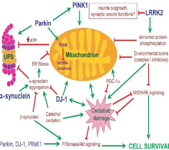
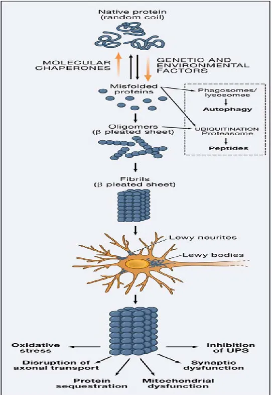
Oligomers are highly toxic for neurons in several ways (Fig. 1,2). They can explete a pore-like activity, inserting themselves within cell membranes, constructing ores and thus disaggregating membranes altering ion and electrolyte equilibrium between the two sides. They can also interact with histones and inhibit histone acetylation. They can also compromise lysosome function, inhibit proteosome function and alter cell cytoskeletal structures, including gap junctions. And finally, they can inhibit neuronal survival factor MEF 2D functioning [2,3]. Alpha synuclein aggregation goes beyond oligomer formation to the stage when insoluble fibrils form, precipitate and give rise to protein aggregates called Lewy bodies when they lie within the cytoplasm and Lewy neurites when located in dendritic spines [2,3]. Some evidence suggests that Lewy bodies reflect an attempt to limit soluble toxic oligomers. In this sense, in vitro studies indicate that alpha-synuclein within Lewy bodies is less toxic than that in soluble oligomer aggregates [2,3]. Alpha-synuclein aggregation is associated also with post-transcriptional changes, primarily a phosphorylated serine residue in position 129 [2,3].
Figure 1. Common intersecting pathways underlying PD pathogenesis (Thomas B, Parkinson’s disease, Hum Mol Gen 2007).
Figure 2. Alpha-synuclein aggregation (Lee V et al, Mechanisms of Parkinson’s disease, Neuron 2006)
Extracellular alpha-synuclein location and passage into the blood
Apart from its intracellular location the alpha-synuclein protein exists also in the extracellulare space. Studies in vivo and in vitro indicate that neurons physiologically secrete alpha-synuclein [4-7]. They do so through two main mechanisms: one is a Golgi dependent mechanism and the other a non-Golgi-dependent mechanism. Alpha-synuclein’s extracellular function remains unclear. Most important, in assessing its use as a biomarker for PD, information is lacking on whether and if so how alpha-synuclein secretion depends on intracellular alpha-synuclein protein accumulation. Several observations show that extracellular secretion involves not only monomeric alpha-synuclein forms but also aggregates [5]. Opinions disagree on whether extracellular secretion changes under conditions of cell stress that could in some way mimic those linked to intracellular alpha-synuclein accumulation. Studying SH-SY5Y human neuroblastoma cells, Lee et al [5] found that proteasome inhibition led to an increase in intracellular alpha-synuclein aggregation. Under this condition, the conditioned medium contained increased levels of both aggregated and monomeric synuclein species [5]. In their study, Jang et al [7] found that alpha-synuclein release consistently increased under various protein misfolding stress conditions in both neuroblastoma and primary neuron models but cells secrete the improperly folded green fluorescence protein alone, not the correctly folded form. Conversely in the study conducted by Emmanoidilou treatment of α-synuclein- SH-SY5Y expressing cells with H2O2, MPP+ and a potent proteasome inhibitor I for 6 h left α-synuclein secretion unchanged [4].
From the extracellular spaces alpha-synuclein could reach the blood plasma in two ways. First, by passing into the CSF and from there into plasma. Amongst its physiological functions, CSF drains interstitial fluids. Because CSF is drained into the venous system [8], alpha-synuclein could later pass into the blood stream. Reports describing alpha-synuclein in
the CSF in healthy subjects could further confirm its presence in the extracellular spaces [9]. Alpha-synuclein might also reach the plasma directly through the blood-brain barrier, a structure that is altered in neurodegenerative diseases and therefore more permeable to the passage of proteins and solutes [8]. No evidence yet shows what exactly passes into the blood and whether the monomeric forms alone or also oligomeric alpha-synuclein forms pass, and whether crossing the blood-brain barrier depends on their phosphorylation state.
Cerebrospinal fluid assay
Studies designed to assay alpha-synuclein in CSF belong in four groups according to the laboratory techniques used.
The early studies used Western Blot. Using Western blot, Jacowec [10] failed to discover any protein compatible with alpha-synuclein in a cohort of 8 parkinsonian patients and 4 healthy control subjects. Borghi [11] identified in CSF samples from 12 patients and 10 healthy control subjects a 19 kDa protein, corresponding to monomeric alpha-synuclein, but found no significant differences between the two groups. Similar findings were reported by El-Agnaf et al [12], who identified a monomeric form containing a 15 kD protein, again both in patients and in controls.
Others using immunoenzymatic methods to assay total alpha-synuclein in CSF generally found lower alpha-synuclein concentrations in patients than in controls. In their study comparing CSF alpha-synuclein concentrations in 33 parkinsonian patients and 38 healthy controls Tokuda [13] reported that the total soluble CSF forms were significantly lower in patients than in controls, a finding the same investigators subsequently confirmed in a later study in 32 patients and 28 controls [14]. These results were confirmed by Mollenahuer et al [15], first in a sample of 8 patients and 13 controls and later in two further populations,
one including 51 patients and 13 controls and the other 273 patients and 23 controls [16]. These findings notwithstanding Park et al, [17 ] in a sample of 23 parkinsonian patients and 29 control subjects found no difference between the two groups and similarly Ohrflet et al, [18] found almost matching alpha-synuclein CSF concentrations in 15 parkinsonian patients and 55 healthy control subjects.
Contrasting results emerged also from studies using immunoenzymatic techniques designed to identify specific alpha-synuclein. For example, Tokuda et al [14] reported finding higher oligomer concentrations in parkinsonian patients in a dual sample, one comparing 32 patients with 28 controls the other comparing 25 patients and 43 controls. In their later study Park et al, [17] reached the same conclusion after comparing 23 parkinsonian patients and 29 control subjects.
Using a Luminax Assay to compare 93 patients and 78 control subjects Wang et al [19], found several differences between the two groups namely, lower total alpha-synuclein concentrations, higher phosphorylated alpha-synuclein concentrations, and a significantly higher ratio between phosphorylated and total alpha-synuclein in patients than in controls. They subsequently confirmed the latter two findings in a sample comprising 116 patients and 126 control subjects. More evidence comes from a study conducted by Hong et al, [20] confirming lower total alpha-synuclein concentrations in 117 parkinsonian patients and 132 controls.
In conclusion, most studies designed to assay alpha-synuclein in CSF indicate reduced total forms, and increased specific forms such as oligomers and phosphorylated monomers in patients with PD. All the studies conducted to date have major limitations, however. For example, all control groups consisted mainly of patients with other neurological disorders,
such as multiple sclerosis, polyradiculoneuropathy, ictus, and sudden onset headache, thus introducing a possible confounding factor. Some studies enrolled small study and control samples thus weakening the study’s statistical power [10-12,15,17,18].
Plasma alpha-synuclein assay
Two studies have used immunoenzymatic techniques to assess plasma alpha-synuclein levels in healthy subjects. In one, Fjorback [21] assayed the protein in 44 healthy subjects (17 women and 27 men, mean age 21-65 years) and found no correlation between age and alpha-synuclein concentrations nor any difference between men and women. In a similar study, Barbour et al [22], assayed plasma alpha-synuclein levels in 4 healthy subjects and reached the same conclusion.
Other studies comparing parkinsonian patients and healthy subjects assayed alpha-synuclein with four techniques.
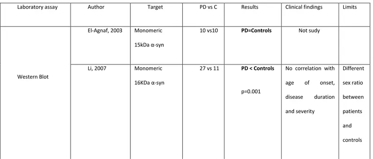
Western Blot (Table A). El-Agnaf et al [12] detected monomeric alpha synuclein both in patients than in controls. Li [23] releaved lower plasma alpha synuclein concentrations in patients than in controls and no correlations between plasma alpha synuclein and sex, disease duration, severity of disease, age of onset. He studied 27 parkinsonian patients and 11 healthy control subjects.
Laboratory assay Author Target PD vs C Results Clinical findings Limits
Western Blot
El-Agnaf, 2003 Monomeric 15kDa α-syn
10 vs10 PD=Controls Not sudy
Li, 2007 Monomeric 16KDa α-syn 27 vs 11 PD < Controls p=0.001 No correlation with age of onset, disease duration and severity Different sex ratio between patients and controls
Table A. Plasma alpha synuclein assay in Parkinson’s disease by Western Blot.
Enzyme-linked immunosorbent assay (ELISA, Table B, next page). In the first study Lee et al, [24] compared 105 parkinsonian patients and 51 control subjects. They found higher total soluble alpha-synuclein levels in parkinsonian patients than in controls but no correlations between alpha-synuclein levels and the duration of PD or Hoehn Yahr stage. Similar results were reported by Duran et al [25], who studied 95 patients and 60 healthy control subjects. Even though the mean age was significantly younger in the control group than in patients the investigators found no correlation between alpha-synuclein levels and age at sampling. Two later studies found no differences between patients and controls [17], one conducted in a sample of 23 parkinsonian patients and 29 healthy control subjects. In the second, conducted in our laboratories in 69 patients and 110 controls, we also found no differences between the two groups [Caranci et al, data submitted]. Last, Mollenahuer et al [16], found lower alpha-synuclein values in a sample of 273 patients than in 23 control subjects.
Laboratory assay
Author Target PD vs C Results Clinical findings Limits
ELISA for total α-syn soluble forms
Lee, 2006 Total α-syn Epitope 117-131
105 vs 51 PD>Controls No correlation with age at study, disease duration and severity (HY)
Characteristics of healthy controls not specified
Duran, 2010 Total α-syn 95 vs 60 PD>Controls No correlation with age at onset, disease duration and severity nor with age at study in both study and control group Significative difference in age at study between patients and controls Park, 2011 Total α-syn
Epitope 121-125
23 vs 29 PD=Controls Not study Controls affected by neurological diseases other than PD and internistic diseases Mollenhauer, 2011
Total α-syn 273 vs 23 PD<Controls Not study Controls affected by neurological diseases other than PD Caranci, 2012 Total α-syn 69 vs 110 PD=Controls Decreased
alpha-synuclein levels in HY stage III only in male patients; associations between a-syn and cognitive deficits, allucinations, psychosis, sleep dist. only in male patients
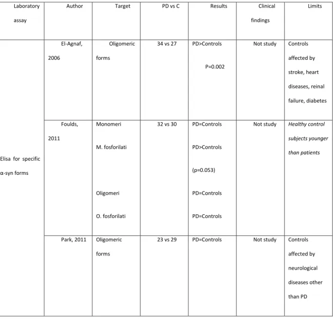
ELISA to detect specific alpha-synuclein forms (Table C). Using an immunoenzymatic technique designed to detect only alpha-synuclein oligomeric forms, El-Agnaf et al [26], found higher concentrations of aggregated species in 34 patients with PD than in 27 healthy controls. Including in the control group persons with cancer and ictus might have acted as a confounding factor. In a study sample comprising 32 patients and 30 controls, Foulds et al [27 ] used a new, complex immunoenzymatic procedure to disclose four alpha-synuclein species: monomers, phosphorylated monomers, oligomers and phosphorylated oligomers. Among these four species the only significant difference between patients and controls was for phosphorylated monomers (p=0.053). The investigators also did a longitudinal assessment to find out whether concentrations vary over time. In samples withdrawn at 1, 2 and 3 months no significant differences were found from baseline in alpha-synuclein plasma concentrations. In their study applying a method specially designed to detect oligomers, Park et al [17] failed to discover any differences between 23 parkinsonian patients and 29 healthy control subjects.
Laboratory assay
Author Target PD vs C Results Clinical findings
Limits
Elisa for specific α-syn forms El-Agnaf, 2006 Oligomeric forms 34 vs 27 PD>Controls P=0.002
Not study Controls affected by stroke, heart diseases, reinal failure, diabetes Foulds, 2011 Monomeri M. fosforilati Oligomeri O. fosforilati 32 vs 30 PD=Controls PD>Controls (p=0.053) PD=Controls PD=Controls
Not study Healthy control subjects younger than patients
Park, 2011 Oligomeric forms
23 vs 29 PD=Controls Not study Controls affected by neurological diseases other than PD
Luminex Assay. Nor did Shi et al [28], find differences between a group of 95 patients and 117 controls when they used a technique designed explicitly to eliminate interference from alpha-synuclein from blood cells. Alpha-synuclein level tended to diminish in the more advance disease stages.
In conclusion, studies designed to assay plasma alpha-synuclein over the past 10 years have provided contradictory results. Our review suggests that these stem mainly on four limitations. First, is the diverse assay techniques used and second that studies using the same technique, for example immunoenzymatic methods, often used different antibody binding alpha-synuclein epitopes. And last, the procedures used for preparing blood samples sometimes differ in relation to centrifugation speeds and duration. Some studies also enrolled small samples [12,17,23,27] thus limiting the study’s statistical power. Several studies selected as “healthy” control subjects patients who had other neurological diseases [12,17]. And finally, some failed to analyze in detail the possible associations between alpha-synuclein concentrations and the main PD clinical and epidemiological features.
Peripheral alpha-synuclein expression
A major problem is that the alpha-synuclein protein is expressed in sites other than neurons. It is expressed also in the periphery and in particular in all blood cells. The cells richest in alpha-synuclein are platelets. The amount of alpha-synuclein physiologically present in plasma from healthy subjects could derive from plasma contamination with intact or lysed red blood cells [22]. No information yet clarifies whether blood cells can secrete alpha-synuclein. The hypothesis that plasma alpha-synuclein might originate from blood cells makes it necessary to clarify whether the various differences between patients and controls reflect different protein expression patterns in the periphery.
Numerous studies have quantitatively assessed alpha-synuclein expression in blood cells. In a study using Western blotting on platelet lysates Li et al [29] compared 13 parkinsonian patients with 11 controls and found full-length alpha-synuclein (16 kDa) and no significant difference in the mean alpha-synuclein levels between the two groups (p = 0.62). They also showed that alpha-synuclein is not secreted by activated platelets. In another study, Miller et al [30] used Western blotting to examine platelets in a single parkinsonian patient with a mutation that causes an alpha-synuclein gene triplication and found higher levels than those in 5 unaffected family members. In a similar study the same investigators analyzed platelets in a patient with an exon 4 deletion in the parkin gene and compared the results with 6 control subjects and three unaffected, heterozygote family members. In the patient with an effective parkin gene knock-out they detected full-length 18 kDa alpha-synuclein and no alteration in steady state α-synuclein protein levels [31]. Tan et al, [32] studied alpha-synuclein mRNA expression in 80 patients with sporadic PD and 80 healthy control subjects. Total RNA was extracted from lymphocytes. There was no difference in mRNA fold expression between PD patients and controls (p=0.15). Miller et al, [33] studied with Western Blot platelet samples of 12 parkinsonian patients and ten healthy control subjects. A band migrating to 19 kDa on Western blot was seen using Syn-1, LB509 and H119, antibodies that recognise different epitopes on α-synuclein, thus confirming its identity. No significance differences was found between patients and controls in alpha-synuclein density. Brighina et al [34] used an immunoenzymatic technique to search for alpha-synuclein in lymphomonocytes in 78 parkinsonian patients and 78 healthy controls. Although they found no difference between patients and controls, alpha-synuclein levels increased significantly with age and were higher in men than in women.
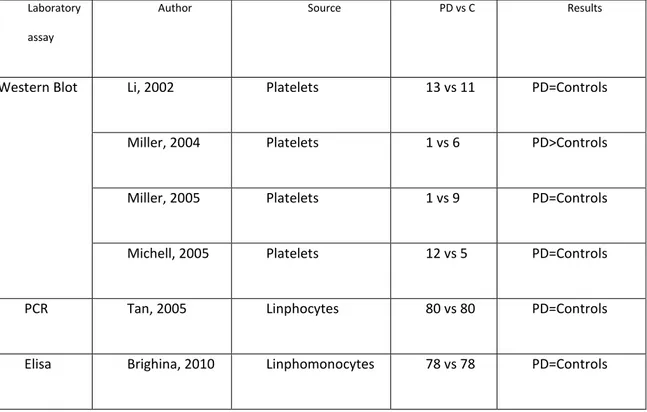
In conclusion, most studies (Table D) found no difference in peripheral alpha-synuclein expression patterns between parkinsonian patients and healthy controls. Some published studies nevertheless suffer limitations from the small study and control samples and few studies have assessed alpha-synuclein RNA expression in red blood cells and last, or aimed to disclose eventual differences in alpha-synuclein gene proteome expression.
Laboratory assay
Author Source PD vs C Results
Western Blot Li, 2002 Platelets 13 vs 11 PD=Controls
Miller, 2004 Platelets 1 vs 6 PD>Controls
Miller, 2005 Platelets 1 vs 9 PD=Controls
Michell, 2005 Platelets 12 vs 5 PD=Controls
PCR Tan, 2005 Linphocytes 80 vs 80 PD=Controls
Elisa Brighina, 2010 Linphomonocytes 78 vs 78 PD=Controls
Table D. Alpha-synuclein assay in peripheral blood cells in Parkinson’s disease and healthy controls
Future Perspectives
To be really useful in clinical practice a biological diagnostic marker must be detectable in a minimally invasive way. Although studies designed to assay alpha-synuclein in CSF have provided immensely interesting data, insofar as lumbar puncture is an invasive procedure and alpha-synuclein can be non-invasively assayed in plasma, plasma studies open up new, intriguing research directions. Several complex problems remain open.
First, research needs to clarify the role played by specific alpha-synuclein species such as oligomeric aggregates and eventual post-translational changes in the protein, most important a phosphorylated serine residue in position 129. If monomers may originate also from blood cells, oligomers and phosphorylated forms, or both, predominantly or exclusively originate from the human brain. Research therefore needs to develop highly sensitive assay techniques. Brain alpha-synuclein might also be present in low concentration ranges, not easily detectable with the experimental techniques currently available for detecting total alpha-synuclein forms.
A second important future research need in identifying a biological diagnostic marker is to ascertain whether eventual differences in plasma alpha-synuclein in patients and controls reflect a different expression pattern for peripheral protein. For this purpose preliminary studies should aim to clarify whether extracellular alpha-synuclein secretion documented in brain neurons takes place also in blood cells. If it does then most of the protein present in plasma probably originates from blood corpuscles. Another useful point to verify is proteomic alpha-synuclein expression in the various blood cell lines and then to compare findings in patients with PD and healthy control subjects. These two research directions serve to verify
further whether eventual differences in plasma alpha-synuclein concentrations arise from differences in peripheral alpha-synuclein gene dosage in patients and controls.
Longitudinal studies are also needed to provide definitive evidence on whether alpha-synuclein might be a marker of disease progression. A question of intense interest is whether alpha-synuclein could become not only a disease marker able to differentiate healthy subjects from those with PD but also reflect disease progression.
Studies are also lacking to show whether alpha-synuclein expression correlates with the various clinical PD phenotypes including patients with hypokinetic-rigid, tremorigenic, mixed, and pure hypokinetic types whose prognosis differs and similarly whether the presence of dyskinesias and motor fluctuations is associated with significant variations in alpha-synuclein concentrations.
Even though oral dopaminergic therapy apparently leaves alpha-synuclein expression unchanged, current evidence comes from only two papers. Hence further studies are need to confirm these data and to find out whether therapies limited to patients with advanced stage PD, such as subcutaneous apomorphine or subthalamic nucleus deep brain stimulation influence alpha-synuclein levels.
Another extremely interesting future direction would be to verify plasma alpha-synuclein protein concentrations in juvenile onset PD. Because these are highly aggressive forms plasma concentrations could differ from those in patients with adult onset PD.
The possibility that gender differences in alpha-synuclein exist, currently supported by our data, needs further investigation.
References
[1] Foulds PG, Mitchell JD, Parker A et al. (2011) Phosphorylated {alpha}-synuclein can be detected in blood plasma and is potentially a useful biomarker for Parkinson's disease. FASEB J. 25(12):4127-37
[2] Wood-Kaczmar A, Gandhi S, Wood NW (2006). Understanding the molecular causes of Parkinson's disease. Trends Mol Med 12:521-8.
[3] Thomas B, Beal MF (2007) Parkinson's disease. Hum Mol Genet. 15; 2:R183-94. [4] Emmanouilidou E, Melachroinou K, Roumeliotis T et al (2010) Cell-produced alpha-synuclein is secreted in a calcium-dependent manner by exosomes and impacts neuronal survival. J Neurosci 30:6838-51.
[5] Lee HJ, Patel S, Lee SJ (2005). Intravesicular localization and exocytosis of alpha-synuclein and its aggregates. J Neurosci 5;25:6016-24
[6] Dixon C, Mathias N, Zweig RM et al (2005) Alpha-synuclein targets the plasma membrane via the secretory pathway and induces toxicity in yeast. Genetics; 170(1):47-59
[7] Jang A, Lee HJ, Suk JE et al (2010) Non-classical exocytosis of alpha-synuclein is sensitive to folding states and promoted under stress conditions. J Neurochem; 113(5):1263-74
[8] Silverberg GD, Mayo M, Saul T et al (2003). Alzheimer's disease, normal-pressure hydrocephalus, and senescent changes in CSF circulatory physiology: a hypothesis. Lancet Neurol 2:506-11.
[9] Tokuda T, Salem SA, Allsop D, et al (2006) Decreased alpha-synuclein in cerebrospinal fluid of aged individuals and subjects with Parkinson's disease. Biochem Biophys Res Commun 13;349(1):162-6
[10] Jakowec M.W., Petzinger G.M., Sastry S et al (1998) The native form of a-synuclein is not found in the cerebrospinal fluid of patients with Parkinson’s disease or normal controls. Neuroscience Letters 253: 13–16
[11] Borghi R, Marchese R, Negro A et al (2000) Full length a-synuclein is present in cerebrospinal fluid from Parkinson's disease and normal subjects. Neuroscience Letters 287: 65-67
[12] El-Agnaf OM, Salem SA, Paleologou KE et al (2003) Alpha-synuclein implicated in Parkinson's disease is present in extracellular biological fluids, including human plasma. FASEB J 17:1945-7
[13] Tokuda T, Salem SA, Allsop D et al ( 2006)Decreased alpha-synuclein in cerebrospinal fluid of aged individuals and subjects with Parkinson's disease. Biochem Biophys Res Commun. 13;349(1):162-6.
[14] Tokuda T, Qureshi MM, Ardah MT et al ( 2010) Detection of elevated levels of α-synuclein oligomers in CSF from patients with Parkinson disease. Neurology. 16;75(20):1766-72.
[15] Mollenhauer B, Cullen V, Kahn I et al ( 2008) Direct quantification of CSF alpha-synuclein by ELISA and first cross-sectional study in patients with neurodegeneration. Exp Neurol. 213(2):315-25
[16 ] Mollenhauer B, Locascio JJ, Schulz-Schaeffer W et al ( 2011)α-Synuclein and tau concentrations in cerebrospinal fluid of patients presenting with parkinsonism: a cohort study Lancet Neurol. 10(3):230-40
[17] Park MJ, Cheon SM, Bae HR, Kim SH, Kim JW (2011) Elevated Levels of α-Synuclein Oligomer in the Cerebrospinal Fluid of Drug-Naïve Patients with Parkinson's Disease. J Clin Neurol 7(4):215-22
[18 ] Ohrfelt A, Grognet P, Andreasen N et al (2009) Cerebrospinal fluid alpha-synuclein in neurodegenerative disorders-a marker of synapse loss? Neurosci Lett. 6;450(3):332-5.
[19] Wang Y, Shi M, Chung KA et al (2012) Phosphorylated α-synuclein in Parkinson's
disease. Sci Transl Med. 15;4(121):121ra20.
[20] Hong Z, Shi M, Chung KA et al (2010) DJ-1 and alpha-synuclein in human cerebrospinal fluid as biomarkers of Parkinson's disease. Brain 133(Pt 3):713-26
[21] Fjorback A. W, Varming K, Jensen H (2007) Determination of a-synuclein concentration in human plasma using ELISA. Scand J Clin Lab Invest 67: 431–435
[22] Barbour R, Kling K, Anderson JP (2008). Red Blood Cells Are the Major Source of Alpha-Synuclein in Blood. Neurodegenerative Dis;5:55–59
[23] Li QX, Mok SS, Laughton KM et al (2007) Plasma alpha-synuclein is decreased in subjects with Parkinson's disease. Exp Neurol 204:583-8.
[24] Lee PH, Lee G, Park HJ et al (2006) The plasma alpha-synuclein levels in patients with Parkinson's disease and multiple system atrophy. J Neural Transm. 113:1435-9
[25] Duran R, Barrero FJ, Morales B et al (2010) Plasma alpha-synuclein in patients with Parkinson's disease with and without treatment. Mov Disord 25:489-93.
[26] El-Agnaf OM, Salem SA, Paleologou KE et al (2006) Detection of oligomeric forms of alpha-synuclein protein in human plasma as a potential biomarker for Parkinson's disease. FASEB J.20 : 419-25.
[27] Foulds PG, Mitchell JD, Parker A et al (2011) Phosphorylated α-synuclein can be detected in blood plasma and is potentially a useful biomarker for Parkinson's disease. FASEB J. 25(12):4127-37
[28] Shi M, Zabetian CP, Hancock AM et al (2010) Significance and confounders of peripheral DJ-1 and alpha-synuclein in Parkinson's disease. Neurosci Lett;480:78-82
[29] Li QX, Campbell BC, McLean CA et al. (2002) Platelet alpha- and gamma-synucleins in Parkinson's disease and normal control subjects. J Alzheimers Dis. 4(4):309-15. [30] Miller DW, Hague SM, Clarimon J et al (2004) Alpha-synuclein in blood and brain from familial Parkinson disease with SNCA locus triplication. Neurology. 2004 25;62(10):1835-8.
[31] Miller DW, Crawley A, et al (2005) Unaltered alpha-synuclein blood levels in juvenile Parkinsonism with a parkin exon 4 deletion. Neurosci Lett. 21;374(3):189-91
[32] Tan EK, Chandran VR, Fook-Chong S et al (2005) Alpha-synuclein mRNA expression in sporadic Parkinson's disease. Mov Disord. 20(5):620-3.
[33] Michell AW, Luheshi LM, Barker RA et al (2005) Skin and platelet alpha-synuclein as peripheral biomarkers of Parkinson's disease. Neurosci Lett. 24;381(3):294-8.
[34] Brighina L, Prigione A, et al (2010),Lymphomonocyte alpha-synuclein levels in aging and in Parkinson disease. Neurobiol Aging. 31(5):884-5.
Study design.
We performed an observational and exploratory cross-sectional study to evaluate plasma alpha synuclein concentrations in patients affected by Parkinson’s disease, Multisystem Atrophy, Corticobasal Degeneration, Essential Tremor, Progressive Sopranuclear Palsy and healthy control subjects.
Primary end points:
1) compare total alpha synuclein concentrations in PD patients and age-matched healthy controls
2) compare total alpha synuclein concentrations in patients affected by Multisystem Atrophy and age-matched healthy controls
3) compare total alpha synuclein concentrations in patients affected by Progressive Sopranuclear Palsy and age-matched healthy controls
4) compare total alpha synuclein concentrations in patients affected by CorticoBasal Degeneration and age-matched healthy controls
5) compare total alpha synuclein concentrations in patients affected by Essential Tremor and age-matched healthy controls
6) evaluate possible associations and correlations between alpha-synuclein plasma expression and the major parkinsonian patients’ clinical and epidemiological characteristics
Parkinsons’ disease.
- Diagnosis of Parkinson’s disease according criteria of the United Kingdom Parkinson’s disease Society Brain bank.
- Good response to levodopa, defined as a reduction>30% of the scores in the motor section of MDS-UPDRS scale, and as a substantially benefit of prolonged treatment with levodopa
- Exclusion of secondary parkinsonisms
- Normal brain MRI
Multisystem Atrophy
Diagnosis according Gilman’s criteria
MSA DIAGNOSTIC CRITERIA
Gilman S et al. Second consensus statement on the diagnosis of multiple system atrophy. Neurology 2008; 71:670-76
Definite Probable Possible
Abundant alpha synuclein positive glial cells inclusions associated with
neurodegeneration in olivo pontocerebellar and striatonigral structures
Sporadic onset after the age of 30 years of disautonomic syndrome associated with parkinsonism poorly levodopa responsive or cerebellar syndrome
Parkinsonism or cerebellar syndrome and almost one symptom expression of autonomic dysfunction.
Essential tremor
Diagnosis according to Bains’ criteria
Criteria for the diagnosis of essential tremor
Bain 2000
Primary Criteria Secondary Criteria Red Flags
Action tremor of the hands
No other neurological signs except for troclea
Possible isolated head tremor without posture abnormalities
Long duration
Familial history
Good response to ethanol
Unilateral tremor, resting tremor, rigidity, bradykinesia
Rapid onset
Assumption of tremor inducing drugs
Corticobasal degeneration
Diagnosis according to Boeve’s Criteria
Diagnostic criteria for corticobasal syndrome
Boeve, 2003
Primary criteria Secondary criteria
Cortical dysfuncton as revealed by: ideomotor apraxia, alien art phenomenon, cortical sensitive deficit, visual or sensitive emineglect, non fluent aphasia
Extrapyramidal syndrome with focal dystonia and asimmetric rigidity without levodopa response
Asymmetrical fronto-parietal cortical cerebral atrophy
Asimmetrical fronto-parietal hypoperfusion or hypometabolism
Cognitive disfunction and preservation of memory
Progressive Sopranuclear Palsy
Diagnosis according to Litvan’s criteria
DIAGNOSTIC CRITERIA FOR PSP
Litvan 1996
Probabile Possibile
Grdaually progressive disorder Onset after 40 years old
Vertical gaze supranuclear palsy Postural instability
Gradually progressive disorder Onset after 40 years old
Almost one of the following criteria: Vertical gaze supranuclear palsy Postural instability
Healthy control subjects had no neurological disease except for tension type headache All study procedures were approved by the institutional review board at the Istituto Neurologico Mediterraneo (via Atinense18, 86077 Pozzilli-IS), Istituto di Ricovero e Cura a carattere Scientifico.
All patients gave informed written consent. The study was conformed to the Declaration of Helsinki.
Venous blood from all patients was drawn in ethylenediaminetetraacetic acid (EDTA) tubes. Hemolyzed blood samples were discarded. Plasma samples were obtained by centrifugation at 2500 rpm for 15 min, at 4°C, aliquoted in 200 μl and stored at -80°C until assay. Total plasma alpha-synuclein concentrations were determined by enzyme-linked immunosorbent assay (ELISA) based on synuclein capture antibody and alpha-synuclein antibody detection according to the manufacturer’s protocol (Invitrogen Corporation, ref. KHB0061). Samples were analyzed in duplicate in blinded experiments. The optical density of the samples was determined at 450 nm with a Tecan Sunrise microplate reader (Tecan, Switzerland) and results were expressed as ng alpha-synuclein /ml plasma.
Experimental results
Introduction
The major histological alteration responsible for Parkinson’s disease (PD) is alpha-synuclein accumulation in the intra-cytoplasmic aggregates known as Lewy bodies. Increasing evidence suggesting that alpha-synuclein passes from the brain to blood [1-7] prompted studies to verify whether plasma alpha-synuclein assay reflects parkinsonian neurodegeneration. Studies in recent years investigating plasma alpha-synuclein as a possible biological marker for PD have provided conflicting results owing partly to the various assay techniques used, protein species assayed and small populations studied [8-13]. Data are also lacking on whether alpha-synuclein plasma expression correlates with parkinsonian patients’ clinical and epidemiological features. Nor have studies investigated possible gender differences in plasma alpha-synuclein concentrations in patients with PD. Having information on alpha-synuclein as a biomarker for PD would help diagnose PD, and monitor disease progression.
In this observational cross-sectional study, using an immunoenzymatic technique we assayed and compared total alpha-synuclein plasma concentrations in men and woman patients with PD and age-matched healthy controls. Second, we sought further information on possible associations and correlations between alpha-synuclein plasma expression and the major parkinsonian patients’ clinical and epidemiological characteristics.
Subjects and methods Subjects
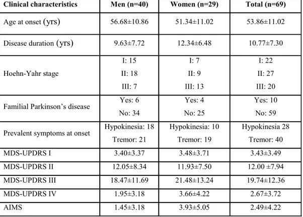
The study included 69 patients with PD and 110 healthy age-matched control subjects, enrolled between January 2010 and July 2011 at the Neuromed Institute. Study procedures were approved by the institutional review board at the Neuromed Institute and all patients gave informed consent. PD was diagnosed in accordance with the criteria of the United Kingdom Parkinson’s Disease Society Brain Bank; patients were included consecutively including those undergoing DBS (deep brain stimulation) or apomorphine pumps. Clinical assessment was based on Movement Disorder Society-Unified Parkinson’s Disease Rating Scale (MDS-UPDRS) and Abnormal Involuntary Movement Scale (AIMS) scores. The healthy control subjects selected had no neurological diseases except for tension type headache or other diseases and came mainly from patients’ families. Among the 69 patients with PD enrolled, 26 had disease classified as predominantly hypokinetic, 30 had tremorigenic disease and 13 a mixed phenotype. Physical examination identified the left body side as involved by major slowing in 41 patients, tremor as the onset symptom in 40 patients and hypokinesia in 29 patients. Among the 25 patients with motor fluctuations, 23 subjects also had dyskinesias. Of the 69 patients enrolled 10 had unilateral or bilateral microelectrodes implanted in the subthalamic nucleus for DBS. Clinical assessment showed that 8 patients were receiving apomorphine alone, 18 patients levodopa alone, 5 patients dopamine agonists alone, 37 patients with levodopa plus dopamine agonists, and 9 patients were receiving no medications. According to Hoehn and Yahr (HY) staging 49 patients were in stage I and II and 20 in stage III. Six patients had familial PD. The mean age at disease onset was 53.86±11.02 years and disease duration was 10.77±7.30 years. Patients’ clinical characteristics and MDS-UPDRS and AIMS scores are summarized in Table 1.
Laboratory Assay
Venous blood from all patients was drawn in ethylenediaminetetraacetic acid (EDTA) tubes. Hemolyzed blood samples were discarded. Plasma samples were obtained by centrifugation at 2500 rpm for 15 min, at 4°C, aliquoted in 200 μl and stored at -80°C until assay. Total plasma alpha-synuclein concentrations were determined by enzyme-linked immunosorbent assay (ELISA) based on synuclein capture antibody and alpha-synuclein antibody detection according to the manufacturer’s protocol (Invitrogen Corporation, ref. KHB0061). Samples were analyzed in duplicate in blinded experiments. The optical density of the samples was determined at 450 nm with a Tecan Sunrise microplate reader (Tecan, Switzerland) and results were expressed as ng alpha-synuclein /ml plasma.
Statistical Analysis
Parametric and nonparametric tests including the t-test or Mann-Whitney test were used to compare means in paired groups. Chi-square test was used for categorical variables. One-way analysis of variance (ANOVA) or Kruskal-Wallis test was used to compare means in multiple groups. Pearson correlation coefficients were obtained to investigate a possible relationship among variables. To find out whether alpha-synuclein values differed according to patients’ clinical variables, we also calculated the median alpha-synuclein value for the 69 patients and analyzed possible differences between two subgroups with upper or lower median alpha-synuclein values. P values <0.05 were considered to indicate statistical significance. Data were analyzed for significance with a commercially available software package (SPSS, version 17.0). A logistic regression model was run to evaluate the relation between upper or lower median alpha-synuclein values and the variables identified as
significant or at borderline level in the univariate analysis. Odds ratios (ORs) with relative 95% confidence intervals (CIs) were estimated. Values are expressed as mean±standard deviation.
Results
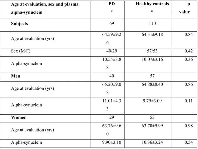
When blood samples were taken for total plasma alpha-synuclein assay, patients with PD and healthy control subjects were comparable for age and sex (p>0.05) (Table 2). No significant differences were found in alpha-synuclein plasma concentrations in patients and healthy control subjects (Table 2). Concentrations tended to differ more widely in men than in women (men: 11.01 ±4.33 vs 9.79±3.09 ng/ml, p=0.11; women: 9.9±3.1 vs 10.36±3.24 ng/ml, p=0.54) (Table 2).
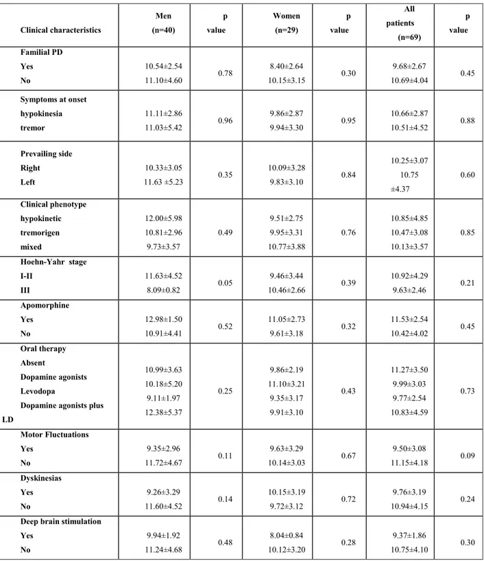
No significant associations were found between plasma alpha-synuclein concentrations and familiarity for PD, symptoms at onset, predominantly affected body side, clinical phenotype, HY stage, oral dopaminergic therapy, subcutaneous apomorphine infusion, unilateral or bilateral DBS, dyskinesias, or motor fluctuations (Table 3). Nor were correlations found between plasma alpha-synuclein concentrations and scores on the MDS-UPDRS section I, II, III, IV, and AIMS scores or with age at onset or disease duration.
Conversely, plasma alpha-synuclein concentrations differed according to PD stage and gender. In men, they were lower at HY scale stage III than at stages I and II (8.09±0.82 vs 11.63±4.52 ng/ml; p=0.05) but in women were similar at all stages (10.46±2.66 vs 9.46±3.44 ng/ml; p=0.39) (Table 3). In men, plasma alpha-synuclein concentrations lower than median value (11.08 ng/ml) were associated with significantly higher scores in the MDS-UPDRS sections II and III (15.29±8.74 vs 8.47±6.32 p=0.01 and 22.48±12.52 vs 14.05±9.07 ng/ml;
p= 0.02 by) (Table 4), whereas no differences were found in women (p = 0.77 and p=0.93). Alpha-synuclein plasma concentrations also differed in men and women according to MDS-UPDRS scores section IV (p=0.06 vs p= 0.68) (Table 4). Plasma alpha-synuclein concentrations also showed gender-related differences according to disease duration. Lower median alpha-synuclein concentrations were associated with a longer disease duration in men than in women (p=0.08 vs p=0.9) (Table 4). Finally, we found a significant inverse correlation between alpha-synuclein levels and MDS-UPDRS IV scores in men but not in women (Men: r = -0.33 p=0.04; Women: r = 0.08 p=0.67).
No gender-related differences were found between alpha-synuclein levels and the other clinical characteristics investigated such as unilateral or bilateral DBS, oral dopaminergic therapy, subcutaneous apomorphine infusion, clinical phenotype, symptoms at onset, familiarity, and predominantly affected body side. Alpha-synuclein concentrations were lower in men with motor fluctuations than in those without (p=0.11) but no difference was found in women (p=0.67). A similar trend was observed in men PD patients with dyskinesias (p=0.14 vs 0.72) (Table 3).
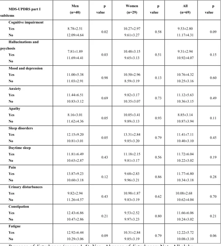
In the 69 patients with PD overall, no significant association appeared between alpha-synuclein levels and characteristics detected in MDS-UPDRS scores for section I, whereas in patients subgrouped by gender alpha-synuclein concentrations were significantly lower in men with cognitive disorders than in those without (8.78±2.51 vs 12.09±4.64 ng/ml, p=0.02 ) (Table 5). Similarly, alpha-synuclein values were significantly lower in men with hallucinations, psychosis and apathy than in those without (7.81±1.89 vs 11.69±4.41 ng/ml, p=0.03; and 8.16±3.01 vs 11.62±4.36 ng/ml, p=0.05), whereas no differences were found in women (p=0.51; and p=0.93) (Table 5). Plasma alpha-synuclein concentrations were significantly higher in men reporting sleep disorders than in those without (12.15±9.20 vs
10.81±3.01ng/ml, p=0.05), but no differences were found in women (p=0.79) (Table 5). Nor were gender-related differences found in the clinical characteristics depression, pain and constipation. Finally, alpha-synuclein levels were higher in men with fatigue than in those without ( p=0.09) whereas no differences were found in women. Nor were significant correlations found between alpha-synuclein plasma concentrations and age in the 179 participants overall (r=0.11; p= 0.15), in the 69 patients (r=0.12; p= 0.33) or in the healthy control group (r=0.10; p= 0.32).
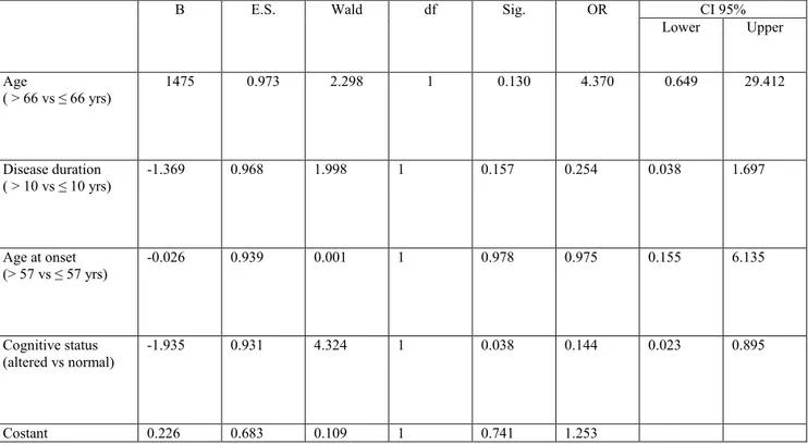
Finally, a regression logistic model showed that the presence of cognitive deficit was associated in men with a lower median value of alpha-synuclein concentrations (≤ 11.08 ng/ml) adjusted for age, age at onset and disease duration (OR= 0.144; CI95% 0.023-0.895) (Table 6). The other clinical variables identified as significant or at borderline level in the univariate analysis in men (hallucinations and psychosis, apathy and sleep disorders) (Table 5) were not significant when included in the regression logistic model.
Discussion
The first new and unexpected finding in this observational cross-sectional study is the significant gender difference in plasma alpha-synuclein concentrations measured using an immunoenzymatic technique in patients with PD. In men, alpha-synuclein concentrations differed significantly according to disease progression: they were lower in HY stage III than in stages I and II. Conversely, in women, although alpha-synuclein levels remained statistically unchanged in the early and later stages, they tended to increase as PD progressed. The second previously unreported finding is the significant association between plasma
alpha-synuclein concentrations and cognitive impairment, hallucinations, psychosis, apathy, sleep disturbances in men but not in women.
Why plasma alpha-synuclein concentrations decrease in men but not in women as PD progresses is complex to explain. An intriguing but possibly controversial explanation is that intracellular alpha-synuclein aggregation differs in men and women owing to a protective hormonal effect in women. Estrogens, the hormones that especially influence women, dose-dependently inhibit alpha-synuclein aggregation. In particular, estriol and estradiol destabilize
preformed fibrillar alpha-synuclein in vitro [14]. Several studiesindicate that alpha-synuclein
is physiologically secreted in the extracellular spaces then reaches the plasma [1, 2, 4]. It could do so by passing directly through the blood-brain barrier, which is structurally altered in neurodegenerative diseases, or by draining into the cerebrospinal fluid and then into the bloodstream. If intracellular alpha-synuclein aggregation impairs protein secretion in the
extracellular spaces and consequently its passage into the bloodstream [11], or if protein
secretion decreases owing to cell death triggered by protein aggregation then the decreased plasma alpha-synuclein concentration we reported in men as PD progressed may reflect greater intracellular accumulation in men than in women. Some studies have nevertheless shown that synuclein secretion increases during cellular stress [15]; extracellular alpha-synuclein protein secretion might therefore increase as a cellular defense mechanism aimed at removing pathological proteins. Finally, in most women patients enrolled in our study PD began in the post-menopausal period when estrogen levels diminished [16]. Some data also indicate that post-menopausal estrogen replacement therapy leaves the risk of PD developing unchanged [17]. Whether the low plasma alpha-synuclein concentrations we observed in men with advanced PD (HY stage III) reflect the estrogen-induced antiaggregant effects on alpha-synuclein fibrils prompts further study.
Because alpha-synuclein tended to increase as the disease progresses rather than decrease in the women with PD included in our study, we conjecture also that intracytoplasmic alpha-synuclein aggregation might differently alter extracellular protein secretion in males and females, possibly causing an opposite effect. This question awaits an answer from laboratory studies investigating gender-related differences in alpha-synuclein secretion in the extracellular space.
Another possible reason why alpha-synuclein concentrations decreased in men as PD progressed is a gender-related difference in quantitative alpha-synuclein expression in the central nervous system, possibly related to hormonal factors. Some evidence suggests that sex hormones may alter alpha-synuclein expression in the brain, for example, oxysterol 27-hydroxycholesterol (27-OHC) increases alpha-synuclein levels in SH-SY5Y cells by modulating estrogen receptors [18]. Studying the gender-associated genetic expression in dopaminergic neurons from post-mortem brains, Cantuti-Castelvetri et al. [17] observed a female-associated gene up-regulation in signal transduction and neuronal maturation . They also observed a men-associated up regulation of genes directly involved in PD pathogenesis, including alpha-synuclein. Men therefore seem to produce more central nervous system alpha-synuclein than women. Conversely, Simunovic et al. [19] showed alpha-synuclein gene downregulation in male dopamine neurons in sporadic PD. These preliminary data suggest that alpha-synuclein gene expression might differ in males and females.
Alternatively, instead of reflecting the neurodegenerative process the gender-related differences in plasma synuclein levels could reflect a sex-related difference in alpha-synuclein expression in peripheral blood cells. Alpha-alpha-synuclein is also expressed in the peripheral tissues and is present in all blood cells, especially in platelets [20]. Hence we
cannot exclude the possibility that plasma alpha-synuclein may derive from blood cell secretion, even though no current data show whether blood cells secrete alpha-synuclein. Several studies on peripheral alpha-synuclein examined qualitative or quantitative protein expression and found no differences between males and females or between patients and healthy control subjects [ 21-23]. For example, Tan et al. [21] studied alpha-synuclein mRNA expression in lymphocytes from 80 PD patients and 80 healthy control subjects, and found no differences between the two groups, even when they adjusted the results for gender. Similarly, Michell et al. [22 ] using western blot experiments on platelet samples found no significant differences in alpha-synuclein density in patients and controls in. Finally, Brighina et al. [23] found no differences in alpha-synuclein protein measured in lymphomonocytes from 78 PD patients and 78 controls.
Precisely what the lower plasma alpha-synuclein concentrations in men with clinical MDS-UPDRS I scores showing cognitive impairment and apathy than in those without reflect remains unclear. Cognitive deficits and apathy in Parkinson's disease may correlate with more widespread neurodegeneration involving the cerebral cortex and in particular the fronto-basal-ganglia circuits starting from the dorsolateral prefrontal cortex and the cingulate gyrus, as well as with multiple cholinergic, noradrenergic and serotonergic neurotransmitter abnormalities [24]. Autopsy studies disclose more numerous cortical Lewy bodies in patients with these deficits than in others [ 25]. The observed differences in plasma alpha-synuclein concentrations between men and women patients with PD may therefore reflect different gender-related protein expression patterns. Some studies have also highlighted gender-related cognitive dysfunction, such as prevalent deficits in verbal fluency and facial expression recognition in males, and visual-spatial deficits in women [26]. The hallucinations and psychosis in our male patients were associated with lower alpha-synuclein levels: even though
these symptoms may be an adverse effect induced by dopaminergic therapy, Duran et al [10] showed that dopaminergic therapy leaves plasma alpha-synuclein values unchanged.
In men, but not in women, alpha-synuclein values differed also according to another clinical characteristic, sleep disorders, and were significantly higher in male patients with sleep disorders than in those without. Sleep disorders including insomnia and REM sleep behavior disorders (RBD), may be an early PD manifestation [27] increasingly studied to identify possible preclinical PD markers before motor symptoms become evident. Sleep disorders probably originate from brainstem degeneration , especially involving nuclei in the ventral midbrain and mesopontine junction, brain areas in which post-mortem studies and animal studies have found abundant Lewy body deposits [28]. The increased plasma alpha-synuclein concentrations we observed only in male patients could reflect the male prevalence in these disorders: several studies have shown an association between REM sleep behavior disorders and male PD patients [17, 27, 29].
Finally, the absence of differences in plasma alpha-synuclein concentrations between patients and controls partially contrasts with results from comparable studies using a similar immunoenzymatic assay [10, 12, 30]. For example, in a study comparing 105 patients with PD and 51 healthy control subjects, Lee et al [12 ] found higher plasma alpha-synuclein values in patients than in controls and no correlations between alpha-synuclein and HY stage, disease duration and age when the study began. The second study [10] using the same immunoenzymatic kit that we use and comparing 95 parkinsonian and 60 control subjects also found significantly higher plasma alpha-synuclein values in patients than in controls, but no correlations between alpha-synuclein and age at onset, PD severity and disease duration. Although alpha-synuclein levels did not correlate with age at the time of sampling controls
were about 15 years younger than patients. Conversely, in a study conducted in a sample of 23 patients and 29 controls Park et al. [30] found no differences in plasma alpha-synuclein concentrations, but the fact that most control subjects had neurological or other diseases may have introduced a confounding factor. Finally, Mollenhauer et al. [31] found lower alpha-synuclein concentrations in patients with PD and multiple system atrophy than in controls with other neurological diseases. None of the aforementioned studies analyzed data for gender [10,12, 30, 31]. Although no differences in plasma alpha-synuclein levels emerged between patients and controls, it cannot be entirely ruled out the possibility that the alpha-synuclein could also represent a marker of disease in men, since the difference in measured concentration between PD males and health controls males (p = 0.11) may become significant increasing the number of subjects.
In conclusion, our findings indicate that alpha-synuclein, as assessed by enzyme immunoassay designed to detect total soluble forms, is a marker of PD progression only in men, and is associated with cognitive disorders, hallucinations, psychosis, and sleep disorders. The possible gender-related role of plasma alpha-synuclein in patients with PD merits
References
1. El-Agnaf OM, Salem SA, Paleologou KE, Cooper LJ, Fullwood NJ, Gibson MJ et al (2003) Alpha-synuclein implicated in Parkinson's disease is present in extracellular biological fluids, including human plasma. FASEB J 17:1945-7
2. Lee HJ, Patel S, Lee SJ (2005). Intravesicular localization and exocytosis of alpha-synuclein and its aggregates. J Neurosci 25:6016-24
3. Dixon C, Mathias N, Zweig RM, Davis DA, Gross DS (2005) Alpha-synuclein targets the plasma membrane via the secretory pathway and induces toxicity in yeast. Genetics 170(1):47-59
4. Emmanouilidou E, Melachroinou K, Roumeliotis T, Garbis SD, Ntzouni M, Margaritis LH et al (2010) Cell-produced alpha-synuclein is secreted in a calcium-dependent manner by exosomes and impacts neuronal survival. J Neurosci 30:6838-51.
5. Silverberg GD, Mayo M, Saul T, Rubenstein E, McGuire D (2003). Alzheimer's disease, normal-pressure hydrocephalus, and senescent changes in CSF circulatory physiology: a hypothesis. Lancet Neurol 2:506-11.
6. Tokuda T, Salem SA, Allsop D, Mizuno T, Nakagawa M, Qureshi MM et al (2006) Decreased alpha-synuclein in cerebrospinal fluid of aged individuals and subjects with Parkinson's disease. Biochem Biophys Res Commun 349(1):162-6
7. Hong Z, Shi M, Chung KA, Quinn JF, Peskind ER, Galasko D et al. (2010) DJ-1 and alpha-synuclein in human cerebrospinal fluid as biomarkers of Parkinson's disease. Brain 133:713-26.
8. Foulds PG, Mitchell JD, Parker A, Turner R, Green G, Diggle P et al. (2011) Phosphorylated α-synuclein can be detected in blood plasma and is potentially a useful biomarker for Parkinson's disease. FASEB J 25:4217-37
9. El-Agnaf OM, Salem SA, Paleologou KE, Curran MD, Gibson MJ, Court JA et al. (2006) Detection of oligomeric forms of alpha-synuclein protein in human plasma as a potential biomarker for Parkinson's disease. FASEB J 20:419-25.
10. Duran R, Barrero FJ, Morales B, Luna JD, Ramirez M, Vives F (2010) Plasma alpha-synuclein in patients with Parkinson's disease with and without treatment. Mov Disord 25:489-93.
11. Li QX, Mok SS, Laughton KM, McLean CA, Cappai R, Masters CL et al (2007) Plasma alpha-synuclein is decreased in subjects with Parkinson's disease. Exp Neurol 204:583-8. 12. Lee PH, Lee G, Park HJ, Bang OY, Joo IS, Huh K (2006) The plasma alpha-synuclein levels in patients with Parkinson's disease and multiple system atrophy. J Neural Transm 113:1435-9
13. Shi M, Zabetian CP, Hancock AM, Ginghina C, Hong Z, Yearout D et al (2010) Significance and confounders of peripheral DJ-1 and alpha-synuclein in Parkinson's disease. Neurosci Lett 480:78-82
14. Hirohata M, Ono K, Morinaga A, Ikeda T, Yamada M (2009) Anti-aggregation and fibril-destabilizing effects of sex hormones on alpha-synuclein fibrils in vitro. Exp Neurol 217(2):434-9
15. Jang A, Lee HJ, Suk JE, Jung JW, Kim KP, Lee SJ (2010) Non-classical exocytosis of alpha-synuclein is sensitive to folding states and promoted under stress conditions. J Neurochem 113(5):1263-74
16. Ragonese P, D'Amelio M, Salemi G, Aridon P, Gammino M, Epifanio A et al (2004) Risk of Parkinson disease in women: effect of reproductive characteristics. Neurology 62(11):2010-4.
17. Cantuti-Castelvetri I, Keller-McGandy C, Bouzou B, Asteris G, Clark TW, Frosch MP et al (2007) Effects of gender on nigral gene expression and parkinson disease. Neurobiol Dis 26(3):606-14
18. Marwarha G, Rhen T, Schommer T, Ghribi O (2011) The oxysterol 27-hydroxycholesterol regulates α-synuclein and tyrosine hydroxylase expression levels in human neuroblastoma cells through modulation of liver X receptors and estrogen receptors--relevance to Parkinson's disease. J Neurochem 119(5):1119-36.
19. Simunovic F, Yi M, Wang Y, Stephens R, Sonntag KC (2010) Evidence for gender-specific transcriptional profiles of nigral dopamine neurons in Parkinson disease. PLoS One 5(1):e8856.
20. Barbour R, Kling K, Anderson JP, Banducci K, Cole T, Diep L et al. (2008) Red blood cells are the major source of alpha-synuclein in blood. Neurodegener Dis. 5(2):55-9.
21. Tan EK, Chandran VR, Fook-Chong S, Shen H, Yew K, Teoh ML et al (2005) Alpha-synuclein mRNA expression in sporadic Parkinson's disease. Mov Disord 20(5):620-3.
22. Michell AW, Luheshi LM, Barker RA (2005) Skin and platelet alpha-synuclein as peripheral biomarkers of Parkinson's disease. Neurosci Lett 381(3):294-8
23. Brighina L, Prigione A, Begni B, Galbussera A, Andreoni S, Piolti R et al (2010) Lymphomonocyte alpha-synuclein levels in aging and in Parkinson disease. Neurobiol Aging 31(5):884-5
24. Ferrer I (2011) Neuropathology and neurochemistry of nonmotor symptoms in Parkinson's disease. Parkinsons Dis 2011:708404.
25. Burack MA, Hartlein J, Flores HP, Taylor-Reinwald L, Perlmutter JS, Cairns NJ (2010) In vivo amyloid imaging in autopsy-confirmed Parkinson disease with dementia. Neurology 74(1):77-84.
26. Miller IN, Cronin-Golomb A (2010) Gender differences in Parkinson's disease: clinical characteristics and cognition. Mov Disord 25(16):2695-703
27. Yoritaka A, Ohizumi H, Tanaka S, Hattori N (2009) Parkinson's disease with and without REM sleep behaviour disorder: are there any clinical differences? Eur Neurol 61(3):164-70. 28. Lai YY, Siegel JM (2003) Physiological and anatomical link between Parkinson-like disease and REM sleep behavior disorder. Mol Neurobiol 27(2):137-52.
29. Scaglione C, Vignatelli L, Plazzi G, Marchese R, Negrotti A, Rizzo G et al (2005) REM sleep behaviour disorder in Parkinson's disease: a questionnaire-based study. Neurol Sci 25(6):316-21.
30. Park MJ, Cheon SM, Bae HR, Kim SH, Kim JW (2011) Elevated Levels of α-Synuclein Oligomer in the Cerebrospinal Fluid of Drug-Naïve Patients with Parkinson's Disease. J Clin Neurol 7(4):215-22
31. Mollenhauer B, Locascio JJ, Schulz-Schaeffer W, Sixel-Döring F, Trenkwalder C, Schlossmacher MG (2011) α-Synuclein and tau concentrations in cerebrospinal fluid of patients presenting with parkinsonism: a cohort study. Lancet Neurol 10(3):230-40.
Table 1 Clinical characteristics of the 69 Parkinson’s disease patients by sex.
MDS-UPDRS part I, II, III, IV: Movement Disorder Society-Unified Parkinson’s disease rating scale. AIMS: Abnormal Involuntary Movement Scale. All MDS-UPDRS and AIMS scores are mean±SD.
Clinical characteristics Men (n=40) Women (n=29) Total (n=69)
Age at onset (yrs) 56.68±10.86 51.34±11.02 53.86±11.02 Disease duration (yrs) 9.63±7.72 12.34±6.48 10.77±7.30
Hoehn-Yahr stage I: 15 II: 18 III: 7 I: 7 II: 9 III: 13 I: 22 II: 27 III: 20
Familial Parkinson’s disease Yes: 6 No: 34
Yes: 4 No: 25
Yes: 10 No: 59
Prevalent symptoms at onset Hypokinesia: 18 Tremor: 21 Hypokinesia: 10 Tremor: 19 Hypokinesia 28 Tremor: 40 MDS-UPDRS I 3.40±3.37 3.48±3.71 3.43±3.49 MDS-UPDRS II 12.05±8.34 11.93±7.50 12.00 ±7.94 MDS-UPDRS III 18.47±11.69 21.48±13.24 19.74±12.36 MDS-UPDRS IV 1.95±3.18 3.66±4.22 2.67±3.72 AIMS 1.45±3.18 3.93±5.05 2.49±4.22
Table 2. Comparison of age at evaluation, sex and plasma alpha-synuclein concentration
in patients with Parkinson’s disease and healthy controls.
° no statistical difference was reported between men and women PD patients for age (p=0.53) and alphasynuclein (p= 0.25).
*no statistical difference was found between men and women health controls for age (p = 0.50) and alphasynuclein (p=0.35).
All alpha-synuclein values are mean±SD ng/ml. Age at evaluation, sex and plasma
alpha-synuclein PD ° Healthy controls * p value Subjects 69 110
Age at evaluation (yrs) 64.59±9.2 6 64.31±9.18 0.84 Sex (M/F) 40/29 57/53 0.42 Alpha-synuclein 10.55±3.8 8 10.07±3.16 0.36 Men 40 57
Age at evaluation (yrs) 65.20±9.0 8 64.88±8.40 0.86 Alpha-synuclein 11.01±4.3 3 9.79±3.09 0.11 Women 29 53
Age at evaluation (yrs) 63.76±9.6 0
63.70±9.99 0.98
Table 3. Plasma alpha-synuclein concentrations in the 69 patients with Parkinson’s disease according to main clinical characteristics by sex.
Clinical characteristics Men (n=40) p value Women (n=29) p value All patients (n=69) p value Familial PD Yes No 10.54±2.54 11.10±4.60 0.78 8.40±2.64 10.15±3.15 0.30 9.68±2.67 10.69±4.04 0.45 Symptoms at onset hypokinesia tremor 11.11±2.86 11.03±5.42 0.96 9.86±2.87 9.94±3.30 0.95 10.66±2.87 10.51±4.52 0.88 Prevailing side Right Left 10.33±3.05 11.63 ±5.23 0.35 10.09±3.28 9.83±3.10 0.84 10.25±3.07 10.75 ±4.37 0.60 Clinical phenotype hypokinetic tremorigen mixed 12.00±5.98 10.81±2.96 9.73±3.57 0.49 9.51±2.75 9.95±3.31 10.77±3.88 0.76 10.85±4.85 10.47±3.08 10.13±3.57 0.85 Hoehn-Yahr stage I-II III 11.63±4.52 8.09±0.82 0.05 9.46±3.44 10.46±2.66 0.39 10.92±4.29 9.63±2.46 0.21 Apomorphine Yes No 12.98±1.50 10.91±4.41 0.52 11.05±2.73 9.61±3.18 0.32 11.53±2.54 10.42±4.02 0.45 Oral therapy Absent Dopamine agonists Levodopa
Dopamine agonists plus LD 10.99±3.63 10.18±5.20 9.11±1.97 12.38±5.37 0.25 9.86±2.19 11.10±3.21 9.35±3.17 9.91±3.10 0.43 11.27±3.50 9.99±3.03 9.77±2.54 10.83±4.59 0.73 Motor Fluctuations Yes No 9.35±2.96 11.72±4.67 0.11 9.63±3.29 10.14±3.03 0.67 9.50±3.08 11.15±4.18 0.09 Dyskinesias Yes No 9.26±3.29 11.60±4.52 0.14 10.15±3.19 9.72±3.12 0.72 9.76±3.19 10.94±4.15 0.24
Deep brain stimulation Yes No 9.94±1.92 11.24±4.68 0.48 8.04±0.84 10.12±3.20 0.28 9.37±1.86 10.75±4.10 0.30