1
Università degli Studi di Catania Scuola Superiore di Catania
International PhD in
Biomedicina Traslazionale XXVI cycle
New therapeutic and diagnostic strategies in glioblastoma: Cancer Stem Cells targeted by monoclonal antibody
Sciuto Maria Rita
CC Coordinator of PhD Tutor
Prof. Daniele Condorelli Prof. RuggeroDe Maria P
2
Acknowledgements
A special thank to my family that encourage me when I am in the dark moment, that makes easy all the difficulty and give me the power to proceed.
To my parents
A s
3 CONTENTS SUMMARY………...4 INTRODUCTION 1 MONOCLONAL ANTIBODIES 1.1 Monoclonal antibodies ………6
1.2 Oncologic application of Monoclonal antibodies………...9
2 GLIOBLASTOMA 2.1 Glioblastoma features………... 12
2.2 Signalling Pathways in Glioblastoma multiforme………. ...14
2.3 Glioblastoma treatment………. 23
3 GLIOBLASTOMA STEM CELLS 3.1 Glioblastoma stem cells features………. ..25
3.2 Altereted pathways in Glioblastoma stem cells………...30
3.3 Glioblastoma stem cells pathogenesis………35
4 INTEGRINS………....36
AIM OF THE WORK……….47
MATERIALS AND METHODS……….48
RESULTS………...54
DISCUSSION……….77
4
SUMMARY
One of the most challenging objective in oncology is the identification of new specific tumor target to design effective therapies.
At present it is widely accepted that the tumors are originated from a undifferentiated cell population able to trigger the cancer establishment and responsible for its
maintenance. These undifferentiated cells are called Cancer Stem Cells (CSC) or tumor-initiating cells. A direct consequence of the CSC hierarchical model applied to clinic is that is currently taking place the idea that the traditional cancer treatment concern only the bulk tumor and the differentiated population without affecting the stem cell compartment. In that scenario the resistant cancer stem cells can proliferate and diffuse resulting in a temporary success of the treatment followed by a relapse. For this reason an effective therapy should be addressed to the eradication of CSCs that represent a gold target from the clinical treatment. CSCs are relatively slow
proliferating in vivo, where they divide asymmetrically and are amplified few time with a massive duplications cycles of division made by progenitors. Therefore CSCs can be resistant to several available therapies which primarily affect actively
duplicating cells. Despite to their relative quiescence, several mechanisms could account for this resistance such as the increased expression of drug pumps (Zhou, Schuetz et al. 2001), the expression of anti-apoptotic proteins, the higher efficiency in repairing DNA damage (Bao, Wu et al. 2006).
The aim of this work was to specifically target CSCs of one of the most important causes of death for cancer, glioblastoma multiforme (GBM) by generating monoclonal antibodies (mAbs). Glioblastoma multiforme is the most common and most aggressive malignant primary brain tumor in humans. Even after treatments based on multiple approaches, as radiotherapy, surgical resection and chemotherapy, the prognosis of GBM patients remains unfavorable. The generation of mAbs against new surface markers against glioblastoma stem cells could have an impact as for the diagnostic as for therapeutic application. New mAbs could help to set up new diagnostic tests or to follow the effect of therapies. The mAbs generated could be employed as new therapeutic tools with selective toxicity against tumor cells, add efficacy to present therapies and represent an additional line of treatment for patients in advanced stages with multiresistant diseases. Moreover the discovery of a common stemness marker as antigen on glioblastoma stem cells may allow to select for cancer stem cells and this
5 help to isolate and characterize them and possibly may be useful for defining novel diagnostic techniques and targeted therapies.
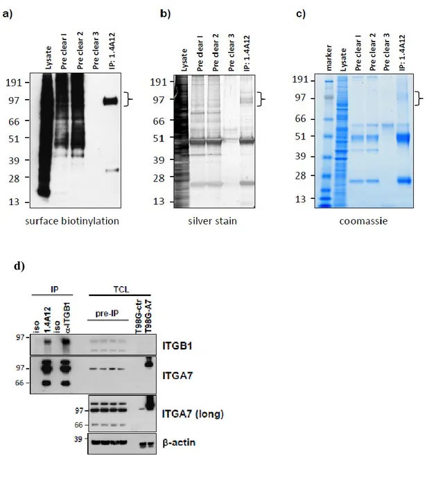
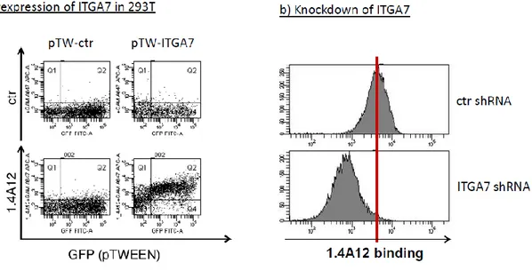
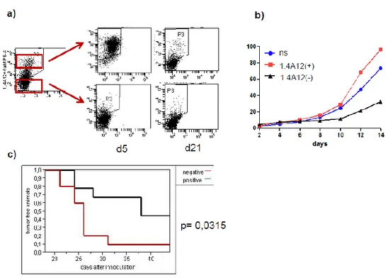
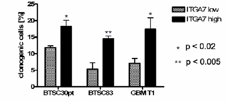
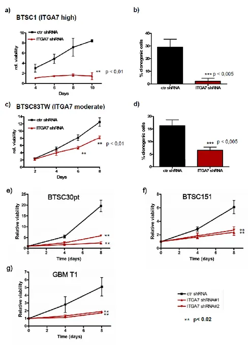
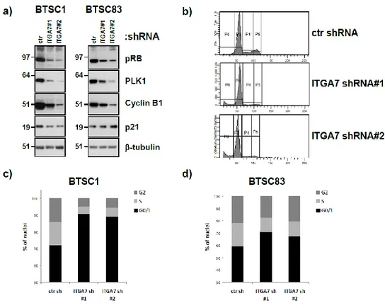
The glioblastoma stem cells maintain the characteristics of tumor initiating cells after prolonged culture in vitro. We used these cells as immunogens in immunocompetent BALB/c mice to develop a hybridoma library. After the initial screening of the library was identified the 1.4A12 antibody that identified integrinα7 (ITGA7) as antigen mostly expressed on brain tumor stem cells. The biological function of the integrinα7 was evaluated the vitro and in vivo. ITGA7 silencing impairs the growth rate proliferation, clonogenicity, invasion in vitro and the tumor growth reduction in vivo. We analyzed the effect of 1.4A12 antibody and its role in counteracting the ITGA7 pathway. The 1.4 anti-integrin alpha7 antibody interferes with the integrin alpha 7 signaling and suppressed tumor invasion in vitro and tumour growth in vivo. All our data indicated that integrin alpha7 is involved in GBM pathogenesis and that 1.4A12 antibody could be of great help in defining new therapeutic options to increase glioblastoma eradication interfering with the tumor growth and with the invasion of cancer cells.
6
INTRODUCTION
1 MONOCLONAL ANTIBODIES 1.1 Monoclonal antibodies features
Over a hundred years have passed from the discovery of the “magic bullet” serum therapy by Behring that led to the awarding of the Nobel Prize in Medicine and Physiology in 1901. Behring and his group demonstrated that the transfer of serum from a guinea pig immunized with diphtheria toxin to another guinea pig offered protection from the toxin. Over the years the serum, from immunized non-human sources (horses or rabbits), was applied to care tetanus toxin bacterial diseases including diphtheria, meningitis, and pneumonia,
demonstrating the action of this system. Many foreign proteins contained in the serum gave a phenomenon that has been called “serum sickness”. Behring recognized these toxic side effects of serum therapy and introduced improved methods for the purification of serum. Köhler and Milstein developed methods for the isolation of mAbs (Monoclonal Antibodies) from hybridoma cells in 1975 (Kohler and Milstein 1975). They showed that the cell fusion technique could be used to produce hybrids between myeloma cells and antibody producing cells. The resulting hybrid lines were permanently adapted to grow in tissue culture and were capable of inducing antibody production in mice (Jerne and Nordin 1963). This discovery led to the awarding of the Nobel Prize in Physiology or Medicine in1984 to Köhler and Milstein. Köhler and Milstein’s method opened the use of hybridoma technology to academics and pharmaceutical fields for generation of antibodies for therapy. The first therapeutic mAb generated was OKT3 by Ortho Biotech in 1984 against CD63 antigen expressed on T cells for the treatment of transplant rejection. Initially, mAbs were from mouse, leading to a relative immunogenic capacity in humans. This issue was progressively solved with the increasing substitution of murine sequences with human ones, which led to the development of chimeric or fully humanized mAbs. The chimeric antibodies have one-third murine sequences (2 VH and 2 VL subunits) fused with two-one-thirds human sequences, including the human Fc part(Morrison, Johnson et al. 1984) (Boulianne, Hozumi et al. 1984). The humanized Abs have the V chains from a murine, or other mammalian antibody, “more human”. Antibodies humanized reduce the human immune response against this murine antibody for the production of human antimouse antibody (HAMA).
The humanized antibodies have the complementarity-determining regions (CDRs) of murine antibodies fused into the closely related human structure, followed by aminoacid changes
7 required to stabilize the engineered constructs (Jones, Dear et al. 1986). The first humanized mAb used as a therapeutic agent was an anti-CD25 (IL-2 alpha subunit) mAb that was humanized and developed to suppress rejection after transplantation from Quen group (Queen, Schneider et al. 1989). In contrast to these in vitro manipulations for humanizing mAb, two research groups independently developed functional human mAbs from
“humanized” transgenic mice. The mice used were unable to produce their own murine antibodies and replacing that function with human antibody genes (Green, Hardy et al. 1994; Lonberg, Taylor et al. 1994). These transgenic humanized mice were immunized with an antigen and fully human antibodies were generated in these mice. The first fully human mAb developed from these humanized systems was Panitumumab, a human IgG2 antibody discovered using Abgenix XenoMouse technology, against EGFR.
Other group proposed another approach to develop antibodies. Smith’s group reported that peptides were able to be displayed as fusions of P3 protein on the tail fibers of filamentous phage M13 (Smith 1985) (Barbas, Kang et al. 1991). This method was applicable for the display of proteins including mAbs (Barbas, Kang et al. 1991). This M13 P3-based phage display technology has become an optimal methodology for selection of antibody fragments (Gram, Marconi et al. 1992). McCafferty et al. reported that was possible build a library of antibody genes, displayed on the P3 protein of M13 phage, using PCR techniques to recover the human gene from either B cells or hybridomas (McCafferty, Griffiths et al. 1990). This method was followed by the construction of huge human libraries from either synthetic repertoires or from multiple naïve human donors (Barbas, Bain et al. 1992) (Marks, Hoogenboom et al. 1991).
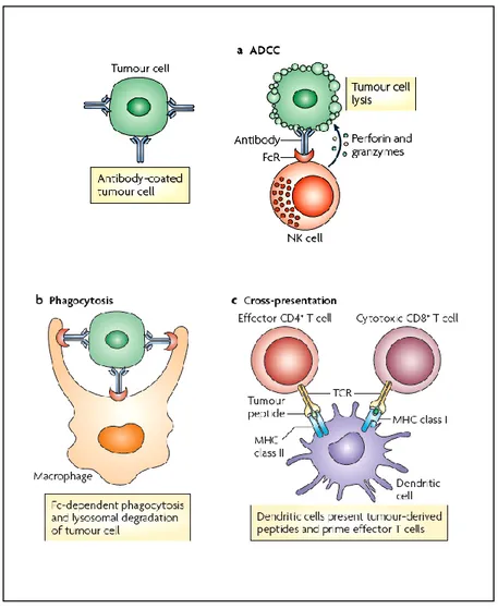
The antibody-based therapy have been developed to target the tumor cells but also to enhance the antitumor immune responses. Many mechanisms have been proposed to explain the antitumor activity of monoclonal antibodies as antibody-dependent cellular cytotoxicity (ADCC) (Fig 1a) and complement-dependent cytotoxicity (CDC). ADCC is initiated by the recognition of Fc part of IgG-coated tumors by Fc receptors (FcγRs) of natural killer (NK) cells, macrophages and neutrophils. These interactions lead to ADCC and to tumor cell apoptosis triggered by cytotoxic granules (containing perforin and granzyme B), released by effector cells. Most clinically approved monoclonal antibodies that mediate ADCC also activate the complement system. IgG can bind to tumor cells and recruit complement factors resulting in tumor cell lysis. Another antitumor mechanism mediated by IgG on tumor cells and FcγR receptor on macrophages interactions is the phagocitosis that induces the
8 lysosomal degradation of the tumor cell (Fig 1b). Dendritic cells (DCs) are also capable of presenting peptides from lysosomal degradation of tumor cells on MHC class I molecules to determinate antigen-specific CD8+ T citotoxic cell responses and on MHC class II leading to the activation of CD4+ T cells, which can activate B cells for the production of tumor-specific host antibodies (Fig 1c) (Weiner, Surana et al. 2010).
Fig 1: Antitumor mechanisms mediated by IgG–FcγR interactions a) Antibody-dependent
cell cytotoxicity (ADCC) is promoted by the recognition of IgG-coated tumors by Fc receptors for IgG (FcγRs) leading to tumor cell apoptosis. b) Fc receptor expressed by phagocytes can bind IgG-coated tumor cells causing Fc-dependent phagocytosis and lysosomal degradation. c) Dendritic cells present peptides from lysosomal degradation of tumor cells on MHC class I molecules and MHC class II activating T cells.
9 1.2 Oncologic application of Monoclonal antibodies
CD20
Rituximab was the first mAb to be approved by the United State Food and Drug
Administration (FDA) for oncological treatment. Rituximab is a mouse-human chimeric mAb against CD20. It derived from mouse anti-CD20 mAb by engineering of human IgG1 and kappa constant regions and the original murine variable regions (Reff, Carner et al. 1994). CD20 is expressed in more than 90% of non-Hodgkin lymphomas and 10% of chronic lymphocytic leukemias. Anti-CD20 mAb is efficient in B cell malignancies treatment because CD20 is expressed at high levels on B cells, is relatively resistant to internalization, thereby allowing the mAb to persist on the cell surface. The binding between CD20 and rituximab induces apoptosis and cell lysis via complement-dependent cytotoxicity (CDC) and ADCC. Clinical studies of rituximab in relapsed or refractory CD20-positive non-Hodgkin B cell lymphomas showed that combination chemo/immunotherapy is superior to treatment alone (Reff, Carner et al. 1994).
Human epidermal growth factor receptor 2 (HER2)
The most known antibodies in solid tumors targeted therapy are the antibodies directed against the EGFR family. Anti-HER2 mAb trastuzumab (Genentech) was derived from murine mAb by humanization of human IgG1 and kappa constant regions fusing the murine complementarity determining regions (Carter, Presta et al. 1992). HER2 is a transmembrane receptor and is a member of the epidermal growth factor receptor (EGFR) family of receptor tyrosine kinases. HER2 normally regulates cell growth and cell cycling via cyclin D and c-myc (Lane, Beuvink et al. 2000). HER2 gene amplification and/or overexpression occurs in around 20–30% of primary breast carcinomas and is associated with reduced survival (Press, Bernstein et al. 1997). Trastuzumab binds to an epitope in the juxtamembrane region of HER2 on breast carcinoma cells, acting as an “anti-cancer reagent” through a direct inhibition of cell growth and receptor internalization and degradation decreasing cell
signaling progression. Trastuzumab inhibits cleavage of an extracellular domain induced by metalloproteinase, induces apoptosis and impairs tumor angiogenesis (Baselga, Albanell et al. 2001; Mohsin, Weiss et al. 2005) (Izumi, Xu et al. 2002). In metastatic breast carcinoma patients, trastuzumab combined with standard chemotherapy produces a better response rate, decreasing the time to disease progression, and overall survival compared with
10 chemotherapy or trastuzumab alone (Esteva, Valero et al. 2002). Better response rates has been observed in patient with overexpression of HER2 as compared to patients with normal expression (Seidman, Fornier et al. 2001). Trastuzumab was approved in 1998 for treatment of breast cancer with HER2 overexpression. The indication of FDA approval recently was as adjuvant treatment of patients with HER2-overexpressing tumors with lymph node
metastasis as part of a regimen of doxorubicin, cyclophosphamide and paclitaxel. Trastuzumab is approved for use as a single reagent in patients with metastatic breast carcinoma whose tumors overexpress HER2 and who have received one or more chemotherapy regimens.
CD33
CD33 is a type I transmembrane sialoglycoprotein that is a member of immunoglobulin-like lectins superfamily (Crocker, Paulson et al. 2007). CD33 is expressed on the cell surface of myeloid and sialic acid -binding early multilineage hematopoietic progenitor cells,
monocytes, and blasts of acute myeloid leukemia (AML) (90%). CD33 is not expressed on normal pluripotent stem cells or non-hematopoietic cells (Brendel and Neubauer 2000). Gemtuzumab ozogamicin (Mylotarg) is a humanized mAb against CD33 conjugated with cytotoxic anti-tumor antibiotic calicheamicin. Gemtuzumab is derived from murine IgG1 mAb generated with human IgG4 and kappa constant regions, while retaining
complementarity determining regions of the murine mAb (Hamann, Hinman et al. 2002). Gemtuzumab ozogamicin binds to the cell surface of CD33-positive leukemic cells with an approximate affinity of 0.08 nM. This mAb acts as a vehicle delivering a conjugated toxic agent to CD33 positive leukemic cells. Binding of the mAb to CD33 results in endocytosis, cleavage of the link between mAb and calicheamicin, and release of calicheamicin that is reduced by glutathione to form a reactive intermediate that binds to DNA, generating a double-strand breaks and inducing apoptosis (Dedon, Salzberg et al. 1993). In a phase II study, 277 patients with CD33-positive AML received monotherapy with gemtuzumab ozogamicin. As a result, 26% achieved remission, 13% of the patients had complete remission (Larson, Sievers et al. 2005). The current FDA approved indications include treatment of patients with CD33-positive AML in first relapse who are more than 60 years old and who are not considered candidates for chemotherapy.
11 Vascular endothelial growth factor (VEGF)
VEGF is an angiogenic growth factor that is activated by the binding with the receptor tyrosine kinase VEGF receptor 2 (also call Flk-1or KDR). These receptors are located on the cell surface of the endothelium of blood vessels and lymphatic vessels. Stimulation of these cells by VEGF leads to cellular growth, inhibition of cell death, and angiogenesis (Ferrara, Gerber et al. 2003). There are several isoforms of VEGF: 121-, 165-, 189-, and 206-amino-acid isoforms that can been formed by alternate splicing. VEGF acts in the regulation of both normal and abnormal angiogenesis, including neoplasm (Ferrara, Gerber et al. 2003). The VEGF is secreted by malignant neoplasm and by tumor-associated stromal cells (Fukumura, Xavier et al. 1998). mAbs, against VEGF signaling, induces growth inhibition of tumor (Kim, Li et al. 1993) (Wood, Bold et al. 2000). Bevacizumab, a humanized mAb against VEGF, was derived from murine mAb against human VEGF165 by Genentech. Bevacizumab has human IgG1 and kappa constant regions as human, while retaining murine complementarity determining regions (Presta, Chen et al. 1997). Bevacizumab reacts and neutralizes all isoforms of VEGF effectively (Kim, Li et al. 1992). Bevacizumab binds an epitope of VEGF that is distinct from the receptor-binding site (Liang, Wu et al. 2006). In a xenograft model with human rhabdomyosarcoma and breast carcinoma, bevacizumab inhibits tumor growth to under 10% of the original tumor weight and impaired tumor vasculature, decreasing microvessel density and permeability (Presta, Chen et al. 1997) (Salnikov, Heldin et al. 2006). The FDA approved bevacizumab for the first-line treatment of metastatic colorectal carcinoma in 2004. Recently FDA approved new indications
including the use of bevacizumab in combination with 5fluorouracil based chemotherapy for first or second line treatment of patients with metastatic carcinoma of the colon or rectum. It is also used in combination with carboplatin and paclitaxel for the first line of treatment for advanced, recurrent, or metastatic nonsquamous cell, non small cell lung carcinoma.
12 2 GLIOBLASTOMA
2.1 Glioblastoma features
Malignant gliomas are tumors that develop in the central nervous system CNS, derived from the glial cells. Gliomas comprise a group of heterogeneous tumors classified for difference of cell type origin and histological features and are divided in astrocytoma,
oligodendroglioma, oligoastrocytoma, ependymoma and choroid plexus tumors (Louis, Ohgaki et al. 2007). The astrocytomas account 75% of all gliomas. They were classified from the WHO into four prognostic grade for their typical characteristics as nuclear atypia, mitotic activity, endothelial proliferation and necrosis (Louis, Ohgaki et al. 2007) (Dolecek, Propp et al. 2012) and comprise: pilocytic astrocytoma (grade I), diffuse or fibrillary
astrocytoma (grade II), anaplastic astrocytoma (Grade III), and glioblastoma multiforme (grade IV). The grade I and II are low grade malignancy, while those of III and IV grade are considered to be high grade malignant (Behin, Hoang-Xuan et al. 2003).
Glioblastoma multiforme (GBM) is the most frequent and the most aggressive malignant brain tumor. GBM derived from astrocytes cells, star shaped cells that support nerve cells, but can be composed from different cells (astrocytes and oligodendrocytes). Sometimes, they evolve from a low-grade astrocytoma or from an oligodendroglioma. In adults, GBM occurs most often in the cerebral hemispheres, especially in the frontal and temporal lobes of the brain. GBM is characterized for the worst survival rates with a median survival ranging from nine to twelve months after the diagnosis (Louis, Ohgaki et al. 2007). The standard therapeutic application, chemotherapy or radiotherapy, and the surgical resection do not improve the patient outcome. The term glioblastoma “multiforme” indicates the
heterogeneous morphology, in which the cellular composition is highly variable. GBM is characterized by high genomic instability, uncontrolled cell proliferation, cells
dedifferentiation, diffused infiltration, propensity for necrosis, strong angiogenesis and resistance to apoptosis (Furnari, Fenton et al. 2007; Shiras, Chettiar et al. 2007; Bonavia, Inda et al. 2011). This complexity combined with the presence of the putative CSCs and the epigenetic lesions makes the GBM one of the most difficult cancer to approach. GBM are subdivided into two subgroups: primary (or the novo GBM) and secondary GBM. The first one account in older patients and with any evidence of a prior symptoms or antecedent low grade, with a short clinical history. The secondary GBM, that represents a lower amount of the GBM cases, occurs in younger patients (less than 45 yr), and derives from a progressive transformation of low grade astrocytomas, generally grade II, that give arise to the high
13 grade. Primary and secondary GBM are morphologically and clinically identical although recent genomic profiles have revealed different transcriptional patterns and recurrent DNA copy number aberrations between primary and secondary GBM as well as new disease subclasses within each category (Maher, Brennan et al. 2006).
14 2.2 Signalling Pathways in Glioblastoma multiforme
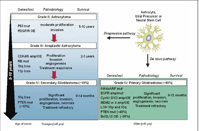
Even if the primary and secondary glioblastoma multiforme are heterogeneous, they share specific cellular pathways of proliferation, survival, differentiation, migration, DNA repair, necrosis and apoptosis. These pathways include p53, pRB, growth factors, PI3K/Akt, apoptosis and angiogenesis signaling (Bonavia, Inda et al. 2011) (Fig 2).
Fig 2: GBM can develop over 5–10 years from a low-grade astrocytoma (secondary GBM), or it can
15 Cell cycle gene disregulation in gliomas
Many mutations in gliomas account for cell cycle genes involved in cellular proliferation and senescence. The RB and p53 pathways are often targets of inactivating mutations in GBM.
RB pathway
The retinoblastoma protein (pRB) is a tumor suppressor that regulates the transition from G1 into S phase of cell cycle. The hypophosphorylated RB, in quiescent cells, sequester the E2F transcription factor preventing the activation of genes involved in the cell cycle progression (Furnari, Fenton et al. 2007). The mitogenic stimulation leads to the activation of the CDK complexes, in particular to the cyclin D1 activation that associated with the cyclin dependent kinase CDK4 and CDK6 phosphorylate RB leaving the E2F to promote the activation of genes to entry in S-phase and the DNA synthesis . The p16INK4a protein inhibits cell cycle progression negatively regulating the formation of the complex cyclin D/CDK (Sherr and McCormick 2002). In gliomas are frequent several alterations or amplifications in these genes. RB1 gene, localized in the chromosome 13q14, is mutated in around 25% of high grade astrocytomas. The 13q is loss in 50% of astrocytomas patients and the loss contributes to the formation of high-grade astrocytomas underlying that Rb inactivation is a tumor suppressor gene in astrocytoma tumorigenesis (Weinberg 1995). Amplification of CDK4 and CDK6 account in 15% of high gliomas determinate the functional inactivation of RB (Serrano, Hannon et al. 1993; Henson, Schnitker et al. 1994). Inactivating mutation by allelic loss or hypermethylation of p16INK4a is also account in 50% - 70% of high grade gliomas bringing to the lost of RB activity (Costello, Berger et al. 1996).
P53 pathway
The p53 (localized in chromosome 17p) gene codify for a tumor suppressor protein containing transcriptional activation, DNA binding and oligomerization domains. Cellular stresses as UV radiation, hypoxia, DNA damage and inappropriate oncogene activation activate p53 response, where p53 acts as a transcription factor promoting the expression of genes that block the cell cycle, as CDNK1A that encodes the protein inhibitor p21. p21 protein (WAF1) acts as inhibitor of CDK2, that is fundamental for the cycle progression in G1-S phase, then the cells blocked in G1-S phase can repair the DNA. If the damage is
16 irreparable p53 actives genes trigger apoptotic pathway, promoting cell death. Mdm2
negatively regulated p53 by ubiquitination and subsequent proteasomal degradation. P53 is also regulated by p14ARFprotein (localized in CDKN2A), that inhibits Mdm2, thus
promoting p53 activation. p53 point mutations that inactive the DNA binding capacity or by loss of chromosome 17p are frequent in secondary GBM and in the progression of
secondary glioblastoma (Louis 1994). The 2q14-15 region containing Mdm2 is
overexpressed in some GBM and the corresponding gene p14ARFis methylated so that its expression is inhibited in malignant gliomas. All these mutations determinate a propagation of cells with genomic instability (Louis 1994).
Mitogenic signaling pathway
The proliferation of normal cells requires the activation of signaling pathways triggered between extracellular matrix (ECM), growth factors, cytokines, cell-cell adhesion and their specific transmembrane receptors. These signals are transduced in the intracellular
compartment by PI3K and MAPK signaling. In gliomas these pathways are constitutively activated.
PI3K/AKT/PTEN signaling
PI3K, phosphatidylinositide 3 kinases, is a family of enzymes divided in three classes (I,II,III), that phosphorylates membrane lipids, as phosphoinositides and
phosphatidylinositols. The class I is formed by phosphatidylinositol- 4,5-bisphosphate (PtdIns(4,5)P2) and phosphatidylinositol 3,4,5-triphosphate (PtdIns (3,4,5) P3). PI3K can be activated after RTK (receptor tyrosine kinase) or integrin signaling.
PI3-kinases have been associated to a diverse group of cellular functions, including cell growth, proliferation, differentiation, motility, survival and intracellular trafficking. Many of these functions are related to the ability of class I PI3-kinases to activate protein kinase B (PKB, aka Akt) as in the PI3K/AKT/mTOR pathway. The class I is formed by two subunits: the p110 catalityc, also divided in three variants p110α, β, or δ, encoded by the PIK3CA, PIK3CB, and PIK3CD genes, and the p85 regulatory subunits, divided in five variants p85α, p55α, p50α, encoded by (PIK3R1) genes and p85β by (PIKR2), and p85γ by (PIKR3). In gliomas 15% frequency of mutation PIK3CA has been observed (Samuels, Wang et al. 2004) and PIK3D gene overexpressed in GBM (Gallia, Rand et al. 2006). The PtdIns (3,4,5)
17 P3 binds to different cytosolic proteins, including Akt which undergoes a conformational change that facilitates its activation through phosphorylation in correspondence of the two amino acid residues T308 and S473 by respectively phosphoinositide-dependent kinases (PDK) 1 and by the phosphatidylinositol 3-kinase-related kinase protein, the mammalian target of rapamycin mTOR, acting in the rapamycin-insensitive TORC2 complex (Mora, Komander et al. 2004). Akt is a key regulator of cell proliferation and survival pathways. It is a Serine/Threonine kinases able to inhibit apoptosis, promote cell proliferation and regulate lipids and glucose metabolisms, cell movements and vesicle trafficking. Akt is also inactivated by PHLPP (PH domain leucine-rich repeat protein phosphatase), which
dephosphorylates S473, that was found to be expressed at very low levels in certain GBM cell lines and enhancing the anti-apoptotic activity of AKT (Maira, Galetic et al. 2001). The class I PI3K enzymes is antagonized by the PtdIns(3,4,5) P3 -phosphatase encoded by the PTEN gene. PTEN is a tumor suppressor that is inactivated in 50% of high grade gliomas by mutations or epigenetic mechanisms determinating the activation of uncontrolled PI3K activities. Deletions or mutations in the PTEN gene and high levels of phosphorylated or activated Akt are the most widespread alterations of the PI3K/Akt signaling in
glioblastomas. These changes characterize GBM features, such as rapid tumor growth, invasiveness, resistance to cytotoxic treatments and massive angiogenesis (Knobbe, Trampe-Kieslich et al. 2005) (Rich and Bigner 2004) (Chakravarti, Zhai et al. 2004).
RTKs signaling
The receptors tyrosine kinase are transmembrane protein that sharethree basic components: the extracellular ligand binding domain, the transmembrane segment and an intracellular protein tyrosine kinase (PTK) domain with a catalytic core and the regulatory sequences. Upon binding of the ligand the receptor changes the inactive monomers into homodimers that activates its intracellular tyrosine kinase domain and autophosphorylate its C-terminal tyrosine residues. This stabilizes the receptor conformation and creates phosphotyrosine-docking sites for adapter proteins that transduce signals within the cell. Among the RTKs there are the Epidermal Growth Factor Receptor (EGFR) and platelet-derived growth factor (PDGFR). In gliomas the alterations of RTK include the overexpression of the ligands (growth factors) or of the receptors that lead to an autocrine activation of the pathways and the mutation of the receptors that are constitutively activated in absence of the specific ligands.
18 Epidermal growth factor receptor is a cell surface receptor family containing four
structurally related members (erb1/EGFR, erb2/HER2, erb3/HER3 and erb4/HER4). Transduction by activated EGFR leads to downstream effect on proteins including phosphatidyl 3-kinase (PI3K), Phospholipase (PL) C-g1, Akt, Ras, Raf and mitogen-activated protein kinase (MAPK) that are associated with cell proliferation, motility and survival.
EGFR gene is amplified in around 40% of all GBMs, and the genes are frequently
rearranged. In 20–30% of all human GBM occurs an EGFR mutant allele with deletion of exons 2–7 known as EGFRvIII (this mutant is also frequent in 50%–60% of those that have amplified wild-type EGFR), EGFR is constitutively actived and enhances tumorigenicity (Louis, Ohgaki et al. 2007). EGFRvIII is a validated target in gliomas and is reported that EGFR mutants have the capacity to enhance the proliferation and reducing the apoptosis and promoting the tumorigenesis in human (Frederick, Wang et al. 2000). EGFR is a target with crucial importance for the GBM therapy. There are small molecule kinase inhibitors, immunotherapy based on antibody, immunotoxins (Huang, Mukasa et al. 2007)and small interfering RNA against allele of EGFRvIII (Jungbluth, Stockert et al. 2003).
The PDGF signalling network consists of four ligands, PDGFA-D, the AB heterodimer ligand, and two receptors, PDGFR alpha and PDGFR beta. The alpha type binds to PDGF-AA, PDGF-BB and PDGF-AB, whereas the beta PDGFR type binds with high affinity to PDGF-BB and PDGF-AB. PDGFR pathway plays a role in embryonic development, cell proliferation, cell migration and angiogenesis.
PDGF-A and PDGF-B are expressed in highgrade glioblastoma and strong expression of PDGFR occurs in endothelial cells in GBM (Fan and Weiss 2005). PDGF-C and PDGF-D are also often expressed in glioma cell lines and in GBM tissues (Westermark, Heldin et al. 1995). The amplification or the rearrangement of PDGFR is much less common but is reported a loss of exons 8 and 9 of PDGFR that determinate a constitutive activation of the receptor enhancing the tumorigenicity (Lokker, Sullivan et al. 2002).
19 Apoptosis
The main feature of glioma cells is its resistance to chemotherapy and to radiotherapy treatment. This property is due to genetic alterations of regulatory molecules involved in mitogenic and/or PI3K/PTEN/Akt pathways and/or in classical cell death networks of extrinsic (death receptor-mediated) and intrinsic (mitochondria-dependent) apoptosis signaling (Clarke and Dirks 2003).
The death receptors are cell surface proteins that after binding of cognate ligands, recruit adapter molecules to create a protein complex that has an autoproteolytic capacity determinating the activation of caspases. The most important system of death receptor include TNFR1, TRAILR1-2 and CD95. These death receptors are often down-regulated and their ligand mutated in glioma pathogenesis (Maher, Brennan et al. 2006).
It was reported that human glioma cell lines and primary glioma-derived cell cultures are sensitive to death ligand-mediated apoptosis in vitro and in vivo (Igney and Krammer 2002). Expression levels of death receptors and of their decoy receptors (antagonistic) were
associated with vulnerability of glioma cells to death ligand-induced apoptosis. The expression of antagonist receptor for CD95 ligand (CD95L), the soluble decoy receptor 3 (DcR3), on malignant glioma cell lines was correlated with the grade of malignancy in human glioma specimens. Even more, the infiltration of CD4+ and CD8+ T cells and microglia/macrophages was significantly decreased in DcR3-driven xenografts, suggesting that a decoy receptor neutralizes CD95L attack by preventing its interaction with the receptor (Maleniak, Darling et al. 2001).
The expression of TRAIL death receptor system has been correlated with survival of patients with primary GBM (Roth, Isenmann et al. 2001). The treatment with TRAIL inhibited growth of human glioma cell xenografts (Kuijlen, Mooij et al. 2006) and acted synergistically with chemotherapeutic drugs (Roth, Isenmann et al. 1999), determinating a up-regulation of TRAIL-R2 and Bak protein and down-regulation of the cFLIPs (caspase-8-specific inhibitor) (Nagane, Pan et al. 2000) . Peptides as Smac, a potent antagonist of the IAP family (Inhibitors of Apoptosis Protein), shown a synergistic effect with TRAIL to induce tumor cell apoptosis in vitro and in vivo without demonstrable neurotoxicity (Song, Song et al. 2003) underling the importance of post-mitochondrial caspase activation for apoptosis propagation in glioma cell lines and its validity as a therapeutic target (Song, Song et al. 2003).
20 Bcl-2 family is composed by pro-apoptotic (Bax, BAD, Bak) or anti-apoptotic (including Bcl-2 proper, Bcl-xL, and Bcl-w, MCL-1) proteins. These proteins control the mitochondrial membrane integrity and the release of cytochrome c, which effects the caspase cascade and the apoptotic program (Fulda, Wick et al. 2002). There is a correlation between tumor grade and expression of anti-apoptotic Bcl-2 proteins (BCL-2 and MCL-1) (Green and Kroemer 2004). For example Bcl-xL was found up-regulated by overexpression of EGFRvIII in glioma cells and this upregulation confers resistance to the chemotherapeutic agent as cisplatin (Krajewski, Krajewska et al. 1997). Bcl2 family may also contribute to
gliomagenesis through enhancement of migration and invasion by altering the expression of metaloproteinases and their inhibitors (Wick, Wagner et al. 1998; Wick, Grimmel et al. 2001).
Necrosis
GBM tumor cells are resistant to therapeutic apoptotic stimuli but show wide cellular necrosis. Necrosis in fact is the major form of spontaneous cell death in GBM (Wick, Wild-Bode et al. 2004). Necrosis is determinated by inadequate blood supply and anoxia due to a microthrombotic process. In this regard was found a new protein Bcl2-like 12 (Bcl2L12) protein, an inhibitor of post-mitochondrial apoptosis signal transduction, that is significantly overexpressed in primary GBMs (Brat and Van Meir 2004). Bcl2L12 is a protein with homology with the BH (Bcl-2 Homology) 2 domain found in several members of the Bcl-2 protein family (Stegh, Kim et al. 2007). Overexpression of Bcl2L12 in primary astrocytes inhibited apoptosis, and its RNAi-mediated knockdown sensitizes human glioma cell lines to drug-induced apoptosis and reduces tumor formation in an orthotopic model system. Bcl2L12 protein neutralizes effector caspase activity determinating an anti-apoptotic effect (Brat and Van Meir 2004). The activities of Bcl2L12 explain the necrotic process in which the suppression of caspase activity from mitochondria redirects the death program from apoptosis to necrosis (Scorilas, Kyriakopoulou et al. 2001).The interconnection between apoptosis and necrosis signaling and Bcl2L12 is represented by the Bcl2L12 expression that induces necrotic cell morphology, as evidenced by substantial plasma membrane
disintegration and enhanced nuclear and subcellular organelle swelling in apoptosis-primed astrocytes (Brat and Van Meir 2004).
21 Angiogenesis
GBMs are the most highly vascular solid tumors. Microvascular hyperplasia consists of endothelial cells in proliferative activity that come out from normal parent microvessels as microaggregates (glomeruloid bodies) accompanied by stromal elements, including
pericytes and basal lamina (Nicotera and Melino 2004). Microvascular proliferation is a features that characterize the evolution from low-grade or anaplastic astrocytomas to secondary GBM (Maher, Brennan et al. 2006) . In the last decade has been reported that tumor-associated angiogenesis is not due only to physiological modification to hypoxia resulting from an increase of tumor cell mass but it appears to be the result of genetic mutations that activate a transcriptional program for angiogenesis with local tumor oxygen status modifying this response. For example mutations, including those in the PTEN, EGFR, and CMYC genes, may act as an “angiogenic switch” by stabilizing HIF-1 (Hypoxia
inducing factor) or one of its downstream targets, VEGF (Stiver, Tan et al. 2004) (Watnick, Cheng et al. 2003) (Blum, Jacob-Hirsch et al. 2005) (Phung, Ziv et al. 2006).
Tumor cell invasion
The brain glioma’s invasiveness is the most important characteristic of low- and high-grade malignant glioma and is the cause of the failure of surgical cure. In most of the cases the tumor develops adjacently to the resection margin. Invasion is determinated by processes that involve cell interactions with the ECM and with adjacent cells and by biochemical proteolytic degradation of ECM and active cell movement.
Glioma cells invade along the white matter tracts and the basement membranes of blood vessels, accessing to a disrupted blood vessel within the tumor. Gliomas are unable to establish robust tumor growth outside the CNS and is unable to metastasize outside the CNS. The invasion and migration of glioma cells in fact differs from other tumors where local spread is very limited and dissemination occurs hematogenously or via the lymphatic system (Lefranc, Brotchi et al. 2005). Several genes have been identified in glioma
invasiveness and include the family of metalloproteases (MMP) and their endogenous tissue inhibitors (TIMPs). Expression of MMP-2 and MMP-9 are associated with invasiveness, proliferation and prognosis in astrocytomas (Wang, Wang et al. 2003). Other non-MMP proteases, including urokinase-type plasminogen activator (uPA) (Landau, Kwaan et al. 1994) and cysteine proteases, as cathepsin B, are elevated in high-grade malignant gliomas
22 (McCormick 1993). The role of proteases in glioma invasion is unclear because low-grade astrocytomas infiltrate throughout the brain, despite relatively normal levels of the proteases. Also integrins, as αVβ3 complexes, are overexpressed in GBM and are important in glioma invasion and angiogenesis (Uhm, Dooley et al. 1997) (Kanamori, Vanden Berg et al. 2004). Other proteins are overexpressed in invasive areas of GBM, such as angiopoietin- 2, the ligand of Tie receptors, which is involved in angiogenesis and determinate infiltration by activating MMP-2 (Hu, Guo et al. 2003).
Ephrin receptors and their ligands, the ephrins, have been shown to control migration and invasion. EphA2 and EphB2overexpression has been associated to poor survival in GBM (Liu, Park et al. 2006) .
23
2.3 Glioblastoma treatment
Glioblastomas is the most difficult cancers to treat for its complexity for the drug distribution within the intracranial space, due to the presence of the blood-brain barrier. GBM grow quickly and show a high resistance to apoptosis (Rich and Bigner 2004; Wen and Kesari 2008).
The standard therapy for malignant gliomas involves surgical resection, when
feasible, radiotherapy, and chemotherapy. GBM is still associated with poor prognosis with the median survival of 1 year (Wen and Kesari 2008). Malignant gliomas cannot be
completely eliminated surgically for their infiltrative nature, but patients undergo to maximal surgical resection whenever possible. Advances in MRI (magnetic resonance imaging) have improved the safety of surgery and increased the extent of resection that can be achieved. Radiotherapy is the primary intervention of treatment for malignant gliomas. The addition of radiotherapy to surgery increases survival among patients with
glioblastomas from a range of 3 to 4 months to a range of 7 to 12 months (Walker, Alexander et al. 1978). Recently has been reported that chemotherapeutic agents may improve the efficacy of radiotherapy, resulting in a modest increase in survival (a 6 to 10% increase in the 1-year survival rate) (Fine, Dear et al. 1993; Stewart 2002). In the last years, the combination of radiotherapy and temozolomide (TMZ), an oral alkylating agent with good penetration of the blood brain barrier, shown an increasing in the median survival (14.6 months vs 12.1 months, p<0.001) (Stupp, Mason et al. 2005). Radiotherapy with concomitant adjuvant temozolomide is a useful combination for newly diagnosed
glioblastomas. Others agents used reflects the prevalence of alterations in EGFR, such as the EGFRvIII deletion mutation (Scott, Lee et al. 2007) and PDGF signaling and modulators of PI3K signaling, as well as the prominence of biological processes such as angiogenesis and invasion. Research is focusing also on others molecules against the integrins as
Etaracizumab, CNTO95, Cilengitide, Volociximab. Vitaxin, the precursor of etaracizumab, is used in Phase I trials showed antiangiogenic activity and a Phase II study showed some efficacy in metastatic melanoma. CNTO95, which targets both αvβ3 and αvβ5 integrins, also had anti-tumour and anti-angiogenic effects in xenograft tumour models. Cilengitide is currently being tested in Phase II trials in patients with lung and prostate cancer, and Phase II and Phase III trials are currently underway in glioblastoma. In the same time the integrins are targets for cancer imaging and drug discovery. Integrins have the ability to deliver diagnostic agents (radionuclides, Cu, F, superparamagnetic iron oxidase suitable in
24 Scintigraphic or PET) and chemoterapeutics or proapoptotic peptides to tumour cells
(Integrin αvβ3 targeted nanoparticles to delivery mutat RAF1 to the tumor vasculature, resulting in apoptosis of endothelial cells and tumor regression or with nanoparticles loaded with doxorubicin) (Desgrosellier and Cheresh 2010) (Guo and Giancotti 2004).
25 3 GLIOBLASTOMA STEM CELLS
3.1 Glioblastoma stem cells features
Accumulating literature, from almost 20 years of research, support the hypothesis of cancer stem cells and the importance to identify them with specific markers.
This hypothesis was initially demonstrated in blood tumors where in acute myeloid leukemia a subpopulation of leukemic cells (CD34+/CD38-) maintain the ability to
propagate the tumor after serial transplantations in recipient immunodeficient mice models, reproducing a tumor with the same characteristic of the original (Bonnet and Dick 1997). The demonstration that, as in hematological malignancies, solid tumors arise from a cancer stem cell (CSC) opens a new scenario for understanding the biology of the tumor. These studies suggest the existence of a subpopulation of cells, the cancer stem cells within the tumor mass (about 2% of the total cancer cell population) with a crucial role in the development of the tumor. This model of cancer growth is called hierarchical model (or cancer stem cell hypothesis). This model implies that the cancer stem cells are at the apex of the hierarchy and for their stemness proprierties, as self renew and the capability to give arise to progenitors cells, are responsible for the origin and for maintenance of cancer
(Maugeri-Sacca, Bartucci et al. 2012).The bulk of cancer cells within a tumor are progeny of CSCs, have not tumorigenic potential, thus cannot regenerate new tumors, and might
represent a mix of partially differentiated cancer progenitor like cells with limited
proliferative capacity, terminally differentiated, an death committed cancer cells (Tang, Ang et al. 2007). On the other site the stochastic clonal model affirm that random occurrence of mutations lead to dominant clones that gain a survival advantage over other cells, with more ability to thrive in a hostile microenvironment and to adapt to microenvironmental
perturbations. More recently is been suggested a new model “clonal- hiercharchical model” within the stem cells population. These model proposed the evidence that are reported in different works. Infact the stemness features has been found to be induced in differentiated cells upon forced expression of embryonic stem cell-specific transcription factors
(Takahashi and Yamanaka 2006), or by exogenous influences as hypoxia, low pH, signals as stimulation with hepatocyte growth factor, and activation of the
epithelial-mesenchymaltransition (EMT) program (Mani, Guo et al. 2008) (Li, Bao et al. 2009) are involved in the process to maintain and to enrich CSCs. This underline that the
27 CSCs, in solid tumor, were first characterized by Al-Hajj and coworkers (Al-Hajj, Wicha et al. 2003), who isolated mammary stem cells (CD44+CD24-) from human breast carcinoma. After many groups attempted in vitro/in vivo characterizations of populations enriched in CSCs isolated from solid tumors.
CD133, also called Prominin-1, a cell surface glycoprotein, localizes on membrane which suggests an involvement in the mechanisms influencing cell polarity, migration and interaction of stem cells with neighbouring cells (Dell'Albani 2008).
CD133 was shown to be expressed on normal human neural stem cells (Uchida, Buck et al. 2000) and was been found on CSC cells isolated from various compartment (Wu and Wu 2009). CD133+ cells isolated and transplanted into NOD/SCID mice recapitulated the heterogeneity of the original tumor. Dirks group in 2004 demonstrate that CD133 positive cell subpopulation from human brain tumors exhibited stem cell properties in vitro and in vivo assay (Singh, Clarke et al. 2003). Recently CD133, as cancer stem cell markers, was in the middle of a debate whereas CD133- cells can shared the same properties of CD133+ cells (Chen, Li et al. 2010).
New markers have been used to identify specifically and target GSCs (Glioblastoma stem cells): SSEA-1, L1CAM, A2B5, Side Population, Nestin, OCT-4, ALDH, Integrinα6 (table 1).
28 SSEA-, also called CD15, is a stage-specific embryonic antigen 1 and was shown that the selection for SSEA-1+ cells enriched for glioma tumor stem cells (TSCs) or tumor-initiating cells (TICs). These SSEA-1+ cells give at least a 100-fold tumorigenic enrichment in mouse xenograft models (Son, Woolard et al. 2009).
L1CAM , called L1 or CD171, has been identified as a potential therapeutic target in neuro-oncology. L1CAM regulates neural cell growth, survival, migration, and axonal outgrowth and neurite extension during central nervous system development. Although the role of L1CAM in the normal adult nervous system is not well defined, L1CAM is overexpressed in gliomas and other solid cancers, including colorectal cancer where L1CAM is used as prognostic indicator (Bao, Wu et al. 2008).
A2B5 is a glial progenitor marker and the authors identified a population of A2B5+ CD133- cells, phenotypically distinct from CD133+ cells, showing a tumorigenic properties (Ogden, Waziri et al. 2008).
A common way to identify CSCs is by exclusion staining with Hoechst 33342 dye. A main feature of CSCs and normal stem cells is the high expression levels of the ATP-binding cassette (ABC) transporters that are responsible for multidrug resistance and able to export the dye. This identify an unlabelled “side population” (SP) highly enriched in stem cells in many tissues, including neural (Kim and Morshead 2003). SP fraction isolated from the rat glioma cell line C6 show stem cell-like properties. The same results were observed for human glioma cell lines. However, there are conflicting data showing that either the sorted SP or non-SP cells were similarly clonogenic in vitro and equally tumorigenic in vivo. Another approach to select the glioblastoma stem cells is the autofluorescence emission from the cells. This was used to enrich a subpopulation with self-renewal ability in vitro, tumor initiating and propagating capacity in vivo (insert Clement et al, 2010). In addition, high aldehyde dehydrogenase (ALDH) activity has been used as a functional marker to isolate CSCs. These enzymes detoxify aldehydic products generated by reactive oxygen species and might therefore participate in cell survival. High ALDH1 activity keep glioma cells in an undifferentiated state. It is reported that ALDH1 inhibition induces differentiation and reduces clonogenicity in vitro (Rasper, Schafer et al. 2010).
Recently Integrin α6 was suggested as new marker for glioblastoma stem cells. Sorting for integrin α6 high alone or in combination with CD133 led to enrichment of cells that display GSC properties. Integrin α6 depletion using short hairpin RNA or treatment with
integrin-29 blocking antibody determinated to growth inhibition in vitro and reduced tumor formation in vivo (Lathia, Gallagher et al. 2010).
Nestin is an intermediate filament (IF) protein expressed in the stem and progenitor
proliferating cells during the central nervous system developmental stages and its expression is down-regulated in differentiated cells. It is involved in the organization of the
cytoskeleton, cell signaling and metabolism, organogenesis, and represents the proliferation, migration and multi-differentiated characteristics of multi-lineage progenitor cells
(Hadjipanayis and Van Meir 2009).
30 3.2 Altereted pathways in Glioblastoma stem cells
The Glioblastoma stem cells differ from normal neural stem cells for a deregulation of the signal involved in fundamental cellular processes such as cell proliferation, differentiation, stemness maintenance, drug and radio resistance (Li, Wang et al. 2009). The main pathways altered in GSCs include Notch, Hedgehog-GLIs, growth factors, BMP, and TGF signalling.
Notch pathway
Notch proteins are a family of single pass transmembrane domain receptors involved in cell-cell communication. After the binding of its ligands (Jagged 1-2, Delta like 1-3-4), Notch is cleaved by the -secretase complex. The Notch intracellular domain is thus released from the plasma membrane, and translocates into the nucleus where it acts as a transcription factor. During development, Notch promotes the proliferation of normal NSCs while suppressing their differentiation. Notch functions were linked to glioma CSCs, as Notch signaling increases expression of the stem cell marker as Nestin. Moreover, activation of Notch signaling in the glioma cell lines increases the formation of neurosphere-like colonies (Huang, Cheng et al. 2010) (Zhang, Zheng et al. 2008).
Hedgehog pathway
The binding of Hedgehog ligands to their receptors activates transducers termed GLIs (named for their discovery in gliomas), which then translocate into the nucleus to activate or repress downstream targets. The Hedgehog pathway is a regulator of embryogenesis, to transmits information to embryonic cells required for proper development, and is involving different types of normal stem cells, including NSCs (Wechsler-Reya and Scott 1999). Hedgehog signaling is also active in gliomas and contributes to GSCs function, favoring cell proliferation, self-renewal, tumorigenicity and drug resistance (Clement, Sanchez et al. 2007). Furthermore, it has been demonstrated that Hedgehog inhibitors, as cyclopamine, improve traditional therapy efficiency against gliomas. In particular, Bar and colleagues demonstrated that cyclopamine treatment improves the effects of radiation on GSC survival (Bar, Chaudhry et al. 2007).
31 Growth factors signaling
GSCs are also characterized by the deregulation of growth factor signalling (EGF, bFGF, PDGF and IGF). GSCs activate these pathways through different mechanisms, such as ligand and/or receptor overexpression, receptor mutation causing its constitutive activation, intracellular messenger activation through mutation or the loss of expression of negative regulators (Huang, Cheng et al. 2010). The signal initiated by the binding between growth factors and their receptors (Receptor Tyrosine Kinases, RTKs) is transduced and amplified through downstream molecule cascades, such as the pro-survival AKT/PI3K pathway. Upon activation, AKT promotes survival, proliferation, invasion, and secretion of proangiogenic factors. It has been recently demonstrated that GSCs are more dependent on AKT signals than non-stem glioma cells. Pharmacologic inhibitors of AKT attenuate the generation of neurospheres, the structures usually formed by GSCs in culture, suggesting that AKT inhibition may specifically target the GSC population to reduce tumor malignancy (Eyler, Foo et al. 2008).
BMP pathway
Bone Morphogenic Proteins (BMPs) are a family of growth factors involved in bone and cartilage development. Upon the binding to the cell-surface receptor kinases, the
BMPRs, Smad1/5/8 proteins are activated and bind to the co-activator Smad4 that translocates into the nucleus and regulate transcription. BMPs regulate the proliferation, apoptosis and differentiation signal in neural stem cells (Li, Wang et al. 2009). It was shown that this pathway is altered in GSCs by epigenetic alterations of BMP receptor leading to a reduced expression and signal transduction; GSCs are able to escape the
differentiation process induced by BMPs maintaining intact their stemness proprieties (Lee, Son et al. 2008).
TGF-β pathway
The TGF-β is a superfamily that includes proteins that regulate differentiation and cellular development processes (Huang, Cheng et al. 2010). It was shown that GSCs produce and release TGF-β into the extracellular microenvironment inducing cell survival and stemness maintenance through the activation of Sox2 signal transduction pathway (Penuelas, Anido et al. 2009).
32 Sox2-Oct4-Nanog
Many intracellular factors are considered important for the regulation of survival,
proliferation and maintenance of stemness proprierties in gliomas cancer cells as Sox-2, Oct-4, Nanog, bm1, c-myc and mirRNA. Sox2, Oct4 and Nanog are transcription factors
fundamental for the regulation between self-renewal and differentiation in embryonic and adult stem cells (Huang, Cheng et al. 2010). The Oct4 expression is correlated with the glioma grade and it is highly expressed in many human glioma specimens and cell lines. Oct4overexpression in rat glioma cells increases the expression level of the stemness marker Nestin suggesting that Oct4contribute to CSC stemness maintenance.
Olig2
Olig2 is a transcription factor expressed in the CNS (central nervous system). It is expressed in both normal neural stem cells and glioma cancer stem cells.
Olig2 is involved in the development of CNS and it is expressed in neural progenitor cells that give rise to oligodendrocytes and substains the multilineage differentiation potential of neural progenitors (Li, Wang et al. 2009). Olig2 was found to be expressed in almost all adult astrocytomas and is required for tumor initiation, suggesting a link to gliomas and CSCs (Ligon, K. L et al. 2004) (Ligon, K. L.et al 2007).
In GSCs Olig2 is involved in the inhibition of the differentiation process through the suppression of the cell cycle regulatory protein p21WAF1/CIP1 (Ligon, K. L.et al 2007).
c-Myc
c-Myc is a transcription factor, considered an oncoprotein, that plays an important role in the proliferation of both normal stem cells and tumor cells. Recently, inducible pluripotent stem cells were generated from differentiated cells through the introduction of several
transcription factors, including c-myc (Takahashi, Ichisaka et al. 2006), supporting a role in core stem cell machinery. c-Myc expression correlates with the grade of malignancy in gliomas (Herms, von Loewenich et al. 1999). This suggests a link between the stemness and malignancy. Glioma cancer stem cells express high levels of c-Myc and is required for maintenance of GSCs in vitro and for GSCs tumorigenic capacity in vivo (Zheng, Ying et al. 2008). It has been demonstrated that c-Myc additionally prevents differentiation and
33 promotes self-renewal of tumor neurospheres derived from a p53/PTEN double knock-out mouse model (Zheng, Ying et al. 2008).
BMI1
BMI1 belongs to the Polycomb group genes, that function as epigenetic silencers. BMI1 has been implicated in determining stem cell fate in multiple tissues and is a positive regulator of neural stem cell selfrenewal. BMI1 is also a known oncogene frequently overexpressed in many cancer types, including gliomas (Li, Wang et al. 2009). BMI1 is overexpressed also in GSCs and is required to sustain the self-renewal process of these cells (Abdouh, Facchino et al. 2009).
miRNAs
miRNAs are small noncoding RNAs that can silence target genes through
post-transcriptional mechanisms on target mRNAs. A single miRNA can regulate many distinct mRNAs. In cancer biology, miRNAs can function as oncogenes or as tumor suppressors. miRNAs play an important role also in glioma CSCs. For example the levels of miR-124 and miR-137 are reduced in grade III and IV malignant gliomas in comparison with normal brain. Overexpression of these two miRNAs inhibits proliferation and induced
differentiation of glioma CSCs, indicating a tumor suppressor role for these two miRNAs in GSCs (Silber, Lim et al. 2008). Similarly, another miRNA, miR-451, is expressed at
lower levels in CD133+ GSCs in comparison with CD133− non-stem glioma cells. In particular, it has been demonstrated that miR-451 inhibits the growth of glioma CSCs and disrupts the formation of cancer stem neurospheres (Gal, Pandi et al. 2008).
Hypoxia
Brain Tumor Stem Cells (BTSCs), similar to their normal stem cell counterpart, are
localized in specific microenvironments or stem cell niches that include hypoxic regions, the perivascular compartment and the invasive edge. CSCs, as NSCs, can recruit vessels during tumorigenesis and seem to have angiogenic properties. It was reported that the number of capillaries correlates with the GBM patients' prognosis (Leon SP et al, 1996). Glioma stem cells are located preferentially in the perivascular area, in fact endothelial cells were shown to interact with CD133+/Nestin+ cells isolated from glioblastoma, medulloblastoma,
34 ependymomas, and oligodendrogliomas and to produce factors that maintain these cells in a self-renewing and undifferentiated state. Co-injection of CSCs and endothelial cells into immunocompromised mice accelerates the initiation and the growth of brain tumors (Calabrese et al, 2007).
GBM cancer stem cells are regulated by microenvironment conditions such as hypoxia that stimulate the growth of the neovasculature by expressing high levels of pro-angiogenic factors such as VEGF (Bao S. et al, 2006a). On the other hand, blood vessels create a vascular niche to maintain CSC population. Endothelial cells express Notch ligands that may stimulate Notch receptors essential for CSCs maintenance (Fan et al, 2010) and may also secrete nitric oxide to activate the Notch pathway (Charles et al, 2010). CSCs are also regulated through ECM receptors, such as integrin α6, that are enriched on GBM CSCs and promote their maintenance (Lathia et al, 2010).
Hypoxia in neural tumors have been well characterized close to areas of necrosis. Low oxygen promotes the mantainance of an undifferentiated cell state in normal tissues and in tumors supporting a niche for CSCs. In hypoxic conditions, non-stem glioma cells acquire self-renewal and long-term proliferative potential, in addition to the expression of genes related to stem cell functions. Hypoxia modulates cell phenotypes via hypoxia-inducible factor (HIF) signaling which drives expression of stem cell-related genes. The expression of HIF2α is specific for CSCs and is associated with poor glioma patient survival.
35 3.3 Glioblastoma stem cells pathogenesis
GBM is one of the most lethal disease with a high rate of recurrence after treatment. In the last decade a lot of literature underlines that GSCs have a crucial role in the GBM malignant behavior, because are involved in the processes of radio and chemo-resistance, recurrence, metastasis and angiogenesis (Xie 2009), (Hadjipanayis and Van Meir 2009) (Salmaggi, Boiardi et al. 2006). GSCs appear to contribute to the radio-resistance. Bao and colleagues demonstrated that GSCs, in response to DNA damage induced by ionizing radiation rapidly activate the response to DNA damage by phosphorylation of proteins of cell cycle pathway as ATM,Rad17, Chk1 and Chk2 determinating cell survival. In particular,
CD133-expressing tumor cells preferentially activate the DNA damage check points in response to radiation compared with the CD133-negative tumor cells (Bao, Wu et al. 2006). The GSCs are resistant also to cytotoxic drugs. This was to be linked to high expression levels of the DNA repair protein MGMT, that remove the methyl groups added to the DNA by alkylating agents. This underlines the resistance of GSCs to pharmacological treatment (Liu, Yuan et al. 2006). GSCs overexpress ABC transporter family (ABCG2 proteins) (Dean, Fojo et al. 2005). These proteins able to transport anticancer drugs outside the cells. This make them resistance to the treatment. It was shown that GSCs have alterations in the apoptotic signaling for their overexpression of apoptosis suppressor such as Bcl2, Bcl-XL, FLIP and several inhibitors of apoptosis, such as IAPs proteins which bind and inhibit caspases 3-7-9, preventing apoptosis (Johnstone, Ruefli et al. 2002; Liu, Yuan et al. 2006). GSCs are also considered responsible of the high angiogenesis that characterize the glioblastoma.
Angiogenesis is a process to provide oxygen and nutrients to the tumor mass and also for metastasis. Bao et all demonstrated that GSCs promote angiogenesis amplifying the
secretion of VEGF, one of the most important pro-angiogenic factor (Bao, Wu et al. 2006). Treatment with bevacizumab, antibody that neutralizes the effect of VEGF, shown a decrease of GSCs to support the angiogenesis. The upregulation of VEGF in GSCs is not clear but it’s supposed that hypoxia could play an important role (Li, Wang et al. 2009).
36 4 INTEGRINS
The “integrins” include (Mitra and Schlaepfer 2006) a family of structurally,
immunochemically, and functionally related cell-surface heterodimeric receptors, that is integrated between the extracellular matrix and the intracellular cytoskeleton to mediate cell migration and adhesion. The subunits account to eight and the at 17 subunits. These subunits interact non covalently in a restricted manner to form more than 20 family members. The diversity of integrins is expanded further by alternative splicing, post-translational modifications, and interactions with other cell-surface and intracellular
molecules (Guo and Giancotti 2004). A hallmark of the integrins is the ability of individual family members to recognize multiple ligands. Table 2 summarizes the major extracellular ligands of integrins. The list includes a large number of extracellular matrix proteins as bone matrix proteins, collagens, fibronectins, fibrinogen, laminins, thrombospondins, vitronectin, and von Willebrand factor, reflecting the primary function of integrins in cell adhesion to extracellular matrices. The preference of any given integrin among its ligands is determined by relative affinity, availability within a specific microenvironment, and the conformational state of the ligand, which controls exposure of its integrin recognition sequence.
Integrin biology
Integrins are clustered in the membrane and recruit various signalling and adaptor proteins to form structures known as focal adhesions. Although integrins lack kinase activity by them self, the clusters that are formed recruit and activate kinases, such as focal adhesion kinases (FAKs) and src family kinases (sFKs), in addition to scaffolding molecules, such as p130 CRK-associated substrate (p130CAs; also known as BCAR1). Integrins also couple the ECM to the actin cytoskeleton by recruiting proteins, including talin, paxillin, α-actinin, tensin and vinculin. Additionally, a ternary complex consisting of an integrin-linked kinase, PInCH (also known as lIMs1), and parvin regulates many scaffolding and signalling
functions required for integrin-mediated effects on cell migration and survival (Legate, Montanez et al. 2006). Furthermore, integrin recruitment to membrane microdomains by tetraspanins might crucially regulate integrin function in tumor cells (Zoller 2009). Regulation of the recruitment and activation of these and other focal adhesion proteins influences cell adhesion and migration on the ECM. In fact, many of these molecules are themselves being investigated as possible targets for cancer therapy. In some cases, the function of an integrin is related to its ligand affinity. Increased affinity or activation can be
37
38 induced by either ligand-mediated integrin clustering on the cell surface or increased
intracellular signaling through molecules, such as the GTPase RAP1A (Han, Lim et al. 2006). Therefore, signaling that is induced by oncogenes or growth factor receptors may influence integrin affinity and function.
Integrin expression in cancer
A wide variety of integrins contribute to tumor progression. Many solid tumors originate from epithelial cells and the integrins expressed by epithelial cells as α6β4, α6β1, αvβ5, α2β1 and α3β1, are generally retained in the tumor and the expression levels may be altered. These integrins are typically involved in epithelial cell adhesion to the basement membrane, but might contribute to migration, proliferation and survival in tumor cells.
However, integrin expression can also vary very much between normal and tumor tissue. Most notably, integrins αvβ3, α5β1 and αvβ6, are usually expressed at low or undetectable levels in most adult epithelia but can be highly upregulated in some tumors. Expression levels of some integrins, such as α2β1, decrease in tumor cells (Kren, Baeriswyl et al. 2007). In fact, re-expression of α2β1 in breast cancer cells reversed some of the malignant
properties of those cells, suggesting that α2β1 could function as a tumor suppressor (Zutter, Santoro et al. 1995). Studies correlating integrin expression levels in human tumors with pathological outcomes, such as patient survival and metastasis, have identified several integrins that might have an important role in cancer progression. Tumor cell expression of the integrins αvβ3, αvβ5, α5β1, α6β4, α4β1 and αvβ6 is correlated with disease progression in various tumor types (Table 3), therefore, these are the most studied integrins in cancer.
Integrin regulation of cell survival and apoptosis
Integrins constantly cooperate with their environment through their capacity to interact with the ECM activating cell survival or apoptosis signals. The balance of these signals maintain the integrity of different organs and tissues by preventing cells from surviving in an
improper environment. Integrin ligation enhances cell survival through several mechanisms, including increased expression of BCl-2 (REFs 13,14) (Matter and Ruoslahti 2001) (Uhm, Dooley et al. 1999) or FlIP (also known as CFlAR) (Aoudjit and Vuori 2001), activation of the PI3K–AKT pathway or nuclear factor-κB (nF-κB) signaling (Courter, Lomas et al. 2005) and/or p53 inactivation (Bao and Stromblad 2004). These cell survival pathways are differentially regulated by specific integrin and growth factor receptor. It was shown that in
40 endothelial cells integrin αvβ3 crosstalk with fibroblast growth factor receptor (FGFR) prevents apoptosis (Alavi, Hood et al. 2003).
Although integrin antagonists directed to αvβ3 and αvβ5 promoted endothelial cell death, which led to decreased angiogenesis, deletion of Itgb3 gene (which encodes integrin β3) or deletion of both Itgb3 and Itgb5 in mice did not inhibit angiogenesis. However, mice deficient in these integrins showed increased VEGF-mediated angiogenesis (Reynolds, Wyder et al. 2002), reflecting a compensatory increase in VEGFR2 in these mice (Reynolds, Reynolds et al. 2004). Itgb3–/– mice did show abnormal cardiac endothelial cell
morphology associated with increased VEGF signaling (Weis, Lindquist et al. 2007). These results indicate a fundamental difference between studies involving genetic deletion of an integrin during early development and studies in which integrin antagonists were used to suppress integrin function in adult animals. It was illustrated the important role that the compensation could have in the interpretation of such knockout studies in mice.
The dual role of integrins in modulating apoptosis or cell survival was proved in some study were αvβ3 and α6β4 enhance tumor progression (Petitclerc, Stromblad et al. 1999),
paradoxically, others such as α5β1 inhibit oncogene-induced transformation (Varner, Emerson et al. 1995). Further experiments showed that the pro-tumorigenic integrin αvβ3 could inhibit tumor progression in some mouse models of glioblastoma (Kanamori, Vanden Berg et al. 2004) and melanoma (Danen, van Kraats et al. 1996). This dual effect could be explained by the unligated integrins that can induce apoptosis (Stupack, Puente et al. 2001) (Zhao, Ross et al. 2005). In a process termed integrin-mediated death (IMD), unligated integrins on adherent cells recruit and activate caspase 8, resulting in apoptotic cell death (Stupack, Puente et al. 2001). IMD differs from anoikis, which is apoptosis that occurs in response to cellular detachment from the ECM (Frisch and Screaton 2001). Further studies demonstrated that the loss of caspase 8 is one mechanism by which tumor cells can avoid IMD, allowing increased metastatic dissemination (Stupack, Teitz et al. 2006). It is still unclear what part IMD plays in the therapeutic effects of integrin antagonists. However, it is thought that by inhibiting adhesion to the ECM, integrin antagonists can induce IMD and therefore have a greater effect in IMD-sensitive tumors. It was shown that in IMD-resistant tumor cells the unligated integrin αvβ3 substantially increases anchorage-independent tumor cell survival in vitro and metastasis in vivo (Desgrosellier, Barnes et al. 2009). These effects specifically required integrin αvβ3 recruitment and the activation of the non-receptor