DOTTORATO DI RICERCA IN NEUROBIOLOGIA
SEDE CONSORZIATA
UNIVERSITÀ DEGLI STUDI DI ROMA,
“Sapienza”
A
NNOA
CCADEMICO2009-2010
XXII ciclo
M
ECCANISMI
N
EUROADATTATIVI E
R
EGOLAZIONE DELLA
S
FERA
C
OGNITIVA
M
ECHANISMS OFN
EUROADAPTATION ANDR
EGULATION OFC
OGNITIONDOTTORANDA
Dott.ssa Isabella Panaccione
TUTOR: CO -TUTOR:
Prof. Ferdinando Nicoletti Prof.ssa Agata Copani
EXTERNAL SUPERVISOR COORDINATORE
Prof. Zafar I. Bashir Prof. Roberto Avola MRC Centre for synaptic plasticity
MECHANISMS OF NEUROADAPTATION AND
R
EGULATION OF
C
OGNITION
Isabella Panaccione
A dissertation for the International PhD in Neurobiology
University of Catania
University of Rome “Sapienza”
Abstract
Perirhinal cortex plays a key role in processing recognition memory. Evidences that repeated exposure to familiar objects produces a decremental response in perirhinal neurones led to the proposal that recognition memory depends on long-term depression. However, long-term potentiation is also expressed in perirhinal cortex. Long-term potentiation is thought to be involved in many form of synaptic plasticity, especially learning and memory. Nevertheless, not much is known on mechanisms maintaining late-phases of long-term depression in perirhinal cortex.
This study shows that LTP in adult perirhinal cortex is maintained by the persistent activity of Protein Kinase Mζ. The inhibition of PKMζ, in fact, completely reverts an established potentiation. This work also focuses on mechanisms that could regulate the persistent activation of PKMζ in perirhinal cortex. The results of the experiments show that synaptic depotentiation appear to down-regulate the activity of PKMζ. Also, the role of PDK1 in regulating the activity of PKMζ is studied. The experiments run provide evidences that the inhibition of PDK1 leads to a decrease of the activity of PKMζ.
This work also explores the mechanisms of synaptic plasticity occurring in perirhinal cortex early in the development. Starting from the observation that it’s not possible to induce LTP in P14 animals, and the only form of potentiation obtainable in P14 perirhinal cortex is de-depression, several experiments have been run to investigate the possible mechanisms underlying this “high levels” of basal synaptic transmission at this stage. PKMζ maintains long-term synaptic potentiation; in P14 perirhinal cortex, the application of the selective PKMζ inhibitor ZIP decreases basal synaptic transmission, but has no effect once LTD has been induced. Moreover, ZIP decreases synaptic transmission in a previously de-depressed pathway,
providing evidences that in P14 perirhinal cortex LTP mechanisms are present but already saturated in a PKMζ-dependent way. This potentiation of the basal synaptic transmission is lost later during the neurodevelopment (i.e. at PND35); at this stage it is possible to induce LTP in perirhinal cortex, and the inhibition of PKMζ completely reverts the potentiation.
Mechanisms regulating the sustained activity of PKMζ in P14 perirhinal cortex are also examined in this work. New PKMζ is synthesized following the induction of LTP via intracellular mechanisms involving different kinases (i.e. PI3K) and ultimately mTOR-dependent translation. The inhibition of PI3K and mTOR in P14 perirhinal cortex produces a PKMζ-dependent decrease in the basal synaptic response. Therefore, our results suggest that synaptic transmission in immature connections in perirhinal cortex relies on PI3K-, mTOR- and PKMζ-dependent mechanisms. Further experiments show that these processes could be regulated by a continuous activity of Group I mGluRs.
Taken together, these results highlight the crucial role of PKMζ in the synaptic potentiation, and suggest that its sustained activity is required to stabilize young synapses during the neurodevelopment.
Acknowledgements
There are many people I feel I should thank for their help and support during these 4 years of PhD, first of all my supervisors Ferdinando Nicoletti, Agata Copani and Zaf Bashir for their support and advice. In particular, I would like to thank Prof. Nicoletti for his amazing scientific contributions, and most of all for always believing in me. But in particular I’d like to offer my most heartfelt thanks to Prof. Bashir for his continual support and remarkable insight. I had a great time in the lab (yes, even while running experiments!), and I am glad I had the opportunity to spend some time in a place where scientific brightness was walking arm in arm with incredible human warmth. I also would like to thank all the people of the Anatomy Department in Bristol for helping me and making me feel at home. Most of all, I am grateful to all my friends in the department for being such a great company. Thanks for the support (scientific and not) you always gave me when I needed it, and also thanks for the great time, the big night outs, the good music and the BBQs. Science tastes better when you’re discussing it beer-in-hand! I realize I should at least name all of you guys one by one, but it would probably take another dissertation of its own! Moreover, I’m sure you know who you are!
Thanks a lot to my closest friends, in Bristol and in Italy, who never fail in making me feel their affection. Thanks for the good and the bad time, for concerts and travels, for good wine and discussions. It means the world to me.
Thanks to the whole of my family, but especially mum, dad and Vale for their love and support, and for always being the safe shore whenever I felt lost in the storm. It goes without saying, I’d be lost without you and I’ll never thank you enough.
Finally, the biggest thanks goes to the one I dedicate these 4 years of study, although what I learnt from him I treasure even more.
ABBREVIATIONS
aCSF, artificial cerebrospinal fluid
AMPA, α-amino-3-hydroxy-5-methyl-4-isoxazolepropionate AMPAR, AMPA receptor
CaMKII, calcium/calmodulin dependent protein kinase II CREB, cyclic adenosine monophosphate response element DA, Dark Agouti
D-AP5, D-2-amino-5-phosphonopentanoate DAG, diacyl glycerol
DMS, delayed matching to sample task DMSO, Dimethyl sulfoxide
DNA, deoxyribonucleic acid
DNMS, delayed non-matching to sample task EC, entorhinal cortex
EPSP, excitatory postsynaptic potential f EPSP, field EPSP
GABA, γ-amino-butyric acid GABAR, GABA receptor
GluR, glutamate receptor subunit GPCR, G-protein coupled receptor HAA, 3-Hydroxyanthranilic acid HCl, Hydrochloric acid
HFS, high frequency stimulation Hz, hertz
iGluR, ionotropic glutamate receptor IP3, inositol (1,4,5)triphosphate KA, kainate
KAR, kainate receptor
LFS, low frequency stimulation LTD, long-term depression LTP, long-term potentiation
MAPK, mitogen-activated protein kinase mGluR, metabotropic glutamate receptor
MPEP 2-Methyl-6-(phenylethynyl)pyridine hydrochloride. mRNA, messenger ribonucleic acid
MTL, medial temporal lobe NMDA, N-methyl-D-aspartate NMDAR, NMDA receptor
NSF, N-ethylmaleimide sensitive factor N-terminus, amino-terminus
P14, Postnatal Day 14 P35, Postnatal Day 35
PDK1, 3-phosphoinositide dependent protein kinase-1 PI3K, phosphatidyl inositol 3-kinase
Pin1 protein interacting with NIMA 1
PICK1, protein interacting with C kinase (PKC) PIP2, phosphatidylinositol (4,5) –bisphosphate PKA, cAMP-dependent protein kinase
PKC, protein kinase C PKM, protein kinase M PLC, phospholipase C PP1, protein phosphatase 1 PP2A, protein phosphatase 2A PPR, paired-pulse ratio
PSD, postsynaptic density
PSD-95, postsynaptic density protein 95 PTK, protein tyrosine kinase
STP, short-term potentiation TE, temporal association cortex TM, transmembrane
Table of Contents
ABSTRACT 4
ACKNOWLEDGEMENTS 6
1 GENERAL INTRODUCTION 12
1.1 INTRODUCTION... 13
1.1.1 The perirhinal cortex ... 14
1.1.2 Connections of the perirhinal cortex ... 16
1.2 THE INVOLVEMENT OF THE PERIRHINAL CORTEX IN LEARNING AND MEMORY... 18
1.2.1 Tests of memory function in laboratory animals... 18
1.2.2 Lesion and cannulation studies of recognition memory... 19
1.2.3 In vivo electrophysiological studies of recognition memory... 21
1.2.4 Immunohistochemical studies of neuronal activation in the perirhinal cortex.. 22
1.2.5 Viral transduction as a technique to study memory ... 23
1.3 EXCITATORY NEUROTRANSMISSION IN THE CNS... 23
1.3.1 AMPA receptors ... 23
1.3.2 Kainate receptors ... 29
1.3.3 NMDARs ... 30
1.3.4 Metabotropic glutamate receptors... 33
1.4 SYNAPTIC PLASTICITY... 35
1.4.1 Short-term plasticity ... 35
1.4.2 Long-term plasticity ... 36
1.4.3 Metaplasticity... 44
1.4.4 Synaptic plasticity in the perirhinal cortex ... 44
1.4.5 Plasticity in humans ... 46
1.5 PKMζ... 47
1.6 AIM OF THE STUDY... 50
2 METHODS AND MATERIALS 51 2.1 ELECTROPHYSIOLOGY... 52
2.1.1 Animals... 52
2.1.2 Preparation of perirhinal cortical and hippocampal slices ... 52
2.1.3 Field electrophysiological recording, equipment and techniques... 54
2.1.4 Pharmacological agents ... 59
2.2 MOLECULAR BIOLOGY... 60
3 ROLE AND REGULATION OF PKMζ IN SYNAPTIC PLASTICITY IN ADULT PERIRHINAL CORTEX 61
3.1 INTRODUCTION... 62
3.2 RESULTS... 63
3.2.1 Induction of LTP in adult perirhinal cortex ... 63
3.2.2 LTP in perirhinal cortex depends on newly synthesized proteins ... 64
3.2.3 Role of PKMζ in the maintenance of LTP in adult perirhinal cortex... 64
3.2.4 Effect of the inhibition of PKMζ on a depotentiated pathway... 65
3.2.5 Effect of the inhibition of PKMζ when the induction of LTD is blocked by AP5... 66
3.2.6 Role of PKMζ in the induction of LTD ... 67
3.2.7 Role of PDK1 in the maintenance of LTP... 68
3.3 DISCUSSION... 79
4 ROLE OF PKMζ IN SYNAPTIC PLASTICITY IN PERIRHINAL CORTEX DURING NEURODEVELOPMENT 83 4.1 INTRODUCTION... 84
4.2 RESULTS... 86
4.2.1 Effect of the inhibition of PKMζ on basal synaptic transmission... 86
4.2.2 Effect of the inhibition of PKMζ in de-depression... 87
4.2.3 Role of PKMζ in P35 perirhinal cortex ... 87
4.3 DISCUSSION... 92
5 REGULATION OF PKMζ IN PERIRHINAL CORTEX DURING NEURODEVELOPMENT 96 5.1 ROLE OF PI3K AND MTOR IN THE REGULATION OF PKMζ ACTIVITY... 97
5.1.1 Introduction ... 97
5.1.2 Results... 99
5.1.3 Discussion...113
5.2 ROLE OF GROUP I METABOTROPIC GLUTAMATE RECEPTORS IN THE REGULATION OF PKMζ DURING NEURODEVELOPMENT...116 5.2.1 Introduction ...116 5.2.2 Results...118 5.2.3 Discussion...130 6 GENERAL DISCUSSION 132 6.1 GENERAL DISCUSSION...133 7 REFERENCES 137
1.1 Introduction
The brain has the ability to acquire novel information as the process of learning and to store and retrieve information as the process of memory. Memory is one of the most fascinating processes happening in the brain, and is crucial in everyday life. Therefore, fully understanding the mechanisms for learning and memory remains an ultimate goal of neuroscience, also to be able to provide treatments and therapies for many people, for example amnesiacs and patients with neurodegenerative diseases, such as Alzheimer’s disease.
Memory can be compartmentalised into a different subtypes, involving specific structural components (Eichenbaum 2002). The two main divisions of memory, declarative and non-declarative, are commonly recognised (Mesulam 1998). Declarative memory includes episodic and semantic memories. Episodic memory refers to the explicit recall of personal experiences, whilst semantic memory refers to the explicit recall of general facts related to the world around us (Tulving, Schacter et al. 1988). Non-declarative memory concerns the unconscious memory for learning skills and procedures and also emotional responses (Mesulam 1998).
Research using amnesiac patients has greatly aided the elucidation of the systems and structures involved in memory in the human brain. The classic example is patient H.M., described in a pioneering paper by Scoville and Milner in 1957, reviewed by Burwell and Amaral (Burwell and Amaral, 1998a, b). H.M. underwent a bilateral resection of the medial temporal lobe (MTL) to relieve severe epilepsy. As a result he sustained severe global anterograde amnesia, manifest as an inability to form new memories. In addition, he suffered some retrograde amnesia covering the decade prior to his surgery (Scoville and Milner 1957). This and subsequent studies have established the vital role of the MTL in the acquisition of declarative memory. The anterograde amnesia suffered by H.M included a loss of recognition memory, which requires a capacity for both identification and judgement of the prior occurrence of what has been identified (Brown and Aggleton 2001).
Earlier work suggested that the contributions of the hippocampus and amygdala were important in recognition memory (Eichenbaum 1999). However, other areas of the brain are now considered to be important, namely the cortical areas surrounding the hippocampus such as the perirhinal cortex and parahippocampal and entorhinal cortices (see Brown and Aggleton, 2001 for review).
1.1.1 The perirhinal cortex
The perirhinal cortex in the rat comprises two narrow strips of cortex (Brodmann’s areas 35, more ventral, and 36, broader and dorsal) located above and below the rhinal sulcus (Burwell et al., 1995; Burwell, 2001). The anterior inferior temporal association cortex (TE) is located dorsorostrally to the perirhinal cortex and the entorhinal cortex is located ventrocaudally. The region is also bordered rostrally by the insular cortex and by the postrhinal cortex caudally, which bears similarities to the parahippocampal cortex in primates (Burwell, Witter et al. 1995). Rat perirhinal cortex has been shown to be highly analogous to primate perirhinal cortex in comparisons carried out by Burwell et al (1995).
Figure 1.1.1 A schematic lateral view of the rat brain showing the perirhinal cortex and a schematic net of the perirhinal and postrhinal cortices of the rat, illustrating subdivisions within this portion of the temporal lobe. Areas 35 and 36 comprise the perirhinal cortex. Numbering refer to Brodmann’s nomenclatures (‘d’, dorsal, ‘v’, ventral, ‘p’, postrhinal, ‘rs’, rhinal sulcus, ‘POR’, postrhinal cortex, ‘Ent’, entorhinal cortex). Modified from Burwell (2001).
The cytoarchitecture of the perirhinal cortex has been thoroughly characterised by Burwell (2001). Areas 35 and 36 were subdivided along differences in cytoarchitectonics into five further areas: dorsal, ventral and posterior (35d, 35v, 36d, 36v and 36p). However, there is no sharp delineation between sub-regions (Burwell 2001). Area 35 is agranular cortex, meaning it lacks a granular layer IV, whilst area 36 is dysgranular cortex, meaning that it has a sparse layer IV. Layer II of area 36 is less dense than layer III and is characterised by aggregates of medium sized round or polygonal cells. Small pyramidal cells are mixed with the round cells and become more numerous as one proceeds
caudally. Layer V cells also form a size gradient such that cells are smaller superficially than at deeper levels.
Figure 1.1.2 Photomicrographs of Nissl stained sections of area TE and perirhinal cortex. Sections from cortical layers of areas (A) TEv, (B) 36d, (C) 36v, (D) 36p, (E) 35d and (F) 35v are shown. The relative size of cortical layers are illustrated in the different sections, with a relatively smaller layer IV in area 36 compared to an absence in area 35. ‘ic’, internal capsule. Panels A-C correspond to –3.80mm and D and F to –6.72mm relative to Bregma. From Burwell (2001).
1.1.2 Connections of the perirhinal cortex
The perirhinal cortex can be considered as a polymodal associational cortex. It receives input from all of the sensory modalities, in addition to input from other polymodal associational areas such as the prefrontal cortex and the entorhinal
cortex. There have been three major studies (Deacon, Eichenbaum et al. 1983; Burwell and Amaral 1998; Burwell and Amaral 1998) demonstrating that afferents to area 36 of the perirhinal cortex arise from postrhinal cortex, entorhinal cortex, temporal association cortex (TE) and from area 35. Area 35 receives afferents from postrhinal cortex, agranular insular cortex, entorhinal cortex and area 36. Area 35 receives its predominant cortical input from polymodal association cortex, particularly from olfactory areas, whilst area 36 receives relatively more of higher cortical input than area 35. Although direct projections to the perirhinal cortex from the visual cortex are generally weak, connectivity is represented to a greater degree between these cortices via the postrhinal cortex. The somatosensory and visuo-spatial cortices are connected to the perirhinal cortex via projections to the postrhinal cortex. This connectivity results in relatively even contributions to the perirhinal cortex from olfactory, auditory, visual and visuo-spatial regions (Suzuki and Amaral 1994; Suzuki and Amaral 1994). The perirhinal cortex sends large projections to the entorhinal cortex, which sends reciprocal connections back to perirhinal cortex. The entorhinal cortex provides the major cortical input to the hippocampus (Burwell and Amaral 1998). The hippocampus projects efferents back to the entorhinal cortex, providing a connection back to the rhinal cortex.
Figure 1.1.3 Schematic diagram illustrating the connectivity of the rat perirhinal cortex. The diagram shows parallel routes by which sensory information reaches the perirhinal cortex and from there the hippocampus. The thickness of the lines indicates the relative size of the projections. Modified from Brown and Aggleton (2001).
1.2 The involvement of the perirhinal cortex in learning and
memory
Numerous studies have attempted to dissect the role of components of the medial temporal lobe (MTL) in learning and memory. The following section will concentrate upon the components involved in object recognition memory, as well as upon the behavioural tests most commonly used to assess this form of memory.
1.2.1 Tests of memory function in laboratory animals
There is a wide range of tests that have been developed to explore different aspects of memory in laboratory animals. However, as this thesis concentrates upon object recognition memory, only tests that rely upon judging the relative familiarity of presented stimuli will be focussed upon. The recognition tasks employed are most commonly variants of delayed matching (DMS) or non-matching (DNMS) to sample tasks with trial unique stimuli, which were originally introduced for monkeys in the 1970s (Gaffan 1974; Mishkin and Delacour 1975). The tests consist of three phases, a sample phase, a delay phase and finally a test phase. In the sample phase the subject is presented with an object, which it can displace to obtain a reward. The delay phase can be variable in length and is followed by the test phase. In this phase the subject is presented with a copy of the object presented in the sample phase which can be considered to be the ‘familiar’ object, and also with a second, ‘novel’ object. A DMS task demands the subject to select the familiar object to receive a reward, whilst a DNMS task demands the subject to select the novel object to receive a reward. A DNMS task provides a better test of recognition memory as, by rewarding selection of the familiar object, a DMS task involves both reward association learning and recognition memory. DNMS tasks only reward objects in the test phase that have not been previously associated with reward in the sample phase and so involves only recognition memory.
The spontaneous test of object recognition (Ennaceur and Delacour 1988) is a one-trial DNMS task and relies on the natural preference of rats to explore
novel objects over familiar objects. During the task, rather than a reward being obtained for exploration of an object, the rat’s spontaneous exploration of the objects is recorded. In addition to eliminating reward association learning, this task does not require extensive pre-training that is required for many other standard DMS/DNMS tasks, such as the Y-maze (Aggleton, Hunt et al. 1986). The delay period within this test can be lengthened to increase the difficulty level and can be run with unique objects each time. In addition, variations of this test can be created to test memory for place (recognition that an object is in a location where previously there had been no object) and memory for object in place (recognition that a specific object has changed position with another object) (Dix and Aggleton 1999).
1.2.2 Lesion and cannulation studies of recognition memory
Lesion studies essentially involve the surgical removal of part of the MTL system and subsequent monitoring of performance on certain recognition memory tasks. A number of studies have examined the effect of lesions in monkeys and rats upon recognition memory for individual objects in DNMS tasks (Meunier, Bachevalier et al. 1993; Suzuki, Zola-Morgan et al. 1993; Mumby and Pinel 1994; Ennaceur, Neave et al. 1996; Meunier, Hadfield et al. 1996; Nemanic, Alvarado et al. 2004; Buckley 2005). In primates, the differential contribution to recognition memory has been demonstrated by the components of the rhinal cortex, namely the entorhinal, perirhinal and parahippocampal cortices. Selective lesions of different areas of the rhinal cortex have revealed that this region does not play a uniform role in recognition memory. Lesions of the perirhinal cortex have been shown to produce severe deficits in DNMS tasks (Meunier et al., 1993; Meunier et al., 1996). Lesions of the entorhinal cortex produced only a mild or transient deficit (Meunier, Bachevalier et al. 1993), whilst parahippocampal lesions did not produce any deficits in DNMS tasks (Meunier, Hadfield et al. 1996). This effect was also shown in rats, with lesions of either the whole rhinal cortex (Mumby and Pinel 1994) or of the perirhinal cortex only (Wiig and Bilkey 1994) resulting in severe deficits in DNMS performance, including the spontaneous test of object recognition memory (Ennaceur, Neave et al. 1996; Aggleton, Keen et al. 1997;
Ennaceur and Aggleton 1997; Bussey, Muir et al. 1999; Nemanic, Alvarado et al. 2004; Winters, Forwood et al. 2004; Buckley 2005). It has been shown that the deficit in recognition memory is not isolated to the visual modality. Lesions of the perirhinal cortex have been shown to impair tactile, olfactory and appetitive recognition memory (Otto and Eichenbaum 1992; Suzuki, Zola-Morgan et al. 1993; Corodimas and LeDoux 1995; Buffalo, Ramus et al. 1999; Fortin, Wright et al. 2004).
Recognition memory may be supported by two independent types of retrieval: the recollection of a specific experience and a sense of familiarity gained from previous exposure to particular stimuli (Brown and Aggleton 2001; Aggleton and Brown 2006). There is considerable debate regarding the exact roles of the hippocampus and perirhinal cortex in object recognition memory; however, perirhinal damage is far more disruptive than hippocampal damage for recognition memory, and hippocampal lesions often have no effect on recognition tests (Aggleton, Hunt et al. 1986; Nemanic, Alvarado et al. 2004; Buckley 2005). On the other hand, hippocampal damage is more disruptive on tests of spatial memory in rats than perirhinal damage (Ennaceur et al., 1996; Glenn and Mumby, 1998; Murray et al., 1998; Aggleton et al., 2004; Winters et al., 2004; Murray et al., 2005). These findings indicate that the role of the perirhinal cortex and hippocampus in recognition memory can be doubly-dissociated. In other words, perirhinal cortex is important for object recognition memory, whereas the hippocampus is crucial for spatial memory. It has been demonstrated that in tasks that require the use of both spatial and object recognition memory, such as the object-in-place task (Dix and Aggleton 1999), lesions of either the hippocampus or the perirhinal cortex affect performance (Gaffan and Parker 1996; Bussey, Duck et al. 2000). Therefore, these structure seem to interact as part of a memory system.
Cannulation studies in the perirhinal cortex have aided the understanding of the mechanisms that are involved in recognition memory. The direct application of pharmacological agents to the perirhinal cortex in vivo via a cannula means that the role of the perirhinal cortex in recognition memory can be examined, rather than a potential global effect of applying antagonists systemically. Studies have ascertained the role for different metabotropic and ionotropic glutamate
receptors (Winters and Bussey 2005; Barker, Bashir et al. 2006; Barker, Warburton et al. 2006) and also the role of cholinergic and GABAergic transmission in recognition memory (Warburton, Koder et al. 2003; Wan, Warburton et al. 2004).
1.2.3 In vivo electrophysiological studies of recognition memory
Recording studies in vivo support a role for the perirhinal cortex in visual recognition memory (Aggleton and Brown 1999). The responses of a subset of neurones in primate perirhinal cortex have been shown to be repetition-sensitive in response to visual stimuli. In optimal recording conditions (i.e. high stimulus repetition frequency, short delay interval) roughly 50% of neurones studied are repetition-sensitive. The response of such neurones is typically maximal to the first stimulus and significantly reduced to repeat presentations (Xiang and Brown 1998). The observed neuronal response reduction has been proposed as a potential neural substrate for familiarity discrimination, with a long-term depression (LTD)-like process at the synaptic level underlying this decremental response. These response reductions are long lasting (at least 24 hours) (Fahy et al., 1993; Xiang and Brown, 1998) and are still seen when stimuli are repeated at very short delays (750ms) (Xiang and Brown 1998). Also, the responses are highly stimulus specific (a neurone that has responded weakly to a stimulus that has been seen before still responds strongly to a novel stimulus (Xiang and Brown 1998) and the response reductions are automatic and do not require any pre-training (Xiang and Brown 1998). This response change to individual stimuli is not seen in the hippocampus (Brown and Aggleton 2001).
It has also been shown in monkeys that these decremental neurones can be further sub-classified into novelty, recency or familiarity neurones according to the circumstances in which a decrement is seen (Brown and Aggleton 2001). In the case of a novelty neurone the decrement is only seen the first time that the stimulus is repeated and not in subsequent repetitions (i.e. when the stimulus becomes familiar). Furthermore, when the stimulus does become familiar, the
response becomes much briefer upon first and repeat viewings. Recency neurones show a decrement in response to repeat stimuli, whether or not it is already familiar to the animal. These neurones therefore only detect whether or not the stimulus has been seen in the recent past. Familiarity neurones show no decrement between the initial first and second presentations of novel stimuli but do show a decrement during first and repeat presentations of familiar stimuli (Fahy, Riches et al. 1993). Approximately 25% of visually responsive neurones in the perirhinal cortex change their response with stimulus repetition (Xiang and Brown, 1998; Brown and Aggleton, 2001). The remaining ~75% of visually responsive neurones are thought to encode information pertaining to the physical characteristics of the stimulus.
1.2.4 Immunohistochemical studies of neuronal activation in the perirhinal cortex
The expression of the immediate early gene c-fos provides a potential marker for changes in neuronal activation (Herrera and Robertson 1996). The counting of Fos stained nuclei (Fos is the protein product of the c-fos gene) in different brain regions after exposure to novel and familiar stimuli has been used to examine the activity of these brain areas in relation to familiarity discrimination (Zhu, Brown et al. 1995). In rat perirhinal cortex, but not in hippocampus, the activation of c-fos is greater when novel objects are seen than when familiar objects are seen (Zhu, Brown et al. 1995). The paired viewing procedure (Zhu, McCabe et al. 1996) is a within subject design where, for each rat, one eye is exposed to a novel object and the other is simultaneously exposed to a familiar object, so the difference in c-fos expression between each hemisphere of a rat can be compared. In a study by Wan et al (1999), neurones in the perirhinal cortex and area TE of the temporal lobe showed significantly greater c-fos expression in response to novel images than familiar images of individual objects. The evidence from these Immunohistochemical studies supports the data from lesion and electrophysiological studies that the perirhinal cortex plays a critical role in familiarity discrimination of individual items. The hippocampus is relatively uninvolved in familiarity discrimination unless spatial factors are involved in the judgment of prior occurrence.
1.2.5 Viral transduction as a technique to study memory
The development of viral vectors to mediate long-term transgene expression in specific cell types has been a relatively recent tool used in studies of memory, behaviour and neuronal gene function (Warburton 1999). Lentiviral vectors have been shown to be highly efficient for in vivo gene delivery and have achieved stable long-term expression in terminally differentiated neurones for up to 16 months (Naldini, Blomer et al. 1996; Dull, Zufferey et al. 1998; Bienemann, Martin-Rendon et al. 2003; Wong, Goodhead et al. 2006).
In the perirhinal cortex, an adenovirus has been used to disrupt binding of the transcription factor CREB (cyclic adenosine monophosphate response element) through the expression of a dominant negative form of CREB (Warburton, Glover et al. 2005). Adenoviral transduction resulted in a block of long-term recognition memory at a behavioural level, a block of LTP at a plasticity level and also a block in the differential neuronal activation at a cellular level (Warburton, Glover et al. 2005).
1.3 Excitatory neurotransmission in the CNS
The amino acid glutamate is the major excitatory neurotransmitter in the mammalian central nervous system (CNS) and it exerts its effects by binding to glutamate receptors. These can be divided into two distinct categories, the ionotropic glutamate receptors (iGluRs) (reviewed by Dingledine et al., 1999) and the metabotropic glutamate receptors (mGluRs), which mediate their effects via coupling to G-protein-coupled second messenger systems (Conn and Pin 1997). The iGluRs can be further subdivided into three categories based upon their pharmacology (reviewed by Dingledine et al., 1999; Mayer and Armstrong, 2004). These are the α-amino-3-hydroxy-5-methyl-4-isoxazolepropionate (AMPA) receptors, N-methyl-D-aspartate (NMDA) receptors and kainate (KA) receptors.
1.3.1 AMPA receptors
In the CNS, AMPARs mediate the majority of fast synaptic transmission and gate Na+, K+ and Ca2+ upon ligand binding. The influx of Na+ ions results in neuronal depolarisation and the generation of an excitatory postsynaptic
potential (EPSP) with the resultant generation of an action potential if threshold is reached (Michaelis, 1998; Dingledine et al., 1999).
1.3.1.1 AMPAR subunit structure
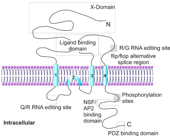
Endogenous receptors are believed to be tetrameric heteromers of subunits (GluR1-4 subunits) and these consist of the extracellular N-terminal and ligand binding domains, transmembrane region and the intracellular C-terminal domain (reviewed by Dingledine et al., 1999; Palmer et al., 2005a).
Figure 1.3.1. The topology of AMPAR subunit. Each subunit consists of an extracellular N-terminal domain, four hydrophobic regions (TM1–4), and an intracellular C-terminal domain. The ligand-binding site is a conserved amino acid pocket formed from a conformational association between the N terminus and the loop linking TM3 and TM4. A flip/flop alternative splice region and R/G RNA editing site are also present within the TM3/TM4 loop. TM2 forms an intracellular re-entrant hairpin loop which contributes to the cation pore channel. The Q/R RNA editing site is present within the TM1/TM2 loop. The intracellular C terminus contains phosphorylation sites and conserved sequences that have been shown to interact with a number of intracellular proteins. Adapted from Palmer et al., (2005a).
Although in NMDA receptors (NMDARs) there is Zn2+ modulation at a similar site, no endogenous ligands have been found to bind at the AMPAR N-terminal domain (Mayer and Armstrong 2004).
Transmembrane (TM) regions 1, 3 and 4 all span the cell membrane but TM2 forms a re-enterant loop on the intracellular side of the cell. The re-enterant loop is thought to contribute to the pore channel, which is permeable to Ca2+, K+ and Na+ (Michaelis, 1998; Dingledine et al., 1999; Palmer et al., 2005a), however the edited form of the GluR2 subunit is Ca2+-impermeable. The orientation of the transmembrane regions was determined by using proteolytic sites, N-glycosylation patterns and specific antibodies (Molnar, McIlhinney et al. 1994; Wo, Bian et al. 1995). The intracellular C-terminal domain of all AMPAR subunits is an interaction site for numerous proteins that are involved in receptor trafficking and synaptic plasticity (reviewed in Song and Huganir, 2002; Malinow and Malenka, 2002; Henley, 2003; Bredt and Nicholl, 2003; Collingridge et al., 2004; Malenka and Bear, 2004; Palmer et al., 2005a), such as PDZ domain-containing proteins, ABP/GRIP, PICK-1, PSD-95 and NFS.
1.3.1.2 Post-transcriptional modification
Functional diversity in AMPARs is largely determined by the expression of the genes that encode the different AMPAR subunits. There is approximately 70% sequence homology between genes encoding each subunit (Hollmann and Heinemann 1994) although further diversity is generated by post-transcriptional modifications. Alternative splicing can occur in the extracellular region of the fourth transmembrane domain to create ‘flip’ and ‘flop’ splice variants (Sommer, Keinanen et al. 1990). Flip variants dominate before birth, whereas flop variants are in low abundance before the eighth postnatal day and are up-regulated to about the same level as the flip forms in adult animals. The flip forms of subunits desensitize four times more slowly than the flop forms (Dingledine et al., 1999; Palmer et al., 2005a). GluR2 and 4 subunits also undergo alternative splicing in the C-terminus to give long and short isoforms, with the short isoforms of GluR2 accounting for 90% of total GluR2 (Kohler, Kornau et al. 1994). The long form of GluR4 is predominant and is largely expressed in the cerebellum (Gallo, Upson et al. 1992).
An additional post-transcriptional modification process is RNA editing. This leads to a single amino acid change at residue 607 in GluR2 subunits from
glutamine (Q) to arginine (R). In the adult rat 99% of GluR2 subunits are the R form and this residue is located in the channel-forming segment of the subunit (Dingledine, Borges et al. 1999). As a result, the edited subunits are Ca2+ -impermeable, because the size and charge of the amino acid side chain in the R form prevents the passage of Ca2+ ions through the channel. Changes in the amount of edited GluR2 subunits have been implicated in a number of diseases due to a link between Ca2+ permeability and excitotoxicity. These include Alzheimer’s disease, Huntington’s disease, schizophrenia, amyotropic lateral sclerosis (ALS) and epilepsy (Akbarian, Smith et al. 1995; Brusa, Zimmermann et al. 1995; Tanaka, Grooms et al. 2000; Kwak and Kawahara 2005).
1.3.1.3 Post-translational modification of subunit isoforms
The primary methods of post-translational modification of AMPAR subunits are glycosylation and phosphorylation. It has been proposed that N-glycosylation is involved in the maturation and transport of the receptor or could protect AMPARs from proteolytic degradation (Standley and Baudry 2000).
The regulated phosphorylation of AMPARs adds yet another level of modulation to an already complex scenario. Phosphorylation can regulate intermolecular interactions, channel properties and trafficking and is intricately linked with synaptic plasticity (reviewed by Smart, 1997; Palmer et al., 2005a). There appears to be a general role for developmental regulation of AMPAR properties by phosphorylation. I.e., an increase in PKC phosphorylation of AMPARs, primarily at S831 on GluR1, in striatal spiny neurones may play a role in the early stages of Parkinson’s disease (Oh, Geller et al. 2003).
1.3.1.4 Subunit composition of AMPARs
The subunit composition of AMPARs is critical in determining the functional and trafficking properties of resulting channels (Malinow and Malenka 2002). AMPARs that lack edited GluR2 are Ca2+-permeable and have an inwardly rectifying current/voltage (IV) relationship, so that at positive membrane potentials there is a voltage-dependent block of the pore channel by polyamines. As opposite, AMPARs that contain edited GluR2 are relatively Ca2+-impermeable (Bowie and Mayer 1995; Dingledine, Borges et al. 1999; Malinow and Malenka 2002). In hippocampal neurones AMPARs comprise
mainly GluR2 with GluR1 (GluR1/GluR2) or with GluR3 (GluR2/GluR3) (Wenthold, Petralia et al. 1996). There have been numerous studies made that examine the changes in AMPARs with different subunit compositions during and following synaptic plasticity (Shi et al., 2001; Lee et al., 2004; Holman et al., 2006; Plant et al., 2006). A general and simplified proposal is that GluR1/2 complexes are driven into the synapse during hippocampal LTP, and are subsequently replaced by GluR2/GluR3 complexes through constitutive recycling (Shi et al., 2001). Relatively little is known about the roles of individual subunits in the removal of AMPARs from synapses but progress is being made in understanding this complex process (Lee et al., 2004; Holman et al., 2006; McCormack et al., 2006).
1.3.1.5 Expression pattern of AMPAR subunits in the brain
Numerous studies have demonstrated the widespread and varied distribution of AMPAR subunits in the brain (reviewed in Hollmann and Heinemann, 1994) using immunocytochemistry, receptor autoradiography and in situ hybridisation studies (Keinanen, Wisden et al. 1990; Petralia and Wenthold 1992; Martin, Blackstone et al. 1993; Beneyto and Meador-Woodruff 2004). The distribution of GluR1, GluR2 and GluR3 are heterogeneous, with differential regional distribution and different levels of expression throughout numerous structures in the brain, with GluR1 being the most ubiquitous subunit (Beneyto and Meador-Woodruff, 2004). GluR4 is enriched in the cerebellum with generally low levels in the rest of the CNS (Petralia and Wenthold, 1992; Martin et al., 1993). Specifically, within the neocortex, (which includes the perirhinal cortex) GluR1-3 are present in all cortical layers except layer I, with GluR1 enriched in layers V and VI. GluR2 has high expression in layers II-III but much less in layer V, whilst GluR3 is highest in layer IV of the neocortex (Xu, Tanigawa et al. 2003; Beneyto and Meador-Woodruff 2004). Within the hippocampal formation there is high expression of GluR1-3 in all areas (Beneyto and Meador-Woodruff, 2004). There are similar developmental changes in regional expression of subunits in both rat and human (Talos, Fishman et al. 2006; Talos, Follett et al. 2006). Rodent cortical pyramidal neurones exhibit a developmental lag in GluR2 and GluR3 expression relative to GluR1, the expression of which is
higher than in adult throughout development and peaks at P10-12 (Talos et al., 2006b). GluR2-4 expression levels progresses with age but at P21 are still lower than adult levels (Talos et al., 2006). This has a clinical significance as the relatively low ratio of the Ca2+-impermeable GluR2 receptors to non-GluR2 receptors in the second postnatal week can lead to greater susceptibility to ischaemic injury. Indeed an equivalent pattern has been found in humans that could potentially lead to a targeted therapeutic strategy (Talos et al., 2006a)
1.3.1.6 AMPA receptor trafficking
The regulation of trafficking of AMPA receptor is of great interest, since it is involved in many aspects of neuronal plasticity. Experience-dependent strengthening of neocortical excitatory synapses in vivo is associated with the delivery of GluR2-lacking AMPARs to the synapse (Clem and Barth 2006). Interestingly, the increase in GluR2-lacking AMPARs after LTP induction is transient and after around 25 minutes they are replaced by GluR2-containing AMPARs during the maintenance phase of LTP (Plant, Pelkey et al. 2006). In addition, the association between LTP and the insertion of AMPARs at the synapse has been demonstrated in vivo. In the barrel cortex, experience drives the delivery of GluR1 subunits into the synapse, shown by an increase in rectification and sensitivity to joro spider toxin, which is selective for GluR2-lacking AMPARs (Clem and Barth 2006). Auditory fear conditioning in the amygdala also drives the trafficking of GluR1-containing AMPARs into the synapse (Rumpel et al., 2005). When trafficking of GluR1 was blocked, short-term and long-short-term memory of the fear conditioning was disrupted (Rumpel, LeDoux et al. 2005). It appears therefore there is a consensus that during LTP a multi-step process is required for the trafficking of AMPARs to the synapse. The endocytosis of AMPARs in response to stimulation occurs initially from extrasynaptic sites and this is then followed by a decrease in synaptic AMPARs In contrast to LTP, where the GluR1 subunit appears to be crucially involved in exocytosis, in LTD the GluR2 subunit appears to have the dominant role since it has been shown to directly interact with adaptor protein 2 (AP2) (Lee, Liu et al. 2002). This protein couples to clathrin, and along with dynamin, plays a pivotal role in clathrin-mediated endocytosis at synapses (Carroll, Beattie et al. 1999;
Wang and Linden 2000). In addition, it has been demonstrated that in the absence of plasticity-inducing stimuli, AMPARs undergo constitutive cycling (Shi et al., 2001). A recent study by McCormack et al (2006) has proposed that there are activity-independent trafficking pathways that serve to maintain the capacity for bidirectional plasticity in neurones (McCormack, Stornetta et al. 2006).
Synaptic AMPAR exchange is slow, with a rate constant of around 17hrs and involves the removal of GluR1 and GluR4 subunits and the addition of GluR2 subunits, which restores the ability for new LTP or LTD (McCormack, Stornetta et al. 2006). In GluR2 knockout mice there is a failure of synaptic AMPAR exchange, but not in GluR1 knockout mice; therefore GluR2 is found to be essential for this process.
It’s interesting to note that PKMζ seems to be involved in the maintenance of LTP through the regulation of NSF/Glu2-dependent AMPA receptor trafficking (Yao, Kelly et al. 2008). Consistently with these remarks, it has been proposed that PKMζ blocks the internalization of AMPA receptor, rather than facilitating their insertion in the membranem through a mechanism depending once again on its interaction with the mGlu2 subunit (Migues, Hardt et al. 2010).
1.3.2 Kainate receptors
The kainate subfamily of iGluRs consists of five subunits, GluR5-7 and KA1 and KA2 (Chittajallu, Braithwaite et al. 1999) The GluR5-7 receptors have a relatively low affinity for kainate and can form homomeric receptors. In comparison, KA1 and KA2 subunits have a higher affinity for kainate and form heteromeric receptors with GluR5-7 subunits (Chittajallu, Braithwaite et al. 1999). KA receptors (KARs) are considered to have a similar transmembrane topology to AMPAR and NMDARs (Michaelis, 1998; Dingledine et al., 1999; Kew and Kemp, 2005). Similar to AMPAR subunits, GluR5 and GluR6 subunits contain the Q/R editing site, the R form of which is impermeable to Ca2+ ions. The lack of specific antibodies has thus far hindered understanding of the exact subunit composition of native KARs and their synaptic localisation, however there is a differential distribution of the mRNA of KA subunits throughout the brain (Chittajallu et al., 1999; Isaac et al., 2004).
The relatively recent development of specific agonists and antagonists to kainate receptors has greatly aided the elucidation of KA receptors’ physiological function (Paternain, Morales et al. 1995; Wilding and Huettner 1995; Clarke, Ballyk et al. 1997; Bleakman and Lodge 1998; More, Nistico et al. 2004). It is now understood that KARs, like AMPARs, mediate fast excitatory transmission and some forms of synaptic plasticity (Isaac, Mellor et al. 2004). Also, KARs can act to depress excitatory transmission in the Schaffer collateral-commissural pathway (Clarke, Ballyk et al. 1997; Clarke and Collingridge 2002) and seem to be involved in the induction and expression of LTD of KAR-mediated synaptic transmission in layer II/III of the perirhinal cortex via a mechanism involving mGluR5, PKC and PICK1 (Park, Jo et al. 2006). KARs have also been shown to play a role in object recognition memory within the perirhinal cortex (Barker et al., 2006b).
1.3.3 NMDARs
In contrast to AMPARs, NMDARs mediate postsynaptic current that has a much slower rise time and decay time. The activation of NMDARs is dependent upon both agonist binding and membrane depolarisation for receptor channel opening. At resting membrane potential, ions cannot flow through the channel due to a block by the Mg2+ ion, rendering NMDARs voltage-dependent. If the cell is depolarised then the Mg2+ block is removed and the current can flow (Dingledine, Borges et al. 1999). NMDARs also require glycine as a co-agonist, so both glutamate and glycine have to be bound before the channel will open. Recently, D-serine has been shown to act as a co-agonist, which is released by astrocytes (Panatier, Theodosis et al. 2006). It is thought that glycine is present at a sufficient concentration in vivo and in vitro to bind all NMDARs (Bashir, Tam et al. 1990; Dingledine, Borges et al. 1999; Wenthold, Prybylowski et al. 2003). NMDARs therefore act as ‘coincidence detectors’ for postsynaptic depolarisation and presynaptic release of glutamate.
An important feature of NMDAR function lies in its permeability to Ca2+ as well as Na+ and K+. Entry of Ca2+ into the cell via NMDARs not only further depolarises the cell, but can also activate many Ca2+ sensitive enzymes. Around 7-18% of inward current through NMDARs is carried by Ca2+ ions
(Skeberdis, Chevaleyre et al. 2006). The influx of Ca2+ has been shown to be very important in the induction of long-term plasticity (Dingledine, Borges et al. 1999).
Structurally, NMDAR subunits have the same membrane topology as AMPAR and KAR subunits, with three transmembrane domains and a re-entrant loop, an intracellular C-terminus and a large extracellular N-terminus that contains a ligand binding domain (Stephenson 2001). A variety of NMDAR subunits have been identified (NR1-4) (Cull-Candy, Brickley et al. 2001). NR1 subunits have eight splice variants and contain the glycine binding site. Four genes encode NR2 subunits (NR2A-D) and the glutamate binding site is found in these subunits. A third subunit exists, NR3, which has two isoforms NR3A and NR3B (Stephenson 2001). Native NMDARs are believed to be tetrameric heteromers of NR1 and NR2 subunits with a stoichiometry believed to be a dimer of dimers, NR1-NR1-NR2-NR2. In receptors containing the NR3 subunit, is seems likely that a NR3 subunit substitutes for one of the NR2 subunits (Dingledine et al., 1999; Cull-Candy et al., 2001; Kew and Kemp, 2005). These receptors function as Ca2+-impermeable excitatory glycine receptors that respond to agonist application with low efficacy (Chatterton, Awobuluyi et al. 2002).
Figure 1.3.2 Schematic representation of the subunit transmembrane topography of NMDARs. A pair of NR1 and NR2 NMDAR subunits is shown to illustrate the transmembrane topography of these subunits. Their arrangement also shows the magnesium block of the pore-forming region made by the M2 regions in fully assembled NMDAR. Modified from Stephenson (2001).
NR1 transcripts are expressed in nearly all neurones, while NR2 subunits are expressed more discretely. The NR2A and NR2B subunits are the major and most widespread NR2 subunits, with NR2C largely restricted to the cerebellum and NR2D most heavily expressed early in development (Monyer, Burnashev et al. 1994; Stephenson 2001; Wenthold, Prybylowski et al. 2003). The NR2B subunit dominates early in development and gradually decreases postnatally and is predominately expressed in the forebrain. NR2C subunits are restricted to the cerebellum and NR2D subunits are expressed prenatally and restricted to the diencephalon and brain stem (Lynch and Guttmann 2001; Stephenson 2001; Molnar and Isaac 2002).
There is evidence that suggests that in adult cortex NR2A subunits are preferentially localised to synaptic sites and NR2B subunits are localised extrasynaptically (Stocca and Vicini 1998; Rumbaugh and Vicini 1999). A study by Massey et al. (2004) has demonstrated that in the perirhinal cortex the
subunit composition and postsynaptic localisation of NMDARs are critical determinants of their roles in synaptic plasticity (Massey, Johnson et al. 2004). NR2A-containing NMDARs are required for LTP induction and depotentiation and NR2B-containing NMDARs are required for de novo LTD (Massey, Johnson et al. 2004). A similar result was also shown in the hippocampus (Liu, Wong et al. 2004)
Numerous studies have been conducted that examine the role of NMDARs in learning and memory. Morris et al. (1990) demonstrated that infusion of the NMDAR antagonist D-AP5 into the hippocampus blocked the acquisition of spatial memory tested by the Morris water-maze (Morris, Davis et al. 1990). Moreover, in perirhinal cortex, the antagonism of NMDARs by D-AP5 impaired the acquisition of recognition memory after a long but not a short delay. However, recognition memory after a 24 hour delay was impaired only when NR2A and NR2B antagonists were infused together, not when either was infused separately (Barker et al., 2006b). This suggests that there could be two independent mechanisms that underlie long-term recognition memory; one dependent on a process used in LTP/depotentiation (requiring NR2A subunits) and another dependent on a process used in LTD (requiring NR2B subunits), either being capable of supporting familiarity discrimination at long delays. Many diseases are proposed to involve excitotoxity, such as stroke, epilepsy, hypoxic injury and also neurological disorders such as Alzheimer’s disease, Huntington’s disease and Parkinson’s disease (Lynch and Guttmann 2001). The NR2B antagonist ifenprodil administered to a rat model of Parkinson’s disease led to a significant improvement in locomotor activity (Loftis and Janowsky 2003).
1.3.4 Metabotropic glutamate receptors
Metabotropic glutamate receptors (mGluRs) were discovered in 1987 (Sugiyama, Ito et al. 1987) and to date there are a total of eight mGluR subunits, named mGluR1-8. These are classified into three groups based on their amino acid sequence identity and signal transduction coupling; mGluR1 and mGluR5 belong to group I; mGluR2 and mGluR3 belong to group II and mGluR4 and mGluR6-8 belong to group III (for a review see Conn and Pin,
1997; Pin et al., 2003). Group I mGluRs couple to phospholipase C (PLC), stimulating the hydrolysis of phosphatidylinositol (4,5) -bisphosphate (PIP2) into
diacyl glycerol (DAG) and inositol (1,4,5)-triphosphate (IP3). This results in the
activation of PKC and the release of Ca2+ from intracellular stores. On the other hand, group II and group III mGluRs are negatively coupled to adenylate cyclase (AC), resulting in a reduction in intracellular levels of cyclic adenosine monophosphate (cAMP) (Conn and Pin 1997; Michaelis 1998; Pin, Galvez et al. 2003). mGluRs of the same group show approximately 70% sequence homology, whereas between groups homology is approximately 45%.
mGluRs form homodimers composed of two mGluR subunits. Each subunit has a large extracellular N-terminal domain, seven transmembrane domains linked by relatively short loops and an intracellular C-terminus of varying length. The glutamate binding site is proposed to exist between two globular extracellular domains with a hinge region. The C-terminus is likely to be involved in the targeting and tethering of mGluRs to specific neuronal compartments and possibly also interaction with the respective G-protein. G-protein coupling is also thought to be made through the intracellular transmembrane loops (Conn and Pin, 1997; Michaelis, 1998; Pin et al., 2003; Kew and Kemp, 2005).
Figure 1.3.3 Schematic representation of the mGluR subunit structure. As is characteristic of metabotropic receptors, mGluRs have seven transmembrane domains. The intracellular loop between transmembrane regions III and IV is important for coupling to G-proteins. Modified from Conn and Pin (1997).
Although mGluR family members can mediate synaptic transmission via activation of slow excitatory postsynaptic potentials, they generally exert a more modulatory role, regulating neuronal excitability, synaptic transmission and plasticity (Kew and Kemp 2005). Group I mGluRs are typically localised postsynaptically in somatodendritic domains, whereas group II and III receptors are predominantly presynaptic, localised in axonal domains and axon terminals (Kew and Kemp 2005). Electrophysiological evidence suggests that mGluRs are located postsynaptically in the perirhinal cortex, though it is not known if presynaptic mGluRs are present (Cho et al., 2000; Cho et al., 2002). The activation of NMDARs and group I mGluRs is necessary for LTD induction (Cho et al., 2000). In the hippocampus, presynaptic mGluRs have been shown to reduce GABA release thereby reducing inhibitory transmission (Conn and Pin 1997).
1.4 Synaptic plasticity
Synapses can be considered dynamic structures that possess the property of being able to change their structure and/or efficiency according to what input they receive. At a basic level, synaptic plasticity can be split into potentiation and depression of synaptic transmission. These are generally defined as changes in the amplitude of postsynaptic potentials that are dependent upon the prior activity of the synapse. Plasticity can last over a period of milliseconds to minutes (short-term) or for hours or days (long-term). Long-term plasticities have attracted great interest as they have been implicated in underlying the brain’s ability to learn and store memories (Bliss and Lomo 1973; Bliss and Collingridge 1993; Malenka and Bear 2004).
1.4.1 Short-term plasticity
The short-term plasticites include facilitation, tetanic potentiation and post-tetanic depression (see Zucker and Regehr, 2002; Shepherd, 1998 for review). Facilitation is usually referred to as ‘paired-pulse facilitation’ (PPF) because it is studied by giving a pair of stimuli to a synaptic pathway and comparing the
amplitude of the second EPSP to the first. This type of plasticity is largely believed to be pre-synaptic in origin (Bear and Malenka 1994). The first pulse leads to depolarisation of the presynaptic terminal and an increase in intracellular Ca2+ that ultimately results in neurotransmitter release. If an optimal interval of ~50ms occurs between the first and second pulse, residual Ca2+ lingering from the first pulse, plus the influx of Ca2+ from the second pulse results in a greater increase in presynaptic Ca2+. This increases the probability of glutamate release from a given synapse, which results in a global increase in amount of transmitter released and therefore a subsequent greater postsynaptic response to the second pulse (Shepherd 1998; Zucker and Regehr 2002). Post-tetanic potentiation is a transient increase in the amplitude of a synaptic response that is seen after a brief train of stimuli. Post-tetanic potentiation, like PPF, is also reliant upon increases in the probability of transmitter release resulting from increases in residual calcium in the presynaptic terminal. Post-tetanic depression is also thought to rely primarily on presynaptic mechanisms (Zucker and Regehr 2002). If pairs of stimuli are delivered, around 50 milliseconds apart, then a depression of the second EPSP can be observed in hippocampal neurones in a phenomenon known as ‘paired-pulse depression’ (PPD). Depression of a synaptic response can occur if there is a repetitive activation of a synapse that leads to a transient depletion of the presynaptic pool of neurotransmitter, or by the action of an inhibitory neurotransmitter such as GABA. Depression may also result from desensitisation of postsynaptic receptors after repeated binding of neurotransmitter (Zucker and Regehr 2002).
1.4.2 Long-term plasticity
LTP is characterised by a long-lasting increase in synaptic efficacy induced typically by a 100 Hz high frequency stimulation (HFS) protocol and is thought to underlie the changes that occur in the brain during learning (Bliss and Lomo, 1973; Bliss and Collingridge, 1993). LTP has been most extensively studied in the CA1 region of the hippocampus and there have been a vast number of studies into the mechanisms of this phenomenon at different synapses and the circuits which operate in the mammalian brain (Malenka and Bear 2004). LTD is
in essence opposite to LTP, in that it is characterised by a long-lasting decrease in synaptic efficacy. It is induced by applying low frequency stimulation (LFS) to a synaptic pathway. Similar to LTD is the process of depotentiation, whereby LFS is given to a pathway that has already been potentiated and is expressing LTP and this increase in synaptic strength is subsequently reversed. De novo LTD itself can be reversed by HFS in the process of de-depression (Kemp and Bashir, 2001; Collingridge et al., 2004). These ‘bi-directional and reversible alterations in synaptic efficiency make possible the dynamic storage of vast amounts of neurally encoded information’ (Collingridge, Isaac et al. 2004).
1.4.2.1 NMDAR-dependent LTP
1.4.2.1.1 LTP induction
Since the important discovery that antagonism of the NMDAR by D-AP5 blocked LTP induction in the CA1 region of the hippocampus (Collingridge, Kehl et al. 1983), there has been a plethora of primary research papers and reviews that aim to elucidate the mechanisms underlying NMDAR-dependent LTP (Bliss and Collingridge 1993; Bear and Malenka 1994; Malenka and Nicoll 1999; Lisman, Schulman et al. 2002; Malinow and Malenka 2002; Lisman 2003; Malenka 2003; Collingridge, Isaac et al. 2004; Malenka and Bear 2004). For the induction of LTP (and LTD) to occur a rise in intracellular calcium (Ca2+) must take place, brought about by NMDAR activation. At resting membrane potentials NMDARs are inactive, due to the Mg2+ block of the channel. But, when a neuron becomes depolarised, typically following the activation of AMPARs, the Mg2+ block of the NMDA channel is relieved. This allows Na+ and Ca2+ to enter the neuron, creating an intracellular rise in Ca2+ (Malenka and Bear 2004). Regardless of how the LTP is induced, there is compelling evidence to indicate that calcium/calmodulin dependent protein kinase II (CaMKII) is required as a mediator for NMDAR-dependent LTP (see Lisman et al., 2002: Lisman, 2003 for an extensive review). During synaptic activity, the activated kinase translocates from the cytoplasm and binds to the NMDAR, where it can sense the very high Ca2+ levels, resulting in the downstream activation of signalling cascades that are involved in LTP expression. Other kinases have been implicated in playing key roles in LTP, although whether they act as mediators or modulators of LTP often remains contentious. There are a large number of protein phosphatase complexes, such as PP1, which have roles in the modulation of CaMKII-dependent signalling, potentially enabling a subtle and diverse modulation of synaptic transmission in the hippocampus, although their exact roles in LTP are not yet fully established (Colbran 2004).
1.4.2.1.2 LTP Expression
There appear to be two major post-synaptic mechanisms that are involved in the expression of NMDAR-dependent LTP. Namely these include the increase in the number of AMPARs at the synapse via trafficking and the modification of AMPARs via the phosphorylation of the GluR1 subunit (Malenka and Nicoll 1999; Malinow and Malenka 2002; Song and Huganir 2002; Bredt and Nicoll 2003; Lee, Takamiya et al. 2003; Malenka and Bear 2004).
The phosphorylation of AMPARs primarily occurs at various sites on the GluR1 subunit by CaMKII and PKC, which results in an increase in single channel conductance of the AMPAR with homomeric GluR1 subunits (Benke, Luthi et al. 1998). A recent study by Boehm et al., (2006) in the hippocampus has identified another PKC phosphorylation site on the GluR1 subunit at serine 818 (S818) (Boehm, Kang et al. 2006). The phosphorylation state of this site controls stable incorporation of GluR1 into the synapse. LTP-inducing stimuli phosphorylate this site, and its phosphorylation is important for the establishment of LTP and they believe this is likely to act by facilitating an interaction with a delivery and/or stabilising protein (Boehm, Kang et al. 2006). This further elucidates the link between the modification by phosphorylation of AMPARs and their trafficking to the synapse during LTP.
1.4.2.1.3 Maintenance of LTP
Much of the work on NMDAR-dependent LTP has focussed upon the mechanisms responsible for the initial increase in synaptic strength lasting 30-60 minutes, although arguably of greater interest and importance are the mechanisms that allow LTP to last for hours, days or even weeks (Malenka and Bear 2004). It is well established that the longer lasting components of LTP require new protein synthesis and gene transcription (Abraham and Williams 2003; Lynch 2004; Miyamoto 2006; Reymann and Frey 2007). As will be described more extensively in following chapters, PI3K and the mammalian target for rapamycin (mTOR) seem to be involved in the maintenance of LTP via protein synthesis and translation. Signalling molecules that are thought to link LTP induction to changes in gene transcription include
calmodulin-dependent protein kinase IV (CaMKIV), mitogen activated protein kinase (MAPK) and PKA, which act downstream to phosphorylate the transcription factor CREB (Lynch, 2004b; Warburton et al., 2005; Miyamoto, 2006; Reymann and Frey, 2007). CREB phosphorylation can lead to the activation of the immediate early gene c-fos and zif268 (Christy and Nathans 1989; Ahn, Olive et al. 1998). The expression product of c-fos is Fos, which can act as an accurate marker for recognition memory processes (Zhu et al., 1996; Wan et al., 1999; Warburton et al., 2003; Wan et al., 2004; Warburton et al., 2005). Inhibition of CREB phosphorylation in the perirhinal cortex (caused by the transduction of a dominant-negative inhibitor of CREB, which prevented the ability of CREB to bind to DNA) blocked LTP and also long-term recognition memory (Warburton, Glover et al. 2005). Other studies have demonstrated that there is a link between the CREB phosphorylation and the maintenance of LTP with memory in other parts of the brain, such as the hippocampus (Pittenger, Huang et al. 2002; Nguyen and Woo 2003; Reymann and Frey 2007).
The proposed link between LTP and other memory systems, such as spatial learning in the hippocampus and fear conditioning in the lateral amygdala has been also extensively studied (Martin and Morris 2002; Morris 2003; Sigurdsson, Doyere et al. 2007). One of the early classic experiments utilised the water maze to establish that spatial memory and LTP in the hippocampus are both NMDAR-dependent (Morris, Anderson et al. 1986). A recent study has used GFP-tagged GluR1 viral constructs to demonstrate that fear conditioning drives synaptic incorporation of GluR1 receptors in the lateral amygdala (Rumpel et al., 2005). Their results indicate that blocking GluR1-receptor trafficking in ~10-20% of neurones undergoing plasticity is sufficient to impair memory formation. This is an elegant set of experiments as it demonstrates a clear link between in vitro plasticity mechanisms, i.e. trafficking of GluR1 to the synapse during LTP and in vivo memory processes during learning.
The PKC isozyme, protein kinase M zeta (PKMξ) and phosphatidyl inositol 3-kinase (PI3K) have been implicated in having roles in the delivery of GluR1-containing AMPARs to synapses that have undergone LTP and in LTP maintenance (Ling, Benardo et al. 2002; Sanna, Cammalleri et al. 2002).