Improvement of pyrazolo[3,4-d]
pyrimidines pharmacokinetic
properties: nanosystem approaches
for drug delivery
Giulia Vignaroli
1,2,*, Pierpaolo Calandro
1,*, Claudio Zamperini
1,2, Federica Coniglio
1,2,
Giulia Iovenitti
1,2, Matteo Tavanti
1, David Colecchia
3, Elena Dreassi
1, Massimo Valoti
4,
Silvia Schenone
5, Mario Chiariello
3& Maurizio Botta
1,2,6Pyrazolo[3,4-d]pyrimidines are a class of compounds with a good activity against several cancer cell lines. Despite the promising anticancer activity, these molecules showed a poor aqueous solubility. This issue could threat the future development of pyrazolo[3,4-d]pyrimidines as clinical drug candidates. With the aim of improving their solubility profile and consequently their pharmacokinetic properties, we have chosen four compounds (1–4) on the base of their anti-neuroblastoma activity and we have developed albumin nanoparticles and liposomes for the selected candidates. Albumin nanoparticles and liposomes were prepared and characterized regarding size and ζ-potential distribution, polidispersity index, entrapment efficiency and activity against SH-SY5Y human neuroblastoma cell line. The most promising nanosystem, namely LP-2, was chosen to perform further studies: confocal microscopy, stability and drug release in physiological conditions, and biodistribution. Altogether, the obtained data strongly indicate that the encapsulation of pyrazolo[3,4-d]pyrimidines in liposomes represent an effective method to overcome the poor water solubility.
Neuroblastoma (NB) is the most common extracranial solid tumor in early childhood (the median age at diag-nosis is 17 months). This tumor arises in the sympathetic nervous system as a result of genetic alterations occur-ring in neural crest cells1. Although NB is characterized by clinical and biological heterogeneity, on the base
of age, tumor stage and genomic rearrangements (MYCN amplification and aneuploidy), the International Neuroblastoma Risk Group Staging System (INRGSS) classification divides patients in four groups, ranging from very low- to high-risk2. Up to 50% of NB patients have very aggressive tumors, associated with high risk of relapse
and very poor prognosis with a 5-year event-free survival rate around 40%. Treatment strategies for high-risk patients are still far from satisfaction, especially considering the severe side effects of the most used drugs (i.e. cisplatin, etoposide, vincristine, doxorubicin and cyclophosphamide)1. Consequently, the search for novel drugs
to improve NB treatment options still represents an outstanding pharmaceutical issue3.
In this context, Tyrosine Kinases (TKs) represent an interesting target for cancer treatment because of their involvement in several altered cellular pathways4. Molecular targeted therapies with Tyrosine Kinase Inhibitors
(TKIs) are designed to disrupt signalling pathways responsible for the abnormal proliferation of cancer cells. Given the importance of this mechanism, several TKIs are currently under preclinical or clinical develop-ment5. A subclass of TKIs is represented by inhibitors of c-Src kinase, which is involved in cell proliferation,
migration, invasion and angiogenesis as well as drug resistance development6,7. Since c-Src kinase has a strong
connection with cancer development, several classes of small molecules (i.e. purines8, anilinoquinazolines9, 1Dipartimento di Biotecnologie, Chimica e Farmacia, Università degli Studi di Siena, via Aldo Moro 2, 53100, Siena,
Italy. 2Lead Discovery Siena S.r.l., via Vittorio Alfieri 31, 53019, Castelnuovo Berardenga, Siena, Italy. 3Consiglio
Nazionale delle Ricerche, Istituto di Fisiologia Clinica and Istituto Toscano Tumori, Core Research Laboratory, Via Fiorentina 1, 53100, Siena, Italy. 4Dipartimento di Scienze della Vita, Università degli Studi di Siena, via Aldo Moro
2, 53100, Siena, Italy. 5Dipartimento di Farmacia, Università di Genova, Viale Benedetto VX 3, 16132, Genova,
Italy. 6Biotechnology College of Science and Technology, Temple University, Biolife Science Building, Suite 333,
1900 N 12th Street, Philadelphia, Pennsylvania 19122, USA. *These authors contributed equally to this work.
Correspondence and requests for materials should be addressed to M.B. (email: [email protected]) Received: 07 October 2015
Accepted: 26 January 2016 Published: 22 February 2016
quinolinecarbonitriles10, benzotriazines11, and thiazole-carboxamides12) have been developed to target this
enzyme. Some outstanding c-Src inhibitors are: (I) Dasatinib, a dual Src/Abl TKI, FDA approved for chronic myeloid leukemia (CML) treatment, (II) Saracatinib13, currently in phase II clinical trials for the treatment of
solid tumors, such as melanoma, prostate and gastric cancer, (III) Bosutinib14, a dual Src/Abl TKI, FDA approved
for CML treatment15. Several studies confirmed that hyperactivated c-Src plays a key role in NB cell
differentia-tion, adhesion and survival16–20. This TK was also associated to the progression of aggressive NB forms21,22. As a
proof of concept of the effectiveness of inhibiting c-Src for NB treatment, recent studies demonstrated that the well-known c-Src inhibitor PP2 was able to inhibit cellular growth and to induce aggregation in NB cell lines23.
Additionally the dual Src/Abl inhibitor Dasatinib was proved to be effective in reducing NB tumor growth in vitro and in vivo24.
The development of pyrazolo[3,4-d]pyrimidines as potential anticancer drugs, represents a major research focus of our group. This family of compounds showed a good cytotoxicity profile against several cancer cell lines: NB25–27, CML28, glioblastoma (GB)29, rhabdomyosarcoma (RMS)30, osteosarcoma (OS)31, prostate cancer (PC)32.
The activity of pyrazolo[3,4-d]pyrimidines has been related to the inhibition of TKs such as Abl and Src-family proteins33. Recently, remarkable results have been obtained in several mouse models of cancer25,28,30. For instance,
a small library of compounds with increased activity towards SH-SY5Y NB cell line was synthesized. The synthe-sis was driven by molecular modelling studies based on the X-ray crystal structure of c-Src in complex with one of our pyrazolo[3,4-d]pyrimidines. The selected lead compound was able to induce a tumor volume reduction greater than 50% in a NB subcutaneous xenograft mouse model25.
Given their unique physicochemical features, albumin nanoparticles and liposomes have showed a great potential as drug-carriers able to modify pharmacokinetic and pharmacodynamic properties of compounds. Encapsulation of a drug might result in an improved biodistribution, a higher stability, a controlled release, an ability to target sites otherwise not accessible, a decreased toxicity, and an enhanced bioavailability34. Oligomers
built with albumin are widely used as nanoparticles for drug delivery35. This small protein, the most abundant in
human plasma, presents very interesting features (i.e. biocompatibility, half-life of 19 days, water solubility and ability to bind a variety of different entities) and accumulates in malignant and inflamed tissues36. A breakthrough
for albumin-based nanotechnology was the commercialization of Abraxane©, a solvent-free formulation of
pacl-itaxel, where albumin binds paclitaxel to carry it through the endothelial cells to the tumor area37.
Liposomes are small vesicles characterized by a hydrophilic core surrounded by a phospholipid bilayer mem-brane whose composition can be adjusted to increase the therapeutic index of the encapsulated drug while min-imizing its side effects. Liposomes are biodegradable and the Mononuclear Phagocyte System (MPS) uptake is strongly reduced when polyethylene glycol (PEG) is included in the membrane composition (Stealth Liposomes). Moreover, liposomes can be selectively trapped by the tumor due to enhanced permeation and retention38. Doxil,
a PEGylated liposome-encapsulated form of doxorubicin, was the first FDA-approved nano-drug. It ensures pro-longed blood circulation and lower cardiotoxicity when compared to free doxorubicin39.
Although the promising anticancer activity, pyrazolo[3,4-d]pyrimidines are usually characterized by a low water solubility profile40,41. Consequently, in this study we have further developed four
pyrazolo[3,4-d]pyrimi-dines (1–4, Fig. 1) previously characterized for their activity against NB by enzymatic, cellular and in vivo assays (Table 1)25–27. Several strategies were applied to overcome this issue in recent years (i.e. introduction of
hydro-philic moieties in solvent-exposed positions42, synthesis of prodrugs43 and inclusion in cyclodextrins)40,41.
With the aim of increasing the low water solubility of this class of compounds, in this study we have explored albumin nanoparticles and stealth liposomes as possible nanotechnologies.
Results
Pyrazolo[3,4-d]pyrimidines 1–4.
The 4-amino substituted pyrazolo[3,4-d]pyrimidine ring represents a very interesting scaffold for the synthesis of molecules with antitumor activity. This structure is an isostere of ATP, the natural phosphorylating agent that binds TK. In our series of derivatives, the C4 amino function, essential for the interaction with the ATP-binding site, is attached to a m-substituted phenyl ring in derivatives 2–4 and a benzyl ring in derivative 1. Among the compounds previously characterized, pyrazolo[3,4-d]pyrimidine 1–4 were chosen for their ability to inhibit c-Src (with Ki values in the submicromolar range) and for their activity againstNB cells25–27. Chemical structures of compounds 1–4 are shown in Fig. 1. In vitro ADME properties were also
taken into account. In fact, the selected compounds demonstrated a very good metabolic stability (greater than 93% after incubation with human liver microsomes). Unfortunately, this favourable property was also associated
with a low aqueous solubility and a medium-low passive permeability through membrane. Enzymatic assays and ADME properties of compounds 1–4, as previously described25–27, are reported in Table 1. Characterization of
compounds 1–4 and assay methods can be found in the supporting material.
Characterization of albumin and liposome nanoparticles.
To improve the poor solubility in aqueous solution and the biodistribution of this family of compounds, albumin nanoparticles (AL-1, AL-2, AL-3 and AL-4) and liposomes (LP-1, LP-2, LP-3 and LP-4) were prepared. The albumin-drug nanoparticles, prepared by disulphide-bond induced self-assembly, were analyzed by Dynamic Light Scattering (DLS) and results are reported in Table 2. The mean diameter ranged between 118.8 nm (AL-3) and 165.6 nm (AL-1). This size param-eter was associated with high polidispersity indexes (close to 1), which indicated broad size distributions. Indeed, morphological analysis by Field Emission Scanning Electron Microscope (FESEM), confirmed the presence of aggregates (Figure S1). The tendency to form aggregates was also suggested by ζ -potential values belonging to the instability range44. The drug loading was around 6% when a very lipophilic compound, namely 1, wasencapsu-lated. However, it rose up to 50% with compounds 2 and 4, both characterized by a better aqueous solubility than compound 1 (Table S1).
Lipid ratio and drug concentration represent two important factors for the encapsulation of drugs in liposomes. Liposomes were prepared by the thin layer evaporation method, using the widely used lipids DPPC:Chol:MPEG-2000-DPPE Na (molar ratio of 20:10:1). The multilamellar vesicles (MLV) were converted by sonication to small unilamellar vesicles (SUV)45,46 to reduce in vivo potential opsonization47,48. The
suspen-sion was filtered through 200 nm filters, to obtain liposomes with a suitable diameter in order to avoid possible occlusion of capillaries in vivo49. The mean diameter and ζ -potential were measured by DLS (Table 2). The size
of nanoparticles ranged between 105 nm and 232 nm (LP-2 and LP-4, respectively), with LP-1 and LP-3 being around 151 nm and 131 nm, respectively. The whole set of liposomes presented good values of ζ -potential (from − 27.4 mV to − 47.6 mV) and polidispersity index (from 0.2 to 0.4). These data suggested the stability of the final suspension. The concentration of drug (1–4) in each liposomal sample was determined by HPLC-UV-MS analysis, after the disruption of the liposomes. The entrapment efficacy was excellent: above 85% for LP-2, LP-3 and LP-4 and 65% for LP-1. The effects of sonication, temperature and lipid composition were analyzed to max-imize the entrapment efficacy and to reduce the variability associated to the solubility of different compounds (Table S2).
Antiproliferative activity on human cell line SH-SY5Y.
The cytotoxicity of nanoparticles (liposomes: LP-1, LP-2, LP-3, LP-4 and albumin: AL-4) was evaluated in SH-SY5Y NB cell line. Taking into account the data regarding albumin nanoparticles previously evaluated, the only sample with satisfactory values of ζ -potential, PDI and EE% values was AL-4. Consequently, it was selected to perform cellular assays in the human NB cell line SH-SY5Y. Pure compounds (DMSO solution), empty liposomes (0.9% NaCl solution, pH 7.4) and albumin (PBS solution, pH 7.4) were included as controls. IC50s of these compounds were evaluated at 24, 48 and 72 h, forCpd Aqueous Solubility (μg/mL) LogPb Src (K
i μM) PAMPA (10−6 cm2/sec) MRc (%) Metabolic Stabilityd (%)
1 0,21 6,555 3.7 3.98 46.8 95.8
2 3,71 5,813 0.13 5.27 46.1 96.0
3 0,22 5,678 0.4 5.51 48.9 97.9
4 0,90 5,992 0.01 4.53 49.6 93.5
Table 1. Compounds 1–4: Activity against Src and ADME propertiesa. aAll the data were previously
reported, see supporting material for experimental details25–27. bCalculated by Qikprop. cMembrane Retention
(MR) expressed as percentage of compound unable to reach the acceptor compartment. dExpressed as
percentage of unmodified drug.
Formulation Sizea (nm) Polydispersity index a
(PDI) EfficiencyEntrapment b (%) ζ-potentiala (mV)
Liposomes 135.2 ± 9.45 0.23 ± 0.01 – − 27.4 ± 1.79 LP-1 151.3 ± 2.06 0.40 ± 0.04 65.0 ± 6.36 − 41.9 ± 5.75 LP-2 105.1 ± 6.39 0.21 ± 0.01 99.1 ± 0.71 − 39.9 ± 0.55 LP-3 131.3 ± 5.14 0.21 ± 0.03 85.2 ± 9.84 − 28.8 ± 0.15 LP-4 232.4 ± 7.35 0.13 ± 0.01 96.3 ± 2.83 − 47.6 ± 0.38 Albumin 9.30 ± 0.15 0.26 ± 0.01 – − 10.3 ± 1.41 AL-1 165.6 ± 6.11 0.97 ± 0.01 6.7 ± 0.91 − 15.2 ± 1.17 AL-2 125.3 ± 3.76 0.68 ± 0.01 51.0 ± 2.88 − 4.47 ± 1.93 AL-3 118.8 ± 3.00 0.58 ± 0.06 24.8 ± 1.23 − 13.4 ± 2.90 AL-4 135.4 ± 15.9 0.55 ± 0.01 52.3 ± 1.82 − 14.9 ± 1.46
Table 2. Properties of liposomes and albumin nanoparticles. aDetermined by DLS (Nano-Zeta Sizer,
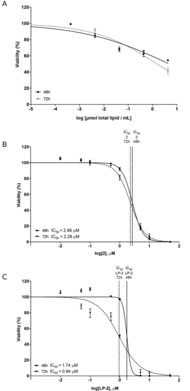
different concentrations (0.1, 1.0, 10, 50 μ M) (Table 3). A greater 24 h-activity was obtained for liposomal sam-ples in comparison with their corresponding drugs. The only exception was pair 3/LP-3 (drug/LP-drug), whose analysis revealed a comparable cytotoxicity. Data collected after 48 h showed a reduced difference in cytotoxicity within each pair. The activity at 72 h was higher for liposomal samples (LP-1 and LP-4) in comparison with the respective free drug (1 and 4). Within the pairs 2/LP-2 and 3/LP-3, a comparable IC50 value between each drug
and its respective liposomal formulation was found. IC50 (72 h) of AL-4 was 12.03 μ M, further demonstrating the
lower activity of albumin nanoparticles in comparison with the free drug 4 (IC50 = 8.63 μ M) and the liposome
LP-4 (IC50 = 1.90 μ M).
Characterization of LP-2.
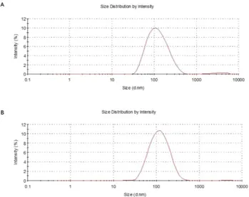
As all the other liposomal nanoparticles, LP-2, was characterized by DLS to assess size and ζ -potential and by cellular assays (SH-SY5Y NB) to determine cytotoxicity (Tables 2 and 3). More in detail, Fig. 2 shows the different size distribution by intensity of unloaded liposomes (Fig. 2A) and liposomes loaded with 2 (Fig. 2B). The average size resulted smaller for the sample with encapsulated drug, 105.1 ± 6.39 nm (LP-2) versus 135.2 ± 9.45 nm (Empty LP). Empty liposomes were tested in SH-SY5Y NB cell line in order to exclude any possible cytotoxic activity (Fig. 3A). This was particularly important because, although DPPC and DPPE are widely used to produce liposomes, recent studies50 demonstrated that saturated fatty acids might have atoxic effect in NB cells. In our assays, however, the obtained curve confirmed that liposomes do not influence sig-nificantly cell viability, indicating that the amount of saturated fatty acids derived from the liposomal preparation is not enough to determine a cytotoxic effect. LP-2 demonstrated an activity comparable with the one of com-pound 2 at 48 h (Fig. 3B). Additionally, the liposomal formulation at 72 h showed an enhanced cytotoxic effect (Fig. 3C). Subsequently, liposomal preparation LP-2 was morphologically analyzed by cryo-Electron Microscopy (cryo-EM) (Fig. 4). The result confirmed the presence of a homogeneous population of unilamellar nanopar-ticles with size around 100 nm. Liposomes were round, smooth and free from drug crystals; while bilamellar, multilamellar or giant-liposomes were not observed in the image from cryo-EM. The average thickness of the phospholipidic bilayer corresponded to 5.77 ± 1.05 nm (12 measurements performed by ImageJ Software, 1.46r).
Cpd/Formulation IC50 24 h (μM) IC50 48 h (μM) IC50 72 h (μM) 1 21.84 ± 1.70 16.5 ± 1.67 13.54 ± 2.00 LP-1 8.03 ± 0.59 7.14 ± 1.90 6.35 ± 1.44 2 12.6 ± 1.6 2.66 ± 0.015 2.28 ± 0.29 LP-2 6.80 ± 0.98 1.74 ± 0.31 0.94 ± 0.68 3 3.04 ± 0.09 2.65 ± 0.08 1.54 ± 1.07 LP-3 3.50 ± 0.17 1.52 ± 0.94 1.54 ± 0.79 4 13.64 ± 0.94 10.06 ± 0.50 8.63 ± 0.75 LP-4 3.87 ± 0.51 1.91 ± 1.46 1.90 ± 0.46 AL-4 20.04 ± 1.04 12.02 ± 0.97 12.03 ± 0.30
Table 3. Cytotoxicity in SH-SY5Y NB cells. SH-SY5Y cells were seeded at 105 cells/well density. The cultures
were maintained at 37 °C in 5% v/v CO2 for 24, 48 and 72 h. IC50s were evaluated by Trypan blue assay and
calculated by GraphPad Prism 6.0 software using the best fitting sigmoid curve.
Delivery of liposomes in neuroblastoma cells.
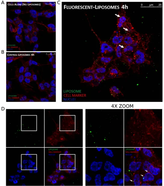
Neuroblastoma cells were incubated with normal and fluorescent liposomes to evaluate their possible uptake. Fluorescent liposomes were prepared incorporating a fluorescent phospholipid (NBD-DOPE) in the lipid mixture. After 4 h of incubation, fluorescent liposomes were internalized into neuroblastoma cells (Fig. 5A,B). Z-stack image confirmed the localization of NBD-labelled liposome inside SH-SY5Y cells (Figure S2).In vitro release.
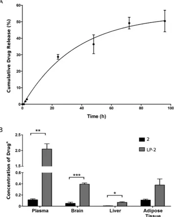
To determine the stability in physiological settings and to confirm the release of the drug 2 from its liposomal formulation LP-2, the release kinetics of 2 was analysed in vitro by measuring the concen-tration of drug released from liposomes into a physiological medium (BSA 50 mg/mL) at 37 °C (Fig. 6A). The cumulative percentage of drug release was determined over a 96 h-period. The results demonstrated the stabilityFigure 3. Viability of SH-SY5Y human NB cells evaluated at 48 and 72 h. (A) Empty liposomes, (B) Free
compound 2 and (C) LP-2. Compound 2 and LP-2 were tested at the following concentrations: 0.01, 0.05, 0.1, 0.5, 1.0, 10 and 50 μ M.
of the sample in physiological conditions at 37 °C. In fact, the percentage of 2 released from LP-2 resulted below 28% after 24 h and 49% over a 72 h-period. The final percentage of drug released was of 50.5%. In addition, the rate of drug release was evaluated during the 24 h (0.96 μ g/h).
Figure 4. Characterization of LP-2: morphological analysis by Cryo-EM.
Figure 5. Confocal microscopy experiments. Neuroblastoma cells (SH-SY5Y) were seeded on glass coverslip
and then incubated for 4 h with: (A) control, (B) normal liposomes, (C) fluorescent liposomes. Liposomes are visualized in green, cellular membranes in red and nuclei in blue. (D) Z-stack projection, on the right 4X zoom of the highlighted regions of the left panel.
Biodistribution at 24 h.
Biodistribution of LP-2 and free drug 2 were evaluated in male Sprague-Dawley rats. The concentration of compound 2 was determined after 24 h in the following tissues: plasma, brain, liver and adipose tissue (Fig. 6B). The concentration of the active compound was one order of magnitude higher in the plasma of rats treated with LP-2, validating the use of liposomes to enhance the plasma-exposure of a drug. In fact, the concentration of the free compound 2 was 0.11 μ g/mL and 2.05 μ g/mL in the groups treated with 2 and LP-2 respectively. The concentration of compound recovered in the brain was 0.05 μ g/g (group treated with 2) versus 0.39 μ g/g (group treated with LP-2). Again, the increase of quantity of compound 2 indicated the improved biodistribution of 2 when liposomes are used as drug delivery systems.Discussion
With the aim determining if the use of albumin nanoparticles and liposomes could represent a possible strategy to improve pharmacokinetic properties of our compounds, four pyrazolo[3,4-d]pyrimidines (1–4), selected from our library of compounds, were encapsulate in these nanosystems. Pyrazolo[3,4-d]pyrimidines have demon-strated a high anticancer activity associated to the inhibition of tyrosine kinases c-Src and Bcr-Abl and we have recently demonstrated that pyrazolo[3,4-d]pyrimidines were able to reduce tumor growth in NB xenograft mouse model25. Thus, we have decided to focus on NB for this study. Compounds 1–4 were selected on the basis of
previously reported data regarding activity and in vitro ADME properties25–27. Nanoparticles were characterized
by DLS regarding their size, polydispersity index and ζ -potential. Particle size has a significant impact on the circulation time51. Furthermore, the dimensions of the smallest capillaries need to be taken into account to avoid
a possible obstruction. Particle size also affects cellular uptake, influencing phagocytosis and endocytosis. In gen-eral, the larger is the nanoparticle, the faster is the clearance by the MPS. Optimal size to facilitate extravasation is about 150 nm or less, i.e. Doxil© has size between 80–100 nm and Myocet© is around 150 nm. In this context,
this study demonstrated that our liposomes are suitable drug-delivery systems with a diameter that ranges from 105 nm to 232 nm. Another important feature for nanoparticle dispersion stability is the ζ -potential that indicates the degree of electrostatic repulsion between particles. In detail, nanoparticles with ζ -potential values greater than + 25 mV or less than − 25 mV typically have high degrees of stability44. Showing a ζ -potential value between
− 28.65 mV and − 48.00 mV, our liposome systems were confirmed to be stable.
On the other side, albumin systems were characterized by ζ -potentials and PDIs into the range of instability (values around − 10 mV and close to 1, respectively). These data suggested that these nanoparticles might form
Figure 6. In vitro release and biodistribution at 24 h. (A) Release of compound 2 from liposomal system LP-2
in physiological conditions, with 50 mg/mL of BSA, at 37 °C. (B) Concentration of compound 2 determined in plasma, brain, liver and adipose tissue, after the administration of the free drug 2 (black) and the liposomal formulation LP-2 (grey). aThe concentration is expressed as μ g/mL for plasma and as μ g/g for brain, liver and
aggregates (hypothesis confirmed by DLS analysis and FESEM study). However, we decided to proceed with the evaluation of the cytotoxicity of the most promising albumin sample, namely AL-4.
Liposomes demonstrated greater 24 h-activity when compared with the appropriate free drug (comparable IC50s of LP-3 and 3, represented the only exception). This difference in cytotoxicity might be related to different
internalization processes. In fact, while the free drug can enter into cells by passive membrane transport, lipos-omes need to be taken up by different processes (i.e. endocytosis, fusion). At 48 h and 72 h, pairs LP-2/2 and LP-3/3 showed similar IC50s. However, LP-1 and LP-4 demonstrated higher cytotoxicity than the respective free
drug both, in 48 h and 72 h measurements. Cellular assays further demonstrated the unfavourable properties of albumin nanoparticles, as the IC50 value was higher than the ones obtained by testing free drug 4 and
lipos-omes LP-4. Given their poor properties (PDI, ζ -potential, EE% and IC50) albumin nanoparticles were not further
characterized.
LP-2 resulted our most promising liposomal candidate because of the following: (I) IC50 value of 0.97 μ M
against NB cells, (II) 99% of entrapment efficiency, (III) ζ -potential value of − 39.9 ± 0.55 mV and (IV) size of 105.1 ± 6.39 nm. Additionally, 2 was the compound previously tested in NB xenograft mouse model25. Thus, LP-2
was further characterized by cryo-EM, confirming the data obtained by DLS analysis. The sample was indeed characterized by a homogenous population of unilamellar liposomes with size around 100 nm. Furthermore, a confocal microscopy-based study was performed to elucidate whether the drug is released from liposomes out-side the cells or inout-side the cytoplasm, after an internalization of the whole liposomal system. The analysis showed the presence of fluorescent liposomes into the cytoplasm of NB cells, suggesting a delivery of the free drug after cellular uptake of liposomes.
The release of compound 2 from the liposomal system LP-2 was evaluated with an in vitro assay that allowed the quantification of compound 2 likely to be released from liposomes into physiological conditions. The result-ing monophasic release curve was a typical feature of unilamellar liposomes, accordresult-ingly with DLS and cryo-EM results. A proof of concept in vivo experiment was then performed to evaluate and compare the biodistribution of free compound 2 and liposomes LP-2 in several tissues, at 24 h. Additionally, this study was important to deter-mine the in vivo stability of our liposomal systems. Data showed a generally improved biodistribution - higher concentration - of the drug 2 when liposomes were used as drug-delivery system. Considerably, when LP-2 was administered a 20-fold increase in plasma-concentration of free compound 2 was observed. In addition, the quantity of compound 2 recovered from the brain tissue after injection of LP-2 was significantly higher than the concentration obtained after administration of compound 2.
In conclusion, this study validated the use of stealth liposomes to improve the biodistribution of pyra-zolo[3,4-d]pyrimidines. Herein we demonstrate that liposomes retain the efficacy of our encapsulated drug against SH-SY5Y NB cells and that this cytotoxic activity is likely to be exerted by the release of the active com-pound in the cytoplasm after uptake of the nanoparticles. Importantly, the liposomal system resulted stable and able to release the encapsulated drug in physiological conditions and the biodistribution assay finally proved the beneficial properties of LP-2. Further preclinical in vivo studies will allow the determination of the full pharma-cokinetic profile and the therapeutic efficacy of this novel formulation.
Methods
Materials.
Pyrazolo[3,4-d]pyrimidine compounds (1–4), were previously synthesized and character-ized by our research group25–27. 1,2-Dipalmitoyl-sn-glycero-3-phosphocholine semisynthetic, ≥ 99% (DPPC),N-(carbonyl methoxypolyethylenglycol 2000)-1,2-dipalmitoyl-sn-glycero-3-phosphoethanolamine sodium salt (MPEG-2000-DPPE Na), 1,2-dioleoyl-sn-glycero-3-phosphoethanolamine,7-nitrobenzofurazan-labeled (NBD-DOPE), 4′,6-diamidino-2-phenylindole (DAPI), cholesterol (Chol), human serum albumin (HSA) (lyophilized, 97%), dialysis tubing cellulose membrane (cut-off 14 kDa), L-cysteine and and D,L-glyceraldehyde and all the solvents were purchased from Sigma Aldrich. Nylon syringe filters with pores of 0.2 μ m (Acrodisc 13 mm Syringe Filter) and PTFE syringe filters with pores of 0.2 μ m (Acrodisc 13 mm Syringe Filter) were from purchased VWR. High purity deionised water was obtained from milli-Q (Millipore, Milford, MA, USA). Wheat germ agglutinin, Alexa Fluor 647 Conjugate (WGA) was purchased from Life Technologies. Fetal Bovine Serum (FBS), Dulbecco’s Modified Eagle Medium: Nutrient Mixture F-12 (DMEM/F12), L-glutamine (L-Glu) and Penicillin-Streptomycin (Pen/Strep) were purchased from Euroclone.
Preparation of albumin-drug nanoparticles.
The albumin nanoparticles were prepared by mixing 100 μ L of compound solution in EtOH (1 mg/mL) with 900 μ L of HSA solution in PBS (pH 7.4, 25 mM) (5 mg/mL)52.Cysteine was added up to a final concentration of 5 mg/mL, and the resulting solution was kept at 37 °C, for 45 min. The excess of cysteine was eliminated by dialysis using a cellulose membrane (capacity 60 mL/ft, cut-off 14 kDa, diameter 16 mm, width 25mm). D,L-glyceraldehyde was added to this solution, to a final concentration of 5 μ M53. The sample was then filtered through a nylon syringe filter with pores of 0.2 μ m (Acrodisc 25 mm Syringe
Filter) to obtain a sterile solution of the drug-albumin nanoparticles. The concentration of the compound in the final sample was determined by HPLC-UV-MS (HPLC-UV-MS method in the supporting material), after dena-turation of the protein by acetonitrile and centrifugation (4500 rpm, 15 min, 4 °C).
Preparation of Liposome-drug nanoparticles.
The method developed by A.D. Bangham, namely thin layer evaporation, was used for the preparation of LP-1, LP-2, LP-3 and LP-4. Lipids DPPC, Chol and MPEG-2000-DPPE Na (molar ratio 20:10:1) were dissolved in a mixture of CHCl3:MeOH = 3:1 (3 mL, round-bottomedflask). A 1 mM solution of each compound (1, 2, 3, 4) in CHCl3:MeOH = 3:1 was prepared. An aliquot was
trans-ferred in the lipid solution to obtain 500 μ M as final concentration of compound. The solvent was removed under reduced pressure to obtain a thin layer of lipids (kept under high vacuum overnight). The thin layer was hydrated with a 0.9% NaCl solution and the mixture was stirred for 1 hour at a temperature higher than the Tm of the
lipids. The suspension was mixed (Vortex, 3 min) and SUV liposomes were formed by sonication for 20 min at room temperature. The solution was filtered with PTFE syringe filters with pores of 0.2 μ m (Acrodisc 13 mm Syringe Filter), to eliminate liposomes with diameter over 200 nm and potentially precipitated drugs. The drug concentration was determined after disruption of liposomes by treatment with ethanol at 40 °C (1:1 = v/v). The compound was extracted and quantified by HPLC-UV-MS (HPLC-UV-MS method in the supporting material).
Measurements of size and ζ-potential.
Size distribution and ζ -potential were determined by Dynamic Light Scattering (DLS) (Zeta Sizer Nano ZS90, Malvern Instruments Ltd, Malvern, UK). Measurements were performed directly after rehydration of the samples with 1 mM NaCl aqueous solution. Samples stability was evaluated by DLS after one week.Antiproliferative activity on human cell line SH-SY5Y.
In vitro experiments were carried out using the human NB cell line SH-SY5Y. Cells were purchased from American Type Culture Collection (ATCC, Manassas, VA, USA) and were cultured in DMEM/F12 1:1 medium supplemented with 10% FBS, 2 mM L-glutamine and 10000 units/mL Penicillin/Streptomycin at 37 °C in 5% CO2 atmosphere. In order to determine theantiprolifer-ative effect of drugs (1–4) and nanosystems, SH-SY5Y cells were seeded at 105 cells/well density and treated with
albumin (PBS solution, pH 7.4) or empty liposomes (0.9% NaCl solution, pH 7.4), compounds encapsulated with albumin (PBS solution, pH 7.4) or liposomes LP-1, LP-3 and LP-4 (0.9% NaCl solution, pH 7.4) and free compounds 1, 3 and 4 (DMSO solution) at increasing concentrations (0.1 μ M, 1.0 μ M, 10 μ M and 50 μ M). IC50
determination for LP-2 and compound 2 was performed testing the following concentrations as well: 0.01 μ M, 0.05 μ M, 0.5 μ M and 5 μ M. The cultures were maintained at 37 °C in 5% v/v CO2 for 24, 48 and 72 h. Cell number
and viability were evaluated using the Bürker chamber, after treatment with Trypan Blue. IC50 was calculated by
GraphPad Prism 6.0 software using the best fitting sigmoid curve.
Morphology study.
Morphology was observed by Cryo Electron Microscopy. 3 μ L of sample were applied on Quantifoil®
holey carbon grids (copper Multi A, Quantifoil®
Micro Tools GmbH, Jena, Germany). Excess fluid was blotted from the grid for 2 sec with Whatman filter paper and then plunge frozen in liquid ethane, using a plunge freezer, to achieve sample vitrification. Frozen samples were stored in liquid nitrogen until EM imaging. Vitrified samples were imaged using a CM200 FEG transmission EM (FEI, Eindhoven, the Netherlands) operated at 200 keV and equipped with a F224HD 2048 × 2048 CCD camera (TVIPS Gauting, Germany). EM images were acquired at 27,500× magnification (pixel size 0,602 nm) at −12, −18 μ m defocus.Confocal microscopy studies on the cellular uptake.
For confocal microscopy experiments, fluores-cent liposomes, carrying NBD-DOPE in the lipid bilayer, were used. DPPC, Chol and MPEG-2000-DPPE were dissolved at a molar ratio of 20:10:1, respectively, CHCl3:MeOH = 3:1 v/v. Then, NBD-DOPE was added to thelipid mixture to a final molar ratio of 2% of total lipids. The lipid mixture was dried and placed in a vacuum desic-cator for at least 24 h to ensure complete solvent removal. Finally, lipid film was rehydrated with NaCl 0.9% solu-tion to obtain a NBD-DOPE final concentrasolu-tion of 440 μ M. SH-SY5Y cells were maintained in DMEM/F12 1:1 medium supplemented with 10% FBS, 2 mM L-glutamine and 10000 units/mL Penicillin/Streptomycin at 37 °C in 5% CO2 atmosphere. SH-SY5Y cells were plated on glass coverslip at a density of 20000 cells per well in 24-well
plates. After 24 h, cells were incubated with liposome formulation, and then fixed in 4% paraformaldehyde in PBS for 20 min. Nuclei and cellular membranes were stained respectively with a 6 μ M solution of DAPI and 2 μ g/mL WGA Alexa Fluor 647 labelled for 10 min. Coverslips were mounted in fluorescence mounting medium (Dako, S3023). Samples were visualized on a TSC SP5 confocal microscope (Leica, 5100000750) installed on an inverted LEICA DMI 6000CS (10741320) microscope and equipped with an oil immersion Plan Apo 63 × 1.4 NA objec-tive. Images were acquired using the LAS AF acquisition software (Leica, 10210). NBD fluorescence was acquired in 500–560 nm range and excited with 476 nm argon laser, DAPI was acquired in 420–480 nm range and excited with 405 nm UV laser and WGA was acquired in 650–730 nm range and excited with 633 nm laser.
In vitro release.
Rates of drug release were studied by dialyzing the nanosystem LP-2 (3.5 mL, 0.4 mM) against PBS (20 mL, pH 7.4, 10 mM) with 50 mg/mL of BSA (physiological plasma concentration). The entire system was stirred at 37 °C and samples (1 mL, from the PBS-BSA) were collected at different time points (0, 1, 2, 3, 24, 48, 72 and 96 h) (1 mL of PBS-BSA was added each time to maintain sink condition). Each sample was treated with 1 mL of ACN and centrifuged at 4000 rpm for 20 min. Then, the supernatant was recovered, concen-trated under reduced pressure and analyzed to determine the concentration of compound 2 by HPLC-UV-MS (HPLC-UV-MS method in the supporting material).In vivo plasmatic distribution.
Male Sprague Dawley rats (Charles River, Milan, Italy) were maintained according to ethical EEC regulations for animal research. The animal protocols used were reviewed and approved by the Animal Care and Ethics Committee of the University of Siena, Italy. The animals were anesthetized (i.p. xylazine hydrochloride, 10 mg/kg, Xilor, Bayer AG, and ketamine hydrochloride 35 mg/kg, Ketavet, Gellini) and treated by i.v. injection through the tail vein. The experiment was performed in triplicate. The two groups received intravenously LP-2 (500 μ L, PBS, pH 7.4) or 2 (50 μ L in DMSO), corresponding to a bolus of 10 mg/Kg. After 24 h, animals were sacrificed and blood and tissues were treated for the quantitative analysis. The blood, previously heparinized, was centrifuged at 4000 rpm for 20 min to separate the plasma fraction and then, 500 μ L were col-lected in a test tube. Tissues were homogenized using a glass/glass Potter-Elvehjem homogenizer. For each sample 1 mL of ACN (in the presence of the internal standard 1-(2-chloro-2-(4-chlorophenyl)ethyl)-N-(3-fluorobenzyl)-1H-pyrazolo[3,4-d]pyrimidin-4-amine, 10 μ M) was added to denature proteins and to extract 2. Samples werecentrifuged at 4000 rpm for 20 min; the supernatant was recovered, evaporated and analyzed by HPLC-UV-MS (HPLC-UV-MS method in the supporting material).
References
1. Hara, J. Development of treatment strategies for advanced neuroblastoma. Int. J. Clin. Oncol. 17, 196–203 (2012).
2. Matthay, K. K. et al. Criteria for evaluation of disease extent by (123)I-metaiodobenzylguanidine scans in neuroblastoma: a report for the International Neuroblastoma Risk Group (INRG) task force. Br. J. Cancer 102, 1319–1326 (2010).
3. Maris, J. M., Hogarty, M. D., Bagatell, R. & Cohn, S. L. Neuroblastoma. Lancet 369, 2106–2120 (2007).
4. Arora, A. & Scholar, E. M. Role of tyrosine kinase inhibitors in cancer therapy. J. Pharmacol. Exp. Ther. 315, 971–979 (2005). 5. Pytel, D., Sliwinski, T., Poplawski, T., Ferriola, D. & Majsterek, I. Tyrosine kinase blockers: new hope for successful cancer therapy.
Anticancer Agents Med. Chem. 9, 66–76 (2009).
6. Brunton, V. G. & Frame, M. C. Src and focal adhesion kinase as therapeutic targets in cancer. Curr. Opin.Pharmacol. 8, 427–432 (2008).
7. Grant, S. & Dent, P. Kinase inhibitors and cytotoxic drug resistance. Clin. Cancer Res. 10, 2205–2207 (2004).
8. Huang, H. et al. Discovery of novel purine derivatives with potent and selective inhibitory activity against c-Src tyrosine kinase.
Bioorg. Med. Chem. 18, 4615–4624 (2010).
9. Green, T. P. et al. N-(5-Chloro-1,3-benzodioxol-4-yl)-7-[2-(4-methylpiperazin-1-yl)ethoxy]-5-(tetrahydro-2H-pyran-4-yloxy) quinazolin-4-amine, a novel, highly selective, orally available, dual-specific c-Src/Abl kinase inhibitor. J. Med. Chem. 49, 6465–6488 (2006).
10. Cao, X. et al. Design and synthesis of 7-alkoxy-4-heteroarylamino-3-quinolinecarbonitriles as dual inhibitors of c-Src kinase and nitric oxide synthase. Bioorg. Med. Chem. 16, 5890–5898 (2008).
11. Palanki, M. S. et al. Development of novel benzotriazines for drug discovery. Expert Opin. Drug Discov. 4, 33–49 (2009).
12. Das, J. et al. 2-Aminothiazole as a novel kinase inhibitor template. Structure-activity relationship studies toward the discovery of N-(2-chloro-6-methylphenyl)-2-[[6-[4-(2-hydroxyethyl)-1-piperazinyl)]-2-methyl-4-pyrimidinyl]amino)]-1,3 thiazole-5-carboxamide (dasatinib, BMS-354825) as a potent pan-Src kinase inhibitor. J. Med. Chem. 49, 6819–6832 (2006).
13. Nam, H. et al. Antitumor activity of saracatinib (AZD0530), a c-Src/Abl Kinase Inhibitor, alone or in combination with chemotherapeutic agents in gastric cancer. Mol. Cancer Ther. 12, 16–26 (2013).
14. Rassi, F. E. & Khoury, H. J. Bosutinib: a SRC-ABL tyrosine kinase inhibitor for treatment of chronic myeloid leukemia.
Pharmacogenomics Pers. Med. 6, 57–62 (2013).
15. Puls, L. N., Eadens, M. & Messersmith, W. Current Status of Src Inhibitors in Solid Tumor Malignancies. The Oncologist 16, 566–578 (2011).
16. Bolen, J. B., Rosen, N. & Israel, M. A. Increased pp60c-src tyrosyl kinase activity in human neuroblastoma is associated with amino-terminal tyrosine phosphorylation of the src gene product. Proc. Natl. Acad. Sci. USA 82, 7275–7279 (1985).
17. O’Shaughnessy, J., Deseau, V., Amini, S., Rosen, N. & Bolen, J. B. Analysis of the c-src gene product structure, abundance, and protein kinase activity in human neuroblastoma and glioblastoma cells. Oncogene Res. 2, 1–18 (1987).
18. Bjelfman, C., Hedborg, F., Johansson, I., NordenskjÖld, M. & Påhlman, S. Expression of the neuronal form of pp60c-src in neuroblastoma in relation to clinical stage and prognosis. Cancer Res. 50, 6908–6914 (1990).
19. Matsunaga, T. et al. Expression of n-myc and c-src proto-oncogenes correlating to the undifferentiated phenotype and prognosis of primary neuroblastomas. Cancer Res. 51, 3148–3152 (1991).
20. Finlay, D. & Vuori, K. Novel noncatalytic role for caspase-8 in promoting SRC-mediated adhesion and Erk signaling in neuroblastoma cells. Cancer Res. 67, 11704–11711 (2007).
21. Takahashi, H. & Simizu, B. Expression of alternatively spliced src messenger RNAs related to neuronal differentiation in human neuroblastomas. Cancer Res. 53, 3179–3185 (1993).
22. Matsunaga, T. et al. Expression of neuronal Src mRNA as a favorable marker and inverse correlation to N-myc gene amplification in human neuroblastomas. Int. J. Cancer 58, 793–798 (1994).
23. Hishiki, T. et al. Src kinase family inhibitor PP2 induces aggregation and detachment of neuroblastoma cells and inhibits cell growth in a PI3 kinase/Akt pathway-independent manner. Pediatr. Surg. Int. 27, 225–230 (2011).
24. Vitali, R. et al. Activity of tyrosine kinase inhibitor dasatinib in neuroblastoma cells in vitro and in orthotopic mouse model. Int.
J. Cancer 125, 2547–2555 (2009).
25. Tintori, C. et al. Combining X-ray crystallography and molecular modeling toward the optimization of pyrazolo[3,4-d]pyrimidines as potent c-Src inhibitors active in vivo against neuroblastoma. J. Med. Chem. 58, 347–361 (2015).
26. Navarra, M. et al. Antiproliferative and pro-apoptotic effects afforded by novel Src-kinase inhibitors in human neuroblastoma cells.
BMC Cancer 10, 602–614 (2010).
27. Radi, M. et al. Identification of potent c-Src inhibitors strongly affecting the proliferation of human neuroblastoma cells. Bioorg.
Med. Chem. Lett. 21, 5928–5933 (2011).
28. Radi, M. et al. Design, synthesis, and biological evaluation of pyrazolo[3,4-d]pyrimidines active in vivo on the Bcr-Abl T315I mutant. J. Med. Chem. 56, 5382–5394 (2013).
29. Ceccherini, E. et al. SRC Family kinase inhibition through a new pyrazolo[3,4-d]pyrimidine derivative as a feasible approach for glioblastoma treatment. J. Cell. Biochem. 116, 856–863 (2015).
30. Casini, N. et al. SRC family kinase (SFK) inhibition reduces rhabdomyosarcoma cell growth in vitro and in vivo and triggers p38 MAP kinase-mediated differentiation. Oncotarget 20, 12421–12435 (2015).
31. Spreafico, A. et al. Antiproliferative and proapoptotic activities of new pyrazolo[3,4-d]pyrimidine derivative Src kinase inhibitors in human osteosarcoma cells. FASEB J. 22, 1560–1571 (2008).
32. Angelucci, A. et al. Pyrazolo[3,4-d]pyrimidines c-Src inhibitors reduce epidermal growth factor-induced migration in prostate cancer cells. Eur. J. Cancer 42, 2838–2845 (2006).
33. Schenone, S., Brullo, C., Musumeci, F. & Botta, M. Novel dual Src/Abl inhibitors for hematologic and solid malignancies. Expert
Opin. Invest. Drugs 19, 931–945 (2010).
34. Saha, R. N., Vasanthakumar, S., Bende, G. & Snehalatha, M. Nanoparticulate drug delivery systems for cancer chemotherapy. Mol.
Membr. Biol. 27, 215–231 (2010).
35. Kratz, F. Albumin as a drug carrier: design of prodrugs, drug conjugates and nanoparticles. J. Control. Release 132, 171–183 (2008). 36. Elzoghby, A. O., Samy, W. M. & Elgindy, N. A. Albumin-based nanoparticles as potential controlled release drug delivery systems.
J. Control. Release 157, 168–182 (2012).
37. Kratz, F. A clinical update of using albumin as a drug vehicle - a commentary. J. Control. Release 190, 331–336 (2014).
38. Allen, T. M. & Cullis, P. R. Liposomal drug delivery systems: from concept to clinical applications. Adv. Drug Deliv. Rev. 65, 36–48 (2013).
39. Barenholz, Y. Doxil
®
–The first FDA-approved nano-drug: Lessons learned. J. Control. Release 160, 117–134 (2012).40. Dreassi, E. et al. 2-Hydroxypropyl-β -cyclodextrin strongly improves water solubility and anti-proliferative activity of
pyrazolo[3,4-d]pyrimidines Src-Abl dual inhibitors. Eur. J. Med. Chem. 45, 5958–5964 (2010).
41. Radi, M. et al. A combined targeted/phenotypic approach for the identification of new antiangiogenics agents active on a zebrafish model: from in silico screening to cyclodextrin formulation. Bioorg. Med. Chem. Lett. 22, 5579–5583 (2012).
42. Vignaroli, G. et al. Exploring the chemical space around the privileged pyrazolo[3,4-d]pyrimidine scaffold: toward novel allosteric inhibitors of T315I-mutated Abl. ACS Comb. Sci. 16, 168–175 (2014).
43. Vignaroli, G. et al. Pyrazolo[3,4-d]pyrimidine prodrugs: strategic optimization of the aqueous solubility of dual Src/Abl inhibitors.
ACS Med. Chem. Lett. 4, 622–626 (2013).
44. Gregoriadis, G. et al. Vaccine entrapment in liposomes. Methods. 19, 156–162 (1999).
45. Lapinski, M. M., Castro-Forero, A., Greiner, A. J., Ofoli, R. Y. & Blanchard, G. J. Comparison of liposomes formed by sonication and extrusion: rotational and translational diffusion of an embedded chromophore. Langmuir 23, 11677–11683 (2007).
46. Cho, N. J., Hwang, L. Y., Solandt, J. J. R. & Frank, C. W. Comparison of extruded and sonicated vesicles for planar bilayer self-assembly. Materials 6, 3294–3308 (2013).
47. Moghimi, S. M. & Szebeni, J. Stealth liposomes and long circulating nanoparticles: critical issues in pharmacokinetics, opsonization and protein-binding properties. Prog. Lipid Res. 42, 463–478 (2003).
48. Yan, X., Scherphof, G. L. & Kamps, J. A. Liposome opsonization. J. Liposome Res. 15, 109–139 (2005).
49. Hallmark, B. et al. Observation and modelling of capillary flow occlusion resulting from the capture of superparamagnetic nanoparticles in a magnetic field. Chem. Eng. Sci. 63, 3960–3965 (2008).
50. Bolognesi, A., Chatgilialoglu, A., Polito, L. & Ferreri, C. Membrane lipidome reorganization correlates with the fate of neuroblastoma cells supplemented with fatty acids. PLoS One 8, e55537 (2013).
51. Laverman, P. et al. Liposomes for scintigrafic detection of infection and inflammation. Adv. Drug Deliv. Rev. 37, 225–235 (1999). 52. Jiang, L. et al. A nontoxic disulfide bond reducing method for lipophilic drug-loaded albumin nanoparticle preparation: formation
dynamics, influencing factors and formation mechanisms investigation. Int. J. Pharm. 443, 80–86 (2013).
53. Vandelli, M. A., Rivasi, F., Guerra, P., Forni, F. & Arletti, R. Gelatin microspheres crosslinked with D,L-glyceraldehyde as a potential drug delivery system: preparation, characterisation, in vitro and in vivo studies. Int J Pharm. 215, 175–184 (2001).
Acknowledgements
The authors would like to thank Prof. Gianni Pozzi and Dr. Francesco Santoro (Dipartimento di Biotecnologie Mediche, Università degli Studi di Siena, Italy) for Dynamic Light Scattering experiments, Prof. Pietro Lupetti (Dipartimento di Scienze della Vita, Università degli Studi di Siena, Italy) for Cryo EM experiments.
Author Contributions
P.C. and D.C. cellular and confocal microscopy studies; C.Z., F.C. and M.T. nanoparticles preparation and characterization; G.I., G.V. and S.S. synthesis and characterization of compounds; G.V., C.Z., D.C., E.D., M.V., S.S., M.C. and M.B. design of methods and experiments, analysis and interpretation of data; G.V., P.C., C.Z., D.C. and M.B. preparation of the manuscript. All authors have approved the final version of manuscript.
Additional Information
Supplementary information accompanies this paper at http://www.nature.com/srep Competing financial interests: The authors declare no competing financial interests.
How to cite this article: Vignaroli, G. et al. Improvement of pyrazolo[3,4-d]pyrimidines pharmacokinetic
properties: nanosystem approaches for drug delivery. Sci. Rep. 6, 21509; doi: 10.1038/srep21509 (2016).
This work is licensed under a Creative Commons Attribution 4.0 International License. The images or other third party material in this article are included in the article’s Creative Commons license, unless indicated otherwise in the credit line; if the material is not included under the Creative Commons license, users will need to obtain permission from the license holder to reproduce the material. To view a copy of this license, visit http://creativecommons.org/licenses/by/4.0/
![Figure 1. Structures of pyrazolo[3,4-d]pyrimidines 1–4.](https://thumb-eu.123doks.com/thumbv2/123dokorg/4659850.42522/2.892.235.825.75.258/figure-structures-of-pyrazolo-d-pyrimidines.webp)