Multidrug-Resistant Tuberculosis
Andrea M. Cabibbe,aPaolo Miotto,aRaquel Moure,bFernando Alcaide,bSilke Feuerriegel,cGianni Pozzi,dVladislav Nikolayevskyy,e,f
Francis Drobniewski,e,fStefan Niemann,cKlaus Reither,g,hDaniela M. Cirillo,aTM-REST Consortium, TB-CHILD Consortium Emerging Bacterial Pathogens Unit, Division of Immunology, Transplantation and Infectious Diseases, IRCCS San Raffaele Scientific Institute, Milan, Italya
; Servei de Microbiologia, Hospital Universitari de Bellvitge-IDIBELL, Universitat de Barcelona, Barcelona, Spainb
; Molecular Mycobacteriology, Research Center Borstel, Borstel, Germanyc
; Laboratory of Molecular Microbiology and Biotechnology, Department of Medical Biotechnologies, University of Siena, Siena, Italyd
; Clinical TB and HIV Group, Blizard Institute, Queen Mary University of London, London, United Kingdome
; Department of Infectious Diseases and Immunity, Imperial College London, London, United Kingdomf
; Swiss Tropical and Public Health Institute, Basel, Switzerlandg
; Ifakara Health Institute, Bagamoyo, United Republic of Tanzaniah
We evaluated the performance of the molecular lab-on-chip-based VerePLEX Biosystem for detection of multidrug-resistant
tuberculosis (MDR-TB), obtaining a diagnostic accuracy of more than 97.8% compared to sequencing and MTBDRplus assay for
Mycobacterium tuberculosis complex and rifampin and isoniazid resistance detection on clinical isolates and smear-positive
specimens. The speed, user-friendly interface, and versatility make it suitable for routine laboratory use.
M
ultidrug-resistant tuberculosis (MDR-TB) requires long
and expensive treatment and often results in poor clinical
outcome in both low- and high-income countries (
1
,
2
). The
World Health Organization (WHO) has endorsed specific
molec-ular diagnostics to improve fast diagnosis of MDR-TB (
3–5
).
However, the genotypic diversity and geographical distribution of
Mycobacterium tuberculosis complex (MTBC), together with the
inability to provide appropriate interpretation of silent mutations
and the limited versatility are some of the restraints undermining
the effectiveness of the current tools on a global scale (
6–13
).
In the present study, we evaluated a lab-on-chip (LoC)
de-vice, developed by STMicroelectronics (Geneva, Switzerland)
and marketed by Veredus Laboratories (Republic of
Singa-pore) as the VerePLEX Biosystem, for the diagnosis of
MDR-TB and detection of common nontuberculous
mycobac-teria (NTM). The molecular assay was evaluated on both
clin-ical isolates and direct specimens in low- and high-burden
set-tings.
We tested 91 MTBC isolates (see Table S1 in the supplemental
material) harboring different patterns of mutations in rpoB, katG,
and inhA genes to evaluate the probes on the array listed in
Table
1
. Eighty respiratory specimens positive for acid-fast bacilli by
smear microscopy and MTBC culture positive were
decontami-nated according to international guidelines and included in the
study (Table S1) (
14
). An additional 116 MTBC culture-negative
specimens were included in the analysis. DNA from isolates and
specimens was extracted by thermal lysis and sonication as
de-scribed elsewhere (
15
). Phenotypic drug susceptibility testing
(DST) for rifampin (RIF) and isoniazid (INH) was performed
according to international recommendations (
16
). Some of the
Received 7 July 2015 Accepted 30 July 2015 Accepted manuscript posted online 5 August 2015
Citation Cabibbe AM, Miotto P, Moure R, Alcaide F, Feuerriegel S, Pozzi G, Nikolayevskyy V, Drobniewski F, Niemann S, Reither K, Cirillo DM, TM-REST Consortium, TB-CHILD Consortium. 2015. Lab-on-chip-based platform for fast molecular diagnosis of multidrug-resistant tuberculosis. J Clin Microbiol 53:3876 –3880.doi:10.1128/JCM.01824-15.
Editor: K. C. Carroll
Address correspondence to Daniela M. Cirillo, [email protected].
Supplemental material for this article may be found athttp://dx.doi.org/10.1128 /JCM.01824-15.
Copyright © 2015, American Society for Microbiology. All Rights Reserved.
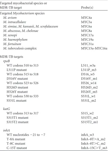
TABLE 1 Probes spotted onto the array and targeted mycobacterial
species and MDR-TB targets included in the assay Targeted mycobacterial species or
MDR-TB target Probe(s) Targeted Mycobacterium species
M. avium MYC4a
M. intracellulare MYC5a
M. simiae, M. kansasii, M. scrofulaceum MYC6a
M. abscessus, M. chelonae MYC8a
M. xenopi MYC17a
M. haemophylum MYC19a
M. fortuitum MYC31a
M. tuberculosis complex MYC15a-MYC16a MDR-TB targets rpoB WT codons 510 to 513 L511_w3a L511P mutant L511P_m3 WT codons 515 to 518 D516_w5 D516V mutant D516V_m1 WT codons 523 to 526 H526_w14 H526D mutant H526D_m2 H526Y mutant H526Y_m5 WT codons 530 to 533 S531L_w1 S531L mutant S531L_m2 katG WT codons 313 to 317 S315_w2 S315T1 mutant S315T1_m2 S315T2 mutant S315T2_m1 inhA WT nucleotides⫺21 to ⫺7 inhA_w3 T-8A mutant InhA–8T⬎A_m2 T-8C mutant InhA–8T⬎C_m2 C-15T mutant InhA–15C⬎T_m3
on September 7, 2016 by guest
http://jcm.asm.org/
specimens were tested in a representative high-burden setting in
Uganda (Nsambya Hospital, Kampala, Uganda), by trained staff.
DNA samples extracted from both isolates and specimens were
tested in parallel, and results were compared with GenoType
MTBDRplus (Hain Lifescience, Nehren, Germany) assay and
Sanger sequencing performed as described elsewhere (
17
).
The VerePLEX Biosystem consists of a single disposable device
comprising microfluidic PCR and microarray modules. The
plat-form includes a temperature control system (TCS) and an optical
reader (OR) which allows automatic analysis of the microarray,
pro-viding a user-friendly diagnostic report (see Fig. S2 in the
supplemen-tal material) (
18
). The protocols for MDR-TB assay are described in
Text S3, and the primers are shown in Table S4. The assay allows
detection of MTBC and other common NTM, together with the most
frequent mutations affecting the rpoB, katG, and inhA genes, which
are involved in phenotypic resistance to RIF and INH in MTBC.
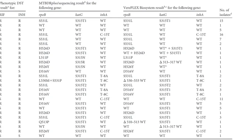
TABLE 2 Phenotypic DST, MTBDRplus, and VerePLEX Biosystem results for the 91 MTBC clinical isolates included in the studyPhenotypic DST resultafor:
MTBDRplus/sequencing resultbfor the
following gene: VerePLEX Biosystem resultb,cfor the following gene:
No. of isolatesd
RIF INH rpoB katG inhA rpoB katG inhA
R R S531L S315T1 WT S531L S315T1 WT 15 R R WT WT WT WT WT WT 1 S R WT WT WT WT WT WT 5 R R S531L WT C-15T S531L WT C-15T 16 R R S531L WT WT S531L WT WT 7 R S S531L WT WT S531L WT WT 2 R R H526D S315T1 WT H526D WT*⫹ S315T1 WT 1 R R H526D S315T1 WT WT⫹ H526D WT⫹ S315T1 WT 1 R R L511P S315N WT L511P WT* WT 1 R R H526D S315R WT H526D ⌬ 313–317 WT WT 1 R R H526Y S315N WT H526Y WT* WT 1 R S D516V WT WT D516V WT WT 1 R R S531L S315T1 T-8A S531L S315T1 T-8A 2 R R L530M⫹S531P S315T1 T-8C ⌬ 530–533 WT S315T1 T-8C 1 R R S531L S315T2 WT S531L S315T2 WT 2 R R D516V S315T1 T-8A D516V S315T1 T-8A 3 R R D516V S315T1 T-8C D516V S315T1 T-8C 1 S R WT WT C-15T WT WT C-15T 11 R R D516V S315T1 WT D516V S315T1 WT 5 S R WT S315T1 WT WT S315T1 WT 5 R R H526D S315T1 WT H526D S315T1 WT 1 R R S531L S315T1 C-15T S531L S315T1 C-15T 3 R R Q513P S315T1 WT ⌬ 510–513 WT S315T1 WT 1 S R WT S315N WT WT ⌬ 313–317 WT WT 1 R R H526Y S315T1 C-15T H526Y S315T1 C-15T 2 S S WT WT WT WT WT WT 1
aThe phenotypic drug susceptibility testing (DST) results for rifampin (RIF) and isoniazid (INH) are given as follows: R, resistant; S, sensitive. b
The results for the 91 MTBC isolates found by the MTBDRplus assay and sequencing or by the VerePLEX Biosystem are shown (wild type [WT] or mutant). cSymbols: *, probe signal was on at the cutoff;⌬, no WT signal.
d
The number of isolates apply to all the test results.
TABLE 3 Diagnostic performance of the phenotypic DST, MTBDRplus, VerePLEX Biosystem, and Xpert MTB-RIF for detecting rifampin resistance
(rpoB) in clinical isolates and specimensa
Parameter
Value (95% CI) for clinical isolates (n⫽ 91) Value (95% CI) for clinical specimensb
Method type and no. of indeterminate results/total (%) MTBDRplus/seq DST MTBDRplus/seq/Xpert MTB-RIF (n⫽ 71) DST (n⫽ 58) Sensitivity (%) 100.00 (94.58, 100.00) 98.53 (92.13, 99.74) 100.00 (77.19, 100.00) 100.00 (75.75, 100.00) Molecular 3/71 (4.23) Specificity (%) 100.00 (86.2, 100.00) 100.00 (85.69, 100.00) 100.00 (93.47, 100.00) 100.00 (91.97, 100.00) Phenotypic 2/58 (3.45) PPV (%) 100.00 (94.58, 100.00) 100.00 (94.58, 100.00) 100.00 (77.19, 100.00) 100.00 (75.75, 100.00) NPV (%) 100.00 (86.2, 100.00) 95.83 (79.76, 99.26) 100.00 (93.47, 100.00) 100.00 (91.97, 100.00) Negative likelihood ratio 0.00 (0.00, ?) 0.01 (0.00, 0.10) 0.00 (0.00, ?) 0.00 (0.00, ?) Diagnostic accuracy (%) 100.00 (95.95, 100.00) 98.90 (94.03, 99.81) 100.00 (95.95, 100.00) 100.00 (93.58, 100.00)
a
The diagnostic performance of the MTBDRplus assay and sequencing (MTBDRplus/seq), phenotypic drug susceptibility testing (DST), and MTBDRplus assay, sequencing, and Xpert MTB-RIF assay (MTBDRplus/seq/Xpert MTB-RIF) for detecting rifampin resistance (rpoB) are shown. The sensitivity, specificity, positive predictive value (PPV), and negative predictive value (NPV), and diagnostic accuracy were calculated according to the Wilson score (www.OpenEpi.com). The positive and negative likelihood ratios were also calculated. The lower and upper limits of the 95% confidence interval (95% CI) are shown in parentheses. The effective number of samples considered for the analysis is reported for each target. The positive likelihood ratio cannot be computed, since specificity is always 100%.
bThere were a total of 80 M. tuberculosis-positive smear-positive clinical specimens and a total of 116 M. tuberculosis-negative clinical specimens.
on September 7, 2016 by guest
http://jcm.asm.org/
Analysis of the diagnostic performance of the LoC assay on
clinical isolates. MTBC was detected in all 91 clinical isolates (
Ta-ble 2
). Concerning the rpoB and inhA targets, 100% concordance
was observed between the MTBDRplus and LoC assay results. In
one case, the LoC assay revealed both wild-type (WT) and
mu-tated signals from probes targeting positions 523 to 526 in rpoB,
which was not confirmed by MTBDRplus assay. A 95.74%
concor-dance was observed between the MTBDRplus and LoC assay
re-sults for the katG target. In two cases, probes complementary to
the WT sequence of codon 315 of katG were detected slightly over
the on/off cutoff, but the MTBDRplus assay showed an absence of
signal from the WT probe. In another two cases, a double pattern
(mutated and WT) was detected by the LoC assay, but only the
mutation was identified by the MTBDRplus assay.
Other mutations identified by sequencing (L530M, S531P, and
Q513 in rpoB and S315N and S315R in katG) were correctly
de-tected on the chip by the absence of signal from respective WT
probes.
Compared with DST, the sensitivity and specificity of the
MTBDRplus assay for RIF were 98.53% and 100%, respectively,
and the sensitivity and specificity for INH were 82.76% and 100%,
respectively (
Tables 3
,
4
, and
5
).
Analysis of the diagnostic performance of the LoC assay on
clinical specimens. DST results for RIF and INH were available
for 58 and 57 samples, respectively. The chips presenting
incom-plete results were repeated once and then included in the analysis
(
Table 6
).
Valid results were obtained in 99.00%, 95.80%, and 95.50% of the
cases for MTBC, rpoB, katG, and inhA targets, respectively. MTBC
was detected with 100% sensitivity and specificity on the LoC, as well
as resistance to RIF (
Tables 3
,
4
, and
5
). One discrepant result was
detected for the katG and inhA genes, leading to a sensitivity of
93.75% and 90.91%, respectively, compared to the MTBDRplus
as-say. Overall, the sensitivity and specificity of katG and inhA targets
were 73.33% and 100%, respectively, compared to DST. Three
spec-imens gave invalid values by the LoC assay. One sample gave an
in-valid result for PCR controls, possibly due to inhibitors affecting the
reaction in the microfluidic environment. The remaining two
speci-mens also yielded invalid results with the MTBDRplus assay. All 116
MTBC culture-negative specimens were classified correctly.
In the current study, we developed and evaluated a LoC-based
assay for the diagnosis of MDR-TB. LoC devices represent
prom-ising tools to fill the diagnostic gap in low-income countries: they
integrate many of the laboratory components on a small chip, thus
reducing infrastructure and technical requirements but
preserv-ing analytical capabilities. In addition, the operatpreserv-ing speed, ease of
modification (addition/removal of probes), and ability to perform
multiplex tests and to scale down costs represent other relevant
features of LoCs (
19
,
20
).
Our results showed high specificity and sensitivity of the
semiau-tomated VerePLEX Biosystem for the MDR-TB targets, thus
suggest-ing an usefulness of the platform for fast and simple diagnosis of
MDR cases in centralized laboratories. The sensitivity and specificity
of the NTM probes on the same platform were evaluated by Lazzeri et
al. (
21
). The assay allowed us to identify correctly MTBC in 100% of
the smear-positive samples tested independently of the smear
mi-croscopy score, with a small number of indeterminate results due
most likely to the low quality of DNA extracted. Resistance to RIF and
INH was detected by the chip with high sensitivity and specificity in
agreement with the minimal requirements established by the WHO
TABLE4 Diagnostic performance of the phenotypic DST, MTBDR plus ,VerePLEX Biosystem, and Xpert MTB-RIF for detecting isoniazid resistance (katG and inhA ) in clinical isolates and specimens a Parameter Value (95% CI) for clinical isolates (n ⫽ 91) Value (95% CI) for clinical specimens b Method type and no. of indeterminate results/total (%) MTBDR plus /seq DST MTBDR plus /seq/Xpert MTB-RIF DST (n ⫽ 57) katG inhA katG (n ⫽ 67) inhA (n ⫽ 67) Sensitivity (%) 95.74 (87.75, 98.83) 100.00 (91.03, 100) 82.76 (73.48, 89.26) 93.75 (71.67, 98.89) 90.91 (62.26, 98.38) 73.33 (55.55, 85.82) Molecu lar 3/67 (4.48) Specificity (%) 100.00 (91.97, 100.00) 100.00 (93.12, 100.00) 100.00 (51.01, 100.00) 100.00 (92.59, 100.00) 100.00 (93.24, 100.00) 100.00 (86.68, 1 00.00) Phenotypic 2/57 (3.5) PPV (%) 100.00 (92.13, 100.00) 100.00 (91.03, 100.00) 100.00 (94.93, 100.00) 100.00 (79.61, 100.00) 100.00 (72.25, 100.00) 100.00 (85.13, 100.00) NPV (%) 95.65 (85.47, 98.90) 100.00 (93.12, 100.00) 21.05 (8.51, 43.33) 97.96 (89.31, 99.64) 100.00 (90.23, 99.67) 75.76 (58.98, 87.17) Negative likelihood ratio 0.04 (0.02, 0.11) 0.00 (0.00, ?) 0.17 (0.15, 0.20) 0.07 (0.009, 0.44) 0.09 (0.01, 0.65) 0.26 (0.21, 0.34) Diagnostic accuracy (%) 97.8 (92.34, 99.4) 100.00 (95.95, 100.00) 83.52 (74.57, 89.75) 98.44 (91.67, 99.72) 98.44 (91.67, 99.72) 85.45 (73.84, 92.4 4) aThe diagnostic performance of the MTBDR plus /seq assays, phenotypic drug susceptibility testing (DST), and MTBDR plus /seq/Xpert MTB-RIF assays for detecting isoniazid resistance (katG and inhA ) are shown. The sensitivity, specificity, positive predictive value (PPV), and negative predictive value (NPV), and diagnostic accuracy were calculated according to the Wilson score ( www.OpenEpi.com ). The positive and negative likelihood ratios were also calcu-lated. The lower and upper limits of the 95% confidence interval (95% CI) are shown in parentheses. The effective number of samples considered for the an alysis is reported for each target. The positive likelihood ratio cannot be com-puted, since specificity is always 100%. bThere were a total of 80 M. tuberculosis -positive smear-positive clinical specimens and a total of 116 M. tuberculosis -negative clinical specimens.
on September 7, 2016 by guest
http://jcm.asm.org/
Downloaded from
for molecular tools, comparable to the sensitivity and specificity of
the MTBDRplus assay (
12
). The limit of detection of the assay was
observed in the range of 10
1genome copies/reaction, as reported in
Table S5 in the supplemental material.
A separate array layout for spoligotyping of MTBC was also
developed in the TM-REST Project (data not shown). The
possi-bility of integrating the probes for spoligotyping, MDR- and
ex-tensively DR-TB in one medium-density microarray layout by
using separate multiplex-PCR would enhance the benefits of the
microarray assays and would enable the reduction of time to
re-sults compared to other available tests (
22–24
).
The ease of customization of the array design makes the LoC a
versatile tool for easy integration of relevant targets for local
ge-netic variants, new genes and/or mutations, and novel key drugs
included in new therapeutic regimens. In addition, the LoC can be
adapted for other diagnostic or research needs, thus providing a
TABLE 5 Diagnostic performance of the phenotypic DST, MTBDRplus, and VerePLEX Biosystem for detecting M. tuberculosis in clinical isolatesand specimensa
Parameter
Value (95% CI) for clinical isolates (n⫽ 91)
Value (95% CI) for clinical specimens (n⫽ 196)bby MTBDRplus/seq/Xpert MTB-RIF No. of indeterminate results/total (%) MTBDRplus/seq DST Sensitivity (%) 100.00 (95.95, 100.00) 100.00 (95.95, 100.00) 100.00 (95.31, 100.00) 2/196 (1.02) Specificity (%) Undefined Undefined 100.00 (96.79, 100.00)
PPV (%) 100.00 (95.95, 100.00) 100.00 (95.95, 100.00) 100.00 (95.31, 100.00) NPV (%) Undefined Undefined 100.00 (96.79, 100.00) Negative likelihood ratio Undefined Undefined 0.00
Diagnostic accuracy (%) Undefined Undefined 100.00 (98.06, 100.00)
a
The diagnostic performance of the MTBDRplus/seq assays, phenotypic drug susceptibility testing (DST), and MTBDRplus/seq/Xpert MTB-RIF assays for detecting M. tuberculosis are shown. The sensitivity, specificity, positive predictive value (PPV), and negative predictive value (NPV), and diagnostic accuracy were calculated according to the Wilson score (www.OpenEpi.com). The positive and negative likelihood ratios were also calculated. The lower and upper limits of the 95% confidence interval (95% CI) are shown in parenthe-ses. The effective number of samples considered for the analysis is reported for each target. The positive likelihood ratio cannot be computed, since specificity is always 100%. b
There were a total of 80 M. tuberculosis-positive smear-positive clinical specimens and a total of 116 M. tuberculosis-negative clinical specimens.
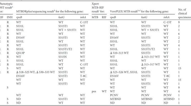
TABLE 6 Phenotypic DST, MTBDRplus, Xpert MTB-RIF, and VerePLEX Biosystem M. tuberculosis results for the 80 smear-positive MTBC
culture-positive clinical specimens included in the study Phenotypic
DST resulta
for: MTBDRplus/sequencing resultbfor the following gene:
Xpert MTB-RIF
resultcfor: VerePLEX MTB resultb,dfor the following gene: No. of
clinical specimense
RIF INH rpoB katG inhA MTB RIF rpoB katG inhA
S R WT WT C-15T WT WT C-15T 9 R R S531L S315T1 WT S531L S315T1 WT 2 R R S531L WT⫹ S315T1 WT S531L WT⫹ S315T1 WT 1 S R WT WT WT WT WT WT 6 R R D516V S315T1 WT D516V S315T1 WT 2 R R S531L WT WT S531L WT WT 2 S R WT S315T1 WT WT S315T1 WT 4 R R S531L S315T1/T2 WT S531L S315T1/T2 WT 1 R R Q513P S315T1 WT ⌬ 510–513 WT S315T1 WT 1 S R WT S315N WT WT ⌬ 313–317 WT WT 1 R S S531L WT WT S531L WT WT 1 R R S531L WT C-15T S531L ⌬ 313–317 WT WT 1 S S WT WT WT WT WT WT 15 R R ⌬ 518–525 WT, ⌬ 530–533 WT S315T1 WT ⌬ 523–526 WT, S531L S315T1 WT 1 D516V S315T1 T-8C D516V S315T1 T-8C 1 WT WT WT WT WT WT 15 WT S315T1 WT WT S315T1 WT 1 S S WT WT WT 9 pos WT WT WT WT 4 WT WT WT PCNV PCNV PCNV 1 S S ND S315T1 WT MTBND MTBND MTBND 1 S S ND WT WT ND ND ND 1 a
The phenotypic drug susceptibility testing results for rifampin and isoniazid are given as follows: R, resistant; S, sensitive.
bThe results for the 80 smear-positive, MTBC culture-positive isolates found by the MTBDRplus assay and sequencing or by the VerePLEX Biosystem are shown (wild type [WT] or mutant).⌬, no WT signal; ND, not detected.
cMTB, M. tuberculosis; pos, positive. d
PCNV, PCR controls not valid; MTBND, M. tuberculosis not detected; ND, not detected. eThe number of smear-positive, MTBC culture-positive clinical specimens applies to all the tests.
on September 7, 2016 by guest
http://jcm.asm.org/
multipurpose platform suitable for other relevant diseases (e.g.,
influenza, malaria, tropical diseases) (
25
,
26
).
ACKNOWLEDGMENTS
This study was supported by FP7 EU grant TM-REST (HEALTH-F3-2008-202145) and European and Developing Countries Clinical Trials Partnership as part of the TB CHILD project (IP.2009.32040.007).
The members of the TM-REST Consortium follow: Patrizia Di Pietro, Floriana San Biagio, Enrico Alessi, and Tony G. Barbuzzi (Analog, MEMS & Sensor Group, HealthCare Business Development Unit, STMicro-electronics, Catania, Italy); Silva Tafaj (University Hospital Shefqet Nd-roqi, Tirana, Albania); Elizabetha Bachiyska (National Center of Infec-tious and Parasitic Diseases, Sofia, Bulgaria); Irina Kontsevaya (Samara TB Service, Samara, Russian Federation); Yanina Balabanova (Clinical TB and HIV Group, Blizard Institute, Queen Mary University of London, United Kingdom, and Department of Infectious Diseases and Immunity, Imperial College London, United Kingdom); and Elisa Lazzeri (Labora-tory of Molecular Microbiology and Biotechnology, Department of Med-ical Biotechnologies, University of Siena, Siena, Italy). The members of the TB-CHILD Consortium follow: Joseph Sserunkuma, Francesco Aloi, and Martin Nsubuga (Laboratory Department, St. Raphael of St. Francis Nsambya Hospital, AISPO, Kampala, Republic of Uganda) and Mo-hamed Sasamalo (Ifakara Health Institute, Bagamoyo, United Republic of Tanzania).
We thank Tanja Ubben and Tanja Struwe Sonnenschein for excellent technical assistance and Enrico Tortoli for valuable support.
REFERENCES
1. World Health Organization. 2014. Global tuberculosis report 2014. World Health Organization, Geneva, Switzerland.
2. European Centre for Disease Prevention and Control/WHO Regional
Office for Europe. 2014. Tuberculosis surveillance and monitoring in
Europe 2014. European Centre for Disease Prevention and Control, Stockholm, Sweden.
3. Drobniewski F, Nikolayevskyy V, Balabanova Y, Bang D, Papaventsis
D. 2012. Diagnosis of tuberculosis and drug resistance: what can new tools
bring us? Int J Tuberc Lung Dis 16:860 – 870.http://dx.doi.org/10.5588 /ijtld.12.0180.
4. Lawn SD, Mwaba P, Bates M, Piatek A, Alexander H, Marais BJ, Cuevas
LE, McHugh TD, Zijenah L, Kapata N, Abubakar I, McNerney R, Hoelscher M, Memish ZA, Migliori GB, Kim P, Maeurer M, Schito M, Zumla A. 2013. Advances in tuberculosis diagnostics: the Xpert MTB/RIF
assay and future prospects for a point-of-care test. Lancet Infect Dis 13: 349 –361.http://dx.doi.org/10.1016/S1473-3099(13)70008-2.
5. Luetkemeyer AF, Kendall MA, Wu X, Lourenço MC, Jentsch U,
Swin-dells S, Qasba SS, Sanchez J, Havlir DV, Grinsztejn B, Sanne IM, Firnhaber C, Adult AIDS Clinical Trials Group A5255 Study Team.
2014. Evaluation of two line probe assays for rapid detection of
Mycobac-terium tuberculosis, tuberculosis (TB) drug resistance, and non-TB
myco-bacteria in HIV-infected individuals with suspected TB. J Clin Microbiol
52:1052–1059.http://dx.doi.org/10.1128/JCM.02639-13.
6. Steingart KR, Schiller I, Horne DJ, Pai M, Boehme CC, Dendukuri N. 2014. Xpert® MTB/RIF assay for pulmonary tuberculosis and rifampicin resistance in adults. Cochrane Database Syst Rev 1:CD009593.http://dx .doi.org/10.1002/14651858.CD009593.pub3.
7. Bwanga F, Hoffner S, Haile M, Joloba ML. 2009. Direct susceptibility testing for multi drug resistant tuberculosis: a meta-analysis. BMC Infect Dis 9:67.http://dx.doi.org/10.1186/1471-2334-9-67.
8. Sanchez-Padilla E, Merker M, Beckert P, Jochims F, Dlamini T, Kahn
P, Bonnet M, Niemann S. 2015. Detection of drug-resistant tuberculosis
by Xpert MTB/RIF in Swaziland. N Engl J Med 372:1181–1182.http://dx .doi.org/10.1056/NEJMc1413930.
9. Köser CU, Feuerriegel S, Summers DK, Archer JA, Niemann S. 2012. Importance of the genetic diversity within the Mycobacterium tuberculosis complex for the development of novel antibiotics and diagnostic tests of drug resistance. Antimicrob Agents Chemother 56:6080 – 6087.http://dx .doi.org/10.1128/AAC.01641-12.
10. Mathys V, van de Vyvere M, de Droogh E, Soetaert K, Groenen G. 2014. False-positive rifampicin resistance on Xpert® MTB/RIF caused by a silent
mutation in the rpoB gene. Int J Tuberc Lung Dis 18:1255–1257.http://dx .doi.org/10.5588/ijtld.14.0297.
11. Ocheretina O, Escuyer VE, Mabou MM, Royal-Mardi G, Collins S, Vilbrun
SC, Pape JW, Fitzgerald DW. 2014. Correlation between genotypic and
phenotypic testing for resistance to rifampin in Mycobacterium tuberculosis clinical isolates in Haiti: investigation of cases with discrepant susceptibil-ity results. PLoS One 9:e90569.http://dx.doi.org/10.1371/journal.pone .0090569.
12. World Health Organization. 2014. High-priority target product profiles for new tuberculosis diagnostics: report of a consensus meeting. World Health Organization, Geneva, Switzerland.
13. Miotto P, Cabibbe AM, Mantegani P, Borroni E, Fattorini L, Tortoli E,
Migliori GB, Cirillo DM. 2012. GenoType MTBDRsl performance on
clinical samples with diverse genetic background. Eur Respir J 40:690 – 698.http://dx.doi.org/10.1183/09031936.00164111.
14. World Health Organization. 1998. Laboratory services in tuberculosis control. Part II. Microscopy. WHO/TB/98.258. World Health Organiza-tion, Geneva, Switzerland.
15. Miotto P, Saleri N, Dembelé M, Ouedraogo M, Badoum G, Pinsi G,
Migliori GB, Matteelli A, Cirillo DM. 2009. Molecular detection of
rifampin and isoniazid resistance to guide chronic TB patient manage-ment in Burkina Faso. BMC Infect Dis 9:142.http://dx.doi.org/10.1186 /1471-2334-9-142.
16. Global Laboratory Initiative. 2014. Mycobacteriology laboratory man-ual, 1st ed. Global Laboratory Initiative, World Health Organization, Ge-neva, Switzerland.
17. Miotto P, Piana F, Penati V, Canducci F, Migliori GB, Cirillo DM. 2006. Use of Genotype MTBDR assay for molecular detection of rifampin and isoniazid resistance in Mycobacterium tuberculosis clinical strains isolated in Italy. J Clin Microbiol 44:2485–2491.http://dx.doi.org/10.1128/JCM .00083-06.
18. Pernagallo S, Ventimiglia G, Cavalluzzo C, Alessi E, Ilyine H, Bradley M,
Diaz-Mochon JJ. 2012. Novel biochip platform for nucleic acid analysis. Sensors
(Basel) 12:8100–8111.http://dx.doi.org/10.3390/s120608100.
19. Sackmann EK, Fulton AL, Beebe DJ. 2014. The present and future role of microfluidics in biomedical research. Nature 507:181–189.http://dx.doi .org/10.1038/nature13118.
20. Foudeh AM, Fatanat Didar T, Veres T, Tabrizian M. 2012. Microfluidic designs and techniques using lab-on-a-chip devices for pathogen detec-tion for point-of-care diagnostics. Lab Chip 12:3249 –3266.http://dx.doi .org/10.1039/c2lc40630f.
21. Lazzeri E, Santoro F, Oggioni MR, Iannelli F, Pozzi G. 2012. Novel primer-probe sets for detection and identification of mycobacteria by PCR-microarray assay. J Clin Microbiol 50:3777–3779.http://dx.doi.org /10.1128/JCM.02300-12.
22. Guo Y, Zhou Y, Wang C, Zhu L, Wang S, Li Q, Jiang G, Zhao B, Huang
H, Yu H, Xing W, Mitchelson K, Cheng J, Zhao Y. 2009. Rapid, accurate
determination of multidrug resistance in M. tuberculosis isolates and spu-tum using a biochip system. Int J Tuberc Lung Dis 13:914 –920. 23. Moure R, Tudó G, Medina R, Vicente E, Caldito JM, Codina MG, Coll
P, Español M, Gonzalez-Martin J, Rey-Jurado E, Salvadó M, Tórtola MT, Alcaide F. 2013. Detection of streptomycin and quinolone resistance
in Mycobacterium tuberculosis by a low-density DNA array. Tuberculosis (Edinb) 93:508 –514.http://dx.doi.org/10.1016/j.tube.2013.07.001. 24. Moure R, Español M, Tudó G, Vicente E, Coll P, Gonzalez-Martin J,
Mick V, Salvadó M, Alcaide F. 2014. Characterization of the embB gene
in Mycobacterium tuberculosis isolates from Barcelona and rapid detection of main mutations related to ethambutol resistance using a low-density DNA array. J Antimicrob Chemother 69:947–954.http://dx.doi.org/10 .1093/jac/dkt448.
25. Tan JJ, Capozzoli M, Sato M, Watthanaworawit W, Ling CL, Mauduit
M, Malleret B, Grüner AC, Tan R, Nosten FH, Snounou G, Rénia L, Ng LF. 2014. An integrated lab-on-chip for rapid identification and
simulta-neous differentiation of tropical pathogens. PLoS Negl Trop Dis 8:e3043.
http://dx.doi.org/10.1371/journal.pntd.0003043.
26. Teo J, Di Pietro P, San Biagio F, Capozzoli M, Deng YM, Barr I,
Caldwell N, Ong KL, Sato M, Tan R, Lin R. 2011. VereFlu™: an
integrated multiplex RT-PCR and microarray assay for rapid detection and identification of human influenza A and B viruses using lab-on-chip technology. Arch Virol 156:1371–1378.http://dx.doi.org/10.1007/s00705 -011-0999-7.