DOI: 10.1093/jnci/djq134 © The Author 2010. Published by Oxford University Press.
Advance Access publication on May 4, 2010. This is an Open Access article distributed under the terms of the Creative Com mons Attribution
Non-Commercial License (http://creativecommons.org/licenses/by-nc/2.5), which permits unrestricted non-commercial use, distribution, and reproduction in any medium, provided the original work is properly cited.
Development of Kaposi sarcoma requires infection with human herpesvirus 8, but not all human herpesvirus 8–infected individuals develop Kaposi sarcoma. There is good evidence of a strong asso-ciation between Kaposi sarcoma and immune suppression because risk of Kaposi sarcoma is increased as immune function is progres-sively impaired in infected individuals (1,2), and HIV-infected individuals with intact immune systems appear to be at a relatively low risk of Kaposi sarcoma (3).
This association has been further supported by the fact that, since the introduction of combination antiretroviral therapy (cART), declines in the risk of Kaposi sarcoma have been reported (1,4–7) as a result of the suppression of HIV viral replication and improving im-mune function, and are in line with declines observed in the rate of other AIDS-defining conditions (8,9) [ie, a set of opportunistic
infections and malignancies according to the definition of the European Centre for the epidemiological monitoring of AIDS (10)]. It is further supported by studies (11,12) that have documented Kaposi sarcoma regression after successful suppression of HIV-1 RNA and increases in CD4+ lymphocyte counts after initiation of cART.
Given that previous reports (13–16) have described Kaposi sarcoma lesions among cART-treated persons whose HIV RNA is not detectable by current assays and who have a high CD4 cell count, the role of immunosuppression and of determinants of risk for Kaposi sarcoma since the introduction of cART remains unknown. A cluster of Kaposi sarcoma cases among nine seemingly unrelated HIV-infected homosexual men, as described (13), ap-pears to indicate that a new form of Kaposi sarcoma may be emerging because the account was akin to the initial reports of
Article
Kaposi Sarcoma incidence and Survival Among HiV-infected
Homosexual Men After HiV Seroconversion
Sara Lodi, Marguerite Guiguet, Dominique Costagliola, Martin Fisher, Andrea de Luca, Kholoud Porter, the CASCADE Collaboration Manuscript received July 10, 2009; revised March 8, 2010; accepted March 25, 2010.
Correspondence to: Sara Lodi, PhD, MSc, Medical Research Council, Clinical Trials Unit, 222 Euston Rd, London NW1 2DA, UK (e-mail: [email protected]).
Background Despite the success of combination antiretroviral therapy (cART) in reducing the incidence of Kaposi sarcoma, HIV-infected individuals who have responded to treatment continue to be diagnosed with Kaposi sarcoma. We examine factors associated with the incidence of Kaposi sarcoma among cART-treated HIV-infected homosexual men and changes in their survival after its diagnosis over calendar time.
Methods Data were from HIV-infected homosexual men with well-estimated dates of HIV seroconversion (ie, change in status from being HIV negative to having HIV antibodies detected). Incidence of Kaposi sarcoma was calculated. We used Kaplan–Meier methods to determine survival after Kaposi sarcoma diagnosis in three calendar pe-riods: before 1996, 1996–2000, and 2001–2006. Poisson models were used to examine the effect of risk factors such as current and nadir CD4 cell count (ie, the lowest CD4 cell count ever recorded for a person), duration of infection, and age at diagnosis for Kaposi sarcoma incidence in cART-treated men. All statistical tests were two-sided.
Results Among the 9473 men, 555 were diagnosed with Kaposi sarcoma in the period 1986–2006, of whom 319 died. The percentage surviving 24 months after Kaposi sarcoma diagnosis rose statistically significantly during the study period from 35% (95% confidence interval [CI] = 29% to 42%) before 1996 to 84% (95% CI = 76% to 90%) in 1996–2000 and to 81% (95% CI = 70% to 88%) in 2001–2006 (P < .001). Seventy men were diagnosed with Kaposi sarcoma after starting cART. Current (ie, within 6 months) CD4 cell count was associated with incidence of Kaposi sarcoma among cART-treated men (rate ratios [RRs] = 18.91, 95% CI = 8.50 to 42.09, for CD4 level category <200 cells per cubic millimeter; RR = 3.55, 95% CI = 1.40 to 9.00, for 200–349 cells per cubic millimeter; and RR = 4.11, 95% CI = 1.74 to 9.70, for 350–499 cells per cubic millimeter; all compared with ≥500 cells per cubic millimeter). After adjustment for current CD4 cell count, HIV infection duration, age, or nadir CD4 cell count was not associated with Kaposi sarcoma incidence.
Conclusions Among cART-treated HIV-infected homosexual men, current CD4 cell count was the factor most strongly asso-ciated with the incidence of Kaposi sarcoma. Survival estimates after Kaposi sarcoma diagnosis have improved over time.
J Natl Cancer Inst 2010;102:784–792
Kaposi sarcoma clusters that were described in the pre-AIDS liter-ature in the 1980s (17). Although Kaposi sarcoma is not rare among cART-treated HIV-infected individuals (15,16), the ques-tion remains as to whether the risk of Kaposi sarcoma persists even when the HIV infection is under control. As the gap in life expec-tancies between people with HIV and uninfected individuals with similar age and sex narrows (18), a deficient immune system over the long term may be reflected in an increase in the incidence of Kaposi sarcoma or other cancers among HIV-infected individuals who have responded to cART.
Although it has been proposed (13,15) that the risk of Kaposi sarcoma among HIV-infected individuals who have responded to cART may be a function of age and/or the duration of HIV infec-tion, this association has not, to our knowledge, been fully explored. We, therefore, used a large dataset of men with well-estimated dates of HIV seroconversion (ie, change in status from being HIV antibody negative to having HIV antibody detected) to evaluate whether the duration of HIV infection and age were asso-ciated with risk of Kaposi sarcoma among HIV-infected homo-sexual men after the initiation of cART. Associations of aspects of immunosuppression with risk were also explored, including current CD4 cell count, current HIV RNA level, nadir CD4 cell count (ie, the lowest CD4 cell count ever recorded for an indi-vidual), and time-weighted average CD4 cell count throughout follow-up since HIV seroconversion. We also explored associa-tions between various CD4 cell levels and Kaposi sarcoma inci-dence among nontreated and cART-treated HIV-infected men to evaluate the extent to which the association between starting cART and risk of Kaposi sarcoma may be mediated through CD4 cell count. Finally, because individuals who are known to have undergone HIV seroconversion are, by definition, diagnosed and come under medical care after infection, we compared our esti-mates of survival after a Kaposi sarcoma diagnosis with those derived from other cohorts of patients with prevalent HIV infec-tion to provide informainfec-tion on whether late diagnosis of HIV is associated with prognosis for Kaposi sarcoma.
Subjects and Methods
Study Subjects and Database
We used data from HIV-infected homosexual men with well- estimated dates of HIV seroconversion from the Concerted Action of Seroconversion to AIDS and Death in Europe (CASCADE) Collaboration. CASCADE has been described in detail elsewhere (19). Briefly, it is currently a collaboration of 25 cohorts of persons with well-estimated dates of HIV seroconversion from Europe, Australia, Canada, and sub-Saharan Africa. The date of serocon-version is estimated by various methods: most frequently (ie, 94% of men) as the midpoint between the last negative and first positive HIV antibody test dates with a maximum of 3 years between test dates, laboratory evidence of seroconversion (real-time polymerase chain reaction positivity in the absence of HIV antibodies or antigen positivity with fewer than four bands on an immunoblot) (ie, 4%), or the date of a seroconversion illness (and an earlier documented negative HIV) (ie, 2%). For 117 homosexual men (148 antibody tests), the last negative and first positive tests were obtained retrospectively (ie, before the availability of the HIV
cONteXt AND cAVeAtS Prior knowledge
Although combination antiretroviral therapy (cART) has reduced Kaposi sarcoma incidence among HIV-infected individuals, some who respond to cART still develop Kaposi sarcoma.
Study design
Data from HIV-infected homosexual men with well-estimated dates of HIV seroconversion were used to calculate Kaposi sarcoma inci-dence and survival after Kaposi sarcoma diagnosis. Poisson models were used to examine associations of risk factors with Kaposi sarcoma incidence.
Contribution
Among the cohort of 9473 men, 555 were diagnosed with Kaposi sarcoma in the period 1986–2006 and 319 of them died. Survival for 24 months after diagnosis rose substantially during the study pe-riod. Seventy men were diagnosed with Kaposi sarcoma after start-ing cART. Low CD4 cell count within 6 months of diagnosis (ie, current CD4 cell count) was associated with increased incidence of Kaposi sarcoma among cART-treated men.
Implications
Among cART-treated HIV-infected homosexual men, current CD4 cell count was the factor most strongly associated with the inci-dence of Kaposi sarcoma. Survival after Kaposi sarcoma diagnosis improved during the study period.
Limitations
No information on treatment adherence was available. Associations between various factors and risk of Kaposi sarcoma were based on 70 patients, and follow-up was 4.5 years. Men who start cART treat-ment may be sicker and at higher risk of Kaposi sarcoma. Cause of death was not available.
From the Editors
antibody test in 1985) from stored blood sample. All cohorts received approval from their individual ethics review boards except for the Danish cohort, which received approval from the National Data Registry Surveillance Agency because Danish law allowed collection and pooling of anonymized clinical data with approval from this agency alone. Two ethics review boards deemed their cohort participants exempt from providing signed informed con-sent. Signed informed consent was obtained from all others. Approval was also given by all ethics review boards to pool anony-mized data for analyses and dissemination.
Because Kaposi sarcoma is caused by human herpesvirus 8 and because the prevalence of this virus is higher in HIV-infected homo-sexual men than in other HIV-infected groups (20,21), we restricted analyses to the men in this dataset who were infected through sex between men, who were aged 16 years or older at seroconversion, and who had at least 1 day of follow-up. Data were pooled in September 2007 and did not include the sub-Saharan African cohorts who were not part of the CASCADE collaboration at that time.
Statistical Methods
We studied HIV-infected homosexual men in our cohort who were diagnosed with Kaposi sarcoma in three calendar periods:
before 1996, which reflects the period before the introduction of cART; 1996–2000, which reflects treatment with a cART reg-imen that included only unboosted protease inhibitors; and 2001–2006, which reflects treatment with a cART regimen that included boosted protease inhibitors and non-nucleoside reverse transcriptase inhibitors. We investigated changes over time in subject characteristics, particularly changes in the median CD4 cell count and the level of HIV RNA at approximately the time of Kaposi sarcoma diagnosis (defined as a period of up to 6 months before and 1 month after Kaposi sarcoma diagnosis). We also explored changes in survival (ie, all-cause mortality) after Kaposi sarcoma diagnosis by using Kaplan–Meier estimates to determine cumulative survival probability in each calendar pe-riod. Time was measured from the date of Kaposi sarcoma diag-nosis. In each calendar period, follow-up for men who remained event free was censored on the last day of that period or the date last known alive, whichever was earlier. If a person’s follow-up extended across more than one of the three calendar periods, the man was censored at the end of the period in which he was diagnosed.
We calculated incidence rates of Kaposi sarcoma per 1000 person-years of follow-up, according to current CD4 cell count for two follow-up groups: the naive follow-up group, who had not been treated with any antiretroviral treatment, and the “cART group,” which included patients who received regimens containing either three or more drugs from at least two classes or at least three nucleoside reverse transcriptase inhibitors, at least one of which was either tenofovir or abacavir. A man who started on cART contributed to both follow-up groups: to the naive follow-up group for the time before he started cART and to cART follow-up group from then on. Follow-up under non-cART regimens was ignored because this is no longer part of clinical care of HIV-infected patients. Follow-up began at the date of estimated sero-conversion or the date of enrollment into a cohort, whichever was later. For the naive follow-up group, follow-up continued until the date of diagnosis of Kaposi sarcoma, the date of last visit, death, or the initiation of any antiretroviral therapy, whichever was earlier. For the cART follow-up group, follow-up continued until the date of Kaposi sarcoma, the date of last visit, or death, whichever was earlier. In a Poisson regression model, we included cART status as an indicator variable, with an interaction term between the CD4 cell count level and cART status and adjustment for current age as a time-updated covariate.
We explored the relationship of duration of HIV infection and current age to the risk of Kaposi sarcoma among those in the cART follow-up group, with adjustment for current (within 6 months) CD4 cell count and current (within 12 months) HIV RNA (defined as time-updated variables), whether the initial cART regimen contained a protease inhibitor (yes or no), whether there was a pre-cART treatment (yes or no), and the time interval since the initiation of cART (≤1 year or >1 year). The prognostic importance of these factors was assessed by use of Wald tests. When a CD4 cell count measurement was not followed by a sub-sequent measurement within 6 months, the man was excluded from the risk set until the date that a new CD4 cell count became available. A longer period for HIV RNA level to remain current was chosen because this marker tends to exhibit smaller changes
over time (22,23). Duration of HIV infection, current age, current CD4 cell count, and current HIV RNA were treated as contin-uous. Because some of the relationships between covariates and the risk of Kaposi sarcoma may not be linear, we transformed the covariates where necessary by using, for example, the natural logarithm, square root, and fractional polynomials to test whether the linearity assumption was appropriate (24). However, to ease the interpretation of the results, current CD4 cell count was cate-gorized into four categories (<200, 200–349, 350–499, or ≥500 cells per cubic millimeter). To avoid selective drop out of patients with worse prognosis (ie, those failing to respond to cART), we assumed that once started, patients remained on treatment and we ignored subsequent changes of treatment and treatment interrup-tion. Men who were diagnosed with Kaposi sarcoma on the date of cART initiation were excluded. All HIV RNA measurements that were less than the detection limit were replaced by half of the assay’s cutoff value, and values that were greater than the upper limit were replaced by the cutoff value.
To explore the role of history of immunosuppression, associa-tions of nadir CD4 cell count at cART initiation and time-weighted average CD4 cell count with the incidence of Kaposi sarcoma after cART initiation were investigated. At each time, average CD4 counts were determined by estimating the area under the curve when CD4 count was plotted over time. The time-weighted average was obtained by dividing the area under the curve by the time since seroconversion and was used as a time-updated covariate in the model. The nadir CD4 cell count may be an important marker if previous immune system damage is related to increased risk of Kaposi sarcoma, even after subsequent im-provement in this cell count, although the time-weighted average CD4 cell count can be viewed as a summary measure of the overall immunodeficiency history. All models were adjusted for the potential confounders described above in the model for current CD4 cell count. Nonnested models were compared by use of Akaike information criterion (25). All statistical tests were two-sided. P values of less than .05 were considered to be statisti-cally significant.
results
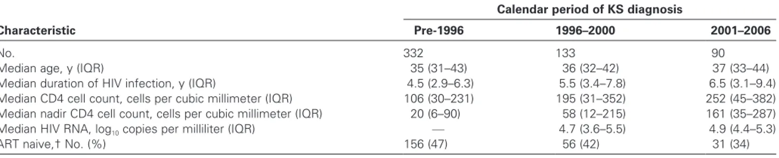
Among the 9473 homosexual men in CASCADE, 555 were diag-nosed with Kaposi sarcoma in the period from January 1, 1986, through December 31, 2006; characteristics of these men at their diagnosis of Kaposi sarcoma are presented in Table 1. As expected, because of the increased survival after the widespread availability of cART in 1996, those diagnosed with Kaposi sarcoma after the introduction of cART had been infected with HIV for increasingly longer periods. Median CD4 cell count at Kaposi sarcoma diagno-sis rose statistically significantly over time from 106 cells per cubic millimeter (interquartile range [IQR] = 30–231 cells per cubic millimeter) before 1996 to 195 cells per cubic millimeter (IQR = 31–352 cells per cubic millimeter) in 1996–2000 and to 252 cells per cubic millimeter (IQR = 45–382 cells per cubic millimeter) in 2001–2006 (P < .001). The nadir CD4 cell count also increased statistically significantly from before 1996 through 2006 (P = .003), but no change in the level of HIV RNA at Kaposi sarcoma diagno-sis was observed during the study period (P = .98).
Effects of Calendar Year on Mortality and Survival Estimates
Among the 555 men diagnosed with Kaposi sarcoma, 319 (57%) had died by the end of 2006. Most deaths (261 [82%]) occurred before 1996, with 42 deaths in 1996–2000 and 16 deaths in 2001– 2006. The proportion of men with at least a 24-month survival after Kaposi sarcoma diagnosis rose markedly over time from 35% (95% confidence interval [CI] = 29% to 42%) in 1986–1995 to 84% (95% CI = 76% to 90%) in 1996–2000 and to 81% (95% CI = 70% to 88%) in 2001–2006 (P < .001, log-rank test). Among patients who were diagnosed with Kaposi sarcoma in 2001–2006, we found cumulative survival probabilities at 24 months of 71% (95% CI = 55% to 83%) for those with a CD4 cell count of less than 300 cells per cubic millimeter and 94% (95% CI = 77% to 98%) for those with a CD4 cell count of 300 cells per cubic milli-meter or more at Kaposi sarcoma diagnosis. Despite the few deaths (13 deaths in the men with a CD4 cell count of <300 cells per cubic millimeter and three deaths in men with CD4 cell count of ≥300 cells per cubic millimeter at Kaposi sarcoma diagnosis), the sur-vival difference between the two follow-up groups was statistically significant (P = .021, log-rank test).
Incidence of Kaposi Sarcoma by cART Status
A total of 7873 homosexual men in the naive follow-up group contributed 23 352.29 person-years of follow-up, of whom 192 developed Kaposi sarcoma during the study period. A total of 4199 patients in the cART follow-up group contributed 19 535.7 person-years, of whom 70 developed Kaposi sarcoma.
We fit a Poisson regression model with an interaction between a cART indicator and the current CD4 count after adjustment for age. According to the fractional polynomial approach (24), the square root transformation of current CD4 count provided an adequate fit for the incidence of Kaposi sarcoma. According to this model, at the same current CD4 level, there is a higher risk of Kaposi sarcoma in the naive follow-up group than in the cART follow-up group but only with borderline statistical significance (P for interaction = .067). For simplicity, we report rates of Kaposi sarcoma by current CD4 cell level (<200, 200–349, 350–499, or ≥500 cells per cubic millimeter) (Table 2). Incidence rates of Kaposi sarcoma increased as the level of CD4 cells decreased in both naive and cART follow-up groups. Overall, the incidence rate of Kaposi sarcoma was lower in the cART follow-up group (3.58
diagnoses of Kaposi sarcoma per 1000 person-years) than in the naive follow-up group (8.22 diagnoses per 1000 person-years), as expected. There were 81 diagnoses of Kaposi sarcoma in the naive follow-up group during 8681.7 person-years and 13 in the cART follow-up group during 1946.1 person-years of follow-up while current CD4 cell count was missing, giving crude incidence rates of 9.33 and 6.68 diagnoses of Kaposi sarcoma per 1000 person-years, respectively. To explore the effect of the missing CD4 cell count data, we took the mean value of the last and first available measurements to obtain median values for the missing CD4 cell counts of 525 cells per cubic millimeter (IQR = 401–682 cells per cubic millimeter) for the naive follow-up group and 522 cells per cubic millimeter (IQR = 348–717 cells per cubic millimeter) for the cART follow-up group.
Factors Associated With the Incidence of Kaposi Sarcoma After Initiation of cART
In the cART follow-up group, 70 men were diagnosed with Kaposi sarcoma, with a median CD4 cell count at diagnosis of 240 cells per cubic millimeter (IQR = 82–405 cells per cubic milli-meter) and a median log10 HIV RNA level of 4.24 (IQR = 2.31– 5.28) during a median follow-up of 4.5 years (IQR = 1.9–7.3 years). The median duration of infection with HIV was 7 years (IQR = 4–9 years), the median age at Kaposi sarcoma diagnosis was 37 years (IQR = 32–43 years), and the time since starting cART was 3 years (IQR = 1–5 years). Among these 70 men, nine were diagnosed with Kaposi sarcoma and had a CD4 level of more than 300 cells per cubic millimeter and a HIV RNA level that had remained less than 500 copies per milliliter for at least 2 years before their diagnosis of Kaposi sarcoma. Among these nine men, the median duration of infection was 5 years (IQR = 2–8 years) and the median age at Kaposi sarcoma diagnosis was 39 years (IQR = 36–43 years).
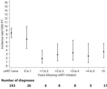
Crude incidence rates of Kaposi sarcoma fell slightly in the first year after starting cART from 8.22 Kaposi sarcoma diagnoses per 1000 person-years in the naive follow-up group (ie, before starting any antiretroviral therapy) to 6.67 per 1000 person-years in the cART follow-up group. This decrease was followed by an even sharper decline thereafter, with the incidence rate of Kaposi sar-coma remaining at approximately 2.5 Kaposi sarsar-coma diagnoses per 1000 person-years from approximately 1 year of follow-up through at least 5 years of follow-up (Figure 1).
Table 1. Characteristics of patients at diagnosis of Kaposi sarcoma (KS) according to calendar period of diagnosis* Characteristic
Calendar period of KS diagnosis
Pre-1996 1996–2000 2001–2006
No. 332 133 90
Median age, y (IQR) 35 (31–43) 36 (32–42) 37 (33–44)
Median duration of HIV infection, y (IQR) 4.5 (2.9–6.3) 5.5 (3.4–7.8) 6.5 (3.1–9.4)
Median CD4 cell count, cells per cubic millimeter (IQR) 106 (30–231) 195 (31–352) 252 (45–382)
Median nadir CD4 cell count, cells per cubic millimeter (IQR) 20 (6–90) 58 (12–215) 161 (35–287)
Median HIV RNA, log10 copies per milliliter (IQR) — 4.7 (3.6–5.5) 4.9 (4.4–5.3)
ART naive,† No. (%) 156 (47) 56 (42) 31 (34)
* CD4 cell count and HIV RNA measurements were recorded up to 6 months before and 1 month after KS diagnosis date. Numbers of individuals with missing data on CD4 cell count (and HIV RNA level) were 58 (—) before 1996, 14 (66) for 1996–2000, and four (three) for 2001–2006. IQR = interquartile range; nadir = lowest value ever recorded.
† These patients had no antiretroviral treatment before their diagnosis of KS.
The duration of HIV infection was not associated with the incidence of Kaposi sarcoma in the cART follow-up group (rate ratio [RR] per additional year = 0.97, 95% CI = 0.90 to 1.05,
P = .44) (Table 3). Current levels of CD4 cells and HIV RNA
were, however, associated with the incidence of Kaposi sarcoma, with lower CD4 cell levels being associated with higher incidence rates of Kaposi sarcoma. More specifically, current (ie, within 6 months) CD4 cell count was associated with crude incidence of Kaposi sarcoma among cART-treated men (RR = 18.91, 95% CI = 8.50 to 42.09, for CD4 level category <200 cells per cubic millimeter; RR = 3.55, 95% CI = 1.40 to 9.00, for 200–349 cells per cubic millimeter; and RR = 4.11, 95% CI = 1.74 to 9.70, for 350–499 cells per cubic millimeter; all compared with ≥500 cells per cubic millimeter). Although the crude incidence rate of Kaposi sarcoma was statistically significantly lower after the first year in which cART was initiated (P = .003), when we adjusted for current CD4 cell level and HIV RNA, the incidence rate of Kaposi sar-coma was not associated with duration of cART of more than 1 year as compared with duration of 1 year or less (RR = 0.87, 95% CI = 0.47 to 1.61, P = .66), the presence of protease inhibitor in the cART regimen (P = .44), or being antiretroviral treatment naive at cART initiation (P = .16).
In multivariable models including other measures of immuno-suppression mediated through CD4 cell count, among the cART follow-up group, the current CD4 cell count was more strongly associated with risk of Kaposi sarcoma than the nadir CD4 cell count or the time-weighted average CD4 count (data not shown). Although there was a borderline statistically significantly associa-tion between the nadir CD4 cell count at cART initiaassocia-tion and the risk of Kaposi sarcoma (P = .06), after adjusting for current CD4 cell count, which appeared to be strongly associated with the risk of Kaposi sarcoma (P < .001), the association disappeared (P = .59) (Table 4), as did that for the time-weighted average CD4 cell count (P = .16). Table 2. Kaposi sarcoma (KS) incidence rates by current CD4 cell count status among the naive follow-up group and the combination antiretroviral therapy (cART) follow-up group* CD4 status, cells per cubic millimeter Naive follow-up group cART follow-up group RR† (95% CI) Person-years KS events, No. Crude incidence rate Person-years KS events, No. Crude incidence rate <200 573.3 42 73.27 1419.1 24 16.91 4.34 (2.65 to 7.27) 200–349 2408.9 23 9.55 3146.9 10 3.18 3.05 (1.45 to 6.42) 350–499 4258.6 28 6.57 4078.9 15 3.68 1.81 (0.97 to 3.42) ≥500 7426.5 18 2.42 8945.2 8 0.89 2.76 (1.19 to 6.36) Missing 8684.0 81 9.33 1945.6 13 6.68 Total 23 351.3 192 8.22 19 535.7 70 3.58 * Crude KS incidence rates are reported as number of KS diagnoses per 1000 person-years of follow-up. CI = confidence interval; RR = rate ratio. † RRs were generated with a Poisson regression model that had an interaction between follow-up group indicator (0 if KS was diagnosed in the cART follow-up group, 1 if KS was diagnosed in the naive follow-up group) and CD4 status with adjustment for age.
Figure 1. Crude Kaposi sarcoma (KS) incidence rates according to time since initiation of combination antiretroviral therapy (cART). Confidence intervals (as shown by error bars) were computed by assuming a Poisson distribution for the number of events. cART naive = KS inci-dence rates in patients who did not start cART; PY = person-years.
Discussion
To our knowledge, this is the first study to examine the associa-tions of HIV infection duration and history of immunosuppression with risk of Kaposi sarcoma. We studied homosexual men with well-estimated dates of HIV seroconversion. In the cART follow-up group, we found no association between duration of infection or age and the incidence of Kaposi sarcoma but we did find that current CD4 cell count and current HIV RNA level were strongly associated with risk of Kaposi sarcoma.
The CD4 cell count at Kaposi sarcoma diagnosis appeared to increase over calendar time from a median of 106 cells per cubic millimeter for the 332 patients who were diagnosed before 1996 to a median of 252 cells per cubic millimeter for the 90 who were
diagnosed in 2001–2006, indicating that approximately 50% of the cohort received their Kaposi sarcoma diagnosis at a CD4 count above that recommended by the contemporaneous treatment guidelines (ie, 200 cells per cubic millimeter) (26,27).
We found an overall reduction in the incidence of Kaposi sar-coma in the cART follow-up group (8.22 Kaposi sarsar-coma events per 1000 person-years) compared with that in the naive follow-up group (3.58 Kaposi sarcoma events per 1000 person-years). At the same CD4 cell count strata, the risk of Kaposi sarcoma was higher in the naive follow-up group than in the cART follow-up group, particularly for a CD4 cell count if less than 200 cells per cubic millimeter, indicating that the risk of Kaposi sarcoma was not completely mediated through CD4 cell count and that other factors contribute to the risk of Kaposi sarcoma. We may have underestimated the incidence rates of Kaposi sarcoma in the high-est CD4 count categories by assuming that the missing CD4 cell levels were more than 500 cells per cubic millimeter; however, this problem is unlikely to have affected the rate ratios.
We also found that over time both the CD4 cell count and the nadir for CD4 cell count at Kaposi sarcoma diagnosis shifted to higher levels. One explanation for these findings is that the increased medical care that generally accompanies treatment with cART increases the opportunity for symptoms and lesions to be identified and examined at an early stage. Other explanations are that cART may not control human herpesvirus 8, which is consid-ered a necessary cause of Kaposi sarcoma, and the causal relation between human herpesvirus 8 and Kaposi sarcoma development may not be directly mediated through the improvement of the immune system. Finally, the observed increase in CD4 cell levels at Kaposi sarcoma diagnosis may be due to a change in Kaposi sarcoma site because it has been reported (28) that the decline in Kaposi sarcoma incidence since the introduction of cART has affected Kaposi sarcoma with visceral involvement more strongly Table 3. Factors associated with Kaposi sarcoma incidence after combination antiretroviral therapy (cART) initiation*
Variable
Univariate analysis Multivariable analysis†
RR (95% CI) P‡ RR (95% CI) P
Per year of HIV infection duration 0.96 (0.90 to 1.02) .16 0.97 (0.90 to 1.05) .44
Per 10-y increase in current age 0.84 (0.63 to 1.13) .26 1.01 (0.74 to 1.39) .93
Current CD4 cell status
≥500 cells per cubic millimeter 1 (ref.) <.001 1 (ref.) <.001
350–499 cells per cubic millimeter 4.11 (1.74 to 9.70) 2.77 (1.12 to 6.85)
200–349 cells per cubic millimeter 3.55 (1.40 to 9.00) 2.77 (1.07 to 7.20)
<200 cells per cubic millimeter 18.91 (8.50 to 42.09) 11.34 (4.63 to 27.80)
Current HIV RNA status 1.31 (1.22 to 1.40) <.001 1.19 (1.10 to 1.29) <.001
Time since cART initiation
1 y 1 (ref.) .003 1 (ref.) .66
>1 y 0.45 (0.27 to 0.76) 0.87 (0.47 to 1.61)
Type of cART
Non-protease inhibitor cART 1 (ref.) .99 1 (ref.) .44
Protease inhibitor cART 1 (0.59 to 1.72) 0.80 (0.46 to 1.41)
Pre-cART status
Pretreated§ 1 (ref.) .67 1 (ref.) .16
Naive to treatment 1.14 (0.63 to 2.05) 1.67 (0.82 to 3.41)
* CI = confidence interval; ref. = referent; RR = rate ratio. † Variable adjusted for all other variables listed.
‡ Wald test. All statistical tests were two-sided.
§ Patients received other or suboptimal antiretroviral treatments before initiation of a combination antiretroviral therapy (cART).
Table 4. Relationship of nadir CD4 cell count to Kaposi sarcoma in homosexual men treated with combination antiretroviral therapy (cART)*
CD4 cell count RR (95% CI) P†
Current
<200 cells per cubic millimeter 11.43 (3.62 to 36.06) <.001 200–349 cells per cubic millimeter 3.17 (1.04 to 9.63)
350–499 cells per cubic millimeter 2.93 (1.04 to 8.27) ≥500 cells per cubic millimeter 1 (ref.) Nadir
<100 cells per cubic millimeter 0.70 (0.23 to 2.13) .59 100–199 cells per cubic millimeter 0.51 (0.17 to 1.52)
200–349 cells per cubic millimeter 0.99 (0.45 to 2.21) ≥500 cells per cubic millimeter 1 (ref.)
* Multivariable models were adjusted for duration of infection, current age, time since cART initiation, whether ART naive (yes or no), and whether treated with a protease inhibitor (yes or no). CI = confidence interval; nadir = lowest level recorded; ref. = referent; RR = rate ratio. † Wald test. All statistical tests were two-sided.
than cutaneous Kaposi sarcoma and that, among cART-treated patients, Kaposi sarcoma with nonvisceral involvement appears to be diagnosed at higher levels of CD4 cells than visceral Kaposi sarcoma.
As previously reported (5,7), we also found that most Kaposi sarcoma diagnoses among patients who were being treated with cART occurred during the first year of therapy (ie, 26 of the 70 Kaposi sarcoma diagnoses), with a high Kaposi sarcoma incidence rate at 6.5 events per 1000 person-years. Some of these events may be due to the immune reconstitution inflammatory syndrome, which has been linked to detection of rare cases of Kaposi sarcoma among individuals after the initiation of cART (29–31). The im-mune reconstitution inflammatory syndrome consists of a clinical deterioration after the initiation of cART that is believed to be a consequence of restored ability to mount an inflammatory response. Connick et al. (30) suggested that Kaposi sarcoma–associated im-mune reconstitution inflammatory syndrome may be more common than is reflected in the literature because Kaposi sarcoma– associated immune reconstitution inflammatory syndrome can be misdiagnosed as Kaposi sarcoma development caused by treatment failure. Given that the development of cancers is preceded by exposure to the causative agent by many years, however, it may not be appropriate to attribute Kaposi sarcoma diagnosis at such an early stage in treatment with cART to cART itself. Indeed, among those in the naive follow-up group, Kaposi sarcoma rate was slightly higher at 8.22 events per 1000 person-years, but we found no evidence to indicate that rates of Kaposi sarcoma increased after cART was initiated. Moreover, in the second year after cART initiation, we detected a sharp fall in the incidence of Kaposi sar-coma that appeared to remain stable thereafter. Our findings, therefore, suggest that Kaposi sarcoma diagnosed in the first year is unlikely to be related wholly to immune reconstitution inflam-matory syndrome.
It may also be that the decline observed after the first year of cART treatment is a result of bias due to underascertainment of men diagnosed with Kaposi sarcoma after the initiation of cART. However, in the multivariable model after adjustment for current CD4 cell count, no association between the rate of Kaposi sar-coma and the duration of cART was observed, indicating that the higher rate in the first year may be explained by the low CD4 cell counts in patients who have recently initiated treatment with cART. Such recording bias is unlikely, therefore, to fully explain this decline.
The risk of Kaposi sarcoma appeared to be similar among men treated with protease inhibitor in their initial cART regimen and those treated with non-protease inhibitor–containing cART. This result is in conflict with results of two previous studies (32,33) that promoted the use of protease inhibitors as potent antiangiogenic and antitumor molecules but is supported by the results of four cohort studies (7,12,28,34), however, that may have been subject to confounding by indication. Our data do not, therefore, support the preferential use of protease inhibitors in cART regimens for the treatment of HIV-infected men at risk of Kaposi sarcoma.
The persistence or development of Kaposi sarcoma in HIV-infected men on cART despite high CD4 counts and undetectable HIV RNA has been reported by several authors (13–16). This observation was confirmed by our finding of nine men who were
diagnosed with Kaposi sarcoma when they had a CD4 cell count of more than 300 cells per cubic millimeter and a level of HIV RNA that had remained at less than 500 copies per milliliter for at least 2 years after the initiation of cART.
Although age is an established risk factor for the development of classic Kaposi sarcoma, which usually occurs in individuals who are older than 50 years (20), increasing age was not associated with risk of Kaposi sarcoma among HIV-seropositive patients who had initiated cART. One explanation for this finding may well be the limited and young age range of our participants (median = 37 years, IQR = 32–42 years) so that we could not study associations among older men. This possibility is supported by a study (35) from the French Hospital Database that found a moderate associ-ation between age and risk of Kaposi sarcoma among individuals who were older than 60 years compared with those who were younger than 30 years, with the strength of the association being only marginally increased among those aged 30–59 years.
Treatment with cART among patients with Kaposi sarcoma has been associated with improved survival (36) and prolonged time to treatment failure (37). Data from this study support this increased survival. The prognosis among HIV-infected cancer patients tends to be worse than that among non-HIV cancer patients (38–40). It is not clear, however, whether this is because of an overall lack of contact with medical services, leading to a late diagnosis of both HIV and cancer, or whether cancer is more aggressive in the pres-ence of HIV infection. Although our survival estimates for patients with chronic HIV infection of 24 months before 1996 (ie, 35% alive) were similar to those from studies (ie, 29%–49% alive) (36,41–44), it is noteworthy that, in the period of 1996–2000, we found more favorable survival estimates (84% alive) than other studies (61%–66% alive). Only one study (45) that was based on data from seroprevalent patients reported better survival at 36 months. Thus, overall, the homosexual men in this study appear to have benefited from being diagnosed and monitored early in HIV infection.
This study had several limitations that should be discussed. First, we did not have information on treatment adherence and, therefore, assumed that patients remained on therapy once it was initiated. If diagnoses of Kaposi sarcoma are largely occurring in individuals who have stopped using cART or have poor adherence to the treatment regimen, then the incidence of Kaposi sarcoma in the cART follow-up group will have been overestimated. To eval-uate this possibility, we performed sensitivity analyses considering follow-up time for those in the cART follow-up group whose HIV RNA level remained at less than 1000 copies per milliliter. This analysis had no effect on estimates of the associations of HIV in-fection duration, age, or current CD4 levels with risk of Kaposi sarcoma (data not shown).
Second, in the cART follow-up group, associations between var-ious factors and risk of Kaposi sarcoma were based on only 70 Kaposi sarcoma events and a limited median follow-up of 4.5 years (IQR = 1.9–7.3 years). Thus, because of the small number of events, this analysis may have lacked the power to detect associations between duration of infection, age, or nadir CD4 cell count and risk of Kaposi sarcoma. However, because we did find associations between current CD4 cell count and risk of Kaposi sarcoma, we concluded that, if an effect of duration of infection, age, or nadir CD4 cell count exists, it
should be smaller than the effect of the current level of immunosup-pression (as measured by CD4 cell count).
Third, when we compared risk of Kaposi sarcoma between the naive follow-up group and the cART follow-up group, our esti-mates may have been biased through confounding by indication (ie, men who start cART are more likely to be sicker and at higher risk of Kaposi sarcoma). Confounding by indication would have led to an overestimate for the risk of Kaposi sarcoma among those in the cART follow-up group and would thus tend toward esti-mating similar rates in the naive and cART follow-up groups.
Finally, the observed increase in survival may have indicated that patients in later years have had less aggressive form of Kaposi sarcoma or that death from non-Kaposi sarcoma causes has been reduced. Unfortunately, information on whether death was caused by Kaposi sarcoma is not recorded in the CASCADE dataset.
In conclusion, in this study of homosexual men with a known date of HIV seroconversion, we found that duration of HIV infec-tion, age, or nadir CD4 cell count was not associated with inci-dence of Kaposi sarcoma in treated men after adjustment for CD4 cell count. We also found that, in the cART follow-up group, Kaposi sarcoma tended to be diagnosed at a higher CD4 cell count than that in the naive follow-up group and that survival after a diagnosis of Kaposi sarcoma has increased in the cART follow-up group. It may also be worthwhile screening at risk men for human herpesvirus 8 so that they can be monitored more frequently for signs of Kaposi sarcoma, even if they have a relatively high CD4 cell count. Finally, given the high survival estimates in this analysis, compared with results from studies (36,41–44) that were based on seroprevalent patients, early diagnosis of HIV infection may be associated with better prognosis of Kaposi sarcoma.
references
1. Biggar RJ, Chaturvedi AK, Goedert JJ, Engels EA. AIDS-related cancer and severity of immunosuppression in persons with AIDS. J Natl Cancer
Inst. 2007;99(12):962–972.
2. Mbulaiteye SM, Biggar RJ, Goedert JJ, Engels EA. Immune deficiency and risk for malignancy among persons with AIDS. J AIDS. 2003;32(5): 527–533.
3. Gates AE, Kaplan LD. AIDS malignancies in the era of highly active antiretroviral therapy. Oncology. 2002;16(5):657–665.
4. Ledergerber B, Telenti A, Egger M. Risk of HIV related Kaposi’s sarcoma and non-Hodgkin’s lymphoma with potent antiretroviral therapy: pro-spective cohort study. BMJ. 1999;319(7201):23–24.
5. Franceschi S, Dal Maso L, Rickenbach M, et al. Kaposi sarcoma incidence in the Swiss HIV cohort study before and after highly active antiretroviral therapy. Br J Cancer. 2008;99(5):800–804.
6. International Collaboration on HIV and Cancer. Highly active antiretro-viral therapy and incidence of cancer in human immunodeficiency virus-infected adults. J Natl Cancer Inst. 2000;92(22):1823–1830.
7. Mocroft A, Kirk O, Clumeck N, et al. The changing pattern of Kaposi sarcoma in patients with HIV, 1994–2003. The EUROSIDA study.
Cancer. 2004;100(12):2644–2654.
8. CASCADE Collaboration. Changes over calendar time in the risk of spe-cific first AIDS-defining events following HIV seroconversion, adjusting for competing risks. Int J Epidemiol. 2002;31(5):951–958.
9. The Antiretroviral Therapy Cohort Collaboration. The changing inci-dence of AIDS events in patients receiving highly active antiretroviral therapy. Arch Intern Med. 2005;165(4):416–423.
10. Ancelle-Park JR. Expanded European AIDS case definition. Lancet. 1993;341(8842):441.
11. Catellan AM, Calabro ML, Gasperini P, et al. Acquired immunodeficiency syndrome-related Kaposi’s sarcoma regression after highly active
antiretro-viral therapy: biologic correlates of clinical outcome. J Natl Cancer Inst
Monogr. 2000;2000(28):44–49.
12. Martinez V, Caumes E, Gambotti L, et al. Remission from Kaposi’s sarcoma on HAART is associated with suppression of HIV replication and is inde-pendent of protease inhibitor therapy. Br J Cancer. 2006;94(7):1000–1006. 13. Maurer T, Ponte M, Leslie K. HIV associated Kaposi’s sarcoma with a
high CD4 count and a low viral load. New Engl J Med. 2007;357(13): 1352–1353.
14. Chan J, Kravcik S, Angel JB. Development of Kaposi’s sarcoma despite sustained suppression of HIV plasma viremia. J AIDS. 1999;22(2): 209–210.
15. Krown SE, Lee JY, Dittmer DP. More on HIV-associated Kaposi’s sarcoma. New Engl J Med. 2008;358(5):535–536.
16. Stebbing J, Powles T, Bower M. AIDS-associated Kaposi’s sarcoma asso-ciated with a low viral load and high CD4 cell count. AIDS. 2008; 22(4):551–552.
17. Hymes KB, Greene JB, Marcus A, et al. Kaposi’s sarcoma in homosexual men—a report of eight cases. Lancet. 1981;2(8247):598–600.
18. Bhaskaran K, Hamouda O, Sannes M, et al. Changes in the risk of death after HIV seroconversion compared with mortality in the general popula-tion. JAMA. 2008;300(1):51–59.
19. CASCADE Collaboration. Changes in the uptake of antiretroviral therapy and survival in people with known duration of HIV infection in Europe: results from CASCADE. HIV Med. 2000;1(4):224–231.
20. Dukers NT, Rezza G. Human herpesvirus 8 epidemiology: what we do and do not know. AIDS. 2003;17(12):1717–1730.
21. Goedert JJ, Vitale F, Lauria C, et al. Risk factors for classical Kaposi’s sarcoma. J Natl Cancer Inst. 2002;94(22):1712–1718.
22. Lyles CM, Dorrucci M, Vlahov D, et al. Longitudinal human immunode-ficiency virus type 1 load in the Italian seroconversion study: correlates and temporal trends of viral load. J Infect Dis. 1999;180(4):1018–1024. 23. Sabin CA, Devereux H, Phillips AN, et al. Course of viral load throughout
HIV-1 infection. J AIDS. 2000;23(2):172–177.
24. Royston P, Altman DG. Regression using fractional polynomials of con-tinuous covariates: parsimonious parametric regression modelling. Appl
Stat. 1994;43(3):429–467.
25. Akaike H. A new look at the statistical model identification. IEEE Trans
Automat Control. 1974;19(6):716–723.
26. British HIV Association (BHIVA) guidelines for the treatment of HIV-infected adults with antiretroviral therapy. HIV Med. 2001;2(4):276–313. 27. Panel on Antiretroviral Guidelines for Adults and Adolescents. Guidelines
for the Use of Antiretroviral Agents in HIV-1-Infected Adults and Adolescents.
Department of Health and Human Services (United States); 2001:1–115. http://www.aidsinfo.nih.gov/ContentFiles/AdultandAdolescentGL.pdf. Accessed April 12, 2009.
28. Grabar S, Abraham B, Mahamat A, Del Giudice P, Rosenthal E, Costagliola D. Differential impact of combination antiretroviral therapy in preventing Kaposi’s sarcoma with and without visceral involvement.
J Clin Oncol. 2006;24(21):3408–3414.
29. Steigbigel RT, Cooper DA, Kumar PN, et al. Raltegravir with optimized background therapy for resistant HIV-1 infection. New Engl J Med. 2008; 359(4):339–354.
30. Connick E, Kane MA, White IE, Ryder J, Campbell TB. Immune recon-stitution inflammatory syndrome associated with Kaposi sarcoma during potent antiretroviral therapy. Clin Infect Dis. 2004;39(12):1852–1855. 31. Feller L, Anagnostopoulos C, Wood NH, Bouckaert M, Raubenheimer
EJ, Lemmer J. Human immunodeficiency virus-associated Kaposi sar-coma as an immune reconstitution inflammatory syndrome: a literature review and a case report. J Periodontol. 2008;79(2):362–368.
32. Sgadari C, Barillari G, Toschi E, et al. HIV protease inhibitors are potent anti-angiogenic molecules and promote regression of Kaposi sarcoma. Nat
Med. 2002;8(3):225–232.
33. Sgadari C, Monini P, Barillari G, Ensoli B. Use of HIV protease inhibitors to block Kaposi’s sarcoma and tumour growth. Lancet Oncol. 2003;4(9): 537–547.
34. Portsmouth S, Stebbing J, Gill J, et al. A comparison of regimens based on non-nucleoside reverse transcriptase inhibitors or protease inhibitors in preventing Kaposi’s sarcoma. AIDS. 2003;25(17):F17–F22.
35. Guiguet M, Boue F, Cadranel J, Lang JM, Lang JM, Rosenthal E, Costagliola D. Effect of immunodeficiency, HIV viral load, and antiretro-viral therapy on the risk of individual malignancies (FHDH-ANRS C04): a prospective cohort study. Lancet Oncol. 2009;10(12):1152–1159. 36. Biggar RJ, Engels EA, Ly S, et al. Survival after cancer diagnosis in
persons with AIDS. J AIDS. 2005;39(3):293–299.
37. Bower M, Fox P, Fife K, Gill J, Nelson M, Gazzard B. Highly active antiretroviral therapy (HAART) prolongs time to treatment failure in Kaposi’s sarcoma. AIDS. 1999;13(15):2105–2111.
38. Tirelli U, Spina M, Sandri S, et al. Lung carcinoma in 36 patients with human immunodeficiency virus infection. Cancer. 2000;88(3):563–569. 39. Spano J, Atlan D, Breau J, Farge D. AIDS and non-AIDS-related
malig-nancies: a new vexing challenge in HIV-positive patients: Part II. Cervical and anal squamous lesions, lung cancer, testicular germ cell cancers and skin cancers. Eur J Int Med. 2002;13(4):227–232.
40. Phelps RM, Smith DK, Heilig CM, et al. Cancer incidence in women with or at risk for HIV. Int J Cancer. 2001;94(5):753–757.
41. Fordyce EJ, Singh TP, Nash D, Gallagher B, Forlenza S. Survival rates in NYC in the era of combination ART. J AIDS. 2002;30(1):111–118. 42. Dore GJ, Li Y, McDonald A, Ree H, Kaldo JM. Impact of highly active
antiretroviral therapy on individual AIDS-defining illness incidence and survival in Australia. J AIDS. 2002;29(4):388–395.
43. Conti S, Masocco M, Pezzotti P, et al. Differential impact of combined antiretroviral therapy on the survival of Italian patients with specific AIDS-defining illnesses. J AIDS. 2000;25(5):451–458.
44. Brodt HR, Kamps BS, Helm EB, Schöfer H, Mitrou P. Kaposi’s sarcoma in HIV infection: impact on opportunistic infections and survival. AIDS. 1998;12(12):1475–1481.
45. Grabar S, Lanoy E, Allavena C, et al. Causes of the first AIDS-defining illness and subsequent survival before and after the advent of combined antiretroviral therapy. HIV Med. 2008;9(4):246–256.
Funding
CASCADE has been funded through grants BMH4-CT97-2550, QLK2-2000-01431, QLRT-2001-01708 and LSHP-CT-2006-018949 from the European Union.
Notes
K. Porter has received a research grant from GlaxoWellcome. A. de Luca has received speakers honoraria and fees for attending advisory boards from GlaxoSmithKline, Gilead, Bristol-Myers-Squibb, Boehringer-Ingelheim, Abbott Virology, Tibotec, and Monogram Biosciences.
The authors had full responsibility in the design of the study, collection of the data, the analysis and interpretation of the data, the decision to submit the manuscript for publication, and the writing of the manuscript.
CASCADE Collaboration: Steering Committee: Julia Del Amo (Chair), Laurence Meyer (Vice Chair), Heiner C. Bucher, Geneviève Chêne, Deenan Pillay, Maria Prins, Magda Rosinska, Caroline Sabin, and Giota Touloumi. Coordinating Center: Kholoud Porter (Project Leader), Sara Lodi,
Kate Coughlin, Sarah Walker, Abdel Babiker, and Janet Darbyshire. Clinical Advisory Board: Heiner C. Bucher, Andrea de Luca, Martin Fisher, and Roberto Muga. Collaborators: Australia Sydney AIDS Prospective Study and Sydney Primary HIV Infection cohort (John Kaldor, Tony Kelleher, Tim Ramacciotti, Linda Gelgor, David Cooper, and Don Smith); Canada South Alberta clinic (John Gill); Denmark Copenhagen HIV Seroconverter Cohort (Louise Bruun Jørgensen, Claus Nielsen, and Court Pedersen); Estonia Tartu Ülikool (Irja Lutsar); France Aquitaine cohort (Geneviève Chêne, Francois Dabis, Rodolphe Thiebaut, and Bernard Masquelier), French Hospital Database (D. Costagliola and M. Guiguet), Lyon Primary Infection cohort (Philippe Vanhems), French PRIMO cohort (Marie-Laure Chaix and Jade Ghosn), SEROCO cohort ((Marie-Laurence Meyer and Faroudy Boufassa); Germany German cohort (Osamah Hamouda, Claudia Kucherer, and Barbara Bartmeyer); Greece Greek Haemophilia cohort (Giota Touloumi, Nikos Pantazis, Angelos Hatzakis, Dimitrios Paraskevis, and Anastasia Karafoulidou); Italy Italian Seroconversion Study (Giovanni Rezza, Maria Dorrucci, and Claudia Balotta), ICONA cohort (Antonella d’Arminio Monforte, Alessandro Cozzi-Lepri, and Andrea de Luca.); Netherlands Amsterdam Cohort Studies among homosexual men and drug users (Maria Prins, Ronald Geskus, Jannie van der Helm, and Hanneke Schuitemaker); Norway Oslo and Ulleval Hospital cohorts (Mette Sannes, Oddbjorn Brubakk, Anne Eskild, and Johan N Bruun); Poland National Institute of Hygiene (Magdalena Rosinska and Joanna Gniewosz); Portugal Universidade Nova de Lisboa (Ricardo Camacho); Russia Pasteur Institute (Tatyana Smolskaya); Spain Badalona IDU hospital cohort (Roberto Muga and Jordi Tor), Barcelona IDU Cohort (Patricia Garcia de Olalla and Joan Cayla), Madrid cohort (Julia Del Amo and Jorge del Romero), Valencia IDU cohort (Santiago Pérez-Hoyos); Switzerland Swiss HIV Cohort Study (Heiner C. Bucher, Martin Rickenbach, and Patrick Francioli); Uganda and Zimbabwe; Ukraine Perinatal Prevention of AIDS Initiative (Ruslan Malyuta); United Kingdom Edinburgh Hospital cohort (Ray Brettle), Health Protection Agency (Valerie Delpech, Sam Lattimore, Gary Murphy, John Parry, and Noel Gill), Royal Free haemophilia cohort (Caroline Sabin and Christine Lee), UK Register of HIV Seroconverters (Kholoud Porter, Anne Johnson, Andrew Phillips, Abdel Babiker, Janet Darbyshire, and Valerie Delpech), University College London (Deenan Pillay), University of Oxford (Harold Jaffe); African cohorts: Genital Shedding Study (United States: Charles Morrison, Family Health International, Robert Salata, Case Western Reserve University; Uganda: Roy Mugerwa, Makerere University; Zimbabwe: Tsungai Chipato, University of Zimbabwe); Early Infection Cohorts (Kenya, Uganda, Rwanda, Zambia, South Africa: Pauli Amornkul, International AIDS Vaccine Initiative).
Affiliations of authors: Medical Research Council, Clinical Trials Unit, London, UK (SL, KP); INSERM, U943, Paris, France (MG, DC); UPMC Univ Paris 06, UMR S 943, Paris, France (MG, DC); Department of HIV/ Genitourinary Medicine, Brighton and Sussex University Hospitals National Health Service Trust, Brighton, UK (MF); Institute of Clinical Infectious Diseases, Catholic University of Sacred Heart, Rome, Italy (AdL); Infectious Diseases Unit, University Hospital of Siena, Siena, Italy (AdL).