Original Article
Corresponding Author
Mario Ganau
https://orcid.org/0000-0002-8676-1147
Division of Neurosurgery, Toronto Western Hospital, University of Toronto, 399 Bathurst St, Toronto, ON, Canada Tel: +1-6472276897
E-mail: [email protected] * These authors contributed equally to this
study as co-first authors. Received: September 14, 2018 Revised: December 2, 2018 Accepted: December 2, 2018
A Pilot Study of Percutaneous
Interlaminar Endoscopic Lumbar
Sequestrectomy: A Modern Strategy
to Tackle Medically-Refractory
Radiculopathies and Restore Spinal
Function
Stefano Maria Priola1,2,*, Mario Ganau2,*, Giovanni Raffa1, Antonino Scibilia1,
Faisal Farrash2, Antonino Germanò1
1Neurosurgical Clinic, Department of Neurosciences, University of Messina, Messina, Italy 2Division of Neurosurgery, Toronto Western Hospital, University of Toronto, Toronto, ON, Canada
Objective: Angled scopes allow 360° visualization, which makes percutaneous endoscopic techniques (percutaneous endoscopic lumbar discectomy, PELD) particularly attractive for sequestrectomies, which entail the removal of extruded lumbar disc fragments that have migrated caudally or cranially between the ligaments, foramina, and neural structures, while preserving the disc. Although many different PELD techniques are currently avail-able, not all of them are suitable for sequestrectomies; furthermore, long-term follow-up data are unfortunately lacking.
Methods: A pilot study was conducted on a cohort of 270 patients with lumbar radiculopa-thy undergoing minimally invasive spine surgery (PELD or microdiscectomy), of whom only 7 were eligible for endoscopic interlaminar sequestrectomy with disc preservation. The patients’ baseline conditions and clinical outcomes were measured with the Oswestry Dis-ability Index and a visual analogue scale. Long-term follow-up was conducted using satis-faction questionnaires that were based on the MacNab criteria and administered by medi-cal/nursing personnel not involved in their primary surgical management.
Results: EasyGo system was eventually used in 5 PELD cases. No dural tears, infections, or nerve root injuries were recorded in patients undergoing sequestrectomy. Surgical events, including blood loss and overall length of hospital stay, did not differ significantly among the 270 patients. In the group treated with endoscopic sequestrectomy, no recurrences or complications were noted during a follow-up of 3 years, and an excellent degree of satisfac-tion was reported.
Conclusion: We provide OCEBM (Oxford Centre for Evidence-Based Medicine) level 3 ev-idence that interlaminar endoscopic sequestrectomy is a tailored and well-tolerated surgical option; nonetheless, a cost-effectiveness analysis assessing the interval until return to work-ing activities and long-term benefits is warranted.
Keywords: Lumbar disc herniation, Endoscopy, Sequestrectomy, Minimally invasive spine surgery, Enhanced recovery after surgery
INTRODUCTION
Following a technological wave toward minimal invasiveness,
in the last decade the rate of percutaneous minimally invasive spine surgery (MISS) steadily increased, eventually overtaking standard open microsurgical approaches in many instances.
This is an Open Access article distributed under the terms of the Creative Commons Attribution Non-Commercial License (http://creativecom-mons.org/licenses/by-nc/4.0/) which permits unrestricted non-commercial use, distribution, and reproduction in any medium, provided the original work is properly cited.
Copyright © 2019 by the Korean Spinal Neurosurgery Society
This trend was implemented at any given spinal segments (cer-vical, thoracic, and lumbar), and for a wide range of patholo-gies, such as: degenerative stenosis, disc herniations, synovial cysts, traumatic or pathological fractures, and even deformities.1-6
The use of the endoscope for lumbar discectomies (percuta-neous endoscopic lumbar discectomy, PELD) was fostered by the development of high-resolution endoscopes, optics, and digital cameras which allowed to gain some edges over MISS performed with the aid of microscope or surgical loupes alone. Many different PELD techniques are available nowadays, they include the microendoscopic discectomy, as well as a number of system for endoscopic spine surgery.7 They differ with regards to the percutaneous entry point (paraspinal or lateral), and the access to the nucleous polposus through the interlaminar, pos-tero-lateral and transforaminal routes, respectively.
Those techniques were initially proposed for standard lum-bar discectomy because of the following advantages: smaller skin incision, less damage to muscular and ligamentous structures, better access to the disc space (even in the axilla), reduced irri-tation to the nerve root, easier visualization of lateral foramina. Although many studies showed the good clinical outcomes of the various PELD techniques (and their combination on mul-tilevel lumbar disc disease), concerns were raised about subop-timal or incomplete removal of the disc’s annulus/nucleus pol-posus, and the long-term results especially in terms of recurren-ces.7-10 Nonetheless, the wider visualization in all axes, favored by the use of angled scopes, makes endoscopic techniques par-ticularly attractive for the removal of the extruded lumbar disc fragment, a maneuver also known as sequestrectomy. This sur-gical concept was introduced in 1978 by Williams11 to describe the decompression of the nerve root obtained by the exclusive removal of the herniated disc fragment, avoiding any surgical penetration of the disc annulus. The shorter hospital stay and earlier return to work activities, were also advocated in the as-sessment of its cost-effectiveness.12,13 To elucidate the safety and efficacy of this approach compared to other MISS techniques, we report the result of a pilot study on the percutaneous inter-laminar endoscopic sequestrectomy with disc preservation, and focus on objective measures of clinical and radiological outcome during a long term follow-up of 3 years.
MATERIALS AND METHODS
Percutaneous endoscopic techniques for lumbar spine sur-gery were introduced in our Department in 2013, becoming a valuable add to the already existing MISS practice. The highly
selective inclusion criteria for the enrollment in this pilot study were the following ones: young adults (>18 and <60 years of age), complaining of radicular pain, refractory to a 3-month course of conservative management (with analgesics, myorelax-ants, and bed rest), with magnetic resonance imaging (MRI) showing all the following specific features: (1) single level, mi-grated lumbar disc herniation, (2) preserved intervertebral height, (3) nearly intact posterior ligament and annulus fibrous. Patients with any Modic type endplate changes,14 spondylolisthesis and other features of spinal instability, defined clinically by mechan-ical pain or radiologmechan-ically with segmental movements in flex-ion-extension X-rays, were not considered eligible for the study. All patients initially assessed for lumbar radiculopathy and sche-duled for MISS intervention were screened for participation in this trial. Whenever the neurosurgical team felt to be in equi-poise for the sole sequestrectomy, either through standard mi-crosurgical or percutaneous interalaminar endoscopic appro-ach, the patient was automatically invited to a thorough discus-sion regarding the surgical options, their benefits and related risks, and therefore offered inclusion in the treatment arm of this pilot study. Preoperative and postoperative clinical data of those patients that resulted not eligible or choose not to accept percutaneous interlaminar endoscopic sequestrectomy were kept as control group (Fig. 1).
An informed consent form (previously approved by the local Ethical Committee), detailing the above, was duly signed by all patients at time of enrollment.
The baseline characteristics of each patient included in the study were duly recorded in the data registry, this included de-mographic information, as well as objective measures of clinical and radiological preoperative conditions. Patients were clinical-ly assessed for radicular and low-back pain using the visual an-alogue scale (VAS) and for functional status using the Oswestry Disability Index (ODI).
A consensus between neurosurgical and neuroimaging team was reached regarding the radiological aspect of disc herniation and migrated fragment, these were evaluated by axial, sagittal, and coronal T1- and T2-weighted MRI, using the classification of Dora et al.15
1. Endoscopic System
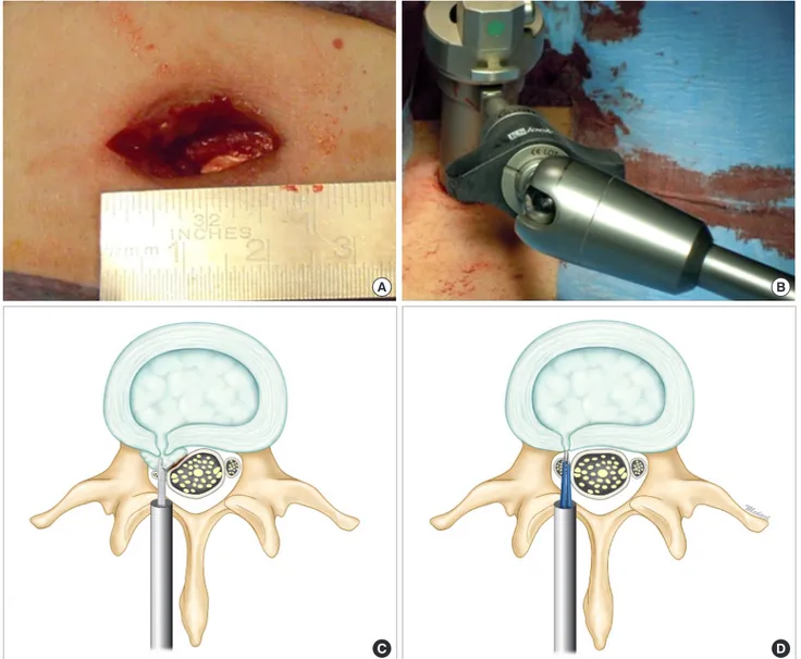
All procedures were performed with a paramedian posterior approach (2 cm in length, along an imaginary line running 1.5 to 2 cm laterally to the supraspinus ligament (Fig. 2A), depend-ing on the spinal level approached) usdepend-ing a Hopkins 10 cm, Ø2.7 mm, telescopes with 30° angle of view and a Trocar 19 mm (Easy
GO [Gaab-Oertel] ENDOSPINE instrumentation - Karl STORZ GmbH and Co. KG, Tuttlingen, Germany). This system consists of a standard tubular dilator for muscular dilation, a 30° Hop-kins rod lens optic, a work sheath of an outer diameter of 19 mm, and an irrigation-suction device, used to keep the surgical site and the optic constantly clean during surgery.
2. Surgical Technique
Upon induction of general anesthesia patients were positioned in a Karlin Frame surgical table, and the surgical site was prepped and draped as usual with povidone iodine and chlorhexidine digluconate. A radiological confirmation of the surgical level was routinely acquired with fluoroscopy, and since none of the patients had allergy to penicillin the prophylactic antibiotic ad-ministered intravenously before skin incision was always ce-fazolin (2 g). The muscle fascia was exposed and punctured, the
paraspinal muscles were subsequently smoothly split by the ap-plication of dilators, and the endoscopic work sheath was intro-duced. The endoscopic procedure was performed with biman-ual surgical technique and the work sheath fixed to the specific endoscope holder (Fig. 2B). A lateral fluoroscopic check, meant to verify the correct entry angle to access the interlaminar space was obtained before introducing the endoscope, the lamina was then identified and the interlaminar space enlarged with a punch or a diamond drill. The ligamentum flavum was exposed and incised. The dura was subsequently protected with a patty and the thecal sac/nerve root explored and gently mobilized to iden-tify the migrated fragment. The sequestrectomy was performed with grasping forceps, leaving intact the annulus and the disc (Fig. 2C, D). At the end of the procedure the surgical team al-ways verified that the disc fragment removed was congruous with the preoperative imaging (Fig. 3). If necessary, epidural
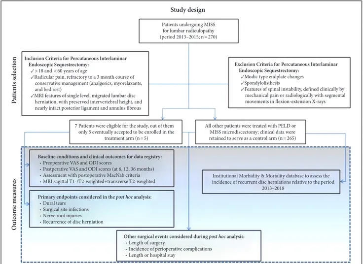
Fig. 1. Study design detailing patients’ selection process and assessment of outcome measures. MISS, minimally invasive spine surgery; MRI, magnetic resonance imaging; PELD, percutaneous endoscopic lumbar discectomy; VAS, visual analogue scale; ODI, Oswestry Disability Index.
Study design Pa tie nts s el ec tio n O ut co me me as ur es
Patients undergoing MISS for lumbar radiculopathy (period 2013–2015; n=270) Inclusion Criteria for Percutaneous Interlaminar
Endoscopic Sequestrectomy: ✓>18 and <60 years of age
✓Radicular pain, refractory to a 3 month course of conservative management (analgesics, myorelaxants, and bed rest)
✓MRI features of single level, migrated lumbar disc herniation, with preserved intervertebral height, and nearly intact posterior ligament and annulus fibrous
7 Patients were eligible for the study, out of them only 5 eventually accepted to be enrolled in the
treatment arm (n=5)
All other patients were treated with PELD or MISS microdiscectomy; clinical data were retained to serve as a control arm (n=265) Baseline conditions and clinical outcomes for data registry:
∙ Preoperative VAS and ODI scores
∙ Postperative VAS and ODI scores (at 6, 12, 36 months) ∙ Assessment with postoperative MacNab criteria ∙ MRI sagittal T1-/T2-weighted+transverse T2-weighted Primary endpoints considered in the post hoc analysis: ∙ Dural tears
∙ Surgical site infections ∙ Nerve root injuries ∙ Recurrence of disc herniation
Other surgical events considered during post hoc analysis: ∙ Length of surgery
∙ Incidence of perioperative complications ∙ Length or hospital stay
Institutional Morbidity & Mortality database to assess the incidence of recurrent disc herniations relative to the period
2013–2018
Exclusion Criteria for Percutaneous Interlaminar Endoscopic Sequestrectomy:
✓Modic type endplate changes ✓Spondylolisthesis
✓Features of spinal instability, defined clinically by mechanical pain or radiologically with segmental movements in flexion-extension X-rays
veins were cauterized with bipolar forceps and divided with mi-croscissors. At the end of the procedure, decompression of the dural sac and the nerve root were meticulously checked, and skin closure performed with 3-0 Nylon.
All sequestrectomies were performed by the same surgical team led by the senior author; the broad experience with MISS techniques including PELD, facilitated the set up and operation-al phases of this clinicoperation-al trioperation-al. Patients were mobilized from bed within the same operative day, and discharged from hospital with instructions within 24 hours. Risk of venous thromboem-bolism was assessed and managed with our standard prophy-laxis protocol.16,17
3. Outcome Measures
Surgical events including length of surgery, incidence of peri-operative complications and length of hospital stay (LOS) were recorded. All patients were followed-up with the same clinical and radiological protocol for at least 3 years after surgery.
The residual degree of disability, the estimated quality of life, and the intensity of residual pain were measured by the use of the VAS at 6, 12, and 36 months postoperatively and ODI (re-ported only at the last available follow-up). During this period patients received a series of phone calls and attended clinical assessments in our outpatient clinic with repeated administra-tion of satisfacadministra-tion quesadministra-tionnaires relying on MacNab criteria18 by medical and nursing personnel not involved in their primary
Fig. 2. Stages of percutaneous interlaminar endoscopic sequestrectomy. (A) Limited paraspinal skin incision. (B) Insertion of work sheath fixed to the endoscope holder. (C) Identification of the disc fragment and its removal. (D) Adequate decompression of thecal sac and nerve roots verified at the end of the sequestrectomy.
A B
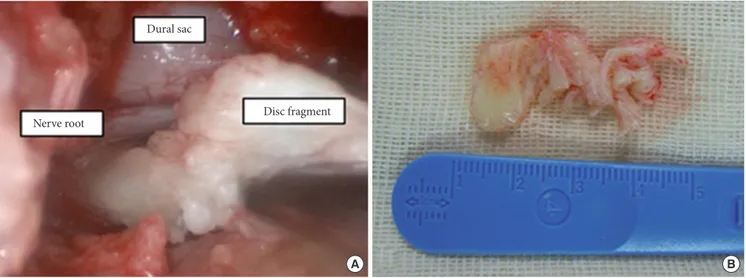
Fig. 3. Removal of extruded fragment and decompression of neural elements. (A) The migrated fragment is completely exposed, carefully mobilized and removed en bloc with grasping forceps. (B) Disc fragment removed en bloc, confirming that its size re-sults congruous with preoperative imaging.
A Nerve root
Dural sac
Disc fragment
B
surgical management. Additionally, all patients underwent post-operative lumbar MRI with a protocol including the acquisition of sagittal T1- and T2-weighted and transverse T2-weighted images.
4. Search Strategy and Criteria for Literature Review
A thorough and systematic literature search was carried out using MEDLINE and Embase databases for studies published in peer-reviewed journals on endoscopic lumbar sequestrecto-my. A search was carried out using the following Boolean search criteria ([endoscopy], [lumbar], and [sequestrectomy or seques-terectomy or fragmentectomy]). The resulting studies were then analysed. Finally, the bibliographies of all selected studies were hand-searched for any study not picked up by the original search.
RESULTS
This trial ran for a total of 14 months (December 2013 to January 2015). Among a cohort of 270 patients referred to our Department for radiculopathy due to lumbar disc herniations and considered for MISS, 7 met the abovementioned inclusion criteria. Two out of 7 patients opted for traditional microscopic approach, while the remaining 5 patients accepted to be enrolled in the treatment arm of this study and underwent a percutane-ous interlaminar endoscopic sequestrectomy. Their mean age was 45.6 years (range, 28–68 years); and the herniated disc frag-ment was localized either at the L5/S1 level (3 cases), or L3–4 level (2 cases). The main presenting symptom was a recent
on-set of radicular pain (usually a 4 months’ history), whose terri-tory of distribution matched the L4 or S1 dermatomes. Neuro-logical examination resulted within normal limits in all cases but two, in which anterior tibialis muscle (ATM) weakness (Grade 4/5, Medical Research Council scale for muscle strength) was observed on clinical examination. All patients denied red flags for cauda equina syndrome.
All surgical procedures in the treatment arm were uneventful (average length of procedure: 67 minutes, average blood loss: 155 mL), no wound infections occurred in the postoperative period. As planned, all patients were allowed to stand-up and walk on the same operative day and discharged home the day after (total LOS, 2 days). The total number of surgical events considered for both treatment and control arms, including length of surgery, blood loss, and overall LOS did not differ significant-ly among the 270 patients. In the control group, a total of 12 du-ral tears were recorded, resulting in a 4.5% incidence; whereas the rate of surgical site infections resulted 3%, specifically 6 cas-es occurred following MISS microdiscectomy and 2 cascas-es fol-lowing PELD. Noteworthy, an audit on the morbidity and mor-tality database of our Department revealed that the incidence of recurrent disc herniation was 4.6% for the total number of cases operated during the period 2013–2018.
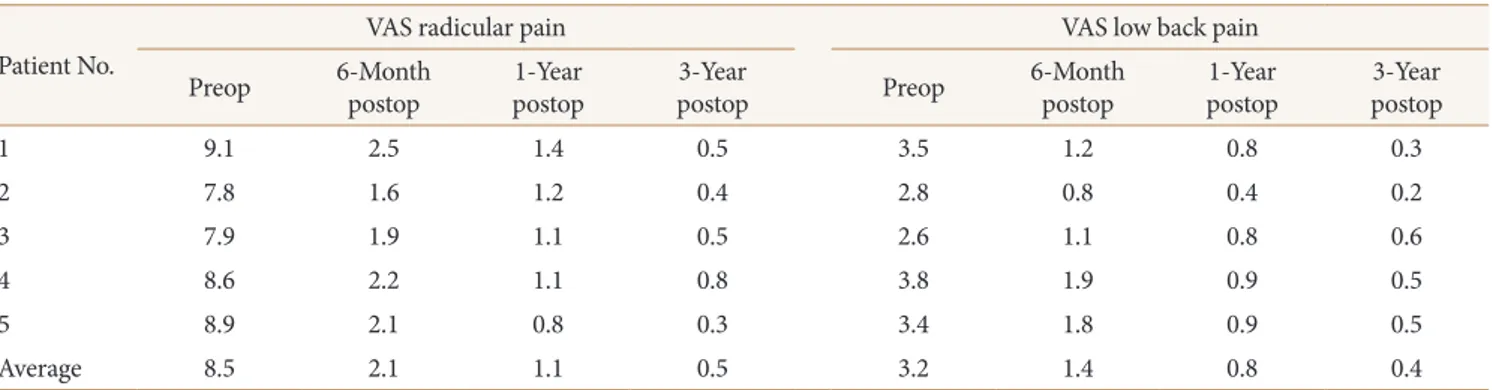
In the sequestrectomy group, the average preoperative VAS score was 8.5 (range, 7.8–9.1) for radicular pain and 3.2 (range, 2.6–3.8) for low-back pain. At the follow-up, the VAS score for radicular pain remarkably decreased reaching 2.1 (range, 1.6– 2.5) at 6 months, 1.1 (range, 0.8–1.4) at 1 year, and 0.5 (range,
0.3–0.8) at 3 years; whereas the VAS for low-back pain were re-ported as follows: 1.4 (range, 0.8–1.9) at 6 months, 0.8 (range, 0.4–0.9) at 1 year, and 0.4 (range, 0.2–0.6) at 3 years (Table 1). The mean preoperative ODI was 33 of 50, 65% (range, 30–34 of 50, 60%–68%), while the mean postoperative ODI at last follow-up (at least 3 years; range, 36 to 51 months) was 5 of 50, 11% (range, 5–6 of 50, 10%–12%) (Table 2).
Both patients with ATM weakness recovered in 2 months, af-ter an intensive rehabilitation period; one patient reported epi-sodes of lumbar or sciatic pain, although significantly reduced compared to before sequestrectomy.
The mean time to return to work was 2.6 months. All patients returned to their previous occupation.
The MRI performed postoperatively ruled out residual or re-current disc herniation in all cases but one, showing persistence of a small disc fragment, without significant conflict with cor-responding nerve root. Of note, this small fragment did not pre-vent the complete resolution of the clinical symptoms.
Using the MacNab Criteria, at 1 year postoperatively, 4 pa-tients were considered in excellent conditions (grade 3), while one patient in good conditions (grade 2). At the last follow-up available (range, 36–51 months), all patients denied radicular
symptomatology and confirmed to be able to carry out their daily living and working activities without pain medication.
DISCUSSION
Although neurosurgeons are nowadays facing pressing re-quests for minimally invasive surgery, conventional microsurgi-cal discectomy still remains a widespread option for treating herniated intervertebral disc. It is well known that the down-sides of open approaches include extensive retraction and dis-section of paraspinal muscles, larger wounds and bone resec-tion. These disadvantages eventually result in slightly longer operative time, blood loss and LOS.
In standard microsurgical lumbar discectomy as described by Caspar and colleagues19,20 more than 30 years ago, the radic-ular decompression is obtained by the removal of the herniated disc followed by curettage of the intervertebral space. In 1978, Williams11 in order to decompress the neural structures with-out entering the disc, described the concept sequestrectomy, which entails only the removal of the migrated disc fragment. The indication for sequestrectomy is very narrow, however this technique is believed to offer significant advantages in terms of minimizing perioperative pain, preserving disc architecture, and protecting against progressive focal disc degeneration.13 In most cases the opening in the annulus fibrosus is expected to seal spontaneously and permanently without further herniation, nonetheless the overall rate of disc recurrence following micro-discectomy is reported to range between 1% and 21%, with a risk that appears to increase proportionally to the degree of an-nular defect, leading some groups to propose in selected cases the use of specific annular closure devices.21-26 In turns, the preservation of the healthy central disc structure is considered crucial to avoid reduction in disc height, decreasing the risk of spinal instability and of postoperative back pain. As such, in
se-Table 1. Preoperative and postoperative VAS scores Patient No.
VAS radicular pain VAS low back pain
Preop 6-Month postop postop1-Year postop3-Year Preop 6-Month postop postop1-Year postop3-Year
1 9.1 2.5 1.4 0.5 3.5 1.2 0.8 0.3 2 7.8 1.6 1.2 0.4 2.8 0.8 0.4 0.2 3 7.9 1.9 1.1 0.5 2.6 1.1 0.8 0.6 4 8.6 2.2 1.1 0.8 3.8 1.9 0.9 0.5 5 8.9 2.1 0.8 0.3 3.4 1.8 0.9 0.5 Average 8.5 2.1 1.1 0.5 3.2 1.4 0.8 0.4
VAS, visual analogue scale; preop, preoperative; postop, postoperative.
Table 2. Preoperative and postoperative Oswestry Disability Index
Patient No. Preoperative, score (%) Long-term postoperative, score (%)
1 34/50 (68) 5/50 (10) 2 32/50 (64) 5/50 (10) 3 30/50 (60) 6/50 (12) 4 33/50 (66) 6/50 (12) 5 34/50 (68) 5/50 (10) Average 33/50 (65) 5/50 (11)
lected cases, this tailored option can provide superior results than standard microdiscectomy. For instance, young patients who present with the radiological features of preserved inter-vertebral height, a disc with otherwise little degenerative chang-es, and nearly intact posterior ligament and annulus fibrosus may be expected to profit from this management option.
Endoscopy has exploded as a successful strategy in various neurosurgical conditions, including spinal surgery.21,22 None-theless, the pros and cons of this MISS variant are still debated: previous reports on PELD suggested that it can provide results comparable to microsurgical technique, allowing for the resolu-tion of sciatica with less paraspinal musculature trauma and smaller surgical accesses.23 Bone removal is often minimal, and this reduces the risks of inducing postoperative spinal instabili-ty; furthermore most PELD variants are reported to cause less epidural bleeding and therefore epidural scarring.23 However, the learning curve is steep, and inexperienced surgeons might encounter perioperative complications and require conversion to open approach: this possibility has to be carefully considered when attempting sequestrectomies, given the risk to get lost and struggle in identifying the extruded fragments between the lig-aments, foramina and neural structures. Given the lack of a su-periority clinical trial, the literature on endoscopic techniques revolves around single centre surgical series indicating that the vast majority of patients were able to return to their previous occupation earlier than those treated with microdiscectomy.24
Given the potential for: (1) a clearer visualization of the mi-grated disc fragments under the guidance of the endoscope, (2) less damage to the paraspinal muscles and other normal tissues, and (3) a reduced patient morbidity with an early return to work; our clinical study was designed to address the research question on the effectiveness of percutaneous interlaminar endoscopic sequestrectomy. Beside the rationale for this technique, already detailed above, another technical aspect deserves attention: it is well known that annular defect size is associated with recurrent disc herniation,25 therefore the simple removal of the migrated disc fragment without formal discectomy is itself a measure to minimize the annular defects or fissures. In fact, our literature search allowed to identify several articles supporting the use of lumbar microscopic sequestrectomy,12,13,27,28 or endoscopic dis-cectomy,22,29-32 nonetheless only few authors proposed endoscop-ic approaches for lumbar sequestrectomies. Of note, some en-doscopic spine systems conceived for posterolateral approach to the lumbar disc through psoas muscle splitting allow for a tailored lateral decompression while sparing the patients from risk of postoperative paraspinal muscles wasting, nonetheless
this surgical route prevents surgeons from being able to per-form selective foraminotomies and sequestrectomies. Other PELD techniques should therefore be considered whenever those are deemed appropriate, in particular microendoscopic discectomy is more appropriate for dorsal pathologies, while interlaminar endoscopic systems allow for an easier approach to lower lumbar segments. In fact, at L4/L5 and L5/S1 the inter-laminar window is large enough to allow optimal visualization of the neural elements and disc material with just a minimal bone removal and very limited resection of the ligamentum fla-vum. Some other variants though exist: in 2005, Suess et al.33 described 41 patients affected by soft extraforaminal disc her-niation who underwent percutaneous endoscopic discectomy using the “extraforaminal targeted fragmentectomy” technique. They reported a good outcome in 39 on 41 patients, concluding that this procedure is safe and effective in selected patients. In 2012 Hirano et al.22 reported a series of 37 patients affected by migrated lumbar disc herniation, treated by targeted fragmen-tectomy through interlaminar, extraforaminal and intraforami-nal approach. The authors postulated that percutaneous endo-scopic interlaminar lumbar discectomy could be considered as safe and alternative procedure to standard approaches in select-ed cases. In 2013, Kim and Park21 reported their experience on a similar approach for disc herniation at L5–S1 and, in order to reduce the rate of recurrence, they proposed the annular seal-ing after fragmentectomy. Based on their experience, Kim and Park21 focused on the relevance of the learning curve and con-cluded that the endoscopic sequestrectomy with annular seal-ing may be a useful technique for reducseal-ing early recurrence. Fi-nally, on 2013, Jasper et al.30 published a case report on the en-doscopic transforaminal removal of the extruded fragment, a technique similar to the one herein described. Similarly to our series, they also reported good results and focused on the po-tential advantages of this technique, such as the maintained spi-nal stability and absence or minimal formation of scar tissue.
The greatest novelty of our study is that it confirms the posi-tive repercussions of endoscopic sequestrectomy in terms of early recovery after surgery, and provides a long-term follow-up without cases of recurrence of disc herniation, instability or failed low back pain. These aspects certainly make the case for the cost benefit effectiveness of this technique.34 As stated by Allen and Garfin,35 although the economic advantages of endo-scopic spinal approaches are yet to be carefully studied, the ex-isting literature suggests its potential as a cost-effective interven-tion, provided that improved clinical outcomes are maintained over time. Although the limited number of cases did not allow
us to perform a proper health economics analysis, the good re-sults recorded in terms of VAS and ODI scores, as well as the excellent degree of satisfaction relying on MacNab criteria up to 51 months following surgery support the consideration of this management option among the spectrum of surgical approach-es available for sequapproach-estered disc fragments. Given the rapproach-espect of muscle and paraspinal tissues, limited intraoperative blood loss, and the short operative time, besides being relevant for all those young patients with sequestered disc fragments, this approach could be potentially suitable in groups of individuals with spe-cial requirements: such as patients with obesity, coagulopathies, or for professional athletes seeking an early return to competi-tions.34-37
Over the years the instrumentation for endoscopic spine sur-gery evolved, and newer models are nowadays available so that beyond the EasyGo system tested in this trial percutaneous in-terlaminar endoscopic approaches can be performed with other full-endoscopic surgical systems. In the future the choice will be even broader due to the continuous competition between producing companies: for the end-user this clearly represents a good news since competition is usually helpful in bringing down costs of initial acquisition or renting. Furthermore, those sys-tems certainly seem to represent a good investment for all spine surgeons performing minimally invasive surgery because of their employability in a number of clinical scenarios beyond the one described in this study. In fact, the endoscopic instru-mentation coupled with other technological aids for minimal invasiveness (neuronavigation, intraoperative ultrasound, etc.) can be used as a viable complement for spinal dysraphisms, de-generative and neoplastic spinal pathologies, including tethered cord, minimally invasive interbody fusion, and surgery for in-tradural tumors.38-40
The major drawback of designing a single centre clinical trial with strict inclusion and exclusion criteria is that such research approach necessarily narrows down the number of potential candidates at time of patient selection. Additionally, if eligible patients prefer not to be enrolled and opt instead for what is considered to be the standard of care, then it is difficult to rule out the influence of a possible selection bias on the results ob-tained in both the treatment and control arms. As such, we be-lieve that the absence of dural tears in the sequestrectomy group does not mean that this technique is far superior in terms of safety when compared to standard PELD approaches, but is simply due to the small sample considered. On the other hand, if the approach tested in this pilot study represents a tailored surgical option, it is also true that a bigger sample size could be
achieved only by keeping the trial open for longer periods, or by reducing the criteria for long-term follow-up. Noteworthy, the greatest usefulness of the data achieved with this small clini-cal trial might become evident when clini-calculating the sample size and power of larger multicentre studies with randomization for percutaneous interlaminar endoscopic sequestrectomy versus other PELD approaches.
CONCLUSION
Although our experience is limited, it provides OCEBM Level 3 evidence that percutaneous interlaminar endoscopic seques-trectomy is a safe and effective option for patients with lumbar radiculopathy caused by single level, migrated lumbar disc her-niation, with preserved intervertebral height, and nearly intact posterior ligament and annulus fibrous. Our long-term follow-up suggests that this option may be considered, in carefully se-lected patients, as a tailored and well tolerated minimally inva-sive technique for removal of migrated lumbar disc herniations. This surgical procedure allows for an immediate relief of the radiculopathy and seems to prevent recurrences. Given the above, and the additional advantages of a shorter LOS and early return to work activities, a comparative cost-effectiveness analysis on percutaneous interlaminar endoscopic sequestrectomies is war-ranted to validate these initial findings.
CONFLICT OF INTEREST
The authors have nothing to disclose.
REFERENCES
1. Zhang Y, Xu C, Zhou Y, et al. Minimally invasive computer navigation-assisted endoscopic transforaminal interbody fusion with bilateral decompression via a unilateral approach: initial clinical experience at one-year follow-up. World Neu-rosurg 2017;106:291-9.
2. Ganau M, Holly LT, Mizuno J, et al. Future directions and new technologies for the management of degenerative cer-vical myelopathy. Neurosurg Clin N Am 2018;29:185-93. 3. Ganau M, Ennas F, Ambu R, et al. Excision of synovial cysts:
pathology matters. J Neurosurg Spine 2013;19:266-7. 4. Chibbaro S, Cebula H, Aldea S, et al. Endonasal endoscopic
odontoidectomy in ventral diseases of the craniocervical junction: results of a multicenter experience. World Neuro-surg 2017;106:382-93.
5. Chibbaro S, Ganau M, Cebula H, et al. The endonasal endo-scopic approach to pathologies of the anterior craniocervi-cal junction: analyticraniocervi-cal review of cases treated at four Euro-pean Neurosurgical Centres. Acta Neurochir Suppl 2019; 125:187-95.
6. Zhang W, Li H, Zhou Y, et al. Minimally invasive posterior decompression combined with percutaneous pedicle screw fixation for the treatment of thoracolumbar fractures with neurological deficits: a prospective randomized study versus traditional open posterior surgery. Spine (Phila Pa 1976) 2016; 41 Suppl 19:B23-9.
7. Pan Z, Ha Y, Yi S, et al. Efficacy of Transforaminal Endoscop-ic Spine System (TESSYS) technique in treating lumbar disc herniation. Med Sci Monit 2016;22:530-9.
8. Lee SH, Kang BU, Ahn Y, et al. Operative failure of percuta-neous endoscopic lumbar discectomy: a radiologic analysis of 55 cases. Spine (Phila Pa 1976) 2006;31:E285-90.
9. Kim JM, Lee SH, Ahn Y, et al. Recurrence after successful percutaneous endoscopic lumbar discectomy. Minim Inva-sive Neurosurg 2007;50:82-5.
10. Choi G, Lee SH, Raiturker PP, et al. Percutaneous endoscop-ic interlaminar discectomy for intracanalendoscop-icular disc hernia-tions at L5-S1 using a rigid working channel endoscope. Neu-rosurgery 2006;58(1 Suppl):ONS59-68.
11. Williams RW. Microlumbar discectomy: a conservative sur-gical approach to the virgin herniated lumbar disc. Spine (Phila Pa 1976) 1978;3:175-82.
12. Baek GS, Kim YS, Lee MC, et al. Fragmentectomy versus conventional microdiscectomy in single-level lumbar disc herniations: comparison of clinical results and recurrence rates. J Korean Neurosurg Soc 2012;52:210-4.
13. Schick U, Elhabony R. Prospective comparative study of lumbar sequestrectomy and microdiscectomy. Minim Inva-sive Neurosurg 2009;52:180-5.
14. Modic MT, Masaryk TJ, Ross JS, et al. Imaging of degenera-tive disk disease. Radiology 1988;168:177-86.
15. Dora C, Wälchli B, Elfering A, et al. The significance of spi-nal caspi-nal dimensions in discriminating symptomatic from asymptomatic disc herniations. Eur Spine J 2002;11:575-81. 16. Chibbaro S, Cebula H, Todeschi J, et al. Evolution of
pro-phylaxis protocols for venous thromboembolism in neuro-surgery: results from a prospective comparative study on low-molecular-weight heparin, elastic stockings, and inter-mittent pneumatic compression devices. World Neurosurg 2018;109:e510-6.
17. Ganau M, Prisco L, Cebula H, et al. Risk of deep vein
throm-bosis in neurosurgery: state of the art on prophylaxis proto-cols and best clinical practices. J Clin Neurosci 2017;45:60-6. 18. Macnab I. Negative disc exploration. An analysis of the causes
of nerve-root involvement in sixty-eight patients. J Bone Joint Surg Am 1971;53:891-903.
19. Caspar W. A new surgical procedure for lumbar disc hernia-tion causing less tissue damage through a microsurgical ap-proach. In: Wüllenweber R, Brock M, Hamer J, et al. Lum-bar disc adult hydrocephalus. Advances in neurosurgery, voulume 4. Berlin: Springer-Verlag; 1977. p. 74-7.
20. Loew F, Caspar W. Surgical approach to the lumbar disc her-niations (the micro-approach to the lumbar disc prolapse). Adv Tech Stand Neurosurg 1978;5:153-71.
21. Kim HS, Park JY. Comparative assessment of different per-cutaneous endoscopic interlaminar lumbar discectomy (PEID) techniques. Pain Physician 2013;16:359-67.
22. Hirano Y, Mizuno J, Takeda M, et al. Percutaneous endo-scopic lumbar discectomy - early clinical experience. Neurol Med Chir (Tokyo) 2012;52:625-30.
23. Peng CW, Yeo W, Tan SB. Percutaneous endoscopic lumbar discectomy: clinical and quality of life outcomes with a mini-mum 2 year follow-up. J Orthop Surg Res 2009;4:20. 24. Natarajan RN, Andersson GB, Patwardhan AG, et al. Effect
of annular incision type on the change in biomechanical properties in a herniated lumbar intervertebral disc. J Bio-mech Eng 2002;124:229-36.
25. Kast E, Oberle J, Richter HP, et al. Success of simple seques-trectomy in lumbar spine surgery depends on the compe-tence of the fibrous ring: a prospective controlled study of 168 patients. Spine (Phila Pa 1976) 2008;33:1567-71. 26. Hahn BS, Ji GY, Moon B, et al. Use of annular closure device
(Barricaid®) for preventing lumbar disc reherniation:
one-year results of three cases. Korean J Neurotrauma 2014;10: 119-22.
27. Kotil K, Köksal NS, Kayaci S. Long term results of lumbar sequestrectomy versus aggressive microdiscectomy. J Clin Neurosci 2014;21:1714-8.
28. Fakouri B, Shetty NR, White TC. Is sequestrectomy a viable alternative to microdiscectomy? A systematic review of the literature. Clin Orthop Relat Res 2015;473:1957-62.
29. Choi KC, Kim JS, Ryu KS, et al. Percutaneous endoscopic lumbar discectomy for L5-S1 disc herniation: transforami-nal versus interlaminar approach. Pain Physician 2013;16: 547-56.
30. Jasper GP, Francisco GM, Telfeian AE. Endoscopic transfo-raminal discectomy for an extruded lumbar disc herniation.
Pain Physician 2013;16:E31-5.
31. Jasper GP, Francisco GM, Telfeian AE. A retrospective eval-uation of the clinical success of transforaminal endoscopic discectomy with foraminotomy in geriatric patients. Pain Physician 2013;16:225-9.
32. Jasper GP, Francisco GM, Telfeian AE. Clinical success of transforaminal endoscopic discectomy with foraminotomy: a retrospective evaluation. Clin Neurol Neurosurg 2013;115: 1961-5.
33. Suess O, Brock M, Kombos T. Motor nerve root monitoring during percutaneous transforaminal endoscopic sequestrec-tomy under general anesthesia for intra- and extraforaminal lumbar disc herniation. Zentralbl Neurochir 2005;66:190-201.
34. Development of an Enhanced Recovery After Surgery (ERAS) approach for lumbar spinal fusion. J Neurosurg Spine 2017; 26:411-8.
35. Allen RT, Garfin SR. The economics of minimally invasive spine surgery: the value perspective. Spine (Phila Pa 1976) 2010;35(26 Suppl):S375-82.
36. Yamashita K, Higashino K, Sakai T, et al. Revision percuta-neous endoscopic lumbar discectomy under the local anes-thesia for the recurrent lumbar herniated nucleus pulposus in a high class athlete: a case report. J Med Invest 2016;63: 135-9.
37. Kapetanakis S, Gkantsinikoudis N, Chaniotakis C, et al. Per-cutaneous transforaminal endoscopic discectomy for the treatment of lumbar disc herniation in obese patients: health-related quality of life assessment in a 2-year follow-up. World Neurosurg 2018;113:e638-49.
38. Ganau M, Syrmos N, Martin AR, et al. Intraoperative ultra-sound in spine surgery: history, current applications, future developments. Quant Imaging Med Surg 2018;8:261-7. 39. Dhandapani S, Karthigeyan M. “Microendoscopic” versus
“pure endoscopic” surgery for spinal intradural mass lesions: a comparative study and review. Spine J 2018;18:1592-602. 40. Youn MS, Shin JK, Goh TS, et al. Full endoscopic lumbar
interbody fusion (FELIF): technical note. Eur Spine J 2018; 27:1949-55.