Emerging diseases of aquatic organisms
Carmelo Iaria
THESIS FOR THE DEGREE OF PHILOSOPHIAE DOCTOR
PHD COURSE IN APPLIED BIOLOGY AND EXPERIMENTAL MEDICINE
UNIVERSITY OF MESSINA DEPARTMENT OF CHEMICAL,BIOLOGICAL,
PHARMACEUTICAL AND ENVIRONMENTAL SCIENCES
University of Messina
THESIS FOR THE DEGREE OF PHILOSOPHIAE DOCTOR
PHD COURSE IN APPLIED BIOLOGY AND EXPERIMENTAL MEDICINE CURRICULUM IN BIOLOGICAL AND ENVIRONMENTAL SCIENCES
XXXII PROGRAM SSD VET/03
Emerging diseases of aquatic organisms
Carmelo Iaria
Supervisor
Professor Fabio Marino
Doctoral Examination Board
Professor Domenico Britti Professor Patrizia Trifilò Professor Vasileios Bakopoulos
Doctor Monique Mancuso
Coordinator: Professor Maria Assunta Lo Gullo ACADEMIC YEAR 2018-2019
To my family,
People who define my world
SOMMARIO
Il presente studio è stato effettuato con l'obiettivo primario di valutare agenti patogeni e malattie nei pesci provenienti da allevamenti di acquacoltura, pesci utilizzati come animali da laboratorio e organismi acquatici selvatici, che sono stati identificati durante il dottorato di ricerca, nell’ambito del progetto di ricerca in Biologia Applicata e Medicina Sperimentale presso il Centro di Ittiopatologia Sperimentale della Sicilia (CISS) e durante l’attività diagnostica routinaria dell’Unità di Patologia Veterinaria dell’Università di Messina.
Sono stati evidenziati gli effetti negativi delle infezioni subcliniche sui risultati della ricerca, sulla salute degli animali utilizzati a fini scientifici, sulla salute degli animali destinati al consumo umano e sulla salute degli organismi acquatici selvatici.
Le malattie dei pesci utilizzati ai fini della ricerca scientifica possono alterare i risultati sperimentali, aumentare la variabilità dei dati e impedire la riproducibilità sperimentale. Le malattie negli organismi di allevamento e selvatici possono causare gravi perdite finanziarie nelle aziende d’acquacoltura e nelle attività di pesca, nonché possono essere indicatori di problematiche ambientali. A tal fine, sono state effettuate analisi istopatologiche su un totale di 3831 organismi acquatici, dei quali 571, appartenenti a sei specie marine (Dicentrarchus labrax, Sparus aurata, Argyrosomus regius, Mugil cephalus, Lithognathus mormyrus, Coelorinchus caelorhincus), otto specie di acqua dolce (Danio rerio, Carassius auratus, Xiphophorus variatus, Poecilia reticulata, Abramis brama, Carassius carassius, Salmo trutta fario e Cobitis bilineata) e due specie di molluschi bivalvi (Pinna nobilis e Pinna rudis), risultavano positivi a differenti patologie.
Numerose malattie sono state riscontrate durante questo studio. In particolare, la micobatteriosi e la mixosporidiosi sono state considerate le patologie più significative nei teleostei destinati alla ricerca scientifica; fotobacteriosi, enteromixiosi, criptocarioniasi e leyomioma sono state considerate le patologie più significative nei teleostei provenienti da impianti di acquacoltura; parassitosi dovute a Clinostomum complanatum, Lophoura edwardsi e Cystidicoloides ephemeridarum, tumori quali papilloma, fibroma e schwannoma e micobatteriosi sono state considerate le patologie più significative negli organismi acquatici provenienti da ambienti selvatici. Questi risultati possono essere utili per ampliare le conoscenze riguardo diverse malattie in varie specie ittiche sia allevate che selvatiche. al fine di comprenderne meglio l’eziologia e i meccanismi di patogenesi, nonché allertare gli operatori del settore sulle emergenze sanitarie nuove e ricorrenti.
ABSTRACT
The present research was undertaken with the prime objective of evaluating pathogens and diseases in fish from aquaculture farms, laboratory fish, and wild aquatic organisms, which were identified during the Ph.D. research project in Applied Biology and Experimental Medicine in the Centre for Experimental Fish Pathology of Sicily (CISS) and the Unit of Veterinary Pathology of the University of Messina.
Adverse effects of subclinical infections on research results, the importance of animal health regarding welfare, the health of animals intended for human consumption and the health of wild aquatic organisms are likewise highlighted. Diseases in fish used for research can alter experimental outcomes, increase the variability of data, and impede experimental reproducibility.
Diseases in farmed and wild organism can cause severe financial losses in aquaculture and fishery companies, as well as being indicators of environmental related distress. For this purpose, 571 diseased fish of different species (out of a total of 3831 fish analysed) that belonged to six marine species (Dicentrarchus labrax, Sparus aurata, Argyrosomus regius, Mugil cephalus, Lithognathus mormyrus, Coelorinchus caelorhincus), to eight fresh water species (Danio rerio, Carassius auratus, Xiphophorus variatus, Poecilia reticulata, Abramis brama, Carassius carassius, Salmo trutta fario and Cobitis bilineata) and two species of bivalve molluscs (Pinna nobilis and Pinna rudis) were examined for histopathological health monitoring. Several diseases have been found during this study. In particular, mycobacteriosis and myxosporidiosis were seen to be the most significant diseases in our research aquatic organisms; photobacteriosis, enteromyxiosis, cryptocarioniasis and leyomioma were seen to be the most significant diseases in our aquaculture aquatic organisms; Clinostomum complanatum, Lophoura edwardsi and Cystidicoloides ephemeridarum parasitosis, papilloma, fibroma and schwannoma tumours and mycobacteriosis were seen to be the most significant diseases in our wild aquatic organisms. These results may be useful in gathering wider knowledge on the occurrence of several diseases in various species and the environment, as well as in alerting industry operators to new and recurring health emergencies. In this way, the pathogenic mechanisms of aetiological agents may be better understood.
ACKNOWLEDGEMENTS
The accomplishment of this thesis work was made possible due to the participation of several co-workers, and different research teams and laboratories.
First of all, I would like to thank my supervisor, Professor Fabio Marino, master of life, science and method, for all the teachings, support and his driving me every day on this path. My sincere thanks also to the PhD Course Coordinator, Professor Maria Assunta Lo Gullo, for help and guidance. To Professor Nunziacarla Spanò, for having been my go-to person during all my student career. Professor Giovanni Lanteri, master of techniques and laughter mate. Professors Battesimo and Francesco Macrì, as well as Professor Giuseppe Mazzullo for all support given during the last few years. I further acknowledge the grant obtained in 2016 from the University of Messina to support my PhD project.
The support and contribution of research teams of veterinary pathology and marine ecology of the University of Messina, in particular Dr. Fabiano Capparucci, Dr. Giovanni De Benedetto, Dr. Sabrina Natale, Dr. Serena Savoca, Dr. Gioele Capillo and Dr. Giuseppe Panarello, in all kinds of daily laboratory activities and sampling procedures, was essential to the success of this thesis.
I am really grateful to all the staff of Keldur, Institute for Experimental Pathology, University of Iceland, Reykjavik, for letting me have such a great, precious time during my PhD abroad internship, in learning skills relating to fish pathology and immunology. A warm thanks to my internship supervisor, Dr. Birkir Þór Bragason, master in fish immunology and molecular biology, for all his teachings, kindness and helpfulness, to Dr. Árni Kristmundsson and Dr. Sigríður Guðmundsdóttir for having kindly pre-reviewed my thesis.
I would also like to thank all co-authors of papers involved in this study, and in particular research teams from the University of Messina in the persons of Professors Gabriella Gaglio, Giuseppe Piccione, Francesco Fazio, Antonio Ieni and Domenico Trombetta; the University of Naples, in the persons of Professor Francesca Carella; the University of Catania, in the person of Professor Maria Violetta Brundo; the Experimental Zooprophylactic Institute of Sicily, in the person of Dr. Daniele Macrì and the Biological Station Anton Dohrn of Messina in the persons of Dr. Teresa Bottari for all support and collaboration in research activities and all analysis provided during the last few years.
I sincerely thank the Reviewers, Professor Giovanni Di Guardo, Professor Gionata De Vico, Professor Jorge Manuel de Oliveira Fernandes and Doctor Sepand Rastegar and the Doctoral Examination Board, Professor Domenico Britti, Professor Patrizia Trifilò, Professor Vasileios
Bakopoulos, and Doctor Monique Mancuso for their interest, expertise contribution and constructive criticism.
A proper thanks is due to journal editorial boards for allowing permission to use the scientific papers to support this thesis, in particular: to the Laboratory Animals SAGE journal, to the Zebrafish journal and Mary Ann Liebert, Inc. publisher, to the Journal of Aquatic Animal Health and The American Fisheries Society, to the Cahiers De Biologie Marine and Station Biologique de Roscoff and to the International Journal of Environmental Research and Springer Nature publisher.
Finally, and not least, my parents for their patience and support during the doctoral years. Also, I wish to express my gratitude to other family members and friends for their encouragement
.
DECLARATION OF CONTRIBUTION
I was responsible for the conception and management of the thesis project. The thesis project (2016-2019) is supported by seven scientific papers and was a joint effort of several research teams from different institutes, mainly: Department of Chemical, Biological, Pharmaceutical, and Environmental Sciences and Department of Veterinary Science, University of Messina; Department of Biology, University of Naples Federico II; Department of Biological, Geological and Environmental Sciences, university of Catania; Zooprophylactic Institute of Sicily (IZS) and Biological Station Anton Dohrn of Messina. I had a major role in the study design and execution of the experiments, in sampling, in the acquisition of most data, their analysis and interpretation as well as in the structuring and writing of the manuscripts in which I am the first author. I particularly worked on all the histopathology and laboratory procedures.
Although permission has been obtained from the journals in using the data to support the thesis, I am responsible for any plagiarism; the thesis was verified with the software Plagiarism Checker X 2019, showing a percentage of similarity lower than 10%, using a five consecutive words method.
TABLE OF CONTENTS
SOMMARIO ... II ABSTRACT ... III ACKNOWLEDGEMENTS ... IV DECLARATION OF CONTRIBUTION ... V TABLE OF CONTENTS ... VI LIST OF ABBREVIATIONS ... IX LIST OF TABLES ... XII LIST OF FIGURES ... XIII LIST OF ORIGINAL PAPERS... XVI1. INTRODUCTION ... 1
1.1 HEALTH MONITORING OF LABORATORY FISH ... 1
1.2 HEALTH MONITORING OF FISH FROM AQUACULTURE... 3
1.3 HEALTH MONITORING OF WILD AQUATIC ORGANISMS ... 6
1.4 COMMON DISEASES OF AQUATIC ORGANISMS... 8
1.4.1 Congenital abnormalities ... 8 1.4.2 Skeletal deformities...10 1.4.3 Bacterial diseases ...13 1.4.3.1 Gram-negative ...16 1.4.2.2 Gram-positive ...24 1.4.4 Viral diseases...27 1.4.3.1 Aquabirnavirus ...28 1.4.3.2 Betanodavirus ...28
1.4.3.3 Infectious Salmon Anaemia Virus ...29
1.4.3.4 Salmonid Alphavirus ...30
1.4.3.5 Infectious Hematopoietic Necrosis Virus...30
1.4.3.6 Epizootic Hematopoietic Necrosis Virus ...31
1.4.3.7 Viral Haemorrhagic Septicaemia Virus ...32
1.4.3.8 Spring viremia of carp. ...32
1.4.3.9 Koi herpesvirus disease...33
1.4.3.10 Lymphocystis disease ...33 1.4.5 Parasitic diseases ...35 1.4.5.1 Protista ...35 1.4.5.2 Myxozoa ...36 1.4.5.3 Trematoda...37 1.4.5.4 Cestoda ...39 1.4.5.5 Nematoda ...40 1.4.5.6 Arthropoda...41 1.4.5.6.1 Copepods ...42 1.4.5.6.2 Isopods ...42 1.4.5.6.3 Branchiurans ...43 1.4.6 Tumours ...44 1.4.6.1 Epithelial tumours ...44 1.4.6.2 Mesenchymal tumours ...45
1.4.6.3 Peripheral nerve sheath tumours ...45
1.4.6.4 Pigmented cells tumours ...46
1.4.7 Diseases of molluscs ...47 1.4.7.1 Marteliosis ...52 1.4.7.2 Bonamiosis ...53 1.4.7.3 Perkinsosis ...54 1.4.7.4 Haplosporidiosis ...55 1.4.8 Zoonoses ...56
2. AIMS OF THE STUDY ... 59
3.1 MATERIALS ...61
3.1.1 Samples from laboratory fish ...61
Fish from CISS laboratory ...61
Zebrafish from aquarium shop...62
Xiphophorus hybrid ...62
3.1.2 Samples from aquaculture ...62
Fish from aquaculture ...62
Dicentrarchus labrax from broodstock ...63
3.1.3 Samples of wild aquatic organisms ...63
Fish from natural environment ...63
Cobitis bilineata from northern Italy...63
Coelorinchus caelorhincus from southern Sicily ...64
Salmo trutta fario from Calabria (southern Italy) ...64
Wild fish presenting unusual masses ...65
Pen Shell from Tyrrhenian sea ...66
3.2 METHODS ...69
3.2.1 Anaesthesia ...69
3.2.2 Euthanasia ...69
3.2.3 Gross pathological examination ...69
3.2.4 Necropsy...70
3.2.5 Radiographic examination ...70
3.2.6 Soft tissue digestion ...71
3.2.7 Histology ...71
3.2.8 Immunohistochemistry ...71
3.2.9 Scanning Electron Microscopy (SEM) ...72
3.2.10 Transmission Electron Microscopy (TEM) ...72
3.2.11 Bacteriological examination...72
3.2.12 Molecular Analysis ...72
C. formosanus DNA isolation and PCR amplification ...72
C. complanatum DNA isolation and PCR amplification ...73
P. nobilis DNA isolation and PCR amplification ...74
Mycobacterium detection in fish ...76
Lymphocystivirus detection...77
3.2.13 Elemental analysis ...77
3.2.14 Gamma Spectroscopy...77
3.2.15 Radioactivity investigation ...78
3.2.16 Mineral contents by AAS ...78
3.2.17 Parasitological analysis ...79
3.2.18 Statistical Analysis ...80
4. RESULTS ... 81
4.1 DISEASES OF LABORATORY FISH...81
Health monitoring in fish from CISS laboratory ...81
Centrocestus formosanus infection in zebrafish ...83
Melanoma in Xiphophorus hybrid ...86
4.2 DISEASES OF FISH FROM AQUACULTURE ...87
Health monitoring in fish from aquaculture ...87
Leyomioma in Dicentrarchus labrax ...90
4.3 DISEASES OF WILD AQUATIC ORGANISMS ...93
Health monitoring in fish from natural environment ...93
Clinostomum complanatum infection in Cobitis bilineata ...94
Lophoura edwardsi infection in Coelorinchus caelorhincus ...96
Cystidicoloides ephemeridarum infection in Salmo trutta fario ... 102
Tumours in wild fishes ... 105
Mycobacteriosis in Pen Shell... 109
5. DISCUSSION ...116
5.1 DISEASES OF LABORATORY FISH...116
Health monitoring in fish from CISS laboratory ... 116
Centrocestus formosanus infection in zebrafish ... 117
5.2 DISEASES OF FISH FROM AQUACULTURE ...118
Health monitoring in fish from aquaculture ... 118
Leyomioma in Dicentrarchus labrax ... 119
5.3 DISEASES OF WILD AQUATIC ORGANISMS ...119
Health monitoring in fish from natural environment ... 119
Clinostomum complanatum infection in Cobitis bilineata ... 120
Lophoura edwardsi infection in Coelorinchus caelorhincus ... 121
Cystidicoloides ephemeridarum infection in Salmo trutta fario ... 122
Tumours in wild fish ... 125
Mycobacteriosis in Pen Shell... 127
6. CONCLUDING REMARKS AND FUTURE PERSPECTIVES ...130
7. REFERENCES ...132
LIST OF ABBREVIATIONS
AIDS Acquired immune deficiency syndrome
API Analytical profile index
Asa Aeromonas salmonicida subsp. Achromogenes BCWD Bacterial Cold-water Disease
BGD Bacterial gill disease BKD Bacterial kidney disease BHI Brain heart infusion
BPNSTs Benign peripheral nerve sheath tumours
CISS Centro di Ittiopatologia Sperimentale della Sicilia
COI Cytochrome c oxidase I
DHA Docosahexaenoic acid
DNA Deoxyribonucleic acid
DO Dissolved oxygen
dpf Days post fertilization
EB Elementary body
EHN Epizootic hematopoietic necrosis EHNV Epizootic hematopoietic necrosis virus
EM Electron microscopy
ENPA Ente Nazionale Protezione Animali
EPA Eicosapentaenoic acid
ERM Enteric red mouth
EU European Union
FAO Food and Agricultural Organization FDA Food and Drugs Administration
GF-AAS Graphite furnace atomic absorption spectrometry
GFCM General Fisheries Commission for the Mediterranean Sea GLP Good laboratory practice
GSA Geographical sub-area
GW Gutted weight
H&E Haematoxylin-eosin
ha Hectare
HE Hemagglutinin-esterase
HIRRV Hirame rhabdovirus
HIV Human immunodeficiency virus
HSV Haemorrhagic septicaemia virus
IB Intermediate body
IF Immune fluorescence
IHN Infectious hematopoietic necrosis IHNV Infectious hematopoietic necrosis virus IPN Infectious pancreatic necrosis
IPN Infectious pancreatic necrosis IPNV Infectious pancreatic necrosis virus
ISA Infectious salmon anaemia
ISPRA Istituto Superiore per la Protezione e la Ricerca Ambientale ITS Internal transcribed spacer
KHV Koi herpesvirus
Krel Relative condition factor
LCD Lymphocystis disease
LCDV Lymphocystis disease virus
LHRH Luteinizing hormone-releasing hormone
LIC Long intermediate cell
LPC Long primary cell
MABV Marine aquabirnavirus
MAS Motile aeromonad septicaemia MCA Multi channel analyzer
MDA Minimum detectable amount
MMEs Mass mortality events
MPA Marine protected area
MPNSTs Malignant peripheral nerve sheath tumours
MRL Maximum residue limit
MRSA Methicillin-resistant S. aureus MS-222 Tricaine methanesulfonate
MSX Multinucleate Sphere unknown X
MT Masson's trichrome
MTCB Mycobacterium tuberculosis complex mtDNA Mitochondrial deoxyribonucleic acid NTM Non-tuberculous mycobacteria
OIE World Organization for Animal Health
PAL Pre-anal length
PAS Periodic acid Schiff PBS Phosphate-buffered saline PCBs Polychlorinated biphenyls PCR Polymerase chain reaction
PD Pancreas disease
PKD Proliferative kidney disease PNSTs Peripheral nerve sheath tumours
PV Parasitophorous vacuole
PWTs Perivascular wall tumours
QX Queensland Unknown disease
RB Reticulated body
ROS Reactive oxygen species
rRNA Ribosomal ribonucleic acid
RT Room temperature
RTFS Rainbow trout fry syndrome
SAV Salmonid alphavirus
SC Small cell
SDV Sleeping disease virus
SHK-1 Infection-susceptible salmon cell line SPF Specific pathogen free
SPVD Salmonid pancreas disease virus SSO Seaside organism disease
ssRNA Single-stranded ribonucleic acid SVC Spring viremia of carp
SVCV Spring viremia of carp virus SVCV Spring viremia of carp virus
TCBS Thiosulfate citrate bile salts sucrose TEM Transmission electron microscopy
TW Total weight
VER Viral encephalopathy and retinopathy VHS Viral Haemorrhagic Septicaemia VHS Viral haemorrhagic septicaemia VHSV Viral haemorrhagic septicaemia virus VNN Viral nervous necrosis
W-E Longitudinal gradient
WD Whirling disease
WHO World Health Organization
XRF X-ray fluorescence
LIST OF TABLES
Table 1. Summary of pathogenic Gram-negative bacteria and their hosts reported in literature
102 ... 14
Table 2. List of pathogenic Gram-positive bacteria and their hosts reported in literature 102 .. 14
Table 3. Commercial vaccines available against major infectious bacterial and viral diseases of fish 105 ... 15
Table 4. Infectious diseases of fish against which vaccines are not available 105 ... 15
Table 5. Summary of potential agents of fish-borne zoonosis 547 ... 56
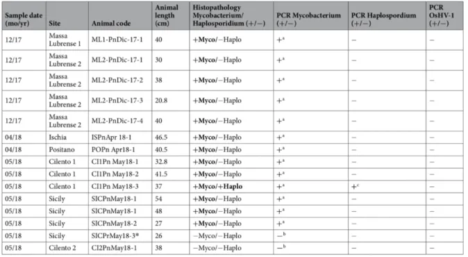
Table 6. Results of PCR analysis of specimens of P. nobilis. a) Moribund animal; with presence of Ziehl-Neelsen-positive bacteria. b) Non- moribund animal. c) Liquid cyst at digestive tissue level. * Specimen of P. rudis586. ... 68
Table 7: IHC antibody details ... 72 Table 8. Spontaneous diseases registered for each species ... 81 Table 9. Evidence of the 517bp amplicon for the 4 positive zebrafish to C. formosanus596. ... 86
Table 10. Measurement of ten metacercariae after surgical removal from C. bilineata. Length and width of oral sucker, length and width of ventral sucker and distance between suckers613.
... 96 Table 11. List of hauls with relative depth range (m) in sampling sites, in Southern Sicily580.
... 96 Table 12. C. caelorhincus. Length and weight of individuals collected from experimental bottom trawl surveys in the south of Sicily. PAL: pre-anal length; TW: total weight; GW: gutted weight; N: number of specimens; SD: standard deviation580. ... 100
Table 13. Summary of results on the specificity of immunoreaction of each antibody in relation to each tumour type612. ... 109
Table 14. Discrimination between neurofibromas and schwannomas depending on tissue patterns612... 126
Table 15. Discrimination between neurofibromas and schwannomas depending on immunohistochemistry antibody activity612. ... 126
LIST OF FIGURES
Figure 1. CISS laboratory ... 60
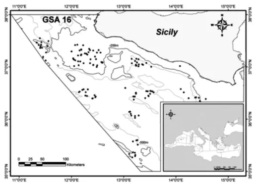
Figure 2. Map of the sampling area (GSA 16, South of Sicily). Haul positions are indicated in grey circles580. ... 64
Figure 3. Map of the two sampling areas in the Tyrrhenian Sea (A, B) (black square) with sampled sites (6 red dots). Mortality reports from SCUBA divers and IUCN are underlined in red. Green lines denote the location of the AMP Punta Campanella586. ... 66
Figure 4. Dead specimens of P. nobilis in Tyrrhenian Sea, Tharros, (OR, Sardinia) over the phanerogame Cymodocea nodosa586. ... 67
Figure 5. 2% agarose gels stained with SYBR® Safe DNA gel stain (Invitrogen)596. ... 73
Figure 6. Neighbour-joining tree of the 16S rDNA sequences. Analysis involved 22 nucleotide sequences and 882 positions in final dataset. Percentages of bootstrap replicates are shown next to the branches. Red asterisks (*) and pink square indicate sequences obtained in this work (ML: Massa Lubrense; SIC: Sicily; PO: Positano; IS: Ischia; CI: Cilento). Blue square and Table show reported cases from literature on mycobacteriosis in mollusc species586. ... 75
Figure 7. Neighbour-joining tree of the 18S rDNA sequence of haplosporidian parasite. Analysis involved 20 nucleotide sequences in final dataset. Percentages of bootstrap replicates are shown next to branches. Red asterisk (*) indicates sequence obtained in this work586. ... 76
Figure 8. Evidence of Myxobolus sp. in C. auratus (May Grunwald Giemsa 40x)611. ... 82
Figure 9. Schwannoma in C. auratus. ... 83
Figure 10. Metacercaria cysts in zebrafish gills. Several nodular lesions in the gills. Fusion of two primary lamellae and thickening and distortion of the primary lamellae. Newborn tissue included several secondary lamellae (arrows), characterized by slight fibroblastic hyperplasia surrounding a central cavity containing parasitic body residues (H&E 10x)611. ... 84
Figure 11. Details of metacercaria inside parasitic cyst (asterisk). Chondrocytes forming the host cyst wall (arrow) (H&E 20)596. ... 85
Figure 12. Details of metacercaria suckers (arrows) (H&E 40)596. ... 85
Figure 13. Xiphophorus spp. hybrid melanoma: (a) lobular brown mass evident on the right dorsal side, close to the dorsal fin; (b) (H&E) confirming the derma (arrow) origin of the neoplastic tissue (asterisk); (c) Melan-A (MEL A) antibody (asterisk) positive reaction612.... 87
Figure 14. Photobacteriosis in seabass. Note enlarged spleen with necrotic/granulomatous foci611. ... 87
Figure 15. Hypertrophic dermal fibroblast with viral cytoplasmic inclusions (H&E 40x)611. . 88
Figure 16. Evidence of E. leei ... 89
Figure 17. Granuloma in A. regius ... 90
Figure 18. Sea bass gastric tumour. ... 90
Figure 19. Section of mass showing slightly lobular, lardaceous appearance, with empty cavities inside. ... 91
Figure 20. Tumour spindle cells arranged in a parallel orientation in interlacing bundles (H&E 20x) ... 92
Figure 21. Muscle fibers surrounding the tumour tissue (MT 20x) ... 93
Figure 22. a). alpha smooth muscle actin (α-SMA) positive reaction; b). vimentin positive reaction; c). desmin positive reaction; d). S100 negative reaction. ... 93
Figure 23. Macroscopical features of the yellow grub disease in C. bilineata613... 94
Figure 24. Clinostomum complanatum at histological analysis. Scale bar 1mm613. ... 95
Figure 25. Map of positive hauls in the trawl survey580. ... 97
Figure 26. Inflammatory infiltrate (asterisk) that deepens in the musculature from the implant point (arrow) (H&E 2.5x)580. ... 98
Figure 27. C. caelorhincus. Histological section of skeletal muscle with portions of the cephalosome (arrow) of L. edwardsi provoking a deep granulomatous reaction (asterisks) (H&E 10x)580. ... 99
Figure 28. C. caelorhincus. Detail of the cephalosome (arrow) of L. edwardsi embedded within a granulomatous (asterisk) tissue reaction in the deep portion of the skeletal musculature (H&E 20x)580. ... 99
Figure 29. C. caelorhincus. Parasite prevalence (P%) as a function of the pre-anal length (PAL)580. ... 100
Figure 30. C. caelorhincus. Prevalence (P%) of L. edwardsi in the South of Sicily580. ... 101
Figure 31. C. caelorhincus. Prevalence (P%) of L. edwardsi as a function of depth (m)580. . 101
Figure 32. Trout showing skeletal deformities ... 102 Figure 33. X-ray photographs showing: a) Six trout with the most severe skeletal deformities from the first two stations; b) Trout coming from the third station with no skeletal deformity ... 103 Figure 34. Histological evidence of skeletal deformity with specular arrangement of contiguous vertebrae showing irregular mineralization as well as unequal bone and cartilage disposition (H&E 5x). ... 103 Figure 35. C. ephemeridarum head (a) and tail (b) and SEM detail. ... 105 Figure 36. Schwannoma in a crucian carp: (a) yellow, compact mass in lateral profile of the lower jaw on the right side of the head (arrow); (b) H&E staining, showing densely packed cells with a storiform pattern typifying Antoni A tissue (label A) was characteristic for most of the tumour masses, although some scanty, almost acellular areas characterizing Antoni B tissue (label B) were also seen; and (c) immunohistochemistry showed strong immunoreactivity for (S100)612. ... 106
Figure 37. Papilloma in a Bream: (a) irregularly round, white, firm mass on the right side of the caudal fin (arrow), with the tissue seeming to emerge from the epidermis, growing out and modifying a portion of the fin; (b) H&E stained histological section, showing a thin connective tissue bundle giving a lobulated aspect (arrow); and (d) strong positive pan-cytokeratin (CK) reaction in the cell membrane of tumour cells612. ... 107
Figure 38. Dermal fibroma in a sand steenbras: (a) multi-centric round mass (arrow), extending on right side of the fish, was smooth on the surface and white-grey, with widespread haemorrhages (asterisks); (b) H&E stained histological section, showing irregularly interspersed spindle cells (arrow), with scanty extracellular matrix; (c) Masson’s trichrome (MT) stained section, confirming the collagen-producing nature of tumour cells; (d) widespread immunohistochemical positivity for vimentin (VIM); and (e) scattered immunoreactivity for muscle actin (ACT; arrows)612. ... 108
Figure 39. Macroscopic appearance of P. nobilis sampled from different areas of Campania. (A) Gill (G) oedema (arrowheads) in samples from Massa Lubrense 1 in December 2017, posterior view. (B) Evident liquid cyst located on the right side of the digestive tissue (DT-arrowheads) and details (insert) of the atrophic and yellowish digestive glands (arrowhead) - frontal view; Go: gonad; Mu: muscle: M: Mantle; By: byssus; K: kidney586. ... 110
Figure 40. Microscopical observation of Mycobacterium sp. within P. nobilis immune cells in the bivalve tissues (arrowheads) on H&E (a and c) and Ziehl-Neelsen stain (b,d–f). (A,B) Connective tissue demarcating gonads presenting nodules filled with Ziehl-Neelse-positive bacteria (arrowheads) (C,D). details of the inflammatory nodules containing bacteria (*) spreading in association with the immune cells and brown cells (Br) (E,F). presence in the mantle epithelium (E) of the mycobacteria (arrowheads); Br: Brown cells586. ... 111
Figure 41. Ziehl-Neelsen-positive mycobacteria (arrowheads) spreading within the digestive gland. DT: digestive tubules; V: vessels; *Inflammation586. ... 112
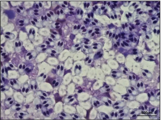
Figure 42. Microscopical observation of the only specimens of P. nobilis parasitized by Haplosporidium sp. Digestive tissue containing high numbers of the pre-sporulation and sporulation stages (arrowheads); DT: digestive tissue. H&E stain586. ... 113
Figure 43. Transmission electron microscopy (TEM) of Mycobacterium sp. (white arrows) free in the cytoplasm of immune cells (black arrowheads) with visible nuclei (N). In the inset: details
of the rod-shaped Mycobacterium with cell walls and containing large vacuoles (arrowhead). Gr: immune cell granules; M: melanin586. ... 114
Figure 44. Histogram showing correlation between length (cm) of trout with skeletal deformities and number of intestinal parasites found in each specimen. ... 123 Figure 45. Histogram showing correlation between parasite burden and calcium levels in samples from the first two stations. ... 124
LIST OF ORIGINAL PAPERS
I. Iaria, C., Saoca, C., Guerrera, M. C., Ciulli, S., Brundo, M. V., Piccione, G., & Lanteri, G. (2019). Occurrence of diseases in fish used for experimental research. Laboratory animals, DOI: 10.1177/0023677219830441.
II. Iaria, C., Migliore, S., Macri, D., Bivona, M., Capparucci, F., Gaglio, G., & Marino, F. (2019). Evidence of Centrocestus formosanus (Nishigori, 1924) in Zebrafish (Danio rerio). Zebrafish, DOI: 10.1089/zeb.2019.1744.
III. Iaria, C., Ieni, A., Corti, I., Puleio, R., Brachelente, C., Mazzullo, G., & Lanteri, G. (2019). Immunohistochemical Study of Four Fish Tumors. Journal of Aquatic Animal Health, 31(1).
IV. Caridi, F., Belvedere, A., D’Agostino, M., Marguccio, S., Scopelliti, F., Iaria, C., & Belmusto, G. (2018). An Investigation on a Possible Correlation Between Skeletal Anomalies of Salmo trutta fario Samples and Internal and External Exposure to Ionizing Radiation. International Journal of Environmental Research, 12(4), 413-417.
V. Bottari, T., Profeta, A., Massi, D., Titone, A., Mobilia, V., Iaria, C., & Lanteri, G. (2018). Lophoura edwardsi (Sphyriidae: Siphonostomatoida), a parasite of Coelorinchus caelorhincus (Macrouridae) from the Central Mediterranean. Cahiers De Biologie Marine, 59(6), 563-569.
VI. Gaglio, G., Reina, V., Caffara, M., Gjurčević, E., Iaria, C., & Marino, F. (2016). Risk of introduction of Clinostomum complanatum (Digenea: Clinostomidae) to Sicily through use of Cobitis bilineata (Canestrini, 1865) as live baits. Bulletin of the European Association of Fish Pathologists, 36(3), 105-110.
VII. Carella, F., Aceto, S., Pollaro, F., Miccio, A., Iaria, C., Carrasco, N., ... & De Vico, G. (2019). A mycobacterial disease is associated with the silent mass mortality of the pen shell Pinna nobilis along the Tyrrhenian coastline of Italy. Scientific reports, 9(1), 2725.
1. INTRODUCTION
1.1 Health monitoring of laboratory fish
Nowadays, there is an increasing interest in fish used for research, especially in zebrafish (Danio rerio) and medaka (Oryzias latipes). Although they are primitive vertebrates, they have shown several advantages in comparison to other animal models as they show particular similarity with human genome and physiology1. This makes them good animal models for
several human pathologies. Moreover, they are easy to breed, fecund and ovulation can be controlled by modifying the day/night cycle. Spawning takes place frequently and there is no limitation to the spawning season. Microinjection of fertilized eggs is relatively easy and cheap. As embryos are transparent, the dynamic gene expression in several tissues and organs in vivo can be monitored, and thus the specimens need not be sacrificed. They are considered the only vertebrates suitable for large-scale mutagenesis as genome sizes are roughly 20-40% of the mammalian genome. Maturation time is a mere 2 ~ 3 months, making it less laborious and quicker for generating transgenic lines. Moreover, several routine techniques of molecular biology and genetics, including knock-in, knockdown and knockout, are well developed in the model fish. Thus, zebrafish and medaka offer novel animal systems for studying vertebrate-specific biology and toxicology2–6. Zebrafish has been shown to be a good model to study
several human diseases, such as congenital and hereditary disease, carcinogenesis, heart disease, Alzheimer’s, leukaemia, Duchenne muscular dystrophy and others1,7,8. Other fish
species are good animal model for different diseases, e.g. Carassius auratus characterises a perfect study model for PNST tumours in fish with particular emphasis on schwannoma9.
Due to this growing interest in using fish for biomedical research, both for human health and aquaculture purposes, it is necessary to focus on health monitoring of fish used as laboratory animals to insure the welfare of these animals and to not compromise results coming from experimental research. Several pathogens are responsible for diseases that can occur in fish husbandry and these could influence research by confounding experimental results. For these reasons, health surveillance of animals used for research is mandatory10.
It has also been reported that many infectious agents can compromise fish welfare status, thereby reducing the breeding performance of animals which are frequently used in laboratory research11.
To date, there is little knowledge on pathogens of fish compared to mice, rats and rabbits and only scanty information on pathological surveys on fish health status during experimental trials. Inadequate attention to health management of laboratory fish, including a lack of suitable
pathogen control in research institutions, is apparent from the relative deficiency of information regarding fish health status, despite the increasing use of fish in experimental trials7,12–16.
Current European Union (EU) Directive 63/2010 aims at improving health management strategies in laboratory fish.
There are only few studies on the incidence and impact of some microorganisms on animal health to date17,18.
The effects of Pseudoloma neurophilia have been studied19,20 and characterized as one of the
most important pathogens in zebrafish husbandry, affecting the nervous system.
Studies on Mycobacterium marinum were carried out in 2009 by Broussard et al.,21 Hegedus et
al. 22 and van der Sar et al.23 Mycobacterial infections, frequently isolated from laboratory fish,
impact greatly on zebrafish health and transcriptome responses24. However, data on pathogens
in other species of laboratory fish are quite scanty.
Today, attention to the application of health monitoring strategies and the use of specific pathogen free (SPF) animals is becoming mandatory in the field of fish research25.
Currently, with the exception of zebrafish and medaka, fish species used for research come from aquaculture plants, and for this reason the absence of pathogens in these fish cannot be guaranteed26–28.
Nevertheless, some SPF zebrafish lines can be obtained from authorized fish production facilities. Pathogen control strategies should be improved to prevent the entry and spread of noxious agents. Thus, more research on the prevalence and distribution of pathogens in fish research plants is suggested. Experimental result predictivity and a decrease in the number of animals used could be improved through evaluation of the effects on anatomy and physiology of fish used in research, when infectious agents are present.
1.2 Health monitoring of fish from aquaculture
Aquaculture is the practice that deals with the breeding of aquatic organisms such as fish, molluscs and crustaceans in a human-controlled environment. It is a very important zootechnic sector, in the production of both freshwater and marine species.
European aquaculture includes the breeding of several fish species, crustaceans and molluscs, but more than 90% of the production is based on the breeding of trout, Atlantic salmon (Salmo salar), European sea bass (Dicentrarchus labrax) and sea bream (Sparus aurata), while for molluscs, the most bred are mussels and clams. Progress in aquaculture in the last few decades has permitted to increase the production and consumption of farmed fish by humans. Between 1961 and 2016, the average annual increase in global fish consumption, destined for human consumption, grew by 1.6%. Moreover, in the last ten years, total aquaculture production has grown by 30% worldwide29. Global aquaculture production almost reached the global total
capture of fisheries production (88%), with a total production of 80 million tonnes out of 90.9 million tonnes of fisheries production in 2016, as seen from the FAO capture database.
The importation of salmon from northern European countries, has exercised strong competition all over Europe.
In recent years, however, greater diversification of the market is underway. In southern Europe, the market for freshwater fish is slowly increasing thanks to the improvement of breeding techniques. It is possible to breed cold water species, even if difficulties due to the high temperature ranges restrict the field to species resistant to strong thermal seasonal excursions. There are various types of aquaculture, which are generally grouped into three categories: extensive, semi-intensive and intensive30.
In extensive breeding the food is totally environmentally responsible and requires large extensions, for example: protected coastal areas, coastal lagoons, ponds and lakes but the production is low, about 10-200kg per ha30.
Semi-intensive farming represents a development of the extensive system. The increase in breeding subjects depends not only on natural food but also on that administered by humans with the function of supplementing the diet. It needs spaces typical of extensive farming, but it can also make use of tanks on shore, and production is measured in tons per ha.
Finally, intensive breeding is totally controlled by man, and food is administered exclusively from breeders. Moreover, the main chemical-physical parameters of the breeding waters are controlled externally. There may be land-based systems and offshore installations (mariculture). With intensive aquaculture, in which high densities are reached, there has been an increase in health problems due to the worsening of environmental parameters, in an effort
to increase production and economic revenues of companies. This has led to the emergence of new diseases and the difficulty of treating them, due, in part, to legislative issues, and partly to issues related to food safety.
With the increase of intensive aquaculture, to increase annual production and therefore also income, there has been a significant increase in pathologies, so much so as to strongly affect the economic efficiency of fishing companies. Losses due to infectious diseases can seriously affect breeding up to the total loss of production lots.
To address this issue, once a pathology has been identified, it is possible to regulate drug administration following EU 37/2010 legislative decree31, which defines the maximum residue
limit (MRL) as the maximum concentration of residues resulting from the use of a veterinary medicinal product permitted by law and recognized as acceptable in foods. MRLs vary according to the drug and species treated. The determination of suitable MRLs makes it possible to calculate the waiting times for the slaughter of the treated subjects. The decree also states that some substances such as Chloramphenicol and Nitrofurans are prohibited due to possible side effects in humans or because they are potentially carcinogenic, for which an MRL has not been established. In aquaculture, the use of disinfectants is allowed only for the disinfection of equipment to reduce the transmission of pathogens. However, direct use on fish bred with any disinfectant is not permitted by current legislation.
Formalin, obtained by 1:10 dilution from 38-40% formaldehyde aqueous solution, is used for its disinfectant properties in cleaning products and as a fixative for biological material. Low concentrations of formalin (15 mg/l) for long periods of time (4-6 hours) have shown good results in treatment against Ichthyobodo necator, Trichodina sp., Gyrodactylus sp., and Ichthyophthirius multifiliis32. Formaldehyde is a very dangerous substance considered to be
potentially carcinogenic as established in EU Regulations 605/2014 and 491/2015. It is included among the pharmacologically active substances that can be found in animals or animal products destined for human consumption, but no MRL has been established. Despite the vast number of drugs (antibacterial and antiparasitic) considered potentially suitable for the treatment of diseases affecting fish species, in reality, few products are allowed. In addition to the economic aspect, attention is increasingly focusing on the possible risks that the intensive use of drugs in aquaculture could represent for human health and the environment33,34.
Furthermore, the legislative decree is further limited according to the different regulations of the EU member states. For example, in Italy, the only authorized treatments in aquaculture are carried out by administering medicated feeds. Thus, antibiotic therapy can only be carried out via food and not by injection or medicated baths. In addition to the limitation in the use of
therapeutic aids, there is also the need to register new molecules for both antibiotics and pesticides in Italy, as those currently used by law are not always effective in treating pathologies.
Vaccination is key to large-scale commercial fish farming and is undoubtedly one of the main reasons for the excellent results obtained in farmed fish. Through the administration of a vaccination, a protective immune response is stimulated in an animal. Antigens are derived from pathogenic organisms, then made non-pathogenic, and converted into vaccines. These vaccines stimulate the immune system of an animal, thereby increasing resistance to a disease to a naturally encountered pathogen. Vaccines for fish can be divided into three main categories; killed whole cell vaccine, live attenuated vaccine and recombinant DNA based vaccines, which have been greatly increased in efficacy with the use of adjuvants, immunostimulants and vaccine carriers35.
To guarantee product quality, competitiveness and growth of the European aquaculture sector must focus on increased scientific knowledge of muscle growth, the impact of environmental factors on growth, the genetics of flesh quality traits and improved health monitoring procedures36.
1.3 Health monitoring of wild aquatic organisms
Globalization, increased demand for fish products and innovative processing, preservation, preparation and sales techniques are all making the fish market one of the fastest growing sectors in the world. The importance of fish is to be found in its nutritional properties37.
Generally, all fish provides proteins of high biological value, balanced in the composition of essential amino acids. It also has a particular composition of polyunsaturated fatty acids of the omega-3 series and, among these, eicosapentaenoic acid (EPA) and docosahexaenoic acid DHA are particularly relevant, with fish products being the only significant food source. These have been reported as good supplements in the prevention of clinical cardiovascular diseases38.
Moreover, fish products are rich in fat-soluble vitamins, such as vitamin A, vitamin E and vitamin D. Also, the content of vitamin B1, B2 and B12 is noteworthy, as well as the classic minerals which this food is rich in, such as, iodine, phosphorus, selenium and zinc.
While the chemical composition makes fish peculiar in comparison to other protein foods, it also contributes to its high perishability.
There are, indeed, some risks related to the consumption of fish products from biological contaminants (bacteria, viruses, algal toxins, parasites, etc.), chemicals (heavy metals, mercury, lead, cadmium, polychlorinated biphenyls (PCBs), dioxins, etc.)39 and plastic pollution40–42.
The presence of harmful substances is mainly due to the influence of humans on the aquatic environment, and the level of contamination depends on the age of the animal, on type of feeding and on the lipid content43 of the species (e.g. dioxins and PCBs are accumulated in
fats)44.
Hence the vital importance of studying fish products, the contaminants contained and also the parasites present, in relation not only to human health, but also to ecological problems45. Fish
can in fact represent direct or indirect vectors of different pathogens that can be zoonotic46.
In relation to ecological problems, parasites are indicative of many aspects related to the host, including diet, provenance and migration, but can also be considered good direct indicators of environmental quality, for example in relation to the concentration of heavy metals47. Parasites
have the potential to interact both directly with the host organism, influencing growth, reproduction and survival, and indirectly affecting predation and competition48.
Several aquatic organisms, e.g. brown trout (Salmo trutta fario) are frequently used in ecotoxicity assessment for organic49 and inorganic substances50,51, which are potentially
dangerous for human health. For this reason, they can be used as ecotoxicological markers. Many of these organisms are often at the top of the aquatic food chain and may accumulate large amounts of pollutants from the surrounding waters serving as a useful indicator of
contamination in aquatic systems, since they respond to aquatic environment changes with great sensitivity52. Mussels, in particular, and other benthonic organisms can accumulate pollutants
due to their filtrative and detritivore nature53. Given the damage caused to aquatic life, water
pollution has become a matter of great concern over the last few decades as many of these contaminants, e.g. heavy metals, possess a biomagnifying potential in the human food chain. Furthermore, some of these possess a pronounced organotropism, accumulating preferentially in well-defined bodily districts52.
Today, there is great interest in knowing the true risks for consumer health stemming from exposure to singular or multiple contaminants. The aim is to quantify the maximum intake to avoid teratogenic, mutagenic and carcinogenic effects starting from levels recommended by the Food and Agricultural and World Health Organizations (FAO and WHO, respectively)54.
1.4 Common diseases of aquatic organisms 1.4.1 Congenital abnormalities
Studies on developmental abnormalities in fish have been largely descriptive and few malformations have been explored beyond the level of association with particular causative factors. However, malformations in fish associated with physical, chemical, environmental and infectious organisms have been commonly observed. Some have also been attributed to crossbreeding between species and nutritional deficiencies. In general, the effects of exposure to harmful or sub-optimal conditions tend to be much deeper at the beginning of development rather than in old age. Even very slight changes can have a significant impact on an entire group of delicate embryos or larvae during the critical phases of differentiation and often result in mortality or severe deformation. Fish that has not yet undergone skeletal ossification is particularly susceptible to negative influences. This can have serious consequences for farmers as most of the fish with visible deformations is not marketable whether it is intended for the food or ornamental animal market. Developmental anomalies are not always harmful.
Most of the large malformations, however, give rise to inefficient swimming motility with a consequent reduction in the intake of food which results in a reduction in the conversion index, uniformity and product value.
Alterations in the shape, and therefore swelling of the swim bladder, in European sea bass give rise to physical deformation and high mortality rate due to decreased predatory efficiency and a simultaneous increase in energy demands of the fry. Among other possible causes, functional insufficiency of the swim bladder can occur as a result of a deficient diet55.
Bred fish and fish from controlled ecosystems seem particularly susceptible to malformations. One reason is the increased survival of less appropriate individuals who would be subject to disease or predation under natural conditions56. It is also clear that some deformations occur in
captivity due to the inherent environmental instability of artificial breeding systems.
Artificial culture systems can expose fish development to a variety of sub-optimal environmental conditions, many of which are associated with induction to malformations. Most nursery culture systems are designed to emulate the natural temperature, lighting and chemistry of water, even if the degree to which they are realized is questionable. In many cases the breeding parameters of the nursery are based on tests and errors or on assumptions about the requests of particular species.
In groups that exhibit a very high incidence of physical abnormalities (>33%), the number of nursery procedures including pre-fertilization conditions, incubation procedures and treatment of eggs and fry should be carefully considered, in addition to the usual set of possible genetic,
toxic and environmental causes. With the exception of a fixed photo-period, tasks in the nursery are generally focused on maintaining constant physical conditions rather than reproducing what happens in natural cycles. For example, some larvae of oceanic species make diurnal migrations of considerable depth, so consequent exposure to increased hydrostatic pressure can have a significant influence on their development57.
The volume, size, surface area and thermal insulation of the tanks used for the cultivation of larvae can also have profound effects on their development and contribute to the thermal and chemical instability of the culture system. The use of exogenous hormones has become practice in aquaculture, even though a variety of malformations have been associated with these treatments. Some have been attributed to the use of excessive doses of hormones or inaccurate treatments. Gilthead seabream eggs obtained with the induction of spawning through the luteinizing hormone-releasing hormone (LHRH) gave rise to larvae that had an increased incidence of detachment of the polar body with consequent buoyancy problems58. High dosages
of anabolic steroids administered to Tilapia accelerate both the growth rate and malformation frequencies59. Administration of exogenous anabolic steroids causes epidermal thickening and
abnormal pigmentation in the short term, followed by morphological changes after prolonged exposure. Other noticeable changes include increases or decreases in the skull, lowering of the jaw, shortening of the caudal peduncle and negative feed-back on the development and function of the gonads. Treatments with thyroid hormones, experimentally used to promote growth and development of larvae60, increase the frequency of spinal curvatures and, in some
circumstances, deformations of the gill opercula. An incorrect larval diet is a fundamental element in the genesis of deformations, as has been noted, in the course of insufficiency of specific nutrients and micronutrients.
Nursery management strategies differ according to their size, often involving extensive treatment with organic and inorganic fertilizers and other chemical components. Such practices can increase the risk of malformations. The size of nurseries is significantly decisive for the chemistry of water. The depth of the nursery, for example, influences physical and temporal models of oxygen stratification. Although these two aspects seem to have little effect on Tilapia during growth61, this may not apply to younger and therefore more sensitive fish. For example,
the daytime migration of larvae and young fish through a poor, dissolved oxygen (DO) concentration could cause a series of developmental problems. Vitamin increases in diets have alleviated some of these malformations62. Furthermore, the possibility has been raised that some
types of abnormalities may be useful indicators of specific classes of environmental toxins or, more generally, indicators of environmental quality63. The constant relationship of
malformations experienced in the laboratory due to environmental toxins was the basis for the use of Japanese Medaka embryos for bioclimatic purposes64. The Medaka embryo responds to
many toxic agents with developmental abnormalities that depend on the chemical type and concentration of the compound65. Excessive insufflation of the swim bladder is one of the most
sensitive developmental processes to toxic insult, although vertebral deformations are also the final key in the bioanalysis of basic development.
Environmental acidification and the resulting exposure to high levels of aluminium can cause spinal deformations. Even in a well-balanced, and presumably optimized, culture system deformations may emerge after successful crop generations if genotypic diversity is compromised. Due to the high fertility rate of many fish species, breeding practices often rely only on a small number of fry with attractive features66.
Developmental deformations have frequently been found in some compromised environments such as contaminated urban lakes. Numerous toxic agents are implicated in these pathologies67.
In many cases, it may be difficult to relate the developmental deformations observed to a specific causative factor as many physical defects have been associated with more than one potential cause. For example, opercula deformations are very common in farmed fish and this has been attributed to infectious diseases, crossings, environmental disturbances, etc56. Very
often multiple deformations have been found in the same fish. When these deformations are found, the diagnosis and corrective action may depend not only on the observation of the nature of the defects but also on their frequency and hereditary model. Changes in gravity and frequency over time can be valuable diagnostic clues for the study of corrective processes56.
Furthermore, detailed knowledge of the physical and chemical environment is an essential component for good nursery management.
1.4.2 Skeletal deformities
Vertebral deformities of varying degrees and severity are very common in breeding sea bass and sea bream. These include scoliosis, lordosis and kyphosis. Scoliosis is one of the most frequent malformations found in fish and has been extensively studied68. Scoliosis is a
debilitating problem that can cause mortality and, if this happens with high frequency, a decrease in the commercial value of fish. The partial or unilateral failure of the formation of the vertebral body can give rise to incomplete or wedge-shaped vertebrae.
Lordosis is a V-shaped dorsal deformity, often found together with scoliosis, probably attributed to genetic causes69 and current velocity. In some cases, lordosis is associated with
lack of functionality of the swim bladder, attributed to compensatory mechanisms due to the lack of a good buoyancy, leading to a progressive twisting of the vertebral column70.
Other anomalies include incomplete dorsal fusion of the vertebrae around the spinal cord called spina bifida, segmentation errors that can give rise to fused vertebrae71. Spinal curvatures and
compressions can also be the result of incorrectly formed vertebrae or vertebral musculature or a variety of fractures.
Other deformities that can be found in farmed fish are borne by the caudal, pectoral and dorsal fins which can present malformations, such as the total or partial absence of the anatomical portion72 or in some cases reduction in size73. When the deformity is severe it can induce
skeletal deformities due to the unnatural movement of the body especially if this occurs during development. Another problem concerns the cephalic portion, and this is frequently found in breeding plants. The deformities often concern the absence, unilateral or bilateral, of the gill operculum74, which can sometimes occur in association with the alteration of the gill arches75.
The cranial portion and mandible are less prone to malformations76.
Causes of deformity
In general, we can say that the greater the difference in environmental characteristics from the natural ones for a species, the higher the number and gravity of skeletal deformities.
Multiple factors have been suggested as possible causes of developmental anomalies:
Environmental factors. Pathological spinal alterations may be the result of environmental
factors such as salinity, temperature and light,77, pH shock78. Thermal shock in farmed fish
larvae for temperatures above or below the optimal temperature for the species may increase the incidence of spinal deformations79. A sudden increase or decrease in water temperature, if
it occurs during a critical phase of early development, can cause 100% incidence of scoliosis in exposed embryos. Apparently high temperatures cause the asynchronous development of organs leading to morphological anomalies.
Nutritional factors. These determine the onset of spinal pathologies such as the deficiency of
ascorbic acid, tryptophan, phospholipids and excess vitamin D and A in the food. Nutritional deficiencies are often associated with scoliosis in fish80. A probable cause is dietary
insufficiency of amino-essential acids such as tryptophan81, although inadequate levels of
intake of vitamin C seem more commonly linked to this problem82. Vitamin C contributes to
spinal development as well as efficient maintenance, and repairs spinal tissues: deficiency has been implicated in a series of vertebral deformations83. Lumps of poorly formed or incomplete
gills have been observed in a wide variety of teleosts. Diets deficient in vitamin C cause weakening of gill cartilage and this leads to distortion of gill filaments84.
Toxic substances (industrial contaminants, heavy metals). Spinal curvatures can be caused
by exposures to relatively low concentrations of some toxic materials. Roberts (2012) made a review of pollutants such as, zinc, chlorine, organophosphates, and carbamates associated with vertebral abnormalities. Heavy metals and pesticides86 cause a breakdown in vertebral
development resulting in spinal curvatures. The green Malachite fungicide, once commonly used in fish farming as a pesticide, was found to be responsible for a number of spinal and skull abnormalities, when the embryos were exposed during the critical developmental phase 87.
Cadmium and zinc can alter the biochemical composition of bones by interfering with mineralization and muscle activity88. Deformed vertebrae and fins are often observed in fish
that have high levels of heavy metals in the liver and muscle 89.
Pathogens. Several pathogens, such as metacercariae (Apophallus sp.), myxozoan (Myxobolus
sp.) and Flavobacterium psychrophilum have been associated with skeletal deformities in fish90. In particular, some Myxozoa parasites can cause skeletal deformities in different fish
species. Myxobolus buckei can parasitize cyprinids and the infected fish show a marked longitudinal compression of the body compared to uninfected individuals of the same class, a characteristic that is pathognomonic for the disease. Histologically, host responses show expansion of the intervertebral membrane to complete hypertrophy and fusion of the vertebrae. The protruding column is present in the intervertebral spaces of the infected fish and the sporogony of the parasite leads to a vigorous focal inflammatory response that involves the proliferation of fibroblasts and osteogenic cells. The parasite causes a radial expansion of the centre and extensive dorsal and ventral growths of the vertebrae leading to compression of the spinal cord and blood vessels91. Myxobolus cerebralis can cause skeletal deformities of
different types and severity in salmonids. Another parasite belonging to the Myxozoa is Enteromyxum leei, which can infect bream in particular, causing serious damage to the gastrointestinal tract and malnutrition syndrome92. P. neurophilia has been reported as a cause
of deformities in zebrafish, characterising the “skinny disease”93. Moreover, massive nematode
infections have been reported as a cause of hypovitaminosis and alteration of bone tissues in human and other vertebrates94–96. Hypovitaminosis was also reported as a cause of skeletal
deformities in fish97. Moreover, nematode helminths have been known to cause alteration in Ca
Genetic factors. Spinal deformations can have hereditary causes83 although they are more often
caused by serious nutritional and environmental problems.
In sea bream, high levels of inbreeding among the reproducers may be responsible for the development of vertebral anomalies, especially in conditions of oxygen deficiency99.
Alterations can occur during the early stages of embryonic development through still unclear mechanisms. During larval development, deformations of the notochord are partly responsible for the development of the spine in sea bream juveniles and adults100.
Mechanical factors and technopathies. Due to manipulation of larvae and fry during
displacement operations from the hatchery to the pre-fattening tanks, anaesthesia and overcrowding can contribute to the alteration of anatomical structures. However, these anomalies do not always reduce performance in terms of growth and resistance to stress and infections. Furthermore, studies report that poor management of the currents caused by movement pumps, determined by the intense muscular activity of the caudal portion, cause an excessive load on the spine, which is still developing, and thus develops malformations101.
It is also very difficult to attribute a deformity to a single factor, very often there are diverse factors among the various biotic and abiotic factors. Generally, in the event of deformity, the operator responsible for the hatchery simply discards the deformed subjects, without looking for the cause of the problem which can almost certainly recur at the next larval stock.
1.4.3 Bacterial diseases
Bacterial diseases provoke significant damage to aquaculture, above all in warm water, even independently of hosts102. Bacterial species from at least 13 genera are considered pathogenic
for aquatic animals, including: (1) Gram-negative bacteria e.g. Aeromonas, Edwardsiella, Flavobacterium, Francisella, Photobacterium, Piscirickettsia, Pseudomonas, Tenacibaculum, Vibrio and Yersinia; and (2) Gram-positive bacteria e.g. Lactococcus, Renibacterium and Streptococcus102. Bacterial diseases are fatal for both fresh-water farmed fish and marine fish,
including common carp (Cyprinus carpio), tilapia (Oreochromis niloticus), catfish (Ictalurus punctatus), sturgeon (Acipenser sturio), raimbow trout (Oncorhynchus mykiss), bass (Micropterus salmoides and D. labrax), common perch (Perca fluviatilis), S. salar and European eel (Anguilla anguilla). Financial loss cause by the three bacterial fish pathogens, Aeromonas hydrophila, Y. ruckeri and Vibrio fluvialis on aquaculture production has been estimated to be anything between hundreds to billions of dollars each year. Tables 1 and 2, as reported by Pridgeon et al., show, respectively, the most important pathogenic Gram-negative
and Gram-positive bacterial species involved in disease outbreaks. Many devastating diseases in aquaculture, including motile aeromonads septicaemia, chryseobacteriosis, francisellosis, pseudomonads septicaemia, nocardiosis, staphylococcosis, streptococcosis caused by Streptococcus agalactiae, along with other emerging diseases, still do not have vaccines, while quite a number of diseases may be reduced through the use of commercially available vaccines104, listed in Tables 3 and 4, as reported by Shefat (2018).
Table 1. Summary of pathogenic Gram-negative bacteria and their hosts reported in literature 102
Table 3. Commercial vaccines available against major infectious bacterial and viral diseases of fish 105
1.4.3.1 Gram-negative
1.4.2.1.1 Motile aeromonad septicaemia (MAS)
MAS is caused by A. hydrophila. While A. hydrophila is not usually considered a primary pathogen it could become so, provoking outbreaks in fish farms leading to high mortality rates and significant financial losses to aquaculture companies106,107. For example, in the USA, nearly
10 years ago A. hydrophila caused a loss of around $6 million103,108. Virulence studies carried
out on that West Alabama outbreak of 2009 showed that the isolates of A. hydrophila are extremely infectious for channel catfish, showing LD50 values of 2x10² CFU/fish by
intraperitoneal injection. MAS disease caused by A. hydrophila, as in the West Alabama isolate case, can cause mortality within 24 h103,108. Vaccines must thus be urgently procured in the
prevention of MAS. Baterins are the most widely examined A. hydrophila vaccines with formalin or heat-killed bacteria of pathogenic A. hydrophila strains109,110. The greatest
hindrance in working on a commercial vaccine against A. hydrophila is its biochemical and serological heterogeneity111. To prevent future disease outbreaks caused by A. hydrophila, a
vaccine against multifaceted serotypes from regions throughout the world is the goal.
1.4.2.1.2 Francisellosis
In the last decade, Francisella sp., aetiological agent of Francisellosis, have led to high mortality in diseased fish, for instance three-line grunt (Parapristipoma trilineatum) in Japan112, giant abalone (Haliotis gigantean) in Japan112, hybrid striped bass (Morone chrysops
x M. saxatilis) in the USA113, G. morhua in Norway114, S. salar in Chile115, and tilapia
(Oreochromis spp.) in Latin America, Costa Rica, the UK and the USA116. The development
of francisellosis is independent of host species, but multiorgan granuloma and high morbidity are common. Francisellosis poses a significant risk to aquaculture farming as Francisella may transfer through live fish movement117.
1.4.2.1.3 Pseudomonas septicaemia or red spot disease
The fish pathogen Pseudomonas spp. has caused disease outbreaks and high mortality in several fish species. Reported for the first time in Japanese eels (Anguilla japonica) P. anguilliseptica provokes haemorrhagic septicaemia118. Pseudomonas spp. is known to be the causative agent
of red spot diseases in several fish, e.g. A. anguilla in the UK and the Netherlands119, S. salar120,
sea trout (S. trutta)120, rainbow trout (S. gairdneri), whitefish (Coregonus sp.) in Finland120, S.
aurata in Spain121 and O. mykiss in Turkey122. When found together with parasites,