UNIVERSITÀ DEGLI STUDI DEL PIEMONTE ORIENTALE
Dipartimento di Medicina Traslazionale
Corso di Dottorato di Ricerca in Scienze e Biotecnologie Mediche
Ciclo XXIXDefining the causal association between human beta
papillomavirus infection, keratinocyte stem cells expansion and
skin cancer development
Coordinatore Tutor
Prof.ssa Marisa Gariglio Prof.ssa Marisa Gariglio
Dottoranda Carlotta Olivero
1
1. SUMMARY ... 2
2. INTRODUCTION... 7
2.1 PAPILLOMAVIRUSES ... 8
2.1.1 General features ... 8
2.1.2 Classification, evolution, tropism and disease association ... 9
2.1.3 HPV structure, genomic organization and life cycle ... 13
2.1.4 High-risk α-HPVs and cervical carcinogenesis ... 18
2.2 β-HPV AND KERATINOCYTE CARCINOMAS ... 20
2.2.1 Epidermodysplasia verruciformis ... 21
2.2.2 β-HPV infection in the immunocompromised host ... 24
2.2.3 Molecular mechanisms underling β-HPV-induced skin cancer ... 27
2.2.4 Mouse models to investigate β-HPV role in keratinocyte carcinoma development ... 30
2.3 KERATINOCYTE STEM CELLS... 32
2.3.1 General characteristics of keratinocyte stem cell (KSC) ... 32
2.3.2 Hair Follicle-Keratinocyte Stem Cells (HF-KSC) features... 35
2.3.3 Interfollicular epidermis - keratinocyte stem cells (IFE-KSC) features ... 38
3. AIMS ... 41
4. RESULTS ... 44
4.1 HPV8 Field Cancerization in a Transgenic Mouse Model is due to Lrig1+ Keratinocyte Stem Cell Expansion (Accepted for publication in Journal of Investigative Dermatology, April 2017) ... 45
4.2 Understanding the natural history of β-HPV infection in skin lesions and their malignant progression using tumourgraft models ... 60
4.3 Establishment of an immunodeficient HPV8tg mouse model that recapitulates the events occurring in OTR... 72
5. CONCLUSIONS ... 79
2
1. SUMMARY
3
1. Summary
Human papillomaviruses (HPVs) are small non-enveloped viruses with a double-stranded DNA genome that infect cutaneous or mucosal squamous stratified epithelia of different body sites. They can establish infections that usually remain latent in healthy individuals but also be responsible for the development of benign or neoplastic proliferative lesions, depending on the specific oncogenic properties of the different types. To date, more than 180 HPV types have been completely sequenced and classified into five genera based on phylogenetic analyses. The Alpha genus (α-HPVs) is the best characterized and comprises mucosa-tropic types associated with the development of genital cancer (e.g. HPV 16, 18). HPVs belonging to the skin-tropic Beta genus (β-HPVs, e.g. HPV 5 and 8) appear to cause widespread unapparent asymptomatic infections without associated disease in the general population. In patients with epidermodysplasia verruciformis (EV), an inherited primary immunodeficiency characterized by a high susceptibility to β-HPV infection, these viruses replicate very efficiently and reveal their full transforming potential, inducing disseminated wart-like lesions and driving their progression to Keratinocyte Carcinomas (KC).
Although their involvement in skin carcinogenesis in both immunocompetent and non-EV immunocompromised patients (either primary or acquired immunocompromised) is still a matter of debate, both experimental and epidemiological evidence suggests a carcinogenic role of these viruses in the skin that takes place at the early stages and contribute to the pathogenesis of KC. Previous studies from our group shown detection of viral gene products and viral genome amplification in precancerous skin lesion derived from OTR (Organ Transplant Recipient). These results greatly strengthen the evidence for an involvement of β-HPVs in the pathogenesis of KC.
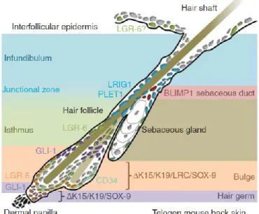
Based on these finding, we focused on understanding the molecular mechanisms underlying β-HPV-induced KC in immunocompromised patients through different approaches, including the HPV8 transgenic mouse model and in vivo tumorigenicity assays of tumours from OTRs. When considering the involvement of β-HPV in the development of KC, it is important to examine where β-HPV is localized. The current hypothesis is that the reservoir for β-HPV resides within long-lived hair follicle keratinocyte stem cells (HF-KSC), however, the precise HF-KSC populations involved in β-HPV latent infection remain to be defined. To determine the role of HF-KSC in β-HPV induced skin carcinogenesis, we utilized a transgenic mouse model in which the keratin 14 promoter drives expression of the entire HPV8 early region
4 (HPV8tg). HPV8tg mice developed thicker skin in comparison to wild type littermates consistent with a hyperproliferative epidermis. HF keratinocyte proliferation was evident within the Lrig1+ KSC population (69 vs 55%, p<0.001, n=6), and not in the CD34+, LGR5+ and LGR6+ KSC populations. This was associated with a 2.8-fold expansion in Lrig1+ keratinocytes and 3.8 fold increased colony forming efficiency. Consistent with this, we observed nuclear p63 expression throughout this population and in the HF infundibulum and adjoining IFE, associated with a switch from p63 TA isoforms to ΔNp63 isoforms in HPV8tg skin. EV keratosis and in some cases actinic keratoses demonstrated similar histology associated with β-HPV virus reactivation and nuclear p63 expression within the HF infundibulum and perifollicular epidermis. These findings would suggest that β-HPV field cancerization arises from the HF junctional zone and predispose to squamous cell carcinoma (SCC).
In addition, we have established an in vivo orthotopic humanized xenograft model using skin tumours from OTR. This technique allow to increase the availability of the tumours -that is usually a limitation in skin cancer research-, to investigate detailed molecular mechanisms of the events driving tumorigenesis and to identify β-HPV infection/expression in these lesions. Therefore, this methodology provides a highly promising ex vivo environment in which to study the progression of the disease from pre-malignancy to malignancy, e.g. from an AK to a SCC as reported in this thesis, which cannot be documented in the human beings due to the necessary excision of such tumours.
However, it remains to be determined whether immunosuppression also facilitates carcinogenesis by inhibiting tumour immune surveillance and killing. To test this hypothesis and generate a mouse model recapitulating the events occurring in OTR, we have crossed the HPV8 transgenic mice (FVB/N genetic background) with Rag2 deficient mice (which lack B and T cells; C57BL/6 genetic background). The results so far obtained with the F2 generation indicate that immunosuppression facilitates carcinogenesis by inhibiting tumour immune surveillance, as the number of skin lesions and their degree of dysplasia in HPV8+:Rag2-/- mice
was found increased when compared to the HPV8tg+:Rag2+/+ mice.
Overall, the results obtained in this study advance our understanding of the molecular mechanisms underlying β-HPV infection and strengthen the hypothesis of a causative role for these viruses in keratinocyte carcinoma development.
5
1. Sommario
I papillomavirus umani (HPV) sono piccoli virus nudi contenenti un genoma a doppia elica di DNA circolare di 8 kb che infettano gli epiteli squamosi stratificati cutanei e mucosali di differenti distretti corporei. Gli HPV possono stabilire infezioni latenti in individui sani, ma inducono anche lesioni neoplastiche benigne o maligne in base alle proprietà oncogene specifiche dei differenti genotipi. Fino ad oggi sono stati identificati più di 180 genotipi differenti di HPV che sono stati classificati in 5 generi sulla base dell’analisi filogenetica. Il genere Alpha (α-HPV) è il più caratterizzato e comprende i genotipi a tropismo mucosale associati all’ insorgenza di carcinoma a livello genitale (ad esempio HPV16 e 18). Gli HPV appartenenti al genere β (β-HPV) sono evoluzionisticamente distinti dal genere α e causano infezioni inapparenti o asintomatiche molto diffuse nella popolazione generale. Nei pazienti affetti da epidermodisplasia verruciforme (EV), una immunodeficienza primaria caratterizzata da un’alta suscettibilità all’infezione da β-HPV, questi virus replicano con alta efficienza, sviluppano il loro pieno potenziale trasformante, inducendo numerose lesioni simil verrucose con alto rischio di progressione a carcinoma cutaneo .
Sebbene il loro coinvolgimento nel processo di carcinogenesi cutanea in pazienti immunocompetenti e immunosoppressi non-EV (con immunosoppressione primaria o acquisita) è ancora materia di dibattito, numerose evidenze epidemiologiche e sperimentali suggeriscono un ruolo carcinogenico di questi virus che inizia nelle fasi precoci e contribuisce alla patogenesi del carcinoma cutaneo. Il nostro gruppo ha dimostrato precedentemente la presenza di prodotti dei geni virali e l’amplificazione del genoma virale in lesioni cutanee precancerose derivate da pazienti trapiantati d’organo (OTR, Organ Transplant Recipient). Questi risultati rafforzano l’evidenza di un coinvolgimento dei β-HPVs nella patogenesi del carcinoma cutaneo.
Sulla base dei dati già acquisiti, abbiamo studiato i meccanismi molecolari alla base dell’induzione del carcinoma cutaneo indotto da β-HPV in pazienti immunocompromessi attraverso differenti approcci, quali il modello di topo transgenico HPV8 e il saggio di tumorigenicita’ in vivo di tumori derivati da OTR.
Pe capire il ruolo di β-HPV nello sviluppo del carcinoma cutaneo, è importante conoscere dove questo virus naturalmente si localizza. L’ipotesi attuale è che il reservoir di β-HPV risieda in cellule staminali con ciclo cellulare rallentato presenti nel follicolo pilifero (HF-KSC); tuttavia, la precisa popolazione di HF-KSC coinvolta nell’infezione latente di β-HPV è ancora sconosciuta. Per determinare il ruolo delle HF-KSC nel carcinoma cutaneo indotto da β-HPV,
6 abbiamo utilizzato un modello di topo transgenico nel quale il promotore della cheratina 14 guida l’espressione della regione precoce di HPV8 (HPV8tg). I topi HPV8tg presentano uno spessore della cute piu’ ampio rispetto ai topi wild type e mostrano una cute iperproliferante. La proliferazione dei cheratinociti del follicolo pilifero è evidente nella popolazione di cellule staminali Lrig1+ (69 vs 55%, p<001, n=6), ma non lo è nelle popolazioni CD34+, LGR5+ e LGR6+. Questo è in linea con l’osservazione dell’espanzione dei cheratinociti Lrig1+ di 2,8 volte e dell’efficienza di formare colonie di 3.8 volte. In accordo con questi risultati, abbiamo osservato l’espressione nucleare di p63 in questa popolazione, nell’infindibulum e nella cute interfollicolare fiancheggiante, associata a un cambiamento di espressione tra la forma TA di p63 in ΔNp63 nella cute dei topi HPV8tg. Le cheratosi dei pazienti EV e alcune cheratosi attiniche (AK) di pazienti non EV mostrano un istologia simile associata alla riattivazione di β-HPV e all’espressione nucleare di p63 nell’ infindibulum e nella cute perifollicolare. Questi dati suggeriscono che il campo di cancerizzazione indotto da β-HPV inizia dalla “junctional zone” e predispone al carcinoma cutaneo..
Inoltre abbiamo realizzato un modello umanizzato di xenotrapianto ortotopico utilizzando tumori cutanei derivati da OTR. Questa tecnica permette di aumentare la disponibilità dei tumori cutanei – che è una delle limitazioni nell’ambito della ricerca sui tumori cutanei-, analizzare in dettaglio i meccanismi molecolari coinvolti nella tumorigenesi e identificare l’infezione e l’espressione di β-HPV in queste lesioni. Inoltre, questa tecnica permette di studiare la progressione della malattia da una situazione pre-cancerosa a una cancerosa, ad esempio da AK a SCC come riportato in questa tesi, che non può essere studiato a livello umano per la naturale necessità di rimuovere la lesione prima dell’evoluzione maligna.
In questo contesto, rimane da stabilire se l’immunosoppressione faciliti la carcinogenesi anche attraverso la riduzione dell’immunosorveglianza. Per verificare questa ipotesi e generare un modello murino che ricapitolasse gli eventi che si verificano nei pazienti trapiantati, abbiamo incrociato i topi transgenici HPV8 (background genetico FVB/N) con i topi deficienti per il gene RAG2 (background genetico C57BL/6), che non producono cellule B e T. I risultati ottenuti fino ad ora con la generazione F2 indicano che l’immunosoppressione facilita la carcinogenesi anche inibendo l’immunosorveglianza del tumore, come dimostrato dai dati ottenuti; infatti,il numero di tumori cutanei nella cute dei topi HPV8+:Rag2-/- e il loro grado di
displasia è risultato significativamente maggiore rispetto ai topi HPV8+:Rag2+/+.
Nell’insieme, i risultati ottenuti in questo studio contribuiscono a chiarire la storia naturale dell’infezione da β-HPV e rafforzano l’ipotesi di un ruolo di questi virus nello sviluppo del tumore cutaneo.
7
2. INTRODUCTION
8
2.1 PAPILLOMAVIRUSES
2.1.1 General features
Papillomaviruses (PV) are small non-enveloped viruses with an icosahedral capsid and a circular double-stranded DNA genome of 8Kb in length. They are species-specific and exhibit a strict tropism for the squamous stratified epithelia where they can induce cutaneous and mucosal hyperplastic lesions in a broad spectrum of vertebrates, including humans, other mammalians, birds and reptiles (Doorbar et al., 2012).
Human Papillomaviruses (HPV) can infect different body sites, such as the upper respiratory, oral and genital mucosae, and the skin according to the specific tropism of the different viral species. HPV prevalence has increased in many parts of the world during the past few decades. They typically establish persistent infections which can either remain in an unapparent silent status or cause clinical manifestations after a latency period of variable duration; reactivation of latent HPVs seems to be favored by several conditions affecting the host, such as immune status, genetics, environmental and unknown factors. HPVs can be responsible not only for benign hyperproliferative lesions (papillomas) in mucosal and cutaneous sites, but also for malignancies (especially squamous cell carcinoma, SCC) depending on their ability to establish chronic infections and stimulate cellular proliferation (Akgül et al., 2006).
HPVs have been implicated in cancers at several sites; in particular, the best documented HPV oncogenic activity concerns cervical cancer, where the association between HPV infection and the onset of precancerous lesions that can subsequently progress to high-grade tumours is well defined. Roughly 610000 new cancer cases per year (among these, 530000 cervical cancer cases) have been attributed to HPV infection, of which more than 80% in developing countries (Crosbie et al., 2013).
Although the causal association of HPV infection with cervical carcinogenesis is epidemiologically and experimentally ascertained, its implication in the development of precancerous skin lesions that can evolve to high-grade tumors of epithelial origin – comprehensively grouped into the Keratinocyte Carcinomas (KCs: basal and squamous cell carcinomas) – has been much less clear to date; such uncertainties partly come from the fact that cutaneous HPVs have been shown to be ubiquitously present in the skin in the general healthy population. Nevertheless, several epidemiologic and experimental evidences are in
9 favour of their potential oncogenic role in the insurgence of skin lesions (Knipe & Howley, “Fields Virology, 4th edition”, 2001; Pfister, 2003).
2.1.2 Classification, evolution, tropism and disease association
Papillomaviruses were originally grouped together with Polyomaviruses (PyVs) in a family called Papovaviridae; this classification was based on similarities in the capsid proteins and some proteins involved in viral replication and on the fact that these were the only viruses with circular double-stranded DNA genomes anxd a non-enveloped icosahedral capsid. Subsequently PVs and PyVs were split into two different viral families (Papillomaviridae and Polyomaviridae) because of substantial differences in genomic sequence, organization and size (de Villiers et al., 2004; Bernard, 2013).
After many years of research and genomic sequencing of thousands of PV isolates, PV classification was based on phylogenetic criteria following hierarchical taxonomic levels (family, genus, species, types, subtypes and variants). The L1 gene, encoding the major capsid protein, represents the most conserved gene of the PV genome, showing clusters of sequence similarity throughout its whole length among different PVs; for this reason, L1 nucleotide sequence is used to perform comparative alignments for PV identification and classification. By this approach, more than 200 PV types have been discovered to date, encompassing more than 180 HPV types and 103 animal PV types (Figure 1; http://pave.niaid.nih.gov/#home).
A novel PV type is established when L1 nucleotide sequence exhibits at least 10% diversity from the closest known PV type; 2-10% diversity define a PV subtype, while less than 2% diversity introduce a PV variant. Different PV genera share less than 60% sequence identity and different PV species belonging to the same genus share 60-70% sequence identity; finally, PV types belonging to the same species share 71-89% sequence identity (de Villiers et al., 2004; Forslund, 2007; Bernard et al., 2010; Bernard, 2013).
PV phylogenetic analyses indicate that PV evolution is linked to their hosts. A major part of PV evolutionary diversification must have proceeded clearly through mechanisms that restrict PVs to their host species; these host-virus associations strongly suggest that PV phylogenetic trees reflect a time scale similar to the host phylogenetic trees and that specific events in host evolution may have created new ecological niches for PV to adopt. However, it has also always been a generally accepted view that beyond host-linked evolution yet other evolutionary mechanisms drive PV diversification, as there exist remotely related PV taxa in
10 the same species, but in part separated by diverse target tissues (skin vs. mucosa); PVs and their hosts have not gone through an identical evolutionary path, even though initial niche sorting followed by host-virus co-speciation was a key determinant of the PV evolutionary history. Recombination may have played an important role during PV evolution, however it is likely that these events mainly occurred early in the evolutionary process; in addition, further mechanisms may have contributed to the current PV diversity, including host-switching and possible extinction of the PV lineage in some hosts. The tight host-virus interactions that have underlain PV evolution are thought to have led over time to a balance between viral replication and immune tolerance, allowing the viruses to become well adapted to their hosts where in most instances can complete their life cycle and be maintained without causing any overt disease (Doorbar et al., 2012).
PV molecular evolution progresses several orders of magnitude slower than that of RNA viruses, possibly even approaching the slow speed of the genome of the host species. This property is shared with other double-stranded DNA viruses infecting vertebrates such as Polyomaviruses and Herpesviruses, reflecting the fact that these viral genome sequences are stabilized by the high-fidelity proof-reading capability of host DNA polymerases; for these reasons, mutations are fixed at a very slow rate in the PV genome, which is therefore highly stable (Bernard, 2013; Van Doorslaer, 2013)
On the basis of L1 nucleotide sequences, HPVs are divided into five genera (Figure 1), with the different species and types exhibiting different sites of infection, life cycle characteristics and functions of specific viral gene products and different disease association: Alpha (α-HPV), Beta (β-HPV), Gamma (γ-HPV), Mu (μ-HPV) and Nu (ν-HPV) (Bernard et al., 2013).
The Alpha genus is the most represented one and comprises both mucosal and cutaneous species. Based on their oncogenic potential and association with the development of malignancies, mucosal α-HPVs are further subdivided into low-risk and high-risk groups. The World Health Organization has defined 12 mucosal α-HPV types (HPV 16, 18, 31, 33, 35, 39, 45, 51, 52, 56, 58, 59) as being high-risk cancer-causing types; they are mainly responsible for the development of cervical cancer and can also be associated with cancers at other sites with much lower incidence (head and neck carcinomas such as oropharyngeal cancers, and cancers of the penis, anus, vagina and vulva). Nevertheless, high-risk α-HPVs do not cause cancer in the vast majority of the individuals they infect.
Low-risk α-HPVs cause benign mucosal lesions. Certain types (e.g. HPV 6 and 11) are associated with the development of respiratory papillomatosis (especially HPV 11), which is a laryngeal disease often occurring in children, and with benign external ano-genital warts
11 (especially HPV 6); these types are sometimes occasionally found to be associated with cancers in these sites especially in individuals with immune defects, where such infections are more difficult to manage. Other low-risk α-HPV types (e.g. HPV 13 and 32) are responsible for the development of oral papillomas as it occurs in the case of oral focal epithelial hyperplasia.
Cutaneous α-HPVs (e.g. HPV 2, 3, 7, 10, 27, 28, 57) are associated with the development of different types of benign skin warts arising on various sites of the hands, face, elbows and knees.
The Beta genus comprises to date 43 types, which exhibit cutaneous tropism (e.g. HPV 5, 8, 9, 14, 17, 20, 21, 23, 36, 38, 47, 49). β-HPVs are evolutionarily distinct from the Alpha genus and establish widespread unapparent asymptomatic infections that remain latent without any clinical manifestation in the general healthy population. In subjects with impaired immune function, it seems that these viruses can spread unchecked and they have been implicated in the development of KCs. In particular, in individuals suffering from epidermodysplasia verruciformis (EV), a primary immunodeficiency associated with abnormal susceptibility to β-HPV infection, they are responsible for the development of disseminated wart-like lesions that often undergo malignant progression; there is also increasing evidence for their involvement in the onset of precancerous skin lesions with potential to evolve to KCs in other immunosuppressed populations (e.g. OTRs, organ transplant recipients). Their involvement in the pathogenesis of KC in immunocompetent individuals is more controversial.
Gamma (e.g. HPV 4), Mu (e.g. HPV 1) and Nu (HPV 41) genera comprise a smaller number of types, exhibiting cutaneous tropism; they are associated with the development of benign palmar and plantar skin warts (de Villiers et al., 2004; Pfister, 2003; Doorbar et al., 2012).
12 Figure 1. The phylogenetic tree of Papillomaviruses, obtained from multiple comparative alignments of the L1 gene. Symbols at the end of each branch define PV types; the external half circles refer to PV genera, while the internal half circles surround PV species (Bernard et al., 2013).
13
2.1.3 HPV structure, genomic organization and life cycle
HPV virions have a 52-55 nm diameter. The icosahedral capsid is made of the two structural proteins L1 and L2, repeated and organized in 72 capsomers. L1 is the major coat protein and accounts for 80% of the total amount of viral proteins; L2 is the minor coat protein and seems to favor the assembly of new viral particles amplifying their infectious ability. The two capsid proteins enclose the double-stranded DNA genome, which is about 8 kb in size (Knipe & Howley, “Fields Virology, 4th edition”, 2001). Genomic organization is highly conserved
among all HPV family members (Doorbar, 2007), with 7-8 open reading frames (ORFs) on a single transcriptionally active DNA strand encoding a larger number of gene products as a result of mRNA splicing (Figure 2). In the HPV genome, three regions can be distinguished: - the early region coding for functional proteins (E1, E2, E4, E5, E6 and E7 for AlphaPV and E1, E2, E4, E6, E7 for BetaPV) expressed in all the phases of the viral life cycle, which are responsible for the persistence of the viral genome in a cell, its replication and the stimulation of cell proliferation necessary to support viral replication itself;
- the late region, coding for the structural coat proteins L1 and L2 that are expressed in the final phases of the productive viral life cycle;
- the long control region (LCR), a non-coding fragment regulating the expression and replication of the viral genome.
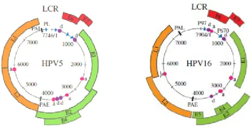
The L1 and L2 proteins are conserved across widely divergent PVs, and along with E1 and E2 are key viral gene products that are thought to have been present in the common ancestor of all PVs; other viral genes, such as E6, E7, E4 and E5, have been acquired or significantly modified during evolution and are not necessarily present or may not have the same function in all PVs. Because of such evolutionary mechanisms, different HPV genera have some exclusive features despite the highly conserved structure of the genome. For instance, the β-HPV genome is relatively short compared with that of the mucosal α-β-HPVs, ranging from 7.4 kb to 7.7 kb; this is due to the considerably reduced size of the LCR, about 400 bp compared with 650–900 bp in other HPVs. β-HPVs also display some differences in the coding region: most of them lack a functional E5 ORF and the E2 and E4 ORFs of all the β-HPV types are much longer compared with the other HPVs. Similarly to β-HPVs, also γ-HPVs lack the E5 gene (Tommasino, 2014; Ciesielska et al., 2012; Quint et al., 2015).
14
Figure 2. The genome organization of the prototype Beta HPV5 and Alpha HPV16. The HPV genome comprises a long control region (LCR) and seven or eight genes that are necessary for different stages of the virus life cycle. These genes encode a larger number of gene products as a result of mRNA splicing. The LCR contains binding sites for cellular transcription factors (e.g., SP1, AP1, Oct1), as well as for the viral E1 and E2 proteins that control viral replication and gene expression. BetaPV types lack an E5 ORF and have a much longer E4 ORF than the AlphaPV types. The positions of major promoters and splice donor/acceptor sites are indicated by arrows (promoters) and circles (d, donor; a, acceptor), respectively, alongside the positions of the early (PAE) and late polyadenylation sites (PAL) (Quint et al, 2015).
The LCR is a significant non-coding fragment of viral DNA located between the L1 and E6 ORFs, accounting for about 10% of the entire genome. It contains an origin of replication and cis-responsive elements for regulatory transcription and replication factors of viral genetic material; in detail, there are binding sites for cellular transcription factors (e.g. SP1, AP1, Oct1), as well as for the viral E1 and E2 proteins that control viral replication and gene expression (Figure 2) (Doorbar et al., 2012). All the LCR regions contain enhancers that provide the virus with specific tropism to the stratified squamous epithelial cells. Other regulatory enhancer elements are positioned within genes. The LCR harbours one of the two well-characterized promoter elements, namely the early promoter upstream of the E6 ORF, while in many HPV types the late promoter is located within the E7 ORF; these promoters regulate the expression of differentially spliced mostly polycistronic mRNAs, being trans-activated in a timely and coordinated differentiation-dependent fashion in specific epithelial layers where the different phases of the viral life cycle take place. The early promoter mainly drives E6 and E7 expression, while the late promoter regulates the expression of all the other viral genes (Ciesielska et al., 2012; Doorbar, 2013; Tommasino, 2014).
15 The E1 protein, an ATP-dependent DNA helicase, is the only enzyme encoded by the PV genome. E1 is essential for the maintenance, replication and amplification of the viral episome in the nucleus of infected cells; it does so by interacting with cellular DNA replication factors and assembling with E2 to the origin of replication to trigger viral DNA replication.
The E2 protein acts both as repressor and activator of viral gene transcription by binding to multiple sites in the LCR; in particular, it negatively modulates the expression of E6 and E7 by down-regulating the activity of the early promoter. Furthermore, E2 recruits E1 to a specific E1-binding motif in the origin of replication and is implicated in the maintenance of the viral genome in its episomal (extrachromosomal) form.
The E4 protein is the most abundant viral gene product and is expressed later than the other early proteins. E4 is thought to be involved in viral genome amplification and suppression of cellular proliferation in the late phases of the productive life cycle; it seems also to be able to form multimers that assemble into amyloid-like fibres that can destabilize the cytokeratin network of the host epithelial cells facilitating the extracellular release of new viral particles (Bergvall et al., 2013; McBride, 2013; Doorbar, 2013).
HPVs encode three proteins with transforming properties: the E5, E6 and E7 oncoproteins. Through combined and cooperative action, they abrogate the activity of tumour suppressor factors, promoting evasion of all cell cycle checkpoints and inducing a deregulated progression of the cell cycle; the result is a stimulation of cell growth, survival and proliferation and a delay of terminal cell differentiation (Doorbar, 2007).
E5 is a transmembrane protein localizing to cell membranes - predominantly to the endoplasmic reticulum and also to the Golgi apparatus and nuclear membrane - suggesting that its activity must be related to the trafficking of membrane proteins through different cellular compartments; it can dimerize in these sites and in certain HPV species trigger cell fusion. It is absent in HPV types from the Beta genus. (Connolly et al., 2014).
E6 and E7 are multimeric proteins with potential to associate with multiple cellular partners. Such functional differences contribute to the respective transforming abilities of various HPV species and types. In the best characterized HPV species, E7 binds and targets for ubiquitin-dependent proteasomal degradation the hypophosphorylated form of members of the Rb family (pRb, p107, p130). In uninfected epithelium, cell cycle entry and cell division in the basal and parabasal cell layers is controlled by growth factors that stimulate the activity of G1 cyclins including cyclinD/Cdk, which phosphorylates Rb family members and displace them from transcriptional activators of the E2F family allowing the trans-activation of genes necessary for S-phase progression (e.g. minichromosome maintenance protein 7 (MCM7), PCNA, Ki67,
16 cyclins); the continual stimulation of these cells physiologically allows renewal of the epithelium as surface cells exfoliate (Tommasino, 2014).
E6 interferes with DNA damage repair, growth arrest and apoptosis. The function of E6 complements that of E7. The efficient binding of Rb proteins by E7 can lead to inhibited cell growth and apoptosis through a p53-dependent pathway; as a result, E6 proteins of many HPV types have evolved to target the tumour suppressor p53 for ubiquitin-dependent proteasomal degradation or other forms of inactivation, resulting in the abrogation of its activity that plays an essential role in protecting genomic integrity. (Akgül et al., 2006; Ciesielska et al., 2012; McLaughlin-Drubin et al., 2012; Moody et al., 2010).
The HPV replicative cycle (summarized in Figure 3) is tightly linked to the differentiation program of stratified squamous epithelia. Most viruses infect a target cell and produce progeny virus from that same infected cell; conversely, in HPV infections, the synthesis of new virions occurs only after the infected cell has undergone mitosis and one of the infected daughter cells has differentiated. HPV replication requires the timely and coordinated expression of the different viral gene products as the infected cell moves towards the epithelial surface; this highly regulated pattern of gene expression allows the different stages of the life cycle to be completed appropriately. For this reason, HPV life cycle takes 2-3 weeks, the time necessary for an epithelial cell to migrate from the basal to most superficial layers, mature, undergo senescence and die. (Doorbar, 2007; Moody et al., 2010; Roman et al, 2013; Crosbie et al., 2013; Doorbar et al., 2012).
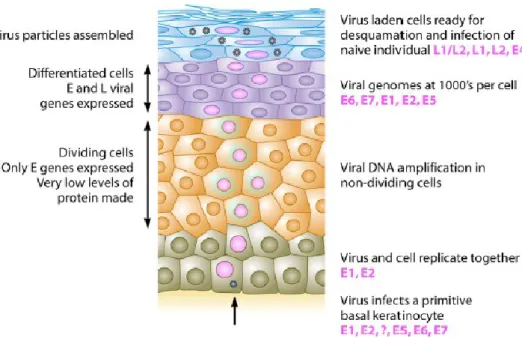
Figure 3. Productive HPV life cycle. HPVs replicate only in fully differentiating squamous epithelia. The life cycle involves both temporal and spatial separation of viral protein expression. The virus first
17 infects a cell in the basal layer of the epithelium where access is naturally facilitated (e.g. microtrauma, epithelial transitional zones, hair follicles). In the lower proliferative compartments of the epithelium, there is a phase of viral episome maintenance al low copy number, in which viral and cellular DNA replicate together. As long as the cell is dividing, HPVs control the expression of their viral proteins very tightly; the E6 and E7 oncogenes are thus expressed at very low levels, along with the genes coding for E1 and E2 replication factors. When the host cells stop dividing and begin to differentiate into mature epithelial cells, this provides a signal to the virus to activate all of its genes to amplify the viral genome copy number to the thousands. In the top layers of the epithelium, all of the viral genes, including those encoding the L1 and L2 proteins, are expressed, and many thousands of viral genomes are encapsidated; finally, the newly assembled infectious viral particles exit the cells in the context of epithelial surface desquamation. The time taken from infection to the generation of new virions is at least 2-3 weeks (Stanley, 2012).
Experimental models suggest that infection requires access of virus particles to the basal lamina and the interaction with heparin-sulphate proteoglycans and possibly also laminin. Structural changes in the virion capsid, which includes cleavage of L2, facilitate transfer to a secondary receptor on the basal keratinocyte, which is necessary for virus internalization and subsequent transfer of the viral genome to the nucleus; although the α-6 integrin and growth factor receptors have been implicated in this process, the precise nature of the entry receptor remains somewhat controversial (Doorbar et al., 2012). Once internalized by a clathrin-dependent endocytic pathway, virions undergo endosomal transport, uncoating and cellular sorting. The L2 protein-DNA complex ensures the correct nuclear entry of the viral genomes, while the L1 protein is retained in the endosome and ultimately subjected to lysosomal degradation (Doorbar et al., 2012).
In many cases, infection is thought to require epithelial wounding or micro-wounding to allow access of the virus to the basal lamina, and a role for the wound healing response in simulating the lateral expansion of the infected cells has been suggested. Some HPV species are thought to infect sites where access to the basal layer is naturally facilitated, such as the base of the hair follicle (HF) for cutaneous HPVs, or sites where columnar and stratified squamous epithelial cells meet each other, such as the cervical or anal squamo-columnar junctions for high-risk α-HPVs. It is thought that lesion formation begins with the infection of a long-lived, slow-cycling keratinocyte stem cell (KSC) and that the longevity of the keratinocyte stem cells is a key factor in the formation of a persistent lesion, with these cells being a reservoir for the infection (Doorbar, 2007; Quint et al., 2015).
Infection of the stem cells is followed by the early non-productive stage of the life cycle, consisting of an initial phase of genome amplification and then by maintenance of the viral episome al low copy number (100-200 copies per cell) (Doorbar et al., 2012).
18 The HPV genome does not encode polymerases or other enzymes necessary for viral DNA replication, which depends on the presence of the cellular DNA replication machinery, with viral DNA replication accompanying cellular DNA replication as the cells progress through S-phase (Stanley, 2012). The only viral proteins that are thought to be necessary for this initial amplification phase are E1 and E2, which are expressed from the late promoter and ensure viral DNA replication and transcription (Doorbar, 2007).
Viral proteins are not readily detectable prior to the onset of genome amplification during normal productive infection; the low level expression of viral proteins in the basal layer is thought to reflect, at least in part, the need of the virus to avoid detection by the host’s immune system (Doorbar et al., 2012). After an initial increase in viral copy number, the infected ‘differentiating’ cells move from an S-like to a G2-like phase, with viral genome amplification occurring primarily in G2 after cellular DNA replication has been completed; the virus at this point fully recruits the cellular DNA replication machinery for its own replication (Doorbar et al., 2012).
A key function of the E6 and E7 proteins in most HPVs is not to promote basal cell proliferation, but rather to stimulate cell cycle re-entry in the mid-upper epithelial layers in order to allow genome amplification (Stanley, 2012). To complete the late productive stage of the life cycle, the infected cells must undergo terminal differentiation in the most superficial epithelial layers. The completion of the life cycle ultimately involves the expression of the minor coat protein L2, the exit of the cell from the cell cycle and the expression of the major coat protein L1 to allow genome packaging (Doorbar, 2007).
2.1.4 High-risk α-HPVs and cervical carcinogenesis
Genital α-HPV infection is the most common sexually transmitted viral infection all over the world, with a peak of incidence in young individuals between 15-25 years of age following the onset of sexual activity and a progressive fall with age; over 80% of sexually active women become infected at some stage in their life with one or several high-risk and low-risk α-HPV types (Crosbie et al., 2013).
The frequency of high-risk α-HPV-associated cervical cancers is of around 30-40 people per 100000 and over 99% of cervical lesions harbor viral sequences, although the proportion associated with specific high-risk α-HPV types is different in different countries and shows demographic, ethnic and socio-economic variation. HPV 16 and 18 cause approximately 50%
19 and 20% of the cases of cervical SCC – arising in the stratified squamous cells of the ectocervix - respectively, and both types are equally associated with around 35% of the cases of cervical adenocarcinoma – arising in the columnar glandular cells of the endocervix and more aggressive (Doorbar, 2007).
Cervical precancerous lesions are named cervical intraepithelial neoplasia (CIN) and are histologically classified for diagnostic purposes according to the grade of dysplasia: CIN1 (mild dysplasia), CIN2 (moderate dysplasia), CIN3 (in situ carcinoma); CIN1 lesions are low-grade lesions (LSIL, low-low-grade squamous intraepithelial lesions), while CIN2 and CIN3 lesions are high-grade lesions (HSIL, high-grade squamous intraepithelial lesions). The accurate identification of lesion grade has prognostic significance, as it has been estimated that around 20% of CIN1 will progress to CIN2, and that around 30% of CIN2 will progress to CIN3 if left untreated; CIN3 are generally considered to be the direct precursors of cervical cancer, and it has been suggested that around 40% of CIN3 lesions will progress to cervical cancer in the absence of intervention. In general, more regions with different histological grade can be found in the context of a cervical lesion; different high-risk α-HPV types are usually associated with discrete areas of disease except at junction regions (where lesions abut or are in close proximity) where more than one type may be detected (Doorbar, 2007).
Most cervical cancers develop in the transformation zone, corresponding to the squamo-columnar junction - the transitional site where the squamo-columnar glandular cells of the endocervical canal meet the stratified squamous cells of the ectocervix. The particular susceptibility of the transformation zone to cancer onset and progression may also be linked to the increased accessibility and proliferation of the basal cell layers at this metaplastic epithelial site, particularly around the time of puberty and the onset of sexual activity; in this case, we can hypothesize that the primary preferential target cells for high-risk α-HPV infection may be cells close to the squamo-columnar junction, such as the epithelial reserve cells, which lie immediately underneath the columnar epithelium of the endocervix, and eventually form the stratified epithelial layers of the transformation zone as the cervix matures (Doorbar et al., 2012). Early acquisition of high-risk α-HPV infections, also facilitated by the low levels in antigen-presenting cells in this site, can disturb the metaplastic changes occurring at this time in the transformation zone and increase the risk of cervical cancer in the future (Cubie, 2013; Tommasino, 2013; Moody et al., 2010).
20
2.2 β-HPV AND KERATINOCYTE CARCINOMAS
While the causative relationship between HPV infection and genital SCCs is well established, the role of HPV in the development of cutaneous malignancy is, as yet, unclear. However, there are an increasing body of evidence suggesting the involvement of cutaneous β-HPV in the development of KC. To demonstrate that a pathogen causes cancer, convincing epidemiologic evidence of association and a plausible biological mechanism for oncogenesis are necessary; while this has been achieved for high-risk α-HPVs and cervical cancer, such requirements have not yet been satisfied for β-HPVs and KC, as there is great lack of consistency among epidemiological studies and no molecular role for these viruses in cutaneous tumorigenesis has been certainly proven so far.
KC is the most common cancer type among Caucasians, where it accounts for around 30% of all malignancies and its rates are increasing by 4–8% yearly. Although the majority of KC cases can be treated surgically and do not usually exhibit high aggressiveness, these cancers are associated with high morbidity and represent a significant burden on the healthcare system. Immunosuppression and UV exposure are the main risk factors for KC – these tumours mostly arise in sun-exposed skin sites (Akgül et al., 2006: Aldabagh et al. 2013).
The major histological types of KCs are basal cell carcinoma (BCC) and cutaneous squamous cell carcinoma (SCC), associated with different underlying mutational patterns. BCC is more common in fair-skinned individuals and originates from transformation of basal epidermal cells. SCC, which is more aggressive than BCC, is more common among darker skin population and arises from transformation of squamous epidermal cells; it is usually preceded by precancerous lesions, namely actinic keratosis (AK), with high potential to undergo malignant progression; other forms of skin tumours that are histologically related to SCC are keratoacanthoma (KA) – a low-grade SCC subtype originating from the hair follicles, able to regress spontaneously and representing a midpoint between a benign wart and invasive SCC – and Bowen’s disease – an early stage in situ intraepidermal form of SCC (Nindl et al., 2007; Knipe & Howley, “Fields Virology, 5th edition”, 2007; Pfister, 2003).
The epidemiological demonstration of a possible causal link between β-HPV infection and KC development is complicated by the fact that these viruses are also present in non-affected skin areas of patients and even ubiquitously widespread in the general healthy population, establishing persistent, asymptomatic latent infections without apparent disease; it is thought that everybody is positive for multiple β-HPV types. With increasing age, the number of infecting β-HPV types seems to rise; β-HPV persistence with the same types is common, and
21 family members often share the same types (“β-HPV signature”). Different studies using different sampling techniques have shown that the most commonly detected β-HPV types are HPV 5, 8 and 23; different ethnicities may harbour different β-HPV types, with HPV5 being the most prevalent type and the only type common to all countries studied (Aldabagh et al., 2013).
β-HPV infection seems to be acquired very early in infancy, with exposure beginning soon after birth; the presence of the viruses on the skin surface of the mother and other people in close contact with the newborn is the most likely source of infection. β-HPV transmission probably occurs by contact with infected skin or its ubiquitously present remnants (e.g. scales, dandruff); the likelihood of a prenatal vertical transmission from mother to fetus has also been proposed. Transmission in later stages of postnatal life is thought to be limited; this may be due to the fact that in the first postpartum days, the thinner horny layer facilitates access to the basal layer, or to the fact that after the sites available to infection have been occupied, the settlement of other β-HPV types is more difficult because of the occurrence of a cross-reactive antiviral response following initial infection. In immunocopetent individuals, this early infection seems to persist in a latency status and without clinical manifestations (Harwood et al., 2002; Bouwes Bavinck et al., 2008; Akgül et al., 2006; Feltkamp et al., 2008).
2.2.1 Epidermodysplasia verruciformis
Epidermodysplasia verruciformis (EV), which is considered to be a primary immunodeficiency (PID), is the only setting where the association between β-HPV infection and skin carcinogenesis has been ascertained. It is a rare autosomal recessive condition, in which selective depletion of specific T-cell clones - although the immunophenotype might be normal in some patients - is associated with an abnormal susceptibility to persistent infection restricted to a subset of about 20 β-HPV types, often with more than one infecting type. It arises in early childhood with the development of disseminated benign plane warts and occurring mainly on the trunk, neck and extremities, and persist lifelong. EV patients also develop wart-like lesions (Figure 4A) that are intraepidermal proliferative lesion that display the typical cytopathic effects of EV skin lesions, including acanthosis, koilocytosis, the presence of enlarged cells with prominent blue–grey pallor and occasional perinuclear halos, with greater malignant potential on light-exposed areas (Figure 4B, left panel). These lesions are associated with HPV 5 and 8, often with more than one β-HPV type being detected, and contain high β-HPV episome copy numbers per cell – indicating a high level of viral replication – and abundant E6 and E7
22 transcripts – indicating viral gene expression in the lesional tissue. In the fourth decade of life, around 30-60% of EV patients develop malignant tumours in sun-exposed sites (Quint et al., 2014), which are usually low grade in situ carcinomas with some features in common with Bowen’s disease, but others are more aggressive SCC; HPV 5 and 8 account for 90% of the tumours with HPV 14, 17, 20 and 47 accounting for the remainder; β-HPV load can reach 100-300 genome copies per cell, even though in a limited number of tumour cells and always in episomal form. β-HPV-induced EV skin tumours are associated with productive infections: the high level of viral replication and subsequent active oncogene expression underlies skin carcinogenesis in these patients.
Our group has throughly studied the association between β-HPV infection and EV patient’s lesions by mapping the β-HPV life cycle events. The data have suggested a progression pattern from benign keratotic skin lesion to Bowen’s Disease and SCC, in which the β-HPV life cycle is progressively disrupted. In addition, our group has demonstrated that the abundant β-HPV seen in some lesions was the result of genome amplification within the carcinoma tissue. E4 protein of β-HPV was very abundant and could be easily visualised either in superficial cells supporting viral genome (cytoplasmic) or in the more basal proliferating cells (nuclear) (Figure 4B, right panel). The loss of staining at the tumour border has suggested that E4 staining could be exploited as a marker of viral expression during β-HPV-associated skin cancer progression (Borgogna et al., 2012; Borgogna, Landini et al., 2014).
A
23 Figure 4. (A, left panel) Clinical manifestation of epidermodysplasia verruciformis on the back of an EV patient. The area surrounded by the black square is enlarged in the right panel. The black arrow shows the characteristic lesions. (B, left pannel) H&E staining of a wart-like lesion displays the typical cytopathic effects of EV skin lesions. The section was double stained in immunofluorescence using antibodies to E4 of β-HPV (red) and the proliferation marker MCM7 (green). The broken lines indicate the position of the basal layer.
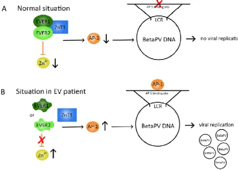
Homozygous truncating mutations in the EVER1 or EVER2 genes have been reported in approximately 50% of EV patients; these two genes encode transmembrane proteins expressed in a variety of cells types, including mostly blood cells. These proteins form a complex localized to the endoplasmic reticulum, where they seem to control intracellular zinc homeostasis and in particular to decrease the nuclear zinc concentration, reducing the activity of zinc-regulated transcription factors necessary for viral gene expression (e.g. AP1). Thus, the EVER proteins may act as restriction factors for β-HPV gene expression and replication in keratinocytes, even though the exact underlying mechanisms are still unclear. EVER mutations disrupt zinc transport between cell compartments; it is thought that this leads to up-regulation of transcription factors that can induce β-HPV transcription and stimulate keratinocyte proliferation, which in turn further amplifies β-HPV oncoprotein expression (Quint et al., 2015; Lazarczyk et al., 2008) (Figure 5)
Figure 5. A. Normally, the EVER/ZnT-1 complex is responsible for a low level of free intracellular Zn2+. This low level of free Zn2+ tonically inhibits the activity of AP-1 transcription factors necessary for BetaPV replication in keratinocytes. B. In EV patients, a dysfunctional EVER/ZnT-1 complex leads to a higher Zn2+ concentration, which causes increased activity of the AP-1 transcription factors, thereby initiating BetaPV replication (Quint et al., 2015)
B A
24 Other HPV species may counteract EVER protein function through different mechanisms in order to facilitate viral gene expression; HPV16 E5 has been reported to be able to bind and inhibit the EVER complex. It has also been postulated that EV-associated imbalance in zinc homeostasis contributes to HPV-specific immune deficiency through the same mutations. However, in a substantial proportion of patients clinically diagnosed with EV, EVER genes are not mutated; it is likely that unknown mutations affecting other genes are involved in the genetic background of the disease (Akgül et al., 2006; Pfister, 2003, Leiding et al., 2012; Cubie, 2013; Arron et al., 2011).
2.2.2 β-HPV infection in the immunocompromised host
The association of cutaneous HPV infection with the development of skin lesions is well established in the case of EV. More recently, mutations in other two genes, RHOH – encoding a Rho-GTPase expressed predominantly in hematopoietic cells involved in intracellular transduction form T-cell and B-cell receptors – and MST1 – encoding a kinase involved in many biological processes in various cell types, including apoptosis, negative regulation of cell growth and proliferation, and differentiation - have been identified in PID patients associated with high susceptibility to β-HPV infections exhibiting an EV-like phenotype. RHOH and MST1 deficiencies lead to T-cells defects, which probably play a role in the pathogenesis of chronic active β-HPV infections (Crequer, Troeger et al., 2012; Crequer, Picard et al., 2012). Other PID syndromes (e.g. SCID, WHIM), whose genetic backgrounds have been described and affect various aspects of immune function, are characterized by recurrent severe bacterial and viral infections since infancy. As a result, these patients have a number of subsequent clinical manifestations, among which diffuse HPV-associated warts and also carcinomas; β-HPV infections have also been reported in EV-like skin lesions from these individuals and might be implicated in their onset and development, leaving open the possibility that these viruses play a role in cutaneous carcinogenesis affecting many of these subjects. Our group reported a patient with an unclassified primary T-cell immunodeficiency (characterized by a low number of naïve T cells) that did not carry any of the genetic mutations associated with EV or WHIM syndrome, but displayed remarkable and specific susceptibility to α and β HPV infection. The HPV susceptibility involved both cutaneous and genital sites as the patient developed either βHPV-positive wart-like lesions or αHPV-positive anal-penile
25 condylomas. This pattern is slightly different from the clinical picture of canonical EV patients where the susceptibility is considered to be restricted to the β genus (Landini et al., 2014). Similarly to PID patients, individuals with acquired T-cell immunodeficiencies, whether secondary (e.g. hematological malignancies, AIDS) or iatrogenic (e.g. OTRs undergoing long-term immunosuppressive therapy to prevent graft rejection) are all at increased risk of developing extensive, persistent and recurrent HPV-associated warty and keratotic skin lesions especially on sun-exposed sites (Leiding et al., 2012; Cubie, 2013; Aldabagh et al., 2013). In the particular case of OTRs, although suppression of T-cell immunity and immunosuppressant drugs per se can exert carcinogenic effects, the most obvious mechanism of post-transplant tumorigenesis is that iatrogenic down-regulation of cell-mediated immunity unmasks the transforming activity of infectious agents resulting in the development of virus-related malignancies (Connolly et al., 2014).
Several epidemiological studies have demonstrated statistically significant associations between β-HPV infection and KC development. For instance, large international case-control studies in OTR and in immunocompetent individuals observed an association between the presence of HPV DNA in eyebrow hairs and the development of KC, as well as between β-HPV serum antibodies and KC (Bavinck et al., 2010). The BCC:SCC ratio is inverted in OTRs compared to the general population, with SCC occurring 4 times more often than BCC; SCC represents the most common de novo malignancy in the OTR setting, who exhibit a risk 60-250-fold as great as that of immunocompetent subjects with a SCC frequency over 50% at 10 years and over 80% at 20 years post-transplantation – while BCC is increased 10-40 fold (Euvrard et al., 2003; Tessari et al., 2010). In addition, SCC arising in OTRs can be multiple and highly aggressive, with increased rates of recurrence and metastasis; they usually develop within the first decade post-transplantation in sun-exposed sites co-localizing with other precancerous lesions, suggesting that their persistence favors malignant progression, and affects large skin areas in a process called field cancerization.
The major weakness of the available studies is that the proposed association is mostly based on the presence of viral DNA in tumour tissues or positive antibody responses. Very few studies have addressed whether the β-HPV detected in these cases are actually localized within the malignant cells or whether they are transcriptionally active, the confirmation of which would greatly strengthen the evidence for a carcinogenic role of these pathogens.
Studies from our group have demonstrated that detection of the abundant viral E4 protein of β-HPV was helpful for the visualization of active β-HPV infection in skin tumours from EV patients (Borgogna et al., 2012; Borgogna, Landini et al., 2014). Based on these finding, our
26
group has decided to investigate whether detection of β-HPV gene products, as defined in EV skin cancer, could also be observed in lesions from OTRs. Using a combination of antibodies against the viral proteins E4 or L1 of the β-genotypes and fluorescent in situ hybridisation (FISH) to detect the viral genome, we were able to visualise viral infection in premalignant lesions, such as actinic keratosis and keratoakantoma as well as in the pathological hyperplastic edges of either SCC or BCC from OTRs. Increased expression of the cellular proliferation marker MCM7, that extended into the upper epithelial layers, was a common feature of all the E4-positive areas, indicating that cells were driven into the cell cycle in areas of productive viral infection (Borgogna, Lanfredini et al., 2014; Figure 6).
Figure 6. (A) Distribution of the viral and cellular markers E4, L1, human papillomavirus 25 (HPV25)
DNA and MCM7 in a case of hypertrophic actinic keratosis from a hand from a kidney transplant recipient. The top picture (scale bar: 1000 mm) shows the scan of the tissue section of the actinic keratosis using hematoxylin and eosin (H&E) staining. The region shown in the lower panels corresponds to the red square highlighted in the over H&E image, reproduced in the lower left-hand picture. The same section was double stained using antibodies to E4 (green) and MCM7 (red) (second picture); serial sections were double stained for the presence of viral genome amplification by HPV25 DNA-fluorescent in situ hybridization (FISH) (red) and for E4 expression (green) (third picture), and also stained with antibodies to L1 (red) (fourth picture). The white arrows indicate nuclear L1 staining. All sections were counterstained with DAPI (blue) to visualize cell nuclei. Scale bars: 50 mm
A
27 (Borgogna, Lanfredini et al., 2014). (B) Schematic table of skin tumours derived from our OTRs cohort analysed by immunofluorescence for detect the presence of β-HPV markers.
These data demonstrated that β-HPV transcription occurs at the site of skin transformation in the organ transplant recipient setting and also point to its possible involvement in the process of skin carcinogenesis (Borgogna et al., 2014).
Based on these results, we have decided to investigate the association between β-HPV infection and OTRs KCs using an orthotopic tumourgraft model developed by Patel et al at the NCI-Bethesda by xenografting fresh skin tumours from OTRs onto a “humanized” stromal bed repopulated with human fibroblasts in nude mice (Patel et al., 2012). This methodology provides advantages in our study for the following reasons: i) skin tumours are usually very small, thus the material left from the routine diagnostic procedure is often insufficient and does not allow for the storage of fresh tissues; ii) the xenograft is usually bigger than the original tumour, it can be exploited in its entirety for molecular investigation and split in half for fresh and fixed storage; iii) this is currently the only way to allow premalignant lesions continue their natural malignant progression once they have been removed from the patient, thus it provides a unique opportunity to analyse them. In addition, in order to recapitulate the events occurring in OTRs and analyse how their immune defects increase virus-induced skin carcinogenesis, we have decided to cross the HPV8 transgenic mice (a model of HPV8-induced skin carcinogenesis; Shaper et al., 2005) with Rag2KO mice (that lack B and T cells). If immunosuppression also facilitates carcinogenesis by inhibiting tumour immune surveillance, we would expect the greatest SCC frequency in the β-HPV8 Rag2KO mice.
2.2.3 Molecular mechanisms underling β-HPV-induced skin cancer
Although both experimental and epidemiological evidence suggests a carcinogenic role of β-HPV, little is known about the initial events of persistent infection. It is generally accepted that tumour initiation primarily involves a population of long-lived cells such as KSC. However, although some specific types of HPV were detected on plucked hairs from different body sites, supporting the idea that the HF (hair follicle) is an important site of infection for HPV, there is as yet no evidence to suggest which cells are initially infected by HPVs.
Recent work has suggested that overexpression of E6 and E7 oncoproteins from β-HPV types 5 and 8 can enhance the stem-like characteristics of transduced keratinocytes (Hufbauer et al., 2013). The capability of these viruses to increase the number of stem cell-like cells present during early carcinogenesis may enable the persistence and accumulation of DNA
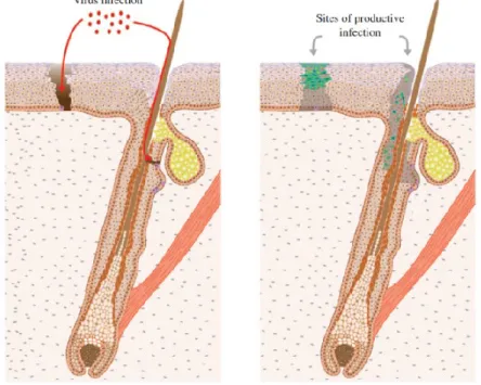
28 damage necessary to generate malignant stem cells. These clinical and molecular observations suggest fundamental differences in the way that β types and high-risk α types cause cancers. To initiate an infection, β-HPV need to gain access to the basal keratinocytes, and probably a KSC within the basal cell layer, which is normally protected by differentiated stratified epithelium. In many cases, it is thought that this is facilitated by an exogenous (micro) trauma or other localized break in the epithelial barrier. However, at particular epithelial sites, infection may be less dependent on epithelial damage. Current thinking suggests that β-HPV infects a long-lived, slow-cycling KSC, such as may be found in the HF bulge region (Figure 7 left panel). Division of these infected cells populates the surrounding basal layer with cells containing viral episomes, with completion of the β-HPV life cycle upon differentiation (Figure 7 right panel).
Figure 7. To initiate infection, HPV virions must gain access to the epithelial basal cells and probably an epithelial stem cell localized in the bulge region (coloured purple), either through a wound or possibly through the hair follicle (left pannel). Once the wound has been repaired and a β-HPV lesion has formed (right pannel), it is thought that the viral genome persists as a low copy number episome in the basal layer, often with limited viral gene expression. Cell cycle entry (cells with red nuclei) can be driven by the viral E6/E7 proteins, with E4 expression (green), genome ampification (dark blue) and L1 expression (yellow) occurring as the infected cell migrates towards the epithelial surface. It is thought that viral gene expression may become deregulated in EV and immunosuppressed individuals (Quint et al., 2015).
Based on the idea that β-HPVs have evolved to persist in a long-lived, slow-cycling stem cell, it is not surprising that, unlike the high-risk α types, the β-HPV episome is incapable of being
29 reliably maintained in faster-cycling cancer cells. This is consistent with the proposed ‘hit and run’ mechanism of carginogenesis. According to this model, the viruses might play a role in early stages of the pathogenesis of KC – particularly SCC, which appear to be the KC type with stronger association to β-HPV infection - as indirect and possibly transient carcinogens. β-HPV could being involved in tumour initiation in the presence of additional risk factors rather than in malignant progression or in the maintenance of the transformed phenotype as on the contrary occurs in high-risk α-HPV-driven cervical tumorigenesis (Borgogna, Lanfredini et al., 2014).
The “hit and run” hypothesis is supported by the following evidences:
the lower transforming potential of β-HPV oncoproteins compared to high-risk α-HPV oncoproteins
the low viral load and minimal gene expression in non-EV tumor tissues, and the fact that even in EV SCC the higher amount of viral DNA and gene expression is confined to few tumor cells
the high prevalence of β-HPV DNA at high viral loads in AK precursor lesions
the lack of evidence for β-HPV DNA integration in tumour cells, which on the contrary is a pivotal event in high-risk α-HPV-induced cervical cancer.
Taken togheter, these evidences suggest that β-HPV oncogene expression in early stages of SCC development could be a condition facilitating a transforming process previously triggered and subsequently carried on by other tumorigenic factors such as UV exposure, causing DNA damage, and immunosuppression, a condition that allows the reactivation of β-HPV.
It seems that β-HPVs, especially by means of the E6 oncoprotein, can inhibit either the apoptotic pathways triggered by UV-induced DNA damage or the repair of DNA damage itself, favouring a condition of genetic instability that may lead to the accumulation of mutations in proto-oncogenes and tumour suppressor genes and subsequently to host cell transformation (Akgül et al., 2006). In this picture, continuous β-HPV persistence could be no longer required when the cells are already initiated toward malignancy and, in more advanced stages of the carcinogenic process, the viruses might finally be lost, substained by the fact that β-HPV is not integrated into the human cellular DNA.
In conclusion, two important factors (UV radiation and immunosuppressive medication) contribute to the development of KC in OTR. In addition to this, a role for β-HPVs in the development of early premalignant lesions and KC has been proposed in individuals with compromised antiviral immune surveillance mechanisms. This might be due to an increased
30 viral replication and gene expression in the context of a reactivation of previously unapparent β-HPV infections kept under control by cell-mediated immunity rather than increased de novo β-HPV infections (Figure 8).
Figure 8. Proposed mechanism for β-HPV infection and keratinocyte carcinoma development. In immunocompetent individuals (left pannel), β-HPV infections are suppressed by the immune system. In this case, only low levels of β-HPV protein will be present to interact with host cell proteins involved in DNA damage repair and apoptosis, and genotoxic damage imposed by UV radiation will be managed adequately. In the presence of immunosuppression, however (right pannel), productive β-HPV infection occurs and sufficient amounts of β-HPV protein, particularly E6, are expressed, inhibiting DNA damage repair mechanisms and apoptosis. As a result, genomic instability develops in the infected keratinocytes, which may lead ultimately to the development of KC (Quint et al., 2015).
These proposed mechanisms may not be relevant in the above epidermal layers where cells have a short lifespan, but such effects could be significant in the KSC. Indeed, the persistent localization of β-HPVs in these cells – which may be the source of skin cancers – is likely to allow the acquisition of a condition of genetic instability that might transform them into cancers stem cells (Akgül et al., 2006; Quint et al., 2015).
2.2.4 Mouse models to investigate β-HPV role in keratinocyte carcinoma development
Different transgenic mice models have been developed so far for investigate the role of HPVs in skin cancer. The first transgenic mouse model for skin-associated HPVs was generated in 1992 at the University of Birmingham Medical School, by Searle’s group. In this model the Mupapillomavirus HPV-1 early region was placed under the control of epidermis specific promoter-keratin 6 gene. Morphological alterations of the normal epithelial differentiation have been observed. The effect was greatest on the tail, where the epithelium
31 became hyperproliferative in appearance, with several layers of irregular or cuboidal cells above the basal layer and an increase in the total number of cell layers with abnormal cornification. A similar transient flaky appearance also associated with epidermal hyperplasia was observed on other regions of the skin around 7 days of age. It is worth mentioning that this transgenic mice were not able to develop any spontaneous skin lesion (Tinsley et al., 1992).
To further evaluate the possible contribution of β-HPV in skin carcinogenesis, Pfister and collegues established a transgenic mouse model where the complete early region of HPV8 (E1, E2, E4, E6, E7) was expressed under the control of the Keratin 14 promoter. In these transgenic mice, the first phenotypic alterations detectable started around 8 weeks after birth, showing the presence of spontaneous tumor like growth on the back with hair loss, hyperkeratosis, and ulceration. Almost all of these HPV8 transgenic mice (91%) developed single or multifocal benign tumors, without any treatment with physical or chemical carcinogens. These tumours are characterized by papillomatosis, acanthosis, hyperkeratosis, and varying degrees of epidermal dysplasia; furthermore 6% of this transgenic mice developed SCC. The rapidity of skin tumor development in these mice is no doubt related to the permanent expression of the HPV8 early genes in all proliferation competent keratinocytes driven by the K14 promoter. With this model it could thus be shown, for the first time, that expression of β-HPV proteins leads to skin cancer development without exposure to any further physical or chemical carcinogens (Schaper et al. 2005).
Using transgenic technology, several others mouse lines have been developed expressing HPV8 E2 protein or also E6/E7 from other β-genotypes, such as HPV38 and HPV20. The HPV38Tg mice had no spontaneous formation of tumours during the lifespan, but they developed actinic keratosis-like lesions after treatment with chronic UV radiation. The same phenotype was observed in HPV20 E6/E7 transgenic mice where again UV radiation was needed to enhance proliferation of the epidermal layers of the skin that was reflected in the higher rate of papilloma formation. Interestingly, E2 seems to act as an oncogene as E2-transgenic mice also develop skin lesions, even though to a lower extent, more slowly and with less severe malignant features. HPV8, so far, has been the only cutaneous HPV type found to induce skin cancer completely on its own in the absence of any additional co-carcinogenic treatment, making it a good candidate to investigate how β-HPV gene expression can influence the keratinocytes differentiation (Schaper et al. 2005; Akgül et al., 2006; Michel et al., 2006; Pfefferle et al., 2008; Viarisio et al. 2011; De Andrea et al. 2010).