Università degli Studi di Foggia
Facoltà di Agraria
D
IPARTIMENTO DI SCIENZE AGRARIE,
DEGLI ALIMENTI E DELL’
AMBIENTE(SAFE)
PhD Course on Management of Innovation in the
Agricultural and Food Systems of the Maditerranean
Region (XXVI Cycle)
NUTRITION AND STRESS: A field study on the
effects of diet on stress-related responses in sheep
Coordinator:
Prof. Giancarlo Colelli
Tutor: PhD student:
Prof. Mariangela Caroprese dr. Maria Giovanna Ciliberti
2
CONTENTS
GENERAL INTRODUCTION ... 5
AIM OF THE PhD THESIS ... 11
REFERENCES ... 12
1. FIRST TRIAL: A mixture of phytosterols from Dunaliella tertiolecta affects proliferation of peripheral blood mononuclear cells and cytokine production in sheep ... 17
1.1 INTRODUCTION ... 18
1.2 MATERIALS AND METHODS ... 20
1.2.1 Separation and purification of total sterols fraction and of ergosterol / 7- dehydroporiferasterol mixture from Dunaliella tertiolecta ... 21
1.2.2 Analyses by gas chromatography-mass (tandem) spectrometry (GC-MS/MS) ... 21
1.2.3 Isolation of PBMC ... 24
1.2.4 Proliferation Suppression Assay and Cytokine Quantification ... 24
1.2.5 Determination of cytokines in culture supernatant by ELISA-test ... 25
1.2.6 Statistical Analysis ... 26
1.3 RESULTS ... 27
1.3.1 Proliferative response to ConA ... 27
1.3.2 Cytokine production by PBMC ... 28
1.4 DISCUSSION ... 31
1.5 CONCLUSION ... 36
1.6 REFERENCES ... 37
2. SECOND TRIAL: Hypothalamic-pituitary-adrenal axis activation and immune regulation in heat stressed sheep after polyunsaturated fatty acids supplementation ... 41
3
2.2 MATERIALS AND METHODS ... 44
2.2.1 Animals and experimental design ... 44
2.2.2 Body condition score and respiration rate ... 47
2.2.3 Plasma cortisol determination ... 47
2.2.4 In vivo cell-mediated immunity ... 48
2.2.5 Humoral immune response ... 48
2.2.6 Determination of cytokines in plasma samples by ELISA ... 49
2.2.7 HSPs determination in white blood cells lysate... 50
2.2.8 Statistical analysis ... 51
2.3 RESULTS ... 52
2.3.1 Meteorological Data ... 52
2.3.2 Dry Matter Intake, Body condition score and Respiration Rate ... 53
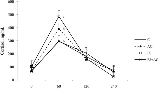
2.3.3 Plasma cortisol ... 54
2.3.4 In vivo cell-mediated immunity and humoral immune response ... 55
2.3.5 Plasma cytokines ... 56
2.3.6 Heat shock proteins ... 58
2.4 DISCUSSION ... 59
2.5 CONCLUSIONS ... 65
2.6 REFERENCES ... 66
3. THIRD TRIAL: Inflammatory responses of sheep peripheral blood mononuclear cells as affected by PUFA supplementation in the diet under high ambient temperature ... 72
3.1 INTRODUCTION ... 73
3.2 MATERIALS AND METHODS ... 75
3.2.1 Animals and Experimental Design ... 75
3.2.2 Isolation of PBMC ... 77
4
3.2.4 Determination of ILs in culture supernatant by ELISA-test ... 78
3.2.5 Statistical Analysis ... 80
3.3 RESULTS ... 81
3.3.1 Proliferative response to PHA ... 81
3.3.2 Cytokines production by PBMC ... 81
3.4 DISCUSSION ... 84
3.5 CONCLUSION ... 89
3.6 REFERENCES ... 90
4. FOURTH TRIAL: Effects of dietary n-3 from flaxseed on immune suppression of dairy ewes during post partum ... 96
4.1 INTRODUCTION ... 97
4.2 MATERIAL AND METHODS ... 98
4.2.1 Animals and experimental treatments ... 98
4.2.2 Sampling and analysis of blood ... 100
4.2.3 ELISA for anti-OVA IgG detection in plasma ... 100
4.2.4 Capture ELISA for IL-6, IL-1β, IL-10 and TNF-α detection in plasma . 102 4.2.5 Statistical analysis ... 104
4.3 RESULTS ... 105
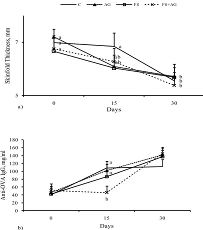
4.3.1 Humoral immune responses... 105
4.3.2 Cytokines detection in plasma ... 106
4.4 DISCUSSION ... 109
4.5 CONCLUSIONS ... 114
5 GENERAL INTRODUCTION
Stress, Nutrition and Hypothalamic-Pituitary-Adrenal (HPA) axis
Animal welfare can be affected by different condition of stress, and kinds of “stressors”: environmental, such as ambient temperature; and physiologicals, such as post partum period, with the starting of the lactation that influences the health status of lactating animals, increasing their susceptibility to the diseases development and reducing the production performances of the animal.
The physiological stress response consists in an activation of the sympathetic nervous system (SNS), a parasympathetic withdrawal, and increased activity of the HPA axis, that separately activates a response (Kunz-Ebrecht et al., 2003). Coupled with endocrine system the immune system interacts via activating different pathways, (Felten and Felten, 1994); as a consequence stress influences also the immune functions (Maier and Watkins, 1998).
Glucose and insulin plasma levels depend from dietary energy in dairy cows (Andersen et al., 2004), in condition of stress it is important the availability of glucose for central nervous system, indeed the glucocorticoids have a central role in energy mobilization during stress (Sapolsky et al., 2002). In ruminants the increase of the HPA-axis activity is connected with acute stress and causes the mobilization of the energy to cope with stress; moreover, the corticotrophin-releasing factor (CRF) system, at level of central nervous system (CNS), is involved in the regulation of feed intake together with behavioural resposes to stress (Koob, 1999). Hypercortisolemia caused by stress can alter the number of circulating lymphocytes (Dhabnar et al., 2009), and change the number and percentage of peripheral blood lymphocytes together with T-lymphocyte subsets, wich have an effect on animal immunocompetence (Caroprese et al., 2010).
6 The understanding of the nutrient requirements of dairy sheep at various stages of lactation and in different physiological conditions in essential for combining various feed ingredients to meet dairy animals needs.
Nutrition and Heat stress
Recent developments in housing and management practices of farm animals under heat stress reflect the increase in moral concerns about animal welfare; few papers discussed the interrelationships between heat stress and animal welfare, while much more it was been studied regarding the interaction between heat stress and livestock productivity in relation to the management systems (Silanikove, 2000). Moreover, the effect of heat stress is substantial in the subtropical-Mediterranean zones, and in farm animals raised in central and western Spain, or in the southern areas of France, Italy and Greece, are exposed annually for 3-5 months to considerable heat stress. Growth, milk production and reproduction are impaired under heat stress as a result of the drastic changes in biological functions caused by stress (Habeeb et al., 1992; Silanikove, 1992). However, these responses are also indications for poor welfare (Broom and Johnson, 1993).
The changes in the biological functions of sheep due to exposure to heat stress include the depression in feed intake and utilization, disturbances in the metabolism of water, protein, energy and mineral balances, enzymatic reactions, hormonal secretions and blood metabolites (Habeeb et al., 1992; Marai et al., 2000, 2003, 2004, 2006a).
Sheep convert fibrous, low quality feedstuffs into meat (protein), milk and other products in a better manner than cattle (Hafez, 1987). However, moderate heat stress reduces intakes and growth in young sheep consuming a high feed intake of medium quality roughage diets; this does not affect the relative
7 responses to supplementation providing principally fermentable Metabolic Energy (ME) or a similar amount of fermentable ME and additional metabolizable protein (Dixon et al., 1999). Supplementation of brown seaweed (Tasco) to post-harvest fescue hay was found to enhance the immune system and protect the wether lambs against prolonged heat-induced oxidative stress (Saker et al., 2004). Exposure to high ambient temperatures augments the efforts to dissipate body heat, resulting in an increase of respiration rate, body temperature and consumption of water, and a decline in feed intake. Heat increment is defined as energy expenditures associated with the digestion and assimilation of food; for this the digestion and the metabolism of nutrient creates heat (Baldwin et al., 1980). A higher heat increment is caused by the specific dynamic action that accompanies the metabolism of feed which is highest in the case of poor quality, fibrous feedstuffs (Marai et al., 2001). It is possible to formulate diets by exploiting different efficiencies of nutrient utilization to decrease the heat increment. (West, 1999) Factors such as water deprivation, nutritional imbalance and nutritional deficiency may exacerbate the impact of heat stress. Sheep, however, recorded a lower sensitivity to heat stress, when compared to cattle, at a maintenance feed level. The provision of shade shelter is suggested as a practical measure applicable under extensive conditions (Silanikove-Nissim, 2000). Feeding excessive quantities of nutrients, such as crude protein, can contribute to reduced efficiency of energy utilization, potentially increasing the level of stress. Is it necessary understanding how dietary modification can minimize heat stress.
Heat stress can affect, moreover, the nutrition of animals by altering the dynamic characteristics of the digestion processes; recent studies on nutrient digestibility in ruminants under heat stress are contradictory: in cattle exposed to
8 hot environments the diet digestibility increased (Weniger and Stein 1992); in contrast, negative or no relationships between high ambient temperatures and diet digestibility have been reported in dairy cattle and small ruminants (Silanikove, 1992). Furthermore, in ewes exposed to chronically heat stress, the diet digestibility was affected in a time-dependent manner, and this change suggested an adaptive response of the digestive tract to heat stress conditions, due to changes in bacterial concentration (Bernabucci et al., 2009); when the exposition to high temperature was finished, to re-establish the function of rumen and intestinal tract, it needed around 2 weeks, even though animals normalize clinical parameters and feed intakes.
Physiological state and nutrient restriction can influence thermoregulatory responses of ewes under heat stress condition (Mohammed et al., 2008).
Nutrition and post-partum period
Dairy animals during peri-partum period need to mobilize adipose tissue to compensate the relative deficit of glucose due to the ongoing of the lactation in concomitance with negative energy balance (Hachenberg at al., 2007).
In dairy cow, this phase of the reproductive life is particularly critical in terms of health, productivity and fertility (Roche et al., 2009). In the past two decades the reproductive efficiency of dairy cows registered a decrease with a concomitant increase of production of milk per cow; to the basis of reduction of reproductive efficiency there is an increasing of disease due to variable and often prolonged state of negative energy balance in dairy cow during peri-partum and early post-partum. It is well known that nutrition has a central role in regulation of reproductive function in cattle; in particular, acute nutritional restriction involved
9 in anovulation and detrimental effects on dominant follicle growth (Roche et al., 2000).
Furthermore, in the peripartum period many hormonal and metabolic alterations occur: a decrease of progesterone and oestradiol level at parturition and an increase of four-fold of plasma cortisol is observed; these alterations lead the functions of polymorphonuclear leukocytes causing immonosuppression (Dosogne et al. 1999) and consequently a major susceptibility of disease.
During post-partum the oxygen consumption increases, with consequently increase of reactive oxygen species (ROS), that caused oxidative stress related disorders, such as puerperal dysfunction, and alterations in colostrums and milk quality (Rizzo et al, 2007 and 2009).
In this metabolic conditions, an adequate dietary supplementation with antioxidant vitamins, such as Vitamin E, β-carotene and vitamin B12 may control
the oxidative disease, improve the immune system, and decrease the post-partum disease (Rizzo et al., 2013). The level of dietary supplementation with vitamins need to be understood because an excess of antioxidant may result in an increase of ROS generation, as demonstrated by Salganik (2001).
Supplementation of diet with fat, such as n3 and n-6 polyunsaturated fatty acids (PUFA) has been showed to be important modulators of immune reactions (Calder et al., 2002), and to supply additional energy requirements for growth as well as for milk production with an alteration of the endocrine profile favoring milk synthesis rather than body reserve replenishment (Drackley et al., 2003).
Linoleic acid (18:2n6) can be converted though enzymatic reactions into arachidonic acid, which is precursor of the pro-inflammatory mediators, prostaglandin E2. The α-linolenic acid (C18:3n3), can be converted in
10 eicosapentaenoic acid, that is precursor, instead, of prostaglandin E3 (PGE3) and
leukotriene B4, that trigger less severe inflammatory reactions than PGE2 and
leukotriene B4 (Yaqoob and Calder, 1995). Dietary supplementation with fat
around parturition was demonstrated to reduce the production of inflammatory factors that could contribute to attenuate the immunesuppressive effect during parturition and could re-establish the immune function against pathogens (Lessard et al., 2004). Dairy cattle fed with whole flaxseed, rich in omega 3 fatty acids, registered a modification of both secretion of prostaglandin and reproduction (Petit et al., 2002). Furthermore, the concentration of progesterone (P4) and PGE2
were affected from the composition of unsaturated fatty acids supplemented in the diet, particularly improving reproductive performance, because both molecules are implicated in the regulation of uterine and systemic immune responses (Lessard et al., 2004). During transition period Caroprese et al., (2006), found that IL-6 level in plasma of dairy ewes can be used to as a reliable indicator of stress connected with lambing; in ewes with multiple lambing were characterized by impaired immune functions and consequently more subjected to invasion of pathogens.
11 AIM OF THE PhD THESIS
The aim of the present thesis was the study of the effects of nutrition on immune system of dairy ewes under different types of stressors.
The experiments were divided in four different trials. The first experiment was undertaken to evaluate the potential effects of phytosterols extracted from a microalga, intented to be used in sheep nutrition as feed supplements, on in vitro immunological responses of cells from dairy ewes. The second and the third trials were undertaken in order to evaluate the effects of PUFA supplementation on welfare and immune responses of sheep under heat stress. The second trial evaluated the effects of supplementation with polyunsaturated fatty acids from seaweed and flaxseed on welfare and in vivo immunological profile and HPA-axis activation during exposition of ewes to high ambient temperature. The third experiment studied the effects of supplementation with polyunsaturated fatty acids from seaweed and flaxseed on ex vivo inflammatory profile of cells from dairy ewes under high ambient temperature. The fourth trial evaluated the effects of supplementation with polyunsaturated fatty acids from flaxseed on immunological profile in dairy ewes during post partum.
12 REFERENCES
Andersen J.B., Friggens N.C., Larsen T., Vestargaaard M., Ingvartsen K.L. 2004. Effects of energy density in the diet and milking frequency on plasma metabolism and hormones in early lactating dairy cows. J. Vet. Med. A 51, 52-57.
Baldwin R.L., Smith N.E., Taylor J., Sharp M. 1980. Manipulating metabolic parameters to improve growth rate and milk secretion. J. Anim. Sci. 51, 1416-1428.
Bernabucci U., Lacetera N., Danieli P.P., Bani P., Nardone A., Ronchi B. 2009. Influence of different periods of exposure to hot environment on rumen function and diet digestibility in sheep. Int. J. Biomet. 53, 387-395.
Broom D.M., Johnson K.G. 1993. Stress and animal welfare. Chapman and Hall, London.
Calder P.C., Yaqoob P., Thies F., Wallace F.A., Miles E.A. 2002. Fatty acids and lymphocyte functions. Brit. J. Nutr. 87, S31-S48.
Caroprese M., Albenzio M., Marzano A., Schena L., Annicchiarico G., Sevi A. 2010. Relationship between cortisol response to stress and behavior., immune profile, and production performance of daity ewes. J. Dairy Sci. 93, 2395-2403. Dhabhar F. S. 2009. Enhancing versus suppressive effects of stress on immune
function: Implications for immunoprotection and immunopathology. Neuroimmunomodulation 16, 300-317.
Dixon R.M., Thomas R., Holmes J.H.G. 1999. Interaction between heat stress and nutrition in sheep and nutrition in sheep fed roughage diets. J. Agric. Sci. 132, 351-359.
13 Dosogne H., Burvenich C., Freeman A.E., Kehrli Jr.M.E., Detilleux J.C., Sulon J., Beckers J-F-, Hoeben D. 1999. Pregnancy-associated glycoprotein and decreased polymorphonuclear leukocyte function in early post-partum dairy cows. Vet. Imm. Immunopat. 67, 47-54.
Drackley J.K., Cicela T.M. LaCount D.W. 2003. Responses of primiparous and multiparous Holstein cow to additional energy form fat or concentrate during summer. J. Dairy Sci. 86, 1306-1314.
Felten S., Felten D. 1994. Neural-immune interactions. Prog. Brain Res. 100, 157-162.
Habeeb A.A.M., Marai I.F.M., Kamal T.H. 1992. Heat stress. In: Phillips C., Pigginns D. (Eds.), Farm Animals and the Environment. CAB International, Wallingford, UK, pp. 27-47.
Hafez E.S.E. 1987. Reproduction in Farm Animals, fifth ed. LEA and Febiger, Philadelphia.
Hebeeb A.A., Marai I.F.M., Kamal T.H. 1992. Heat stress. In: Philips C., Piggens D. (Eds.), Farm Animals and the Environment C.A.B. International, pp. 27-47. Koob G.F., 1999. Corticotropin-releasing factor, norepinephrine and stress. Biol.
Psych. 46, 1167-1180.
Kunz-Ebrecht Sabine R, Mohamed-Ali V., Feldman P.J., Kirschbaum C., Steptoe A. 2003. Cortisol responses to mild psychological stress are inversely associated with proinflammatory cytokines. Brain Behav. Immun. 17, 373-383. Lessard M., Gagnon N., Godson D.L. Petit H.V. 2004. Influence of parturition
and diets enriched in n-3 or n-6 polyunsatured fatty acids on immune response of dairy cows during the transition period. J. Dairy Sci. 87, 2197-2210.
14 Maier S.F., Watkins L.R. 1998. Cytokines for psychologists: Implications of bidirectional immune-to-brain communication for understanding behaviour, mood, and cognition. Psychol. Rev. 105, 83-107.
Marai I.F.M., Bahgat L.B., Shalaby T.H., Abdel-Hafez M.A. 2000. Fattening performance, some behavioural traits and physiological reactions of male lambs fed concentrates mixture alone with or without natural clay, under hot summer of Egypt. Ann. Arid Zone 39, 449-460.
Marai I.F.M., Ayyat M.S., Abd El-Monem U.M. 2001. Growth performance and reproductive traits at first parity of New Zealand White female rabbit as affected by heat stress and its alleviation, under Egyptian condition. Trop. Anim. Health Prod. 33, 457-462.
Marai I.F.M., El-Darawany, A.A., Abou-Fandoud E.M., Abdel-Hafez, M.A.M. 2003. Alleviation of heat stressed Egyptian Suffolk rams by treatment with selenium, melatonin or prostaglandin F2α during hot summer of Egypt. J.
Anim. Vet. Adv. 2, 215-220.
Marai I.F.M., El-Darawany A.A., Abou-Fondoud E.M., Abdel-Hafez M.A.M. 2004. Reproductive traits and the physiological background of the seasonal variation in Egyptian Suffolk ewes under the condition of Egypt. Ann. Arid. Zone (India) 42, 1-9.
Marai I.F.M., El-Darawany A.A., Abou-Fandoud, E.I. Abdel-Hafez M.A.M. 2006. Serum blood components during pre-oestrus, oestru and pregnancy phases in Egyptian Suffolk as affected by heat stress, under the conditions of Egypt. Egypt. J. Sheep Goats Desert Anim. Sci. 1, 47-62.
15 Mohammed E.E., Abdalla M.A., Shraf Eldin A.M. 2008. Thermoregulation and reproductive performance of grazing Desert ewes (Ovis aries) as influenced by concentrate supplementation. Pak. J. Biol. Sci. 11, 2209-2216.
Petit H.V. 2002. Digestion, milk, production, milk composition, and blood composition of dairy cows fed whole flaxseed. J. Dairy Sci. 85, 1482-1490. Rizzo A., Minoia G., Trisolini C., Manca R., Sciorsci R.L. 2007. Concentration
free radicals and beta-endorphins in repeat breeder cows. Anim. Repr. Sci. 100, 257-263.
Rizzo A., Minoia G., Trisolini C., Mutinati M., Spedicato M., Jirillo F., Sciorsci R.L. 2009. Reactive Oxygen Species (ROS): involvement in bovine follicular cyst etiopathogenesis. Immunopharmacol. Immunotoxicol. 31 631-635. Rizzo A., Pantaleo M., Mutinati M., Minoia G., Trisolini C., Ceci E., Sciorsci
R.L. 2013. Blood and milk oxidative status after administration of different antioxidants during early post partum in dairy cows. Res. Vet. Sci. 95, 1171-1174.
Roche J.F., Mackey D., Diskin M.D. 2000. Reproductive management of postpartum cows. An. Repr. Sci. 60-61, 703-312.
Roche J.R., Friggens N.C., Kay J.K:, Fisher M.W., Stafford K.J., Berry D.P. 2009. Invited review: body condition score and its association with dairy cow productivity, health, and welfare. J. Dairy Sci. 92, 5769-5801.
Hachenberg S., Weinkauf C., Hiss S., Sauerwein H. 2007. Evaluation of classification modes potentially suitable to identify metabolic stress in healthy dairy cows during the peripartal period. J. Anim. Sci. 85, 1923-1932.
16 Saker K.E., Fike J.H., Veit H., Ward D.L. 2004. Brown seaweed-(TascoTM) treated conserved forage enhances antioxidant status and immune function in heat-stressed wether lambs. J. Anim. Pgysiol. a. Anim. Nutr. 88, 122-130. Salganik R.I. 2001. The benefit and the hazards of antioxidants: controlling
apoptosis and other protective mechanisms in cancer patients and the human population. J. Am. Coll. Nutr. 30, 464-472.
Sapolsky R.M. 2002. Endocrinology of the stress-response. In: Becker J. B., Breedlove S. M., Crews D., McCarthy (eds.) Behavioral Endocrinology. A Bradford Book, The MIT Press (London) pp. 409-451.
Silanikove N. 1992. Effects of water scarcity and hot environment on appetite and digestion in ruminants: a review. Livest. Prod. Sci. 30, 175-194.
Silanikove N. 2000. Effects of heat stress on the welfare of extensively managed domestic ruminants. Livest. Prod. Sci. 67, 1-18.
Silanikove-Nissim, 2000. Effects of heat stress on the welfare of extensively managed domestic ruminants. Livest. Prod. Sci. 67 1-18.
Weniger J.H., Stein M. 1992. Einfluss von Ungebungstemperatur und Luftfeuchte auf die Nahrstoffverdaulichkeit beim Schaf. 1. Problemstellung, Durchfuhrung der Untersuchungen, Verdaulichkeit. Zuchtungskunde. 64, 148-155.
West J.W. 1999. Nutritional strategies for managing the heat-stressed dairy cow. J. Anim. Sci. 77, Suppl. 2/J Dairy Sci. 82, Suppl. 2/1999.
Yoqoob P. and Calder P.C. 1995. The effect of dietary lipid manipulation on the production of murine T-cell derived cytokines. Cytokine 7, 548-553.
17
FIRST TRIAL
A mixture of phytosterols from Dunaliella tertiolecta affects
proliferation of peripheral blood mononuclear cells and cytokine
18 1. INTRODUCTION
Plants, fungi, and algae are a source of biological active compounds which have a key role in the production of nutraceutical foods and of pharmaceutical products (Lordan et al., 2011). Among the biological compounds extracted from plants, fungi, and algae, phytosterols have a key role for human nutrition. Sterol biosynthesis lead to different final products in different organisms: plants produce sitosterol, stigmasterol, campesterol and spinasterol (Salimova et al., 1999), fungi produce mainly ergosterol. Green algae are characterized by a wide range of sterols in their composition such as chondrillasterol, poriferasterol, 28-isofucosterol, ergosterol, cholesterol and others (Patterson, 1982). Phytosterols have been investigated in human studies for their role in lipid metabolism because of their cholesterol-lowering activity. This biological effect is due to the structure of phytosterols that are very similar to cholesterol (Ling et al., 1995). It is generally assumed that LDL cholesterol reduction results directly from inhibition of cholesterol absorption through displacement of cholesterol from lipidic micelles during intestinal absorption of phytosterols contained in the diet without changes in HDL cholesterol level (Methiev et al., 2008). Beside their action on LDL cholesterol, however, phytosterols have been proposed as therapeutic compounds for their immunomodulatory properties. Ergosterol and ergosterol peroxide extracted from mushrooms have been shown to suppress proliferation of human and mouse cells, when stimulated with mitogens, to inhibit the growth of some cancer cells, and to induce apoptosis of human leukemia cells (Kobori et al., 2007; Kuo et al., 2003). Ergosterol is a precursor of vitamin D2 and can be
converted to vitamin D2 by UV irradiation (Jasinghe and Perera, 2003).
19 production of some cytokines. It has been shown (Kuo et al., 2011) that, when tested on LPS-stimulated macrophages in vitro, ergosterol can inhibit the production of TNF-α and the expression of COX-2. Ergosterol, together with a large variety of other phytosterols, is contained also in algae and microalgae. The utilization of algae and microalgae for phytosterols purification and extraction could be an opportunity for finding new molecules of particular interest for human health or a mixture of molecules able to enhance the nutraceutical activities of single phytosterols by synergistic mechanisms.Recently the microalga Dunaliella tertiolecta was found to be a source of phytosterols, among which ergosterol and 7-dehydroporiferasterol were the most abundant; as a consequence its utilization for commercial production of phytosterols has been suggested (Francavilla et al., 2010). To the best of our knowledge no studies have investigated the immunological activity of a mixture of ergosterol and 7-dehydroporiferasterol on animals or human cells. Based on previous findings, the study of the suppressive effects of a mixture of phytosterols on the immunological responses of ovine PBMC could be remarkable and of practical use for reducing tissue damaging and uncontrolled immune activation resulting from cytokine release during inflammatory diseases, such as fly strike, mastitis and acute parasitoses in sheep. The evaluation of sheep PBMC responses in vitro to a mixture of phytosterols purified and extracted from Dunaliella tertiolecta could be preliminary to the use of microalgae extracts in sheep feeding, as feed supplements. The choice of the sheep model to study the effects of a mixture of phytosterols from Dunaliella tertiolecta could have a twofold advantage: firstly, the use of bioactive mixtures of proven immunosuppressive properties extracted from Dunaliella tertiolecta could be effective to control inflammatory diseases in sheep production systems;
20 secondly, the supplementation of sheep diet with bioactive mixtures could contribute to the potential enrichment of sheep dairy products in phytosterols. From this point of view studies on sheep immune response could be used to better understand human immune responses to enriched food. Nowadays, no information, however, on the possible bio-hydrogenation occurring in the rumen environment due to rumen microorganisms are available. The mixture extracted from Dunaliella tertiolecta, however, could be microencapsulated by spray-drying or spray-congealing, as reported in Eldem et al. (1991) to improve its intestinal absorption and to escape rumen bio-hydrogenation.
We hypothesized that a mixture of phytosterols extracted from the microalga Dunaliella tertiolecta might have immunomodulatory properties on sheep cells. Furthermore, it could be crucial to evaluate the dose of phytosterols at which their immunomodulatory properties become apparent for future in vivo applications in sheep feeding strategies. This study, therefore, was undertaken to evaluate the effects of a mixture of total phytosterols, or a mixture of only ergosterol and 7-dehydroporiferasterol extracted from the microalga Dunaliella tertiolecta on the proliferative response and cytokine production of sheep peripheral blood mononuclear stimulated cells.
1.2 MATERIALS AND METHODS
1.2.1 Separation and purification of total sterols fraction and of ergosterol / 7- dehydroporiferasterol mixture from Dunaliella tertiolecta
Total sterols were extracted and separated from Dunaliella tertiolecta as described by Francavilla et al. (2010). Briefly, total lipids were extracted, according to Bligh and Dyer (1959), from 0.5 g of freeze dried biomass of D.
21 tertiolecta grown in the Walne’s culture medium (modified from Laing, 1991), at a salt concentration of 0.6 M NaCl. Extracted lipids were saponified and the unsaponified fraction (containing sterols not esterified) was separated and concentrated. Total sterols from unsaponified material were isolated by preparative thin layer chromatography (TLC 20 20 cm, silica gel 60 Å, layer thickness 500 m) developed in one dimension in n-hexane /ethyl acetate 8:2 (v/v).
Silver Ion Flash Chromatography (Ag-FLC) was used for purification of ergosterol (22E,24R)-methylcholesta-5,7,22-trien-3β-ol) and 7-dehydroporiferasterol ((22E,24R)-ethylcholesta-5,7,22-trien-3β-ol) as described by Francavilla et al. (2012). The column was eluted in isocratic conditions using as eluent n-hexan- ethyl acetate 8:2 (v/v). Fractions containing only ergosterol and 7-dehydroporiferasperol were combined and weighed after the solvent was removed on a rotary evaporator (Büchi Rotavapor).
1.2.2 Analyses by gas chromatography-mass (tandem) spectrometry (GC-MS/MS)
Purified sterols fraction of D. tertiolecta were analyzed by gas chromatography-mass spectrometry. A Varian Saturn 2200 GC/MS/MS ion trap (Varian Analytical Instruments, Walnut Creek, CA) was used. Identification of sterols was based on the comparison of their retention times relative to authentic standards, mass spectra of authentic standards, and available spectra in NIST05 and Wiley 07 mass spectral libraries as described by Francavilla et al. (2010).
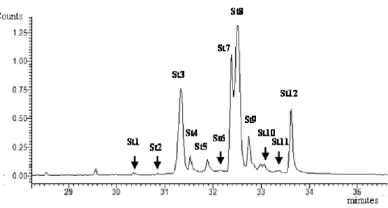
22 Figure 1.1 represents a ion chromatogram (GC-MS) of Total Sterol Fraction extracted from Dunaliella tertiolecta.
Figure 1.1 Ion Chromatogram (GC-MS) of Total Sterol Fraction extracted from Dunaliella
tertiolecta: St1=Cholesta-5-en-3β-ol[Cholesterol], St2=(22E,24R)Methylcholesta-5,7,9(11),22-tetraen3βol[9(11)Dehydroergosterol], St3=(22E,24R)-Methylcholesta-5,7,22-trien-3β-ol
[Ergosterol], St4= (22E,24R)-Methyl-5-cholesta-7,22-dien-3β-ol [5-Dihydroergosterol],
St5=(24)-Methyl-5-cholesta-8(14)-en-3β-ol,
St6=(22E,24R)-Ethylcholesta-5,7,9(11),22-tetraen-3β-ol, St7=(24S)-Methyl-5-cholesta-7-en-3β-ol [Fungisterol],
St8=(22E,24R)-Ethylcholesta-5,7,22-trien-3β-ol [7-Dehydroporiferasterol], St9=(22E,24R)-Ethyl-5 -cholesta-7,22-dien-3β-ol [Chondrillasterol], St10=(24)-Ethyl-5-cholesta-8(14)-en-3β-ol, St11=(24 )-Ethylcholesta-5,7-dien-3β-ol, St12=(24S)-Ethyl-5 -cholesta-7-en-3β-ol[22-Dihydrochondrillasterol].
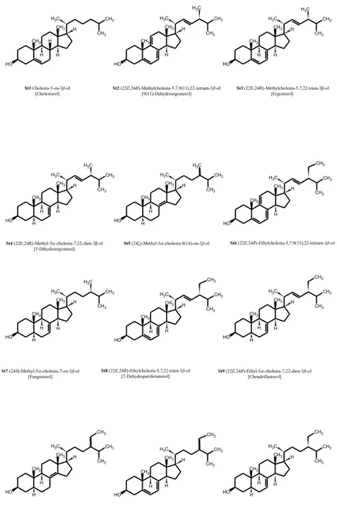
23 The molecular structures of twelve sterols purified from Dunaliella tertiolecta are reported in Figure 1.2.
Figure 1.2 Molecular structures of twelve sterols purified from Dunaliella tertiolecta.
CH3 CH3 C H3 H H H H CH3 CH3 O H CH3 CH3 C H3 H H CH3 CH3 O H C H3 CH3 CH3 C H3 H H H CH3 CH3 O H C H3 CH3 CH3 C H3 H H H CH3 CH3 O H C H3 H CH3 CH3 C H3 H H CH3 CH3 O H C H3 H CH3 CH3 C H3 H H CH3 CH3 O H CH3 CH3 CH3 C H3 H H CH3 CH3 O H C H3 H H CH3 CH3 C H3 H H CH3 CH3 O H CH3 H CH3 CH3 C H3 H H CH3 CH3 O H H H CH3 CH3 CH3 C H3 H H CH3 CH3 O H H CH3 CH3 CH3 C H3 H H CH3 CH3 O H CH3 H CH3 CH3 C H3 H H CH3 CH3 O H H CH3 H St2 (22E,24R)-Methylcholesta-5,7,9(11),22-tetraen-3β-ol [9(11)-Dehydroergosterol] St3 (22E,24R)-Methylcholesta-5,7,22-trien-3β-ol [Ergosterol] St1 Cholesta-5-en-3β-ol [Cholesterol] St4 (22E,24R)-Methyl-5-cholesta-7,22-dien-3β-ol [5-Dihydroergosterol]
St5 (24)-Methyl-5-cholesta-8(14)-en-3β-ol St6 (22E,24R)-Ethylcholesta-5,7,9(11),22-tetraen-3β-ol
St7 (24S)-Methyl-5-cholesta-7-en-3β-ol [Fungisterol] St8 (22E,24R)-Ethylcholesta-5,7,22-trien-3β-ol [7-Dehydroporiferasterol] St9 (22E,24R)-Ethyl-5-cholesta-7,22-dien-3β-ol [Chondrillasterol]
St11 (24)-Ethylcholesta-5,7-dien-3β-ol St12 (24S)-Ethyl-5-cholesta-7-en-3β-ol [22-Dihydrochondrillasterol] St10 (24)-Ethyl-5-cholesta-8(14)-en-3β-ol CH3 CH3 C H3 H H H H CH3 CH3 O H CH3 CH3 C H3 H H CH3 CH3 O H C H3 CH3 CH3 C H3 H H H CH3 CH3 O H C H3 CH3 CH3 C H3 H H H CH3 CH3 O H C H3 H CH3 CH3 C H3 H H CH3 CH3 O H C H3 H CH3 CH3 C H3 H H CH3 CH3 O H CH3 CH3 CH3 C H3 H H CH3 CH3 O H C H3 H H CH3 CH3 C H3 H H CH3 CH3 O H CH3 H CH3 CH3 C H3 H H CH3 CH3 O H H H CH3 CH3 CH3 C H3 H H CH3 CH3 O H H CH3 CH3 CH3 C H3 H H CH3 CH3 O H CH3 H CH3 CH3 C H3 H H CH3 CH3 O H H CH3 H St2 (22E,24R)-Methylcholesta-5,7,9(11),22-tetraen-3β-ol [9(11)-Dehydroergosterol] St3 (22E,24R)-Methylcholesta-5,7,22-trien-3β-ol [Ergosterol] St1 Cholesta-5-en-3β-ol [Cholesterol] St4 (22E,24R)-Methyl-5-cholesta-7,22-dien-3β-ol [5-Dihydroergosterol]
St5 (24)-Methyl-5-cholesta-8(14)-en-3β-ol St6 (22E,24R)-Ethylcholesta-5,7,9(11),22-tetraen-3β-ol
St7 (24S)-Methyl-5-cholesta-7-en-3β-ol [Fungisterol]
St8 (22E,24R)-Ethylcholesta-5,7,22-trien-3β-ol
[7-Dehydroporiferasterol] St9 (22E,24R)-Ethyl-5[Chondrillasterol] -cholesta-7,22-dien-3β-ol
St11 (24)-Ethylcholesta-5,7-dien-3β-ol St12 (24S)-Ethyl-5-cholesta-7-en-3β-ol [22-Dihydrochondrillasterol]
24 1.2.3 Isolation of PBMC
Ten Comisana sheep were randomly selected from the breeding flock at Segezia research station of the Council for Research and Experimentation in Agriculture (CRA-ZOE).
Isolation of PBMC was performed by density gradient centrifugation according to Wattegedera et al. (2004). Briefly, blood from each animal was diluted in cold PBS (pH 7.4), and centrifuged at 670g at 4°C for 20 min. The buffy coat was recovered, layered over a Ficoll gradient (1.077g/ml), and centrifuged at 1130g at 15°C for 30 min. The mononuclear cell band was recovered, washed and finally resuspended at a final concentration of 2x 10⁶ cells/ml in Iscove’s Modified Dulbecco’s medium (Sigma-Aldrich, Italy) supplemented with FBS and gentamicin.
1.2.4 Proliferation Suppression Assay and Cytokine Quantification
Lymphocyte proliferation assays were performed adding 100 μl of cell suspension into quadruplicate wells of 96 well U-bottom plates. PBMC were treated with synthetic ergosterol (E) (Sigma-Aldrich, Milan, Italy), a mixture of eleven Algae sterols extracted and purified from Dunaliella tertiolecta (Algae Extract, AE), a mixture of ergosterol and 7-dehydroporiferasterol extracted and purified from Dunaliella tertiolecta (Purified Extract, PE), and 50 μl of ConA (Sigma-Aldrich, Italy) at a final concentration of 5μg/ml. For each treatment, 0.0mg/ml, 0.2 mg/ml, 0.4mg/ml and 0.8mg/ml were tested on PBMC to verify the effects of sterols on their proliferation. Negative Control wells contained 100 μl of PBMC suspensions without mitogen (NC). Positive Control wells contained 100 μl of PBMC suspensions with ConA (Stimulated Cells, SC). Cell viability was greater than 90-95% in all the assays, as determined by the Trypan Blue exclusion
25 test. The plates were incubated at 37°C and 5% CO2 in a humidified incubator for
96 h. In order to test lymphocyte proliferation after 96 h of incubation a BrDU test was performed using a commercial kit (Roche, Milan, Italy). After 18h of incubation, BrDU incorporation during DNA synthesis was measured by reading optical density with a titer-ELISA spectrophotometer (Power Wave XS, Biotek, UK) at 450nm.
To determine the suppressive effects of Algae sterols on inflammatory responses, cytokines production by PBMC was evaluated: 100 μl of cell suspension were treated with 50 μl of LPS at a final concentration of 1μg/ml (Sigma-Aldrich, Milan, Italy), 50 μl of ConA at a final concentration of 5μg/ml (Sigma Aldrich, Milan, Italy), and 50 μl of E, AE, or PE (0.0mg/ml, 0.2 mg/ml, 0.4mg/ml and 0.8mg/ml) in 96 well U-bottom plates (Kobori et al., 2007). Control wells contained 100 μl of PBMC suspensions without LPS and mitogen. Positive Control wells contained 100 μl of PBMC suspensions with both LPS and ConA. The plates were incubated at 37°C and 5% CO2 in a humidified incubator for four
days. After 96 h of incubation, cell suspensions were centrifuged at 300 g at 4°C for 10 min, and cell-free supernatants from each well were collected and stored at-20 °C until ELISA-test time to measure cytokine production.
1.2.5 Determination of cytokines in culture supernatant by ELISA-test The levels of IL-6 and IL-1β in cell-free supernatants were determined by capture ELISA performed on 96-well microtiter plates, according to Caroprese et al. (2006). The presence of bindings for IL-6 and IL-1β was detected using sheep anti-rabbit IgG conjugated to horseradish peroxidase (HRP, Sigma Aldrich, Italy). The determination of IL-10 and TNF-α in cell-free supernatants was carried out by an ELISA test according to Kwong et al. (2002) and Hope et al. (2003),
26 respectively. Plates were read by a titer-ELISA spectrophotometer (Power Wave XS, Biotek, UK). Culture supernatants were read for IL-1 β detection against a standard curve obtained using scalar dilution of recombinant ovine IL-1β (CAB, Australia). Data were expressed as ng of IL-1β /ml. IL-6 data were expressed as optical density (OD). The ELISA for IL-10 detection was standardized using biologically-active recombinant ovine IL-10 expressed in Chinese hamster ovary (CHO) cells using the Gln synthetase (GS) expression vector™ (Lonza, UK). Production of recombinant ruminant cytokines using this system has been described in detail elsewhere (Graham et al., 1995; Entrican et al., 1996). Recombinant ovine IL-10 was provided by the BBSRC/RERAD Immunological Toolbox; recombinant bovine TNF-α was purchased from Serotec (UK). Data were expressed as bU of IL-10 /ml and pg of TNF-α/ml.
1.2.6 Statistical Analysis
All variables were tested for normality using the Shapiro-Wilk test (Shapiro and Wilk, 1965) and transformed into logarithm form to normalize their frequency distribution, when necessary. Then, data were processed by ANOVA using the GLM procedure of SAS (1999).
The model utilized was: yijkl = μ + αi + βij + γk + (αγ)ik + εijkl
Where μ=the overall mean; α=treatment β=animal effect within treatment; γ=dose; αγ=interaction of treatment x dose and ε=error. When significant effects were found (at P<0.05), the Student t-test was used to locate significant differences between means.
27 1.3 RESULTS
1.3.1 Proliferative response to ConA
On average proliferation of PBMC stimulated with Con A after 96 h of incubation was significantly higher than proliferation of PBMC stimulated with Con A and treated with E, AE and PE (P<0.001); in particular, on average the proliferation of PBMC stimulated with Con A in the presence of E was significantly lower (P<0.05) than proliferation of PBMC stimulated with only Con A.
After 96 h of incubation, the cells stimulated with Con A and PE showed a significantly different proliferation rate according to the dose of PE administrated (P<0.01, Fig. 1.3).
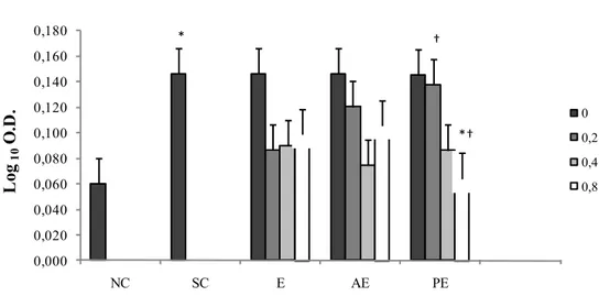
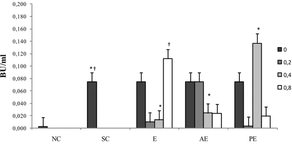
Figure 1.3 Proliferation of sheep PBMC following in vitro stimulation (Least Squares means ± SEM). PBMC were treated with 0.0mg/ml, 0.2 mg/ml, 0.4mg/ml and 0.8mg/ml of ergosterol (E), a mixture of eleven Algae sterols extracted and purified from Dunaliella tertiolecta (Algae Extract, AE), a mixture of ergosterol and 7-dehydroporiferasterol extracted and purified from Dunaliella tertiolecta (Purified Extract, PE), and ConA. Negative Control wells contained PBMC without mitogen (NC). Positive Control wells contained PBMC with ConA (Stimulated Cells, SC). (*) Shows a significant difference ( P < 0.05) between PBMC proliferation treated with 0.8 mg/ml PE and ConA stimulated cells (SC). (†) Shows a significant difference between PBMC proliferation treated with 0.2 mg/ml and 0.8 mg/ml PE.
* † *† 0,000 0,020 0,040 0,060 0,080 0,100 0,120 0,140 0,160 0,180 NC SC E AE PE L og 10 O .D . 0 0,2 0,4 0,8
28 Con A-stimulated cells showed higher proliferation than cells treated with Con A and PE at a concentration 0.8 mg/ml. In addition, cells treated with Con A and PE at a concentration of 0.8 mg/ml showed a lower proliferation than cells treated with Con A and PE at a concentration of 0.2 mg/ml (P=0.07).
1.3.2 Cytokine production by PBMC
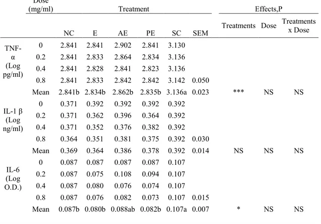
The production of TNF-α, IL-6, IL-1β at different concentrations of sterols, in supernatant by stimulated cells is shown in Table 1.1.
Means followed by different letters are significantly different at P<0,05. NS: non significant.
*** P<0.001 *P<0.05
Table 1.1 TNF-α, IL-1β and IL-6 production (Least Squares means ± SEM) in LPS and ConA
stimulated sheep pheripheral blood mononuclear cells (PBMC). PBMC were treated with 0.0mg/mL, 0.2 mg/mL, 0.4mg/mL and 0.8mg/L of ergosterol (E), a mix of eleven Algae sterols extracted and purified from Dunaliella tertiolecta (Algae Extract, AE), a mix of ergosterol and 7- dehydroporiferasterol extracted and purified from Dunaliella tertiolecta (Purified Extract, PE), LPS and ConA. Negative Control wells contained PBMC without mitogens (NC). Positive Control wells contained PBMC with LPS and ConA (Stimulated Cells, SC).
Dose
(mg/ml) Treatment Effects,P
NC E AE PE SC SEM Treatments Dose
Treatments x Dose TNF-α (Log pg/ml) 0 2.841 2.841 2.902 2.841 3.130 0.2 2.841 2.833 2.864 2.834 3.136 0.4 2.841 2.828 2.841 2.823 3.136 0.8 2.841 2.833 2.842 2.842 3.142 0.050 Mean 2.841b 2.834b 2.862b 2.835b 3.136a 0.023 *** NS NS IL-1 β (Log ng/ml) 0 0.371 0.392 0.392 0.392 0.392 0.2 0.371 0.362 0.396 0.364 0.392 0.4 0.371 0.352 0.376 0.382 0.392 0.8 0.364 0.351 0.381 0.375 0.392 0.030 Mean 0.369 0.364 0.386 0.378 0.392 0.014 NS NS NS IL-6 (Log O.D.) 0 0.087 0.087 0.087 0.087 0.107 0.2 0.087 0.075 0.108 0.094 0.107 0.4 0.087 0.080 0.076 0.074 0.107 0.8 0.087 0.076 0.082 0.073 0.107 0.015
29 Cells stimulated with LPS and Con A in presence of E, AE and PE had a highly significantly lower mean production of TNF-α than cells stimulated with LPS and Con A (P<0.001). TNF-α produced by cells in the presence of E, AE and PE was similar to the production of TNF-α by unstimulated cells. In addition, no differences were detected among TNF-α produced by cells in the presence of different doses of E, AE and PE.
Mean values of IL-6 production by cells stimulated with LPS and Con A in the presence of both E and PE was significantly lower (P<0.05) than production of IL-6 by cells stimulated with LPS and Con A (0.080 ± 0.015 and 0.082 ± 0.015 vs 0.107 ± 0.015, respectively). No differences emerged for IL-6 production when cells were treated with different doses of E, PE, and AE.
Mean values of IL-1β production by cells stimulated with LPS and Con A or stimulated with LPS, Con A and different doses of E, PE, and EA did not show differences.
Cells stimulated with LPS and Con A produced on average significantly higher IL-10 in the presence of PE (P<0.1). Furthermore, significant differences emerged with regard to the dose of PE used in contact with cells. In fact, the IL-10 produced by cells stimulated with LPS and Con A in the presence of PE significantly increased (P<0.05, Figure 1.4) when the concentration of PE increased from 0.2 mg/ml to 0.4 mg/ml. IL-10 produced by cells stimulated with LPS and Con A in the presence of 0.4 mg/ml PE was higher than IL-10 produced by cells stimulated with LPS and Con A without Algae sterols or in the presence of 0.4 mg/ml E, and of 0.4 mg/ml AE.
30
Figure 1.4 IL-10 production (Least Squares means ± SEM), in LPS and ConA stimulated sheep
peripheral blood mononuclear cells (PBMC). PBMC were treated with 0.0mg/ml, 0.2 mg/ml, 0.4mg/ml and 0.8mg/ml of ergosterol (E), a mixture of eleven Algae sterols extracted and purified from Dunaliella tertiolecta (Algae Extract, AE), a mixture of ergosterol and 7-dehydroporiferasterol extracted and purified from Dunaliella tertiolecta (Purified Extract, PE), LPS and ConA. Negative Control wells contained PBMC without mitogens (NC). Positive Control wells contained PBMC with LPS and ConA (Stimulated Cells, SC). (*) Shows significant differences (P < 0.05) between IL-10 production by PBMC treated with 0.4 mg/ml PE and 0.4 mg/ml AE, with 0.4 mg/ml PE and 0.4 mg/ml E, and 0.4 mg/ml PE and LPS and ConA stimulated cells (SC). (†) Shows a significant difference between IL-10 production by PBMC treated with 0.8 mg/ml E and LPS and ConA stimulated cells (SC).
The production of IL-10 by cells stimulated with LPS and Con A in the presence of 0.8 mg/ml E was significantly higher than the production of IL-10 in the presence of 0.4 mg/ml E (P<0.05). Furthermore, IL-10 produced by the cells stimulated with LPS and Con A in the presence of 0.8 mg/ml E was higher (P<0.05) than IL-10 produced by cells stimulated with only LPS and Con A.
*† * * * † 0,000 0,020 0,040 0,060 0,080 0,100 0,120 0,140 0,160 0,180 0,200 NC SC E AE PE B U /m l 0 0,2 0,4 0,8
31 1.4 DISCUSSION
This study was undertaken to evaluate the immunomodulatory and anti-inflammatory activities of a particular mixture of phytosterols extracted from Dunaliella tertiolecta on sheep cells. Proven immunomodulatory properties of a mixture of phytosterols extracted from Dunaliella tertiolecta could validate their use to sustain animal health.
Ergosterol is the major sterol in the cell membrane of mushrooms, representing about 70% of the sterols in fungi (Kuo et al., 2011). In their studies, Kuo et al. (2011) and Kobori et al. (2007) demonstrated that ergosterol has anti-inflammatory properties on LPS-stimulated RAW 264.7 macrophages in vitro by inhibiting TNF-α production and COX-2 expression. TNF-α is a highly proinflammatory cytokine which is responsible for heightened inflammatory responses produced by a number of different cell types, including macrophages, lymphocytes, neutrophils, and epithelial cells (Angelini et al., 2005). TNF-α production can be induced by fungi, viruses, parasites, and bacterial wall products, such as bacterial lipopolysaccharide of the Gram-negative bacteria. TNF-α production can be also induced by bacterial toxins as well as by some cytokines, as IL-1 and IFN-γ. TNF-α has a wide range of biological effects including cell differentiation, tissue development and death, and proliferation. Kuo et al. (2011), studying the proteomic profile of cells incubated with ergosterol, found that the expression of NF-κB p65, which is identified as the dominant transcription factor for induction of TNF-α expression, was suppressed. As a result, ergosterol was able to suppress the expression of TNF-α in LPS-stimulated RAW 264.7 macrophages. Ergosterol peroxide, which is also extracted by Ascomycetes, is able to suppress proliferation of human lymphocytes under
32 stimulation with PHA (Kuo et al., 2003). No studies evaluated the biological effects of 7-dehydroporiferasterol, as well as of a mixture of sterols extracted by Dunaliella tertiolecta. However, it has been hypothesized that the biological effects of 7-dehydroporiferasterol could be similar to those of ergosterol because the two sterols differ only for an extra methyl group (Francavilla et al., 2010). In our study ergosterol (E), the mixture of sterols from Dunaliella tertiolecta (AE) and the purified Algae extract of 7-dehydroporiferasterol and ergosterol (PE) were able to suppress cell proliferation in sheep PBMC stimulated with ConA. These results seemed to suggest a similarity between the mixture of 7-dehydroporiferasterol, ergosterol, and the mixture of sterols based on their effects on PBMC. The increase of suppressive effects exerted at 0.8 mg/ml on sheep cell proliferation only by the mixture of ergosterol and 7-dehydroporiferasterol extracted and purified from Dunaliella tertiolecta, however, led us to the hypothesis that the presence of 7-dehydroporiferasterol may strengthen the inhibitory effects of ergosterol on cell proliferation with increasing concentrations of both sterols. A possible explanation of our results may be the synergic effects exerted by 7-dehydroporiferasterol and ergosterol at increasing concentrations. Suárez et al. (2005) demonstrated that the effects of sterols on cell proliferation depend on their structural features. Results from our study suggested that 7-dehydroporiferasterol and ergosterol, when administrated together, have synergic effects in reducing sheep cell proliferation. Another possible explanation of the suppressive effects of the mixture of ergosterol and 7-dehydroporiferasterol on cell proliferation could be their cytotoxic effects on ovine PBMC, caused by massive overload of their ambient environment. Anyway, we excluded this hypothesis considering that other authors tested higher concentration of
33 phytosterols on cell proliferation without finding effects on cells viability (Valerio and Awad, 2011; Kobori et al., 2007). The proved suppressive effects of the mixture of ergosterol and 7-dehydroporiferasterol on the immunological responses of ovine PBMC could be of some relevance when used as a feed supplement in sheep production systems to control inflammatory diseases and reduce tissue damages derived from uncontrolled immune activation.
When the effects of different sterols from Dunaliella tertiolecta on cytokine production by stimulated PBMC were tested, we found that ergosterol, the mixture of sterols from Dunaliella tertiolecta, and the purified 7-dehydroporiferasterol and ergosterol from Dunaliella tertiolecta were able to suppress TNF-α production by LPS and ConA stimulated sheep cells, thus supporting previous findings on the anti-inflammatory effects of sterols (Kobori et al., 2007; Kuo et al., 2011).IL-6 is a pleiotropic cytokine and has a wide range of immunological functions including stimulation of B cells and cytotoxic T cells. Both the expression and production of IL-6 are strongly controlled and mainly occur under inflammatory conditions, with IL-6 being responsible for the increase of acute phase proteins. IL-1 is known as an inflammatory cytokine involved in lymphocyte activation and in acute-phase response. Bacterial lipopolysaccaride, cytokines, such as TNF-α, interferon-γ, and IL-2, are able to induce IL-1 production (Stylianou and Saklatvala, 1998). In our study PBMC production of IL-6 in response to stimulation with LPS and ConA was reduced in presence of both ergosterol and of the mixture of ergosterol and 7-dehydroporiferasterol extracted and purified from Dunaliella tertiolecta, confirming a potential anti-inflammatory effect of both sterols and of the purified extract from Dunaliella tertiolecta. Kobori et al. (2007) found suppressive effects of ergosterol peroxide
34 on LPS-stimulated cells after 6 hour of incubation by a reduction of IL-1β expression. Our results on the absence of effects of sterols on IL-1β production may be caused by a general suppressive effects exerted by IL-6 on production of IL-1β by stimulated cells. In humans, IL-6 provided a negative feed-back signal on IL-1β production after 48 hours of cells incubation with LPS (Schindler et al., 1990). IL-6 has the ability to inhibit also the expression of IL-1β and TNF-α, and to stimulate expression of IL-1-receptor antagonist and soluble TNF-receptor (Bannerman, 2008). As a consequence, discrepancy between our and previous results on suppressive effects of sterols on IL-1β expression by stimulated cells may be attributed to different times of incubation of cells and subsequent increase in IL-6, which acted on cells with a reduction of IL-1β production. Furthermore, different results can be attributed to differences in the type of cells stimulated (sheep PBMC vs mouse RAW 264.7 macrophages).
IL-10 is a regulatory cytokine with anti-inflammatory properties which refer to its ability to down-regulate the production of proinflammatory cytokines and the activity of both macrophages and dendritic cells. In our study, cells stimulated with LPS and ConA displayed an increase in IL-10 production in presence of both ergosterol and of the mixture of ergosterol and 7-dehydroporiferasterol purified from Dunaliella tertiolecta, thus further suggesting the anti-inflammatory role of the extract containing the two sterols by Dunaliella tertiolecta. A number of studies demonstrated that the presence of phytosterols is able to up-regulate the IL-10 production in different cell lines (Valerio and Awad, 2011, Nashed et al., 2005). In our study both ergosterol and the mixture of ergosterol and 7-dehydroporiferasterol extracted from Dunaliella tertiolecta dose-dependently increased the production of IL-10 in LPS-stimulated sheep cells. In particular, our
35 results suggest that the optimal concentration of the mixture of ergosterol and 7-dehydroporiferasterol from Dunaliella tertiolecta to obtain the highest production of IL-10 was 0.4 mg/ml; further increase in the mixture of ergosterol and 7-dehydroporiferasterol from Dunaliella tertiolecta did not result in a further increase in IL-10 secretion. On the contrary, ergosterol alone achieved the highest production of IL-10 at 0.8 mg/ml. Kobori et al. (2007) reported an inhibition of the C/EBPβ DNA-binding activity in the nuclear extracts of macrophages incubated with both ergosterol peroxide and ergosterol at a concentration of 30 and 60 μM. Macrophages have been demonstrated to require C/EBPβ for IL10 production, when exposed to E. coli (Csóka et al., 2007). Based on previous findings a tentative explanation of the reduction of IL-10 production by sheep cells at increasing concentrations of the mixture of ergosterol and 7-dehydroporiferasterol from Dunaliella tertiolecta could be the possible inhibition of C/EBPβ DNA-binding activity exerted at increased concentrations of the mixture. It is worth noting that the inhibitory effects of both ergosterol peroxide and ergosterol on C/EBPβ DNA-binding activity were obtained by the highest concentrations tested by Kobori et al. (2007), which were similar to the highest concentration of sterols tested in our study. As a consequence, the increased IL-10 production by sheep cells in the presence of 0.4 mg/ml of the mixture of ergosterol and 7-dehydroporiferasterol from Dunaliella tertiolecta could be attributed to the absence of inhibition on C/EBPβ DNA-binding activity; on the contrary the increased concentration of the mixture up to 0.8 mg/ml could have inhibited the C/EBPβ DNA-binding activity, thus leading to a reduction of IL-10 production. The increase of IL-10 production by PBMC in our study can help to explain the reduction in IL-6 production in stimulated cells both in presence of
36 ergosterol and of the mixture of ergosterol and 7-dehydroporiferasterol purified from Dunaliella tertiolecta. It has been demonstrated that IL-10 can control inflammatory processes by regulating the production of inflammatory cytokines (Wattegedera et al., 2004). Caroprese et al. (2012) in cows found a relation between IL10 production and the decrease in IL-6 secretion. In addition, IL-10 has been demonstrated to strongly regulate proliferation of sheep PBMC (Wattegedera et al., 2004) and this could help to explain the reduction in PBMC proliferation exerted by the mixture of ergosterol and 7-dehydroporiferasterol purified from Dunaliella tertiolecta observed in our study.
1.5 CONCLUSIONS
We studied the immunomodulatory properties of a mixture of phytosterols, and a mixture of ergosterol and 7-dehydroporiferasterol extracted from Dunaliella tertiolecta, in relation to the immunomodulatory properties of ergosterol, on peripheral blood mononuclear cells isolated from sheep. The results of the present study evidenced a role of phyotosterols from Dunaliella tertiolecta on immune modulation and anti-inflammatory activity in sheep. The immunomodulatory and anti-inflammatory activities were more evident in the purified extract represented by a mixture of ergosterol and 7-dehydroporiferasterol, and might depend on the existence of a synergic effect of the structures of the two phytosterols.
The use of a mixture of sterols, purified and extracted from Dunaliella tertiolecta, could have particular innovative implications on the modulation of the immune reactions in sheep production systems to control tissue damages deriving from uncontrolled inflammatory reactions, when administrated as feed additives in sheep diet.
37 1.6 REFERENCES
Angelini, D.J., Hasday, J. D., Goldblum, S. E., Bannerman D. D., 2005. Tumor necrosis factor-α-mediated pulmonary endothelial barrier dysfunction. Curr. Respir. Med. Rev. 1, 233–246.
Bannerman D.D., 2008. Pathogen-dependent induction of cytokines and other soluble inflammatory mediators during intramammary infection of dairy cows. J. Anim. Sci. 87, 10-25.
Bligh, E.G., Dyer, W.J., 1959. A rapid method of total lipid extraction and purification. Can. J. Biochem. Phys. 37, 911–917.
Caroprese M., Albenzio M., Annicchiarico G., Sevi A., 2006. Changes Occurring in Immune Responsiveness of Single- and Twin-Bearing Comisana Ewes During the Transition Period. J. Dairy Sci. 89, 562-568.
Caroprese M., Albenzio M., Marino R, Santillo A., Sevi A., 2012. Immune response and milk yield of dairy cows fed graded levels of rumen-protected glutamine. Res. Vet. Sci. 93, 202-209.
Csóka B., Németh Z. H., Virág L., Gergely P., Leibovich S.J., Pacher P., Xiao C. Sun, Blackburn M.R., Vizi E. S., Deitch E. A., Haskó G., 2007. A2A adenosine receptors and C/EBPβ are crucially required for IL-10 production by macrophages exposed to Escherichia coli. Blood 110, 2685-2695.
Eldem, T., Speicer, P., Hincal, A., 1991. Optimization of spray-dryed and congealed lipid micropellets and characterisation of their surface morphology by Scanning Electron Microscopy. Pharm. Res. 8, 47-54.
Entrican, G., Deane, D., MacLean, M., Inglis, L., Thomson, J., McInnes, C., Haig, D.M., 1996. Development of a sandwich ELISA for ovine
38 granulocyte/macrophage colony-stimulating factor. Vet. Immunol. Immunopathol. 50, 105-115.
Francavilla M., Colaianna M., Zotti M., Morgese M. G., Trotta P., Tucci P., Schiavone S., Cuomo V., Trabace L., 2012. Extraction, Characterization and In Vivo Neuromodulatory Activity of Phytosterols from Microalga Dunaliella Tertiolecta. Curr. Med. Chem. 19, 3058-3067.
Francavilla M., Trotta P., Luque R., 2010. Phytosterols from Dunaliella tertiolecta and Dunaliella salina: A potentially novel industrial application. Biores. Technol. 101, 4144-4150.
Graham S.P., Jones G.E., MacLean M., Livingstone M., Entrican, G., 1995. Recombinant ovine interferon gamma inhibits the multiplication of Chlamydia psittaci in ovine cells. J. Comp. Pathol. 112, 185 -95.
Hope J.C., Whelan A.O., Hewinson R.G. Vordermeier M., Howard C.J., 2003. Maturation of bovine dentritic cells by lipopeptides. Vet. Immunol. Immunopathol. 95, 31-31.
Jasinghe, V.J., Perera C.O., 2003. Distribution of ergosterol in different tissues of mushrooms and its effect on the conversion of ergosterol to vitamin D2 by UV irradiation. Food Chem. 92, 541–546.
Kobori M., Yoshida M., Ohnishi-Kameyana M., Shinmoto H., 2007. Ergosterol peroxide from an edible mushroom suppresses inflammatory responses in RAW264.7 macrophages and growth of HT29 colon adenocarcinoma cells. Brit. J. Pharmacol. 150, 209-219.
Kuo C.F., Hsieh C.H., Lin W.Y., 2011. Proteomic response of LAP-activated RAW 264.7 macrophages to the anti-inflammatory property of fungal ergosterol. Food Chem. 126, 207-212.
39 Kuo Y.C., Weng S.C., Chou C.J., Chang T.T., Tsai W.J., 2003. Activation and proliferation signals in primary human T lymphocytes inhibited by ergosterol peroxide isolated from Cordyceps cicadae. Brit. J. Pharmacol. 140, 895-906. Kwong, L.S., Hope J.C., Thom M.L., Sopp P., Duggan S., Bembridge G.P.,
Howard C. J., 2002. Development of an ELISA for bovine IL-10. Vet. Immunol. Immunopathol. 85, 213–223.
Laing, I., 1991. Cultivation of marine unicellular algae. MAFF Laboratory. Leaflet No.67. Directorate of Fisheries Research Lowestoft, UK.
Ling W.H., Jones P.J.H., 1995. Minireview dietary phytosterols: a review of metabolism, benefit and side effects. Life Sci. 57 (3), 195-206.
Lordan S., Ross S. R., Stanton C., 2011. Marine Bioactives as Functional Food Ingredients: Potential to Reduce the Incidence of Chronic Diseases. Mar. Drugs 9(6), 1056–1100.
Methiev A.R., Misharin A. Yu., 2008. Biological activity of phytosterols and their derivates. Biochemistry (Moscow) Supplement Series B: Biomedical Chem. 2, 1-17.
Nashed B., Yeganeh B., HayGlass K.T., Moghadasian M.H, 2005. Antiatherogenic effect of dietary plant sterols are associated with inhibition of proinflammatory cytokine production in Apo E-KO Mice. J. Nutr. 135, 2438-2444.
Patterson G.W., 1982. Steroids of some algae. In: Zaborsky, O.R. (Ed.), Handbook of Biosolar Resources, vol.1, part.1, CRC Press Inc., Boca Raton, pp. 433-444.
40 SAS User’s Guide. Statistics, Version 8.1 Edition. 1999. SAS Inst., Inc., Cary,
NC.
Schindler R., Mancilla J., Endres S., Ghorbani R., Clark S. C: Dinarello C. A., 1990. Correlations and interactions in the production of interleukin-6 (IL-6), IL-1, and TNF in human blood mononuclear cells: IL-6 suppresses IL-1 and TNF. Blood 75, 40-47.
Stylianou E., Saklatvala, J.,1998. Interleukin 1. Intern. J. Biochem. Cell Biol. 30,
1075-1079.
Suárez Y., Fernández, Ledo B., Martín M., Gómez-Coronado D., Lasunción M.A., 2005. Sterol stringency of proliferation and cell cycle progression in human cells. Bioch. Biophys. Acta, 1734, 203-213.
Valerio M., Awad A.B., 2011. β-sitosterol down-regulates some pro-inflammatory signal transduction pathways by increasing the activity of tyrosine phosphatase SHP-1 in J774A.1 murine macrophages. Intern. Immunopharmacol. 11, 1012-1017.
Wattegedera, S., Sills, K., Howard, C.J., Hope, J.C., McInnes, C.J., Entrican, G., 2004. Variability in cytokine production and cell proliferation by mitogen-activate ovine peripheral blood mononuclear cells: modulation by interleukin (IL)-10 and IL-12. Vet. Immunol. Immunopathol. 102, 67–76.
41
Second trial
Hypothalamic-pituitary-adrenal axis activation and immune
regulation in heat stressed sheep after polyunsaturated fatty acids
42 2.1 INTRODUCTION
Stressors of different natures and intensity can induce production of extracellular and intracellular mediators to modulate cell responses. Heat stress stimulates signal transduction pathways to alter gene expression of immune cell mediators (i.e. cytokines) and can activate heat shock response. Heat shock response evokes large changes in the secretion and activity of heat shock proteins (HSPs), which are strictly connected to the endocrine and immune system (Collier et al., 2008); HSPs are divided into a set of different families according to their molecular weights; those of approximately 90, 70 and 27 kDa are referred to as HSP 90, HSP 70, and HSP 27. Cultured sheep lymphocytes have been demonstrated to produce HSP 90 and HSP 70 in response to thermal stress (Guerriero and Raynes, 1990). The engagement of extracellular HSPs also includes the promotion of cytokine activity (Martin et al., 2009). Distinct cytokine patterns are responsible for the effector functions and development of T-helper1 (Th1) and T-helper 2 (Th2) cell responses. Th1 cells activate cellular immunity and inflammatory responses, whereas Th2 cells control humoral immunity and promote anti-inflammatory responses. Animals’ ability to produce selectively Th1 cytokines (interferon-γ (IFN-γ), and 12), and Th2 cytokines (10, 4, IL-13), and thereby to regulate Th1/Th2 cytokine balance, is highly temperature dependent (Park et al., 2005). Hyperthermia can down regulate Th1 cytokines in favour of the secretion of Th2 cytokines, thus suppressing cell-mediated immunity (Murzenok et al., 1997; Elenkov and Chrousos, 1999; Webster et al., 2002). Ewes subjected to heat stress displayed an impairment of their cellular immune response after the intradermal injection of mitogens, and increased cortisol concentrations (Sevi et al., 2002; Caroprese et al., 2012). It has been demonstrated that
43 corticosteroids, whose rise can be caused by stress, bind to DNA and inhibit the expression of genes involved in T-cell activation and cytokine production (Sgorlon et al., 2012). Anti-inflammatory properties of corticosteroids lead to decreased phagocytic cell activity, which in turn alters lymphocyte function (Rosen at al., 2001). A number of ruminant diseases, including Johne’s disease, require a strong cell-mediated immune response in sheep to protect against pathogenic bacteria (Begg et al., 2005). Therefore, the maintenance of the proper Th1/Th2 balance could be a critical factor to face immunological challenges, in particular if occurring during the summer season.
Few studies have focused on the effects of heat stress on both nutrient utilization and circulating hormones to cope with immune challenges in sheep. Recently, Caroprese et al. (2012) found that in heat stressed ewes the supplementation of whole flaxseed, rich in linolenic acid (C18:3, ALA), which is a n-3 PUFA, sustains humoral responses and induces an increase in cortisol secretion. The precise mechanisms underlying cellular immune and humoral functions in sheep under high temperatures, however, remain undefined, particularly with regard to cytokine profiles in vivo and to HPA activation.
To overcome heat stress in lambs, Saker et al. (2004) suggested the use of a supplementation with a brown seaweed, Ascophyllum nodosum (Tasco). The high antioxidant content of Ascophyllum nodosum was found partially responsible for the enhanced immune function and antioxidant status in heat stressed lambs. In addition, Ascophyllum nodosum contains n-3 PUFA, such as eicosapentaenoic acid (C20:5, EPA), which are considered health promoting molecules, and fucoidan, which has anti-inflammatory and anti microbial activities.