IL-33 promotes recovery from acute colitis by inducing
miR-320 to stimulate epithelial restitution and repair
Loris R. Lopetusoa,b,c,1, Carlo De Salvoa, Luca Pastorellia,d,e, Nitish Ranaa, Henry N. Senkfora, Valentina Petitoa,b,c, Luca Di Martinof, Franco Scaldaferrib,c, Antonio Gasbarrinib,c, Fabio Cominellia,f, Derek W. Abbotta,
Wendy A. Goodmana, and Theresa T. Pizarroa,f,1
aDepartment of Pathology, Case Western Reserve University School of Medicine, Cleveland, OH 44106;bUnità Operativa Complessa Medicina Interna e Gastroenterologia, Area Gastroenterologia ed Oncologia Medica, Dipartimento di Scienze Gastroenterologiche, Endocrino-Metaboliche e Nefro-Urologiche, Fondazione Policlinico Universitario A. Gemelli Istituto di Ricovero e Cura a Carattere Scientifico (IRCCS), 00168 Rome, Italy;cIstituto di Patologia Speciale Medica, Università Cattolica del Sacro Cuore, 00168 Rome, Italy;dDepartment of Biomedical Sciences for Health, University of Milan, 20133 Milan, Italy;eGastroenterology Unit, IRCCS Policlinico San Donato, 20097 San Donato Milanese, Italy; andfDepartment of Medicine, Case Western Reserve University School of Medicine, Cleveland, OH 44106
Edited by Lora V. Hooper, The University of Texas Southwestern, Dallas, TX, and approved August 20, 2018 (received for review March 2, 2018)
Defective and/or delayed wound healing has been implicated in the pathogenesis of several chronic inflammatory disorders, in-cluding inflammatory bowel disease (IBD). The resolution of inflammation is particularly important in mucosal organs, such as the gut, where restoration of epithelial barrier function is critical to reestablish homeostasis with the interfacing microenvironment. Although IL-33 and its receptor ST2/ILRL1 are known to be in-creased and associated with IBD, studies using animal models of colitis to address the mechanism have yielded ambiguous results, suggesting both pathogenic and protective functions. Unlike those previously published studies, we focused on the functional role of IL-33/ST2 during an extended (2-wk) recovery period after initial challenge in dextran sodium sulfate (DSS)-induced colitic mice. Our results show that during acute, resolving colitis the normal function of endogenous IL-33 is protection, and the lack of either IL-33 or ST2 impedes the overall recovery process, while exoge-nous IL-33 administration during recovery dramatically accel-erates epithelial restitution and repair, with concomitant improvement of colonic inflammation. Mechanistically, we show that IL-33 stimulates the expression of a network of microRNAs (miRs) in the Caco2 colonic intestinal epithelial cell (IEC) line,
espe-cially miR-320, which is increased by>16-fold in IECs isolated from
IL-33–treated vs. vehicle-treated DSS colitic mice. Finally, IL-33– dependent in vitro proliferation and wound closure of Caco-2 IECs is significantly abrogated after specific inhibition of miR-320A. Together, our data indicate that during acute, resolving colitis, IL-33/ST2 plays a crucial role in gut mucosal healing by inducing epithelial-derived miR-320 that promotes epithelial repair/restitu-tion and the resolurepair/restitu-tion of inflammarepair/restitu-tion.
IL-33/ST2
|
miR-320|
IBD|
DSS colitis|
mucosal healingT
he resolution of inflammation at mucosal surfaces is a fun-damental physiological process that promotes restitution of the epithelial barrier and appropriate tissue repair in an attempt to restore normal organ function and homeostatic conditions with the interfacing microenvironment. Dysfunction and/or delay in mucosal healing has been implicated in the pathogenesis of several chronic inflammatory disorders, including psoriasis, idi-opathic pulmonary fibrosis, and inflammatory bowel disease (IBD) (1). Crohn’s disease (CD) and ulcerative colitis (UC), two main forms of IBD, are chronic, relapsing inflammatory disor-ders of the gastrointestinal (GI) tract resulting from dysregulated immune responses toward environmental factors in genetically predisposed individuals. In this setting, achieving efficient reso-lution of inflammation and mucosal healing is one of the most important goals to achieve to maintain long-term remission in patients with IBD.Although the precise etiology is currently unknown, it is widely accepted that an imbalance of pro- and antiinflammatory me-diators is a key mechanism in the pathogenesis of IBD (2);
among these, a wealth of data supports the role of the IL-1 family of cytokines (3). IL-33/IL-1F11 is the most recently identified member of this family and is widely expressed in var-ious organs, particularly those positioned at mucosal surfaces, such as the GI tract (4). The main cellular sources of IL-33 within the GI tract are predominantly nonhematopoietic in nature and include intestinal epithelial cells (IECs), endothelial cells, subepithelial myofibroblasts (SEMFs), and smooth muscle cells (4–9); however, IL-33 is also expressed in cells of hemato-poietic origin, particularly professional antigen-presenting cells, such as macrophages (4, 5). IL-33 serves as a protein with dual functions that can act both as a classic cytokine and as an intracellular nuclear factor (4, 10). Similar to IL-1α, IL-33 functions as an endogenous danger signal, or“alarmin,” that is released from stressed, damaged, or necrotic cells and alerts the innate immune system to tissue injury during trauma or infection (11). As a classic signaling cytokine, IL-33 exerts its biological effects through binding to its cognate receptor, IL-1 receptor-like 1 (IL1RL1)/IL-1R4, also known as“ST2L,” pairing with its
Significance
We clarify that the normal, inherent function of IL-33 following acute, resolving colitis is protection, inducing proliferation and restitution of ST2L-bearing intestinal epithelial cells (IECs). Im-portantly, this response occurs in otherwise healthy, immuno-competent C57BL/6J (B6) mice and may be different in other models possessing genetic and/or immunologic abnormalities that predispose to colitis, similar to patients with inflammatory bowel disease. Mechanistically, although the molecular pro-cesses responsible for control of microRNA (miR) biogenesis in response to challenge remain largely unknown, we report that IL-33 augments epithelial miR-320, which increases IEC pro-liferation and wound closure that is significantly diminished upon specific miR-320 inhibition. This study provides the ra-tionale for the potential therapeutic use of either IL-33 or miR-320A to obtain optimal gut mucosal healing and the resolution of inflammation.
Author contributions: L.R.L. and T.T.P. designed research; L.R.L., C.D.S., L.P., N.R., H.N.S., and V.P. performed research; L.R.L., C.D.S., L.D.M., F.S., A.G., F.C., D.W.A., W.A.G., and T.T.P. analyzed data; L.R.L. and T.T.P. wrote the paper; and L.D.M. performed endoscopic assessment.
The authors declare no conflict of interest. This article is a PNAS Direct Submission. Published under thePNAS license.
1To whom correspondence may be addressed. Email: [email protected] or [email protected].
This article contains supporting information online atwww.pnas.org/lookup/suppl/doi:10. 1073/pnas.1803613115/-/DCSupplemental.
coreceptor, IL-1 receptor accessory protein (IL1RAcP)/IL-1R3, and activating MAPK and NF-κB pathways (4, 12). A secreted, soluble isoform of ST2 (sST2) also exists, which results from differential splicing of IL1RL1 and which down-regulates IL-33’s bioactivity by functioning as an antagonist decoy receptor (13). Several cell types within the GI tract express ST2, most preva-lently type 2 innate lymphoid cells, eosinophils, mast cells, al-ternatively activated M2 macrophages, Th2 lymphocytes, Tregs, IECs, SEMFs, and adipocytes (4, 5, 14–16). While one of the first reported and main functions of IL-33 is to promote the Th2 immune responses (4), IL-33 is now well recognized as playing a role in several biological functions aside from immune regulation (17).
The role of the IL-33/ST2 axis in the pathogenesis of IBD was first reported in 2010, revealing a strong association with UC (5– 8). In subsequent attempts to determine the precise mechanistic role of IL-33 in IBD, investigations have mainly been performed using chemically induced mouse models of colitis (SI Appendix,
Table S1). The most commonly used of these is acute
adminis-tration of dextran sodium sulfate (DSS), which has long been established as an effective model of epithelial damage that re-sults in a highly reproducible acute colitis with weight loss, bloody diarrhea, and mucosal ulceration (18). Interestingly, in-vestigations into the role of IL-33 in the development of colitis using variations of this model have generated ambiguous results, revealing both protective and pathogenic functions (SI Appendix,
Table S1). Nevertheless, although DSS-induced colitis does not
recapitulate all features of IBD [e.g., colitis occurs in the absence of adaptive immune responses that are a hallmark feature of the human condition (19)], several variations are routinely used to study different aspects of colitis. In fact, studying the recovery phase following DSS challenge is a useful approach to specifically evaluate potential mechanism(s) of epithelial restitution and repair as well as mucosal healing. Therefore, unlike previously published studies investigating the role of the IL-33/ST2 axis in DSS-induced colitis, we focused on an extended 2-wk recovery period after DSS challenge to mechanistically evaluate how IL-33 and ST2 affect the resolution of inflammation and mucosal healing.
In the present study we show that, although IL-33 can initially sustain colonic inflammation in mice immediately after acute DSS challenge, its primary role is to promote mucosal wound healing during recovery. In fact, both Il33 and St2 deficiency considerably dampen epithelial restitution and repair and exac-erbate ulcer formation up to 2 wk into recovery. Conversely, exogenous IL-33 administration during recovery is potently ef-fective in accelerating mucosal healing and decreasing colitis severity. In vitro studies demonstrate that IL-33 has a direct ef-fect on the Caco-2human colonic IEC line by inducing cell proliferation and promoting wound closure. Microarray analysis of IL-33–stimulated IECs confirmed the activation of intracel-lular proliferative pathways and showed increased expression of a network of microRNAs (miRs), of which MIR320A was one of the most highly expressed. Mechanistically, miR-320A inhibition in Caco-2 cells significantly decreases IL-33–dependent cell proliferation and wound closure, while increased Mir320 is ob-served in IECs isolated from IL-33–treated vs. vehicle-treated DSS colitic mice. Taken together, our data indicate that during acute, resolving colitis, the IL-33/ST2 axis plays a critical role in gut mucosal wound healing by inducing epithelial-derived miR-320, promoting epithelial repair and restitution, overall restoration of barrier integrity, and the resolution of inflammation.
Results
IL-33 and Its Receptor ST2 Are Up-Regulated and Localized to the
Epithelium During Recovery from DSS-Induced Colitis.Using a
highly reproducible model of epithelial damage and repair with extended recovery, we found that colonic Il33 increased dra-matically (by 4.1± 0.2-fold, P < 0.001) in C57BL/6J (B6) mice
(SI Appendix, Fig. S1) immediately after DSS challenge
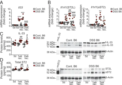
(here-after, “DSS B6 mice”) compared with control B6 mice admin-istered regular drinking water and was further augmented (by 5.1± 0.5-fold vs. control mice, P < 0.001) after 1-wk recovery during peak inflammation (Fig. 1A). After 2-wk recovery, when mucosal healing was evident, Il33 in DSS-induced colitic mice decreased markedly (by 80.8%, P< 0.001) compared with levels at 1-wk recovery and was similar to levels in uninflamed con-trol mice (Fig. 1A). Similar trends were observed for colonic ST2 mRNA levels in DSS-treated mice (Fig. 1B). Il1rl1 expres-sion of cell-surface ST2L was elevated after 5 d of DSS admin-istration (2.4 ± 0.2-fold vs. control mice, P < 0.001) and increased, reaching peak levels at 1-wk recovery (3.5± 0.2-fold vs. control mice, P< 0.001 and 1.5 ± 0.3-fold vs. 5-d DSS chal-lenge, P< 0.01). After 2-wk recovery, Il1rl1 (ST2L) was reduced compared with 1-wk recovery and compared with 5-d DSS challenge (by 62.8 and 45.9%, respectively, both P< 0.001) and reached baseline levels close to those of control mice (Fig. 1B, Left). Similarly, Il1rl1 (sST2) increased at 5-d DSS challenge (3.3± 0.6-fold vs. control mice, P < 0.01), rose further after 1-wk recovery (5.2 ± 0.9-fold vs. control mice, P < 0.01), and sub-sequently decreased (by 58.4% vs. 1-wk recovery, P< 0.05) but, interestingly, remained elevated compared with control mice at 2-wk recovery (2.2± 0.6-fold, P < 0.05) (Fig. 1B, Right).
At the protein level (Fig. 1 C and D), while the abundance of colonic IL-33 and total ST2 remained relatively constant throughout the experimental period in control mice, both were considerably increased in colitic mice, reaching peak levels at 1-wk recovery and remaining elevated after 2-wk recovery (P< 0.001 and P < 0.01, respectively, vs. control mice), when epi-thelial restitution and repair is achieved, eventually decreasing from 1-wk to 2-wk recovery (P < 0.05) (Fig. 1 C and D, Left). Western blots for IL-33 showed the presence of 30 and 20– 22 kDa bands, corresponding to full-length (f) IL-33, the most bioactive form, and cleaved (c)-IL33, a less bioactive isoform (20), respectively. f-IL-33 was up-regulated in the colons of DSS-challenged mice compared with control mice, with little post-translational modification into c-IL-33, which was equally low in all groups (Fig. 1C, Right). The difference in total ST2 protein was mainly contributed by sST2 (∼60 kDa), which was the prevalent form up-regulated in colons of DSS colitic mice, while cell-surface ST2L (∼120 kDa), the predominant form in control mice, diminished considerably after 5-d DSS challenge but began to reappear during recovery (Fig. 1D, Right).
IL-33 and ST2 immunolocalized to colon tissues from both DSS-treated and control mice, although the intensity and dis-tribution varied among experimental groups (Fig. 2). In unin-flamed control mice, IL-33 localized primarily to IECs but also to lamina propria mononuclear cells (LPMCs), while ST2 was found mainly in surface IECs (Fig. 2, Left). Following 5-d DSS challenge and at 1-wk recovery, IL-33 increased dramatically in inflamed colonic tissues (Fig. 2, Upper Middle), with intense staining primarily localized to IECs, particularly restituting epi-thelium (black arrows). ST2 was similarly augmented in inflamed tissues after 5-d DSS challenge (Fig. 2, Lower Middle) but was most notably localized to ulcerated lesions and areas of reepi-thelialization upon recovery and initial reformation of crypts (Fig. 2, Lower Right).
Overall, these data suggest that IL-33 is potently up-regulated following acute DSS challenge and also during active recovery; as such, the overall functional effects of IL-33 and whether its expression is a consequence of inflammation or healing are still unclear. However, the potential role of ST2 is better defined. Total ST2 is also elevated, which functionally likely represents the observed increase in sST2 in an attempt to decrease the pathogenic inflammatory activity of IL-33 or, alternatively, up-regulation of epithelial ST2L to promote IL-33–dependent
re-pair and restitution of barrier integrity. IMM
UNOLOG
Y
AND
INFLAM
Il33−/−andSt2−/−Mice Are Deficient in Their Ability to Recover from
Acute DSS-Induced Colitis. To address whether the role of
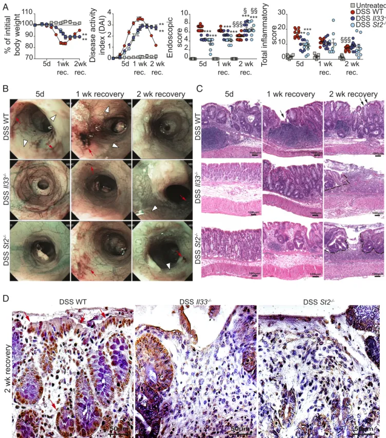
IL-33 during recovery from DSS-induced colitis is protective or pathogenic, we performed the same experiment using Il33- and/ or St2-deficient mice (Fig. 3). Il33−/−and St2−/−mice exposed to DSS (DSS Il33−/−and DSS St2−/−mice, respectively) lost less of their initial body weight and displayed a decreased disease ac-tivity index (DAI) than DSS-challenged Il33+/+St2+/+(DSS WT) controls early during DSS challenge and after 1-wk recovery (both P < 0.01), consistent with the observation that early
IL-33 expression appears to possess pathogenic functional effects (Fig. 3A, Left and Left Center). At 2 wk, however, DSS Il33−/− and St2−/−mice continued to lose weight and sustained an ele-vated DAI compared with DSS WT mice (both P< 0.01), which, like DSS B6 mice (SI Appendix, Fig. S1 A and B), showed effi-cient recovery as indicated by both weight gain almost back to baseline and a dramatic drop in DAI (Fig. 3A, Left and Center Left). Similarly, endoscopic examination of the colonic mucosa revealed progressive worsening of disease in DSS Il33−/− and St2−/−mice, peaking at 2-wk recovery (P< 0.05 and P < 0.01,
mRNA expression (fold-change) Il33
A
5 4 3 2 1 0 Il1rl1 (ST2L) 10 8 6 4 2 0 DSS B6 Cont. B6 mRNA expression (fold-change) 10 8 6 4 2 0 Il1rl1 (sST2) mRNA expression (fold-change) DSS B6 Cont. B6 IL-33 *** ** ** *** **C
Protein levels (pg/mg) 400 300 200 100 0 Total ST2 5d 1wk rec. 2wkrec. 5d 1wk rec. 2wkrec. 800 600 400 200 0 § § DSS B6 Cont. B6D
Protein levels (pg/mg) Cont. B6 DSS B6 31 kDa 24 kDa 17 kDa 38 kDa f-IL-33 c-IL-33 β-Actin 1wk rec. ST2L sST2 β-Actin rmST2 76 kDa 52 kDa 38 kDa DSS B6 Cont. B6 Cont. B6 DSS B6 5d 2wkrec. 5d 1wkrec. 2wkrec.
1wk rec.
5d 2wk
rec. 5d 1wkrec. 2wkrec.
rmIL-33 ## # ## *** *** §§§ 5d1wk rec.2wkrec. 0d *** *** §§§ ## ### 5d1wk rec.2wkrec. 0d ** ** * § 5d1wk rec.2wkrec. 0d
B
Fig. 1. IL-33 and ST2 are modulated, with differential expression of protein isoforms, during acute DSS challenge and recovery from colitis. (A and B) Il33 (A) and Il1rl1 (B) encoding ST2L (B, Left) and sST2 (B, Right) in colons from DSS B6 mice at baseline/steady state (day 0), after 5 d of DSS challenge, and at 1- and 2-wk recovery (n= 5–7). Vehicle-treated B6 mice served as controls (Cont. B6) and were killed at the same time points as DSS-treated mice. (C and D) IL-33 (C) and total ST2 (D), evaluated quantitatively (n= 7–8) (Left) and for expression of protein isoforms (Western blots representative of four separate experiments) (Right) to distinguish between full-length (f)- and cleaved (c)-IL-33, at 30 and 20–22 kDa, respectively (C, Right), and between ST2L (∼120 kDa) and sST2 (∼60 kDa) (D, Right). Either recombinant mouse IL-33 (rmIL) (∼18 kDa) or ST2/IL-33R (∼63 kDa) proteins were used as positive controls. Data are expressed as mean± SEM with relative fold differences compared with control B6 mice at 5-d DSS challenge (arbitrarily set as 1); *P < 0.05, **P < 0.01, ***P < 0.001 vs. time point-matched control B6 mice;§P< 0.05,§§§P< 0.01 vs. 1-wk recovery in DSS B6 mice;#P< 0.05,##P< 0.01,###P< 0.001 vs. DSS B6 mice at 5-d DSS challenge.
IL-33 ST2 DSS B6 Cont. B6 5d 1 wk recovery 2 wk recovery 5d
Fig. 2. IL-33 and ST2 are predominantly localized to the colonic epithelium during acute DSS challenge and recovery from colitis. Representative photo-micrographs of colonic tissues stained for IL-33 (Upper Row) and ST2 (Lower Row) show primary localization to IECs in uninflamed (no DSS) 5-d control mice (Far Left), which is unchanged after 1 and 2 wk of recovery. During DSS challenge and 1 wk of recovery, both IL-33 and ST2 are potently increased, particularly in restituting epithelium (black arrows) overlying ulcer lesions (Center). After 2 wk of recovery (Far Right), crypt formation becomes more prominent, with IL-33–responsive, ST2-expressing cells limited to regenerating epithelium (n = 8) (original magnification: 20× + 1.25).
A
% of intitial
body weight
5d 1wk rec. 2wk rec. 5d 1 wk rec. 2 wk rec.
Disease activity
index (DAI)
5d 1 wk rec. 2 wk rec.Endoscopic
score
5d
1 wk recovery
2 wk recovery
DSS WT DSS Il33 -/-DSS St2-/-T
otal inflammatory
score
110 100 90 80 70 4 3 2 1 0 *** *** *** §§ §§§ §§§ § *** *** *** ***** 5d 1 wk rec. 2 wk rec.B
2 wk recovery
DSS WT DSS Il33-/- DSS St2-/-D
30 20 10 0 10 8 6 4 2 0 DSS St2 -/-DSS Il33 -/-DSS WTUntreated ** ** ** **C
5d
1 wk recovery
2 wk recovery
DSS WT DSS Il33 -/-DSS St2-/-Fig. 3. Absence of the IL-33/ST2 receptor–ligand pair disrupts the ability to recover effectively from DSS-induced colitis. (A) Body weight, DAI, and endoscopic and histologic analyses of colons from Il33−/−, St2−/−, and Il33+/+St2+/+(WT) mice (color-coded circles) after 5-d DSS challenge and at 1 and 2 wk of recovery; untreated mice not exposed to DSS (gray squares), regardless of genotype, are grouped since no significant differences were observed among groups or with untreated B6 mice (SI Appendix, Fig. S1). (B) Representative endoscopic images; red arrows indicate frank bleeding, and white arrowheads indicate edema. (C) Representative photomicrographs of H&E-stained colonic tissues (original magnification: 10× + 1.25); black dashed lines demarcate ulcerated mucosal lesions (also evident on corresponding endoscopic images), and black arrows indicate reepithelialization and healing of mucosa. (D) Representative photomicro-graphs of colonic tissues stained for BrdU at 2-wk recovery (original magnification: 40× + 1.25); red arrows indicate actively proliferating BrdU+cells. For each time point, n= 7–10 untreated mice, n = 18 = 21 DSS WT mice, n = 8–12 DSS Il-33−/−mice, and n= 8–10 DSS St2−/−mice; data are expressed as mean± SEM. **P< 0.01, ***P < 0.001 vs. time point-matched DSS WT mice;§P< 0.05,§§P< 0.01,§§§P< 0.001 vs. strain-matched mice challenged with DSS for 5 d. IMM
UNOLOG
Y
AND
INFLAM
respectively, vs. 5-d DSS challenge) and compared with DSS WT mice at 2-wk recovery (both P< 0.001) (Fig. 3A, Center Right). These mice showed persistent bleeding (red arrows) and edema (white arrowheads), with abundant and large mucosal ulcera-tions, particularly at 2-wk recovery (Fig. 3B, Bottom Right), while DSS WT mice displayed improved overall endoscopic scores and mucosal healing (Fig. 3B, Top Right ).
Histologic analyses confirmed these results: Like DSS B6 mice
(SI Appendix, Fig. S1 A–C), WT mice during DSS challenge
showed severe colitis, which improved progressively over 2-wk recovery (P < 0.001 vs. 5-d DSS challenge) (Fig. 3A, Right). Conversely, both Il33−/− and St2−/− mice displayed moderate colitis during DSS challenge, which did not change significantly even after 2 wk of recovery (Fig. 3A, Right). In fact, at 1-wk re-covery, areas of regenerating epithelium (black arrows) overlying ulcerated inflammatory lesions were present in DSS WT mice (Fig. 3C, Top Center) but were much less evident in DSS Il33−/− and St2−/−mice even at 2-wk recovery (Fig. 3C, Bottom Right). Moreover, BrdU staining showed reduced expression and de-creased epithelial distribution of proliferating cells in both DSS Il33−/− and St2−/− mice as compared with WT mice, with no apparent differences between Il33−/−and St2−/−mice (Fig. 3D). In addition, mice not exposed to DSS (untreated), independent of genotype (here, grouped together for Il33−/−, St2−/−, and WT controls), did not show any significant changes (Fig. 3A) and were similar to results obtained in (untreated) control B6 mice
(SI Appendix, Fig. S1). Overall, these data indicate that the lack
of either IL-33 or ST2 impedes the overall recovery process after acute DSS colitis and appears to affect epithelial-specific pro-liferation and repair.
Exogenous Administration of IL-33 During Recovery After DSS Challenge Enhances Mucosal Healing and the Resolution of Colitis.
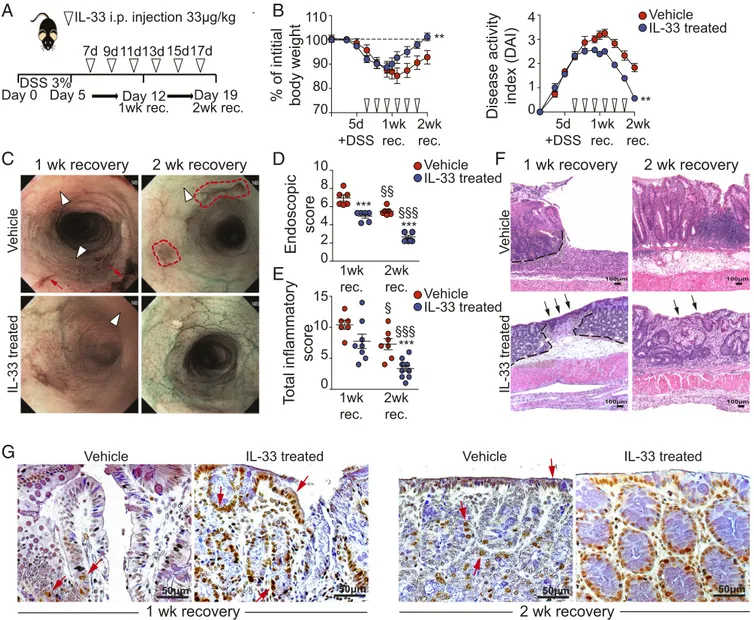
To confirm our results thus far and to test the potential thera-peutic role of IL-33 in promoting epithelial repair/restitution and ultimate wound healing, we exogenously administered a phar-macological dose (33 μg/kg) of recombinant IL-33 (rIL-33) during the recovery period following acute DSS colitis (Fig. 4A). IL-33–treated mice showed accelerated recovery from body weight loss compared with vehicle-treated controls (P< 0.01), reaching initial baseline levels after 2-wk recovery, and lowered DAI (P< 0.01), decreasing disease activity almost to zero (Fig. 4B). The general endoscopic appearance of the colonic mucosa was also dramatically improved upon IL-33 treatment (P < 0.001 at both 1- and 2-wk recovery vs. vehicle-treated controls), with greater magnitude and efficacy between 1 and 2 wk of re-covery (Fig. 4C). Less edema (white arrowheads) and bleeding (red arrows) were observed at 1-wk recovery (Fig. 4C), and in-creased translucency of the mucosal surface, prominent vascu-larization, and centralized, tubular-shaped lumens became evident at 2-wk recovery (Fig. 4D).
Histologic analyses provided further support for the efficacy of IL-33 treatment in promoting epithelial repair/restitution and mucosal healing, showing decreased inflammation in treated DSS-induced colitic mice at 1- or 2-wk recovery (P< 0.001) (Fig. 4E). In particular, the emergence of restituting epithelium (black arrows), goblet cells, and organized colonic crypts (Fig. 4F, Lower Row), as well as increased expression of BrdU+cells (Fig. 4G, red arrows), mainly localized to epithelial crypt cells, was evident in IL-33–treated colitic mice as compared with untreated controls after 1- and 2-wk recovery. Taken together, these find-ings indicate that pharmacologic treatment of DSS-induced colitic mice with rIL-33 during recovery dramatically acceler-ates epithelial restitution and repair, with concomitant im-provement of intestinal inflammation.
IL-33 Directly Induces IEC Proliferation and Wound Healing by Specific
Up-Regulation of miR-320.To mechanistically determine the direct
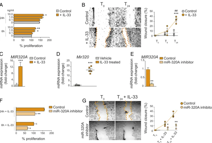
effects of IL-33 on IECs, we performed a series of in vitro ex-periments using the colonic Caco-2 IEC line stimulated with human rIL-33. IL-33–treated Caco-2 cells showed increased proliferation compared with (untreated) controls in a dose-dependent manner after both 6 and 12 h, with maximum ef-fects (21.7% vs. control, P< 0.01) at 6 h using 10 ng/mL IL-33 (Fig. 5A). More dramatic results were observed when measuring in vitro wound healing, demonstrating that IL-33 accelera-ted IEC wound closure by 33.1% (P < 0.01 vs. control), with maximum effects at 24 h (Fig. 5B). Microarray analysis of IL-33–regulated genes in IECs revealed gene targets primarily involved in cell-cycle function, cell morphology, cell-to-cell sig-naling and interaction, and cellular development and mainte-nance (SI Appendix, Table S2). Among these, a specific network of highly expressed miRs was identified that are associated with the activation of intracellular proliferative pathways (SI
Appen-dix, Table S3). To confirm these results, we performed in vitro
experiments that were identical to the previous experiments ex-cept for miR enrichment. Our results showed that after miR enrichment, MIR320A was the most highly expressed miR in colonic IECs stimulated with IL-33 (by 12.58 ± 2.63-fold vs. controls, P< 0.02) (Fig. 5C). These findings were verified in vivo; IECs isolated from DSS-induced colitic mice treated with IL-33 during recovery (Fig. 4A) expressed 16.63 ± 1.42-fold ele-vated Mir320 after 2 wk than vehicle-treated DSS-induced colitic mice (P< 0.001) (Fig. 5D).
To determine the functional role of miR-320 in IECs, we transfected Caco-2 cells with small ssRNA molecules designed specifically either to inhibit miR-320A (mirVana miR-320A in-hibitor) or to serve as a negative control (scrambled mirVana). After stimulation with IL-33, reduced MIR320A was confirmed after specific inhibition of miR-320A (by 84.8% vs. control, P< 0.01) (Fig. 5E), and IL-33–dependent proliferation was partially, albeit significantly, blocked, by 35.7% vs. control after 6 h (P< 0.05) and by 32.9% vs. control at 24 h (P< 0.01) (Fig. 5F). More impressively, specific miR-320A inhibition decreased in vitro wound closure by 66.4% vs. control after 6 h (P< 0.05) and by 74.5% vs. control at 24 h (P< 0.01) (Fig. 5G). Taken together, these data indicate that IL-33 has a direct effect on epithelial proliferation and wound healing that is due, in part, to miR-320 regulation.
Discussion
While the increased expression of IL-33 and its association with IBD has been firmly established, dissecting the precise mecha-nistic role of IL-33 during intestinal inflammation has generated ambiguous results, suggesting both protective and pathogenic functions. In the great majority of studies using experimental models of colitis, healthy immunocompetent mice have been challenged with 1.5–5% DSS continuously for 5–13 d (in some cases, DSS exposure was repeated with a 5- to 7-d recovery pe-riod between cycles), and colitis was evaluated either immedi-ately without recovery or after a recovery period of up to 7 d (SI
Appendix, Table S1). Under these conditions, IL-33 was
gener-ally shown to have pathogenic effects when colitis was evaluated without recovery or after up to 7 d of recovery and when acute DSS was administered (without cycling) at a moderate to high dose (2–5%). These results are consistent with our observation that deletion of either Il33 or St2 in mice in which colitis was induced by 3% DSS (which allowed almost 100% survival) was effective in decreasing DAI, improving endoscopic appearance of the colonic mucosa, and dampening overall disease severity when evaluated either during DSS challenge or after 7 d of re-covery. Importantly, however, when allowed to recover for an extended period of 2 wk, DSS Il33−/−and St2−/−mice were de-ficient in their ability to appropriately restitute epithelium, ab-rogate mucosal ulcerations, and effectively heal the colonic mucosa as compared with WT controls. These data suggest that
in healthy mice, when assaulting factors (e.g., DSS) are removed, the normal function of endogenously produced IL-33 within the gut mucosa is protection.
This concept is further supported by our results showing that exogenous supplementation of rIL-33 at pharmacological doses during recovery from DSS is able to significantly decrease DAI, promote ulcer healing, restore normal epithelial architecture, and globally improve gut mucosal wound healing. This finding may also account for results from previously published studies in which IL-33 administered during recovery periods between DSS cycling, commonly used to produce chronic colitis, demonstrated protective effects (21, 22). In fact, other studies treating colitis with exogenous, supraphysiological doses of IL-33 generally resulted in disease improvement in acute and chronic models (14, 23). Our results, however, conflict directly with those of
Sedhom et al., (24), who reported that inhibiting the IL-33/ ST2 pathway either by genetic ablation or by treatment with a specific blocking antibody against ST2 ameliorated colitis in two models of colitis [i.e., DSS- and 2,4,6-Trinitrobenzenesulfonic acid (TNBS)-induced] by enhancing mucosal healing. Unlike our study that allowed recovery for 2 wk after ceasing DSS challenge, experimental mice in the study by Sedhom et al. were not per-mitted to recover, and colitis was assessed on the last day of chemical insult, which may, as in our results, represent a time point when IL-33–dependent disease activity is still increasing and colonic inflammation is most severe. As such, treatment with an anti-ST2 antibody during this time period or performing the same protocol in ST2-deficient mice would predictably decrease colitis severity. In the same study, exogenous IL-33 treatment was shown to exacerbate colitis and decrease effective wound
A
% of intitial
body weight
Disease activity
index (DAI)
5d +DSS 1wk rec. 2wk rec.B
Endoscopic
score
1wk rec. 2wk rec. IL-33 treated Vehicle 5d +DSS 1wk rec. 2wk rec.T
o
tal inflammatory
score
1wk rec. 2wk rec.2 wk recovery
1 wk recovery
2 wk recovery
V ehicle IL-33 treatedC
15 10 5 0 *** § 10 8 6 4 2 0 *** ***1 wk recovery
V ehicle IL-33 treatedDay 0 Day 5 Day 12 Day 19
1wk rec. 2wk rec. DSS 3% 7d 9d11d13d 15d17d 1 2D
G
2 wk recovery
1 wk recovery
Vehicle IL-33 treated
110 100 90 80 70 4 3 2 1 0 §§
F
Ve hD
E
IL-33 treated Vehicle IL-33 treated Vehicle ** ** IL-33 treated Vehicle §§§ §§§Fig. 4. IL-33 treatment during recovery from DSS accelerates mucosal healing and decreases colitis severity. (A) Strategy for IL-33 treatment (open arrow-heads) in the DSS colitis model with extended recovery. (B) Resulting body weight (Left) and DAI (Right). (C) Representative images showing areas of frank bleeding (red arrows) and edema (white arrowheads). Red dashed lines outline healing ulcers. (D) Semiquantitative scores. (E and F) Histologic analysis showing total inflammatory scores (E) and representative photomicrographs (original magnification: 10× + 1.25) (F) of H&E-stained colonic tissues. Black dashed lines in F outline ulcerated mucosal lesions, and black arrows indicate single-layer reepithelialization overlying healing ulcers. (G) Representative photomicrographs of colonic tissues stained for BrdU at 1- and 2-wk recovery, comparing vehicle- and IL-33–treated DSS-induced colitic mice (original magnification: 40× + 1.25). Red arrows indicate actively proliferating BrdU+cells, with greatest abundance at 2-wk recovery following IL-33 treatment. For each time point, n= 6–7 vehicle-treated mice and n = 8–13 IL-33–treated mice; data are expressed as mean ± SEM. **P < 0.01 and ***P < 0.001 vs. vehicle;§P< 0.05,§§P< 0.01,§§§P< 0.001 vs. the same treatment at 1-wk recovery.
IMM UNOLOG Y AND INFLAM MATION
healing (24). However, an additional observation to note is that during and immediately after DSS challenge, lamina propria enriched with activated innate immune cells is maximally ex-posed to microbial antigens, since epithelial injury and de-nudation peak, with loss of ST2L-bearing IECs that then are unable to respond to IL-33 and promote proliferation and repair. This concept is further supported by our Western blot data demonstrating the specific loss of ST2L during DSS challenge. Interestingly, ST2 was also found to be absent or decreased in the epithelium of UC and Crohn’s colitis patients but was pre-sent and increased within LPMCs during active disease (5), which may explain the skewing toward immune activation (vs. epithelial-induced repair and protection) that promotes chronic inflammation such as found in IBD.
In fact, the apparent discrepancies in dissecting the precise role of IL-33 using variations of DSS colitis, as well as other acute and chronic models of colitis, may simply be explained by the fact that DSS colitis in healthy, immunocompetent mice represents the response of a normal animal to acute intestinal injury, whereas other models possess genetic and immunologic abnormalities that predispose these mice to chronic intestinal inflammation, similar to patients with IBD. Such is the case with the SAMP1/YitFc (hereafter, “SAMP”) mouse strain, which
possesses multifactorial genetic, immunologic, and regional GI epithelial barrier defects predisposing these mice to spontane-ously occurring chronic CD-like ileitis that escalates in severity over time (25). In SAMP mice, IL-33 has deleterious effects overall, inducing eosinophil infiltration and activation of patho-genic Th2 immune responses, resulting in chronic intestinal in-flammation that is dependent on the gut microbiome (26). In fact, neutralization of IL-33 with an anti-ST2 antibody in this model was effective in preventing the massive influx of eosino-phils into the gut mucosa and decreasing the overall severity of ileal inflammation when used as either a preventive treatment before the onset of inflammation or a therapeutic treatment of established disease. However, although the effect was consistent and significant, disease severity was reduced by only 30% (26), which, while reflecting efficient blockade of inflammation, may also compromise effective IL-33–dependent epithelial restitution/ repair and mucosal healing. This concept of the dichotomous roles of IL-33 during chronic intestinal inflammation is consis-tent with other innate-type cytokines, including several members of the IL-1 family such as IL-1, IL-18, and IL-36, which can possess both protective and proinflammatory functions, depending upon the immunological status of the host and/or the type and phase of the ongoing inflammatory process (3, 27, 28).
MIR320A Mir320 miRNA expression (fold-change) miRNA expression (fold-change) 25 20 15 10 5 0 16 12 8 4 0 IL-33 treated Vehicle + IL-33 Control 50 40 30 20 10 0 W o und closure (%) T6 T24 Control + IL-33 *** ***
T
24 ***T
0 ## 0 6h 24h 50 100 150 200 + IL-33 Control 10.0 1.0 0.1 0 10.0 1.0 0.1 0 % proliferation ng/ml * ** * T0 + IL-33 Control 50 40 30 20 10 0 W ound closure (%) T6 + IL-33 ControlT
24+ IL-33
T
0 0 6h + IL-33 24h + IL-33 50 100 150 200 % proliferation ** miR-320A inhibitor T24 + IL-33 T0 ** * miR-320A inhibitor Control miRNA expression (fold-change) 1.5 1 0.5 0 miR-320A inhibitor Control ** miR-320A inhibitor Control MIR320A *A
C
F
G
D
E
B
Fig. 5. IL-33 promotes epithelial-specific proliferation and wound healing through up-regulation of miR-320. (A) IL-33 dose–response on IEC percentage proliferation at 6 and 24 h after IL-33 stimulation; *P< 0.05, **P < 0.01 vs. time-matched (vehicle) control. (B) Representative images (Left) and quantitation (Right) of IEC wound closure at baseline (T0) and after 6 h (T6) and 24 h (T24) of IL-33 stimulation; ***P< 0.001 vs. time-matched control,##P< 0.01 vs. baseline. (C) MIR320A in IECs± IL-33 IL-33 after miR enrichment. Data are shown as relative fold difference compared with vehicle (control) (arbitrarily set as 1). (D) Mir320 in IECs isolated from in vivo IL-33–treated induced colitic mice. Data are shown as relative fold difference compared with vehicle-treated DSS-induced colitic mice (arbitrarily set as 1); n= 5. (E–G) MIR320A expression (E), percentage IEC proliferation (F), representative images (G, Left), and quan-titation (G, Right) of wound closure in IL-33–stimulated IECs transfected with either a miR-320A–specific inhibitor or scrambled (negative) control. *P < 0.05, **P< 0.01, ***P < 0.001 vs. control for all in vitro studies; n (number of repeated measures for each condition for single experiment) = 3–4 and are rep-resentative of three or four separate experiments (A–G).
In the present study, transcriptional profiling of the Caco-2 human colorectal epithelial cell line revealed that IL-33 stimulation resulted in the up-regulation of several miRs, in particular miR-320A, that have the ability to modify molecular patterns involved in various functions associated with cell pro-liferation and cell maintenance (SI Appendix, Table S2). In general, miRs are small, noncoding RNAs of ∼20–24 nt that posttranscriptionally repress the expression of target genes (29). Although the exact function of miRs in human development and physiology remains largely unknown, differential expression of given miRs during disease progression suggests that miRs have an especially crucial role in human pathologic conditions, but they also have been shown to modulate GI mucosal growth and repair after injury (30, 31). In fact, several GI-specific miRs are highly expressed in the intestinal epithelium and are critical for the maintenance of normal barrier integrity and regulation of tight junction proteins during disease states (30, 31).
Relevant to our results, MIR320A was shown to be abnormally expressed in mucosal biopsies from IBD patients, and MIR320A was decreased in UC patients as compared with uninflamed, healthy controls (32). These findings were later confirmed in a pediatric population with early-onset IBD, in which MIR320A was decreased in inflamed vs. uninflamed colonic biopsies from both UC and CD patients (33). The latter study further showed in vitro evidence that NOD2 served as a target for MIR320A, which negatively regulated its expression, specifically in IECs and under inflammatory conditions (33). NOD2 encodes a cytosolic protein receptor that recognizes the bacterial cell wall product muramyl dipeptide and normally promotes its clearance by ac-tivating a proinflammatory cascade and/or by autophagy to maintain normal gut homeostasis. Mutations in NOD2 were the first and strongest reported genetic associations shown to confer susceptibility to CD (34, 35), suggesting that carriers of such mutations have a dysfunction in handling normal bacterial loads. The aforementioned findings, along with the results from our current study, pose conceptually interesting possibilities re-garding the potential role of epithelial-derived miR-320 in the setting of IBD. First, decreased miR-320 can lead to NOD2 overexpression and amplification of aberrant NOD2-induced proinflammatory events, influenced by carriage of NOD2 muta-tions. Alternatively, inherent decreased miR-320 may result in the inability to achieve appropriate epithelial repair and restitution, leading to impaired gut mucosal healing. Both processes have the ability to promote uncontrolled immune responses and impair effective resolution of inflammation.
At present, the role of IL-33–dependent regulation of miR-320A during IBD is unknown and is the major focus of ongoing studies in our laboratory at Department of Pathology, Case Western Reserve University (CWRU). Although our current results definitively indicate that in normal, immunocompetent BL6 mice IL-33 induces increased epithelial miR-320 expression that promotes epithelial repair/restitution and confers normal mucosal healing, it has yet to be revealed how this process is defective in inherently colitis-prone mice and/or in patients with IBD. In fact, the molecular processes responsible for the control of miR biogenesis in response to challenge or stress remain largely unknown and are an area of growing investigation. Based on our results and prior studies, it is tempting to speculate that dysfunction of IL-33 as a prototypic alarmin and/or increased epithelial-specific sequestration of IL-33 may negatively regulate or repress miR-320 synthesis, since primary processing of all animal miRs is achieved first in the nucleus, with subsequent, secondary processing occurring within the cytoplasm. As such, the elevated levels of IL-33 in both the nuclear and cytoplasmic compartments of IECs commonly observed in IBD patients (5, 6, 8) would potentially allow increased, and easy, accessibility of IL-33 to a number of different proteins involved in miR processing and specifically in miR-320 processing. An alternative hypothesis
to consider is that IL-33 can also function as an intracellular nuclear factor with potent transcriptional-repressor properties (10), and in this setting IL-33 may regulate various proteins that impact miR-320 processing or may affect miR-320 transcription directly. In both scenarios, IL-33–dependent down-regulation of miR-320 could be achieved.
Additionally, we cannot rule out the possibility that intestinal tissue protection may require cooperative action from both hema-topoietic and stromal cell lineages that could be initiated by rapid epithelial damage-induced alarmins, such as IL-33. One potential mechanism involves type 2 innate lymphoid cells (ILC2s), which have the ability to produce amphiregulin (AREG), an EGF re-ceptor ligand, in response to IL-33 induced by intestinal injury that consequently mediates epithelial repair and eventual healing (23). This process can be further enhanced by IL-33–dependent differ-entiation of Tregs and tolerogenic CD103+dendritic cells, initiated by IEC activation and release of IL-33 and the subsequent pro-duction of Th2 cytokines, ultimately leading to intestinal mucosal protection (14–16). As such, IL-33/ST2 may orchestrate an inte-grated framework involving IECs, intestinal ILC2s, and Tregs through modulation of miR-320A, AREG, and Th2 cytokines, re-spectively, with the end goal of promoting gut health.
In summary, the results of the present study indicate that, upon challenge, the inherent role of endogenous IL-33 within the gut mucosa is protection, potentially through a mechanism that augments miR-320 expression, inducing epithelial restitu-tion and repair and overall epithelial barrier integrity. In the setting of IBD, particularly during early disease stages, this process may be defective, leading to impaired healing and ex-acerbation of colitis into a more chronic and sustained in-flammatory phenotype. Results from this study also provide the rationale for the potential therapeutic use of either IL-33 or miR-320A to obtain optimal gut mucosal wound healing and to promote the resolution of inflammation.
Materials and Methods
Mice. All experimental mice were bred and maintained under specific pathogen-free conditions in the Animal Resource Center at Case Western Reserve University. For a full description, seeSI Appendix, Materials and Methods. All procedures performed were approved by the Institutional Animal Care and Use Committee at CWRU and followed the American As-sociation for Laboratory Animal Care guidelines.
Model of DSS-Induced Colitis with Extended Recovery. Induction of colitis was performed on adult C57BL/6J (B6), Il33−/−, and St2−/−mice and their WT (Il33+/+St2+/+) littermate controls by 5-d administration of 3% DSS, as pre-viously described (36), but with an extended 2-wk recovery period. Il33−/−, St2−/−, and WT mice not exposed to DSS served as untreated controls. In separate experiments, acute colitis was induced in B6 mice, and these mice were injected i.p. with either murine rIL-33 (33μg/kg) or vehicle (PBS) control during the 2-wk recovery period. For a full description, seeSI Appendix, Materials and Methods.
Colonoscopy. Macroscopic progression of colitis was assessed by endoscopy of left colon and rectum following a standardized protocol (37). For a full de-scription, seeSI Appendix, Materials and Methods.
Tissue Harvest and Histologic Assessment of Colitis. When mice were killed, whole colons were taken for analysis and histology. Disease severity was evaluated as previously described (18, 36). For a full description, seeSI Ap-pendix, Materials and Methods.
Immunohistochemistry. Immunohistochemistry (IHC) was performed as pre-viously described (5) using specific antibodies against murine IL-33 and ST2, and in select experiments IHC was performed for BrdU to detect cell pro-liferation. For a full description, seeSI Appendix, Materials and Methods.
ELISA and Western Blot Analysis. IL-33 and ST2 protein levels were quantified by ELISA and evaluated by Western blots to detect specific IL-33 and ST2 isoforms as previously described (5). For a full description, seeSI Ap-pendix, Materials and Methods. IMM
UNOLOG
Y
AND
INFLAM
IEC Isolation. Freshly isolated colons from vehicle- and IL-33–treated DSS colitic mice were harvested and processed as previously described (5, 38). For a full description, seeSI Appendix, Materials and Methods.
Cell Culture and in Vitro Assays. Caco-2 cells (HTB-37; ATCC) were grown to 80% confluency and were cultured with or without human rIL-33. Cells were collected after 6 and 24 h and were evaluated for cell proliferation using the XTT Cell Proliferation Assay Kit (ATCC) and measuring wound closure. For full description, seeSI Appendix, Materials and Methods.
Microarray Analysis. Caco-2 cells were cultured with or without human rIL-33, and total RNA was prepared as described below. RNA (100 ng/μL) was sub-mitted to the Case Comprehensive Cancer Center’s Gene Expression and Genotyping Core at Case Western Reserve University for microarray analysis. For a full description, seeSI Appendix, Materials and Methods.
Total RNA Extraction, miR Enrichment, and qPCR. Total RNA extraction and/or miR enrichment were performed on tissue homogenates and IEC prepara-tions, and qPCR was done using specific primers for Il33, Il1rl, MIR320A, and Mir320. For a full description, seeSI Appendix, Materials and Methods.
Inhibition of miR-320A. Single-stranded inhibitor RNA molecules (mirVana inhibitors), designed to specifically target miR-320A, miR-Let7c (positive control), or scrambled sequences (negative control), were transfected into Caco-2 cells. Transfection efficiency was confirmed by qPCR analysis of the miR-Let7c target gene, HMGA2, in miR-Let7c–targeted Caco-2 cells. For a full description, seeSI Appendix, Materials and Methods.
Statistical Analysis. Statistical analyses were performed using one-way ANOVA with Bonferroni correction for multiple comparisons and individu-al t test comparisons adjusted for unequindividu-al variance (Welch’s correction), as appropriate.
ACKNOWLEDGMENTS. This work was supported by Cleveland Digestive Diseases Research Core Center Grant P30 DK097948; NIH Grants DK056762 (to T.T.P.), DK042191 (to T.T.P. and F.C.), and DK091222 (to T.T.P., D.W.A., and F.C.); Crohn’s and Colitis Foundation Research Fellowships (to L.R.L. and C.D.S.) and a Career Development Award (to W.A.G.); an American Gastro-enterological Association Research Foundation Eli and Edythe Broad Student Research Fellowship Award (to H.N.S.); and an Italian Group for the Study of Inflammatory Bowel Disease Research Internship (to V.P.).
1. Leoni G, Neumann PA, Sumagin R, Denning TL, Nusrat A (2015) Wound repair: Role of immune-epithelial interactions. Mucosal Immunol 8:959–968.
2. Cominelli F (2004) Cytokine-based therapies for Crohn’s disease–new paradigms. N Engl J Med 351:2045–2048.
3. Lopetuso LR, Chowdhry S, Pizarro TT (2013) Opposing functions of classic and novel IL-1 family members in gut health and disease. Front Immunol 4:IL-18IL-1.
4. Schmitz J, et al. (2005) 33, an interleukin-1-like cytokine that signals via the IL-1 receptor-related protein ST2 and induces T helper type 2-associated cytokines. Immunity 23:479–490.
5. Pastorelli L, et al. (2010) Epithelial-derived IL-33 and its receptor ST2 are dysregulated in ulcerative colitis and in experimental Th1/Th2 driven enteritis. Proc Natl Acad Sci USA 107:8017–8022.
6. Beltrán CJ, et al. (2010) Characterization of the novel ST2/IL-33 system in patients with inflammatory bowel disease. Inflamm Bowel Dis 16:1097–1107.
7. Kobori A, et al. (2010) Interleukin-33 expression is specifically enhanced in inflamed mucosa of ulcerative colitis. J Gastroenterol 45:999–1007.
8. Seidelin JB, et al. (2010) IL-33 is upregulated in colonocytes of ulcerative colitis. Immunol Lett 128:80–85.
9. Sponheim J, et al. (2010) Inflammatory bowel disease-associated interleukin-33 is preferentially expressed in ulceration-associated myofibroblasts. Am J Pathol 177: 2804–2815.
10. Carriere V, et al. (2007) IL-33, the IL-1-like cytokine ligand for ST2 receptor, is a chromatin-associated nuclear factor in vivo. Proc Natl Acad Sci USA 104:282–287. 11. Moussion C, Ortega N, Girard JP (2008) The IL-1-like cytokine IL-33 is constitutively
expressed in the nucleus of endothelial cells and epithelial cells in vivo: A novel ‘alarmin’? PLoS One 3:e3331.
12. Chackerian AA, et al. (2007) 1 receptor accessory protein and ST2 comprise the IL-33 receptor complex. J Immunol 179:2551–2555.
13. Hayakawa H, Hayakawa M, Kume A, Tominaga S (2007) Soluble ST2 blocks in-terleukin-33 signaling in allergic airway inflammation. J Biol Chem 282:26369–26380. 14. Duan L, et al. (2012) Interleukin-33 ameliorates experimental colitis through
pro-moting Th2/Foxp3+regulatory T-cell responses in mice. Mol Med 18:753–761. 15. Schiering C, et al. (2014) The alarmin IL-33 promotes regulatory T-cell function in the
intestine. Nature 513:564–568.
16. He Z, et al. (2017) Epithelial-derived IL-33 promotes intestinal tumorigenesis in ApcMin/+mice. Sci Rep 7:5520.
17. Liew FY, Girard JP, Turnquist HR (2016) Interleukin-33 in health and disease. Nat Rev Immunol 16:676–689.
18. Okayasu I, et al. (1990) A novel method in the induction of reliable experimental acute and chronic ulcerative colitis in mice. Gastroenterology 98:694–702. 19. Dieleman LA, et al. (1994) Dextran sulfate sodium-induced colitis occurs in severe
combined immunodeficient mice. Gastroenterology 107:1643–1652.
20. Cayrol C, Girard JP (2009) The IL-1-like cytokine IL-33 is inactivated after maturation by caspase-1. Proc Natl Acad Sci USA 106:9021–9026.
21. Groβ P, Doser K, Falk W, Obermeier F, Hofmann C (2012) IL-33 attenuates develop-ment and perpetuation of chronic intestinal inflammation. Inflamm Bowel Dis 18: 1900–1909.
22. Zhu J, et al. (2015) IL-33 alleviates DSS-induced chronic colitis in C57BL/6 mice colon lamina propria by suppressing Th17 cell response as well as Th1 cell response. Int Immunopharmacol 29:846–853.
23. Monticelli LA, et al. (2015) IL-33 promotes an innate immune pathway of intestinal tissue protection dependent on amphiregulin-EGFR interactions. Proc Natl Acad Sci USA 112:10762–10767.
24. Sedhom MA, et al. (2013) Neutralisation of the interleukin-33/ST2 pathway amelio-rates experimental colitis through enhancement of mucosal healing in mice. Gut 62: 1714–1723.
25. Pizarro TT, et al. (2011) SAMP1/YitFc mouse strain: A spontaneous model of Crohn’s disease-like ileitis. Inflamm Bowel Dis 17:2566–2584.
26. De Salvo C, et al. (2016) IL-33 drives eosinophil infiltration and pathogenic type 2 helper T-cell immune responses leading to chronic experimental ileitis. Am J Pathol 186:885–898.
27. Medina-Contreras O, et al. (2016) Cutting edge: IL-36 receptor promotes resolution of intestinal damage. J Immunol 196:34–38.
28. Ngo VL, et al. (2018) A cytokine network involving IL-36γ, IL-23, and IL-22 promotes antimicrobial defense and recovery from intestinal barrier damage. Proc Natl Acad Sci USA 115:E5076–E5085.
29. Krol J, Loedige I, Filipowicz W (2010) The widespread regulation of microRNA bio-genesis, function and decay. Nat Rev Genet 11:597–610.
30. Xiao L, et al. (2011) Regulation of cyclin-dependent kinase 4 translation through CUG-binding protein 1 and microRNA-222 by polyamines. Mol Biol Cell 22:3055–3069. 31. Xiao L, Wang JY (2014) RNA-binding proteins and microRNAs in gastrointestinal
epithelial homeostasis and diseases. Curr Opin Pharmacol 19:46–53.
32. Fasseu M, et al. (2010) Identification of restricted subsets of mature microRNA ab-normally expressed in inactive colonic mucosa of patients with inflammatory bowel disease. PLoS One 5:e13160.
33. Pierdomenico M, et al. (2016) NOD2 is regulated by Mir-320 in physiological condi-tions but this control is altered in inflamed tissues of patients with inflammatory bowel disease. Inflamm Bowel Dis 22:315–326.
34. Hugot JP, et al. (2001) Association of NOD2 leucine-rich repeat variants with sus-ceptibility to Crohn’s disease. Nature 411:599–603.
35. Ogura Y, et al. (2001) A frameshift mutation in NOD2 associated with susceptibility to Crohn’s disease. Nature 411:603–606.
36. Scaldaferri F, et al. (2007) Crucial role of the protein C pathway in governing micro-vascular inflammation in inflammatory bowel disease. J Clin Invest 117:1951–1960. 37. Kodani T, et al. (2013) Flexible colonoscopy in mice to evaluate the severity of colitis
and colorectal tumors using a validated endoscopic scoring system. J Vis Exp 80:e50843. 38. Olson TS, et al. (2006) The primary defect in experimental ileitis originates from a


