Ca
2⫹influx through
␣
1SDHPR may play a role in regulating
Ca
2⫹release from RyR1 in skeletal muscle
Alexander Shtifman,1Cecilia Paolini,2Jose´ R. Lo´pez,1,3 Paul D. Allen,1and Feliciano Protasi1,4 1Department of Anesthesia Research, Brigham and Women’s Hospital, Harvard Medical School,
Boston, Massachusetts 02115;2Department of Cell and Developmental Biology, University of Pennsylvania School of Medicine, Philadelphia, Pennsylvania 19104;3Centro de Biofı´sica y Bioquı´mica, Instituto Venezolano de Investigaciones Cientı´ficas, Caracas, Apartado 21827, Venezuela; and4Laboratory of Cellular Physiology, CeSI, Center for Research on Aging, University G. d’Annunzio School of Medicine, 66023 Chieti, Italy
Submitted 12 May 2003; accepted in final form 31 August 2003
Shtifman, Alexander, Cecilia Paolini, Jose´ R. Lo´pez, Paul D. Allen, and Feliciano Protasi. Ca2⫹influx through␣1SDHPR may
play a role in regulating Ca2⫹release from RyR1 in skeletal muscle.
Am J Physiol Cell Physiol 286: C73–C78, 2004. First published September 3, 2003; 10.1152/ajpcell.00194.2003.—Differentiated pri-mary myotubes isolated from wild-type mice exhibit ryanodine-sensitive, spontaneous global Ca2⫹oscillations as well as spontaneous
depolarizations in the plasma membrane. Immunolabeling of these myotubes showed expression of both␣1Sdihydropyridine receptors (DHPRs) and ryanodine-sensitive Ca2⫹-release channel 1 (RyR1), the
two key proteins in skeletal excitation-contraction (E-C) coupling. Spontaneous global Ca2⫹oscillations could be inhibited by addition
of 0.1 mM CdCl2/0.5 mM LaCl3or 5M nifedipine to the extracel-lular bathing solution. After either treatment, Ca2⫹oscillations could be restored upon extensive washing. Although exposure to DHPR antagonists completely blocked Ca2⫹oscillations, normal orthograde
signaling between DHPRs and RyRs, such as that elicited by 80 mM KCl depolarization, was still observed. In addition, we showed that spontaneous Ca2⫹oscillations were never present in cultured mdg
myotubes, which lack the expression of ␣1SDHPRs. These results suggest that under physiological conditions in conjunction with the mechanical coupling between the␣1SDHPRs and RyR1, the initiation of Ca2⫹oscillations in myotubes may be facilitated, in part, by the
Ca2⫹influx through the␣
1s-subunit of the DHPR.
calcium-induced calcium release; dihydropyridine receptors; excita-tion-contraction coupling; ryanodine receptors; skeletal muscle
EXCITATION-CONTRACTION (E-C) coupling in muscle involves a
rapid cascade of events that transform the surface membrane depolarization into Ca2⫹release and generation of force. This
sequence of events is initiated by an action potential that upon reaching the transverse tubules (TT) activates the L-type Ca2⫹
channels, the dihydropyridine receptors (DHPRs). Activation of the DHPRs triggers a sudden release of Ca2⫹ from the
sarcoplasmic reticulum (SR) via Ca2⫹ release channels
(ryan-odine receptors, RyRs) that reside in the junctional region of the SR immediately adjacent to the TT membrane. The result-ing release of Ca2⫹ produces a transient increase in
intracel-lular [Ca2⫹], which activates the contractile apparatus of
mus-cle fibers (for review, see Ref. 20). Although the major com-ponents of the signal transduction between the two membrane systems have been identified, the precise mechanisms that govern this process are not fully resolved.
DHPRs and RyRs are the two primary proteins involved in the E-C coupling in skeletal, cardiac, and smooth muscle. However, it has become increasingly evident that the commu-nication between these two proteins in skeletal muscle employs a different mechanism compared with the cardiac and smooth muscle. In cardiac and smooth muscle, activation of the ␣1DHPR generates a large inward flux of Ca2⫹ from the
extracellular space, which induces the opening of RyRs and a consequent massive release of Ca2⫹ into the myoplasm. This
mechanism has been defined as calcium-induced calcium re-lease, or CICR (6). In skeletal muscle, however, initiation of a contraction can be achieved in the absence of the extracellular Ca2⫹ (3, 4). In fact, it has been generally accepted that in
skeletal muscle␣1SDHPR functions predominantly as the
volt-age sensor (24, 30) that activates the RyRs though a physical interaction, known as the orthograde signaling. Although the role of Ca2⫹influx in skeletal muscle E-C coupling is unclear,
Ca2⫹ ions do participate in modulation of the RyR activity
through a process similar to that of cardiac CICR (5a, 17–19). In the present work, we report the existence of spontaneous, RyR-mediated Ca2⫹ transients, as well as spontaneous
depo-larizations of the plasma membrane in primary myotubes of skeletal origin. The ability of Ca2⫹ channel blockers to
elim-inate these events indicates that the repetitive pattern of this activity is dependent upon the influx of Ca2⫹ through the
DHPRs and suggests that Ca2⫹ current through ␣
1SDHPRs
may play a relevant role in developing skeletal muscle cells.
MATERIALS AND METHODS
Cell culturing. Forelimb and hindlimb muscles were removed from wild-type and dysgenic neonatal mice. Satellite cells were selected as described elsewhere (28). Briefly, cells were enzymatically dissoci-ated from minced muscle by the addition of a dispase solution (2 ml/g of tissue, grade II, 2.4 U/ml; Boehringer Mannheim, Indianapolis, IN) and collagenase (class II, 1%; Boehringer Mannheim) supplemented with CaCl2to a final concentration of 2.5 mM. The cell slurry was maintained at 37°C for 30–45 min, triturated every 15 min with a 5-ml plastic pipette, and filtered through 80m of nylon mesh (NITEX; Tetko, Monterey Park, CA). The filtrate was spun at 350 g to sediment the dissociated cells. The obtained pellet was resuspended in growth medium, and the suspension was plated on collagen-coated dishes. The cell culture was expanded on enhanced chemiluminescence (ECL) coated dishes at 37°C in a growth medium composed of low-glucose Dulbecco’s modified Eagle’s medium (DMEM)
Address for reprint requests and other correspondence: A. Shtifman, Dept. of Anesthesia Research, Brigham and Women’s Hospital, Harvard Medical School, 20 Shattuck St., Boston, MA 02115 (E-mail: shtifman@zeus. bwh.harvard.edu).
The costs of publication of this article were defrayed in part by the payment of page charges. The article must therefore be hereby marked “advertisement” in accordance with 18 U.S.C. Section 1734 solely to indicate this fact.
(GIBCO, Invitrogen, Grand Island, NY), supplemented with 20% fetal bovine serum (FBS), 100 U/ml penicillin, 100g/ml streptomy-cin, 2 mML-glutamine, and 20 mM basic fibroblast growth factor (bFGF) (Promega, Madison, WI). After ⬃36/48 h, the cells were detached and plated on either 1) 35-mm dishes containing thermanox coverslips for immunocytochemistry (Nunc, Naperville, IL) or 2) 96-well plates with ultrathin, clear bottoms (Corning, Costar, NY) coated with Matrigel (BD Bioscience, Bedford, MA). When cells reached ⬃40% confluence, the growth medium was replaced with differentiation medium that was composed of DMEM supplemented with 5% heat-inactivated horse serum, 1%L-glutamine, 1% penicillin, and 1% streptomycin. All media were changed daily.
Immunohistochemistry. The cells were fixed in methanol for a minimum of 20 min at⫺20°C. To avoid nonspecific detection, cells were blocked for 1 h in PBS supplemented with 1% BSA and 10% goat serum. Cells were incubated at room temperature with the appropriate primary antibody for 2 h and then washed three times for 10 min with PBS/BSA before being incubated for 1 h with secondary antibodies. Code, specificity, working dilution, and the sources of primary antibodies used in single staining experiments are as follows: anti-RyR 34C, 1:30 (2) (Developmental Studies Hybridoma Bank, The University of Iowa); sheep anti-␣1SDHPR, 1:500, Upstate Bio-technology, Lake Placid, NY. Secondary antibodies were conjugated with either cyanine 3 or cyanine 5 (Jackson ImmunoResearch Labo-ratories, Lexington, KY). The specimens were viewed on a laser scanning confocal microscope (Zeiss LSM510, Specifics) interfaced with an inverted Zeiss Axiovert microscope.
Fluorescence measurements. Intracellular Ca2⫹imaging was
per-formed as described previously (22, 26). Briefly, the differentiation media were removed and cells were washed twice with imaging buffer (IB) containing 125 mM NaCl, 5 mM KCl, 1.2 mM MgSO4, 6 mM glucose, 25 mM HEPES, 0.05% BSA, 2 mM CaCl2, pH 7.4 (for those conditions where depolarization of the myotubes was required, IB contained 50 mM NaCl and 80 mM KCl). Cells were then loaded for 30 min with Ca2⫹ indicator dye (fluo 4-AM, 10 M) and washed
several times with IB to terminate further loading. Whole cell fluo-rescence changes were detected using PTI delta-RAM as the light source with a 12-bit digital intensified charge-coupled device (Stan-ford Photonics) interfaced with an inverted microscope equipped with an Olympus Uapo/340 ⫻40 oil immersion objective. Changes in intracellular Ca2⫹were characterized as changes in fluo 4
fluores-cence intensity. All experiments were conducted at room temperature (22°C). Solution exchange within each well was achieved via pressure controlled perfusion system (Automate Scientific, Berkley, CA). The perfusion inlet was positioned close to the cells to allow a very efficient and rapid change of solution. Detected changes in fluores-cence from the regions of interest within each cell were analyzed using QED imaging software (QED Software, Pittsburgh, PA). The resulting fluorescence changes were corrected for the background fluorescence within individual cells by dividing the value of the fluorescence intensity at each measured interval by the mean fluores-cence intensity of a 30-s quiescent period within that cell to give the F/F0values.
Microelectrode preparation and membrane potential recording. Microelectrodes used in the recording of the membrane potential were prepared from thin-walled 1.5/1.12 mm internal diameter borosilicate glass capillaries with internal filaments (WPI-TW150-4). Before pull-ing, the capillaries were washed with 1 M HCl and distilled water and dried at 150°C for 3 h. The clean glass capillaries were drawn into microelectrodes by using a Flaming Brown puller model P-87 (Sutter Instruments, San Francisco, CA). The microelectrodes were backfilled with filtered 3 M KCl immediately before use and had a tip resistance ranging from 10 to 15 M⍀. The bath reference electrode was an Ag-AgCl pellet.
Single myotubes were carefully impaled with the aid of an inverted compound microscope (Axiovert 10) fitted with a⫻10 eyepiece and a⫻40 dry objective. The potential from the 3 M KCl barrel (Vm) was
recorded with a WPI high-impedance amplifier F-223A (Sarasota, FL). The Vmpotential was filtered at 5–10 KHz to improve the signal to noise ratio and was stored for further analysis. All membrane potential recordings were carried out at 22°C.
RESULTS
RyR-mediated spontaneous activity in skeletal myotubes. We have observed that cultured primary myotubes isolated from mouse limb muscle exhibit spontaneous, regenerative macro-scopic Ca2⫹ release activity (Fig. 1A). The frequency of
appearance of this phenomenon increased with the duration of time that the cells were kept in differentiation media, but it was always present in at least 25–30% of the cells in each culture. Changes in intracellular Ca2⫹were detected as changes in the
fluorescence intensity of a Ca2⫹ indicator, fluo 4-AM. These
spontaneous events had a distinctly different morphology than the Ca2⫹transients elicited by either KCl depolarization (Fig.
1B) or caffeine stimulation (not shown). One of the major differences between these spontaneous events and the KCl-induced Ca2⫹transients is that application of KCl generates a
depolarization that sets the membrane potential to a prolonged, stable level that is dependent on the extracellular KCl concen-tration ([KCl]o), whereas events reported here are initiated by
brief, spontaneous depolarizations and repolarizations of the plasma membrane (see Fig. 3). The apparent frequency of regeneration and the amplitude of the oscillations differed among the cells but appeared to be fairly consistent within each individual cell. This activity was also distinctly different from spontaneously occurring Ca2⫹ waves. Whereas Ca2⫹ waves
typically originate either within the central region of the cell and slowly propagate outward or initiate at the outer edges of the myotubes and migrate toward the center of the cell, this type of activity appeared as uniform oscillations in fluores-cence throughout the entire cell body.
Fig. 1. Ryanodine receptor (RyR)-mediated spontaneous oscillations in intra-cellular Ca2⫹concentration ([Ca2⫹]i) in primary myotubes. A: representative fluorescent time courses of primary myotubes exhibiting spontaneous oscilla-tions in [Ca2⫹]i. (n ⫽ 82 cells). B: representative response of primary myotubes to KCl-induced depolarization. Cells were depolarized by exposure to 80 mM KCl. C: representative fluorescent time courses of primary myotubes treated with 0.5 mM ryanodine (n⫽ 21 cells). Horizontal bar represents time (s), and the vertical bar represents F/F0fluorescence intensity (arbitrary units, au).
To determine whether the spontaneous activity was gener-ated by Ca2⫹ release through the RyRs, the myotubes were
incubated with 0.5 mM ryanodine for 30 min at 37°C. Ryan-odine is a plant alkaloid that binds specifically to the open state of the RyRs and at concentrations above 10 M locks the channel in a conformation that does not allow for the release of Ca2⫹ (35). As demonstrated in Fig. 1C, application of the
ryanodine resulted in complete elimination of all detectable Ca2⫹release activity, suggesting that the observed oscillations
in Ca2⫹occurred as a direct result of the opening of the RyRs.
Expression of key E-C coupling proteins and formation of Ca2⫹release units. It has been previously reported that
immu-nolabeling of either RyR1 or ␣1SDHPRs in skeletal muscle
myotubes results in a characteristic punctate pattern localized at the periphery of the cell. This pattern indicates clustering of the RyRs and DHPRs and corresponds to the formation of junctions, or calcium release units (CRUs), between SR and exterior membranes in developing myotubes (10, 25, 27). Colocalization of␣1SDHPRs and RyR1s is also an indication
of correct assembly of skeletal CRUs (10, 27). As demon-strated in Fig. 2, A and B, wild-type myotubes exhibit a punctate pattern of fluorescence when immunolabeled with either anti-RyR1 or anti-␣1SDHPRs antibodies, respectively.
The two proteins are not only clustered in bright foci but are also colocalized as demonstrated in Fig. 2C, indicating the formation of functional CRUs.
Spontaneous depolarizations in myotubes. As demonstrated in Fig. 1, the observed spontaneous Ca2⫹oscillations generally
appeared at the same frequency and amplitude within each given cell. The uniformity of the release suggests that this activity might be controlled in part by changes in the plasma
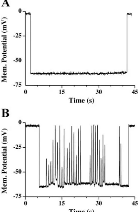
membrane potential. We have observed that in addition to the cells exhibiting stable resting membrane potential, a subset of cells (⬃30%) exhibited spontaneous fluctuations in the mem-brane potential. Figure 3, A and B, shows memmem-brane potential recordings from a myotube that does not exhibit spontaneous fluctuations in the membrane potential, and from one that does, respectively. Consistent with previous reports (13, 15, 34), the average value of the resting membrane potential was⫺62 ⫾ 0.44 mV (n⫽ 48). The recorded oscillations in the membrane potentials did not appear to resemble action potentials, because the magnitude of the depolarization, 26 ⫾ 1 mV (n ⫽ 146), was insufficient to reach the action potential threshold. How-ever, on the basis of previous reports, these depolarizations should be sufficient to activate Ca2⫹influx through the L-type
Ca2⫹ channels (32). The percentage of cells tested in each
culture exhibiting spontaneous membrane depolarizations cor-related with the percentage of cells that exhibited spontaneous intracellular Ca2⫹ oscillations as described in Fig. 1.
Analo-gous to the changes in the intracellular Ca2⫹, the spontaneous
depolarizations occurred without any stimulation of the cells. Role of DHPR and Ca2⫹influx in spontaneous oscillations.
Because membrane depolarizations activate the voltage sensors in the surface membrane/TT, we sought to determine whether DHPR is directly involved in eliciting these events. To do so, we applied 5M nifedipine to the extracellular bathing solu-tion. Nifedipine is a DHP-specific antagonist, which promotes the inactivation of the channel (21, 29) and has a blocking effect on the Ca2⫹current through the TT membrane (21). As
demonstrated in Fig. 4, application of nifedipine abolished the spontaneous RyR-mediated Ca2⫹release. However, nifedipine
could not abolish the KCl-induced Ca2⫹ transients, which
Fig. 2. RyR1 and␣1SDHPR (dihydropyridine receptor) are clustered in foci and colocalized in correspondence of calcium release units. A and B: both RyR1 and␣1SDHPR are clustered in discrete foci indicating the targeting of both proteins to junctions between sarcoplasmic reticulum and external membranes. C: double immunolabeling for RyR1 and␣1SDHPR shows a striking colocal-ization of the 2 proteins. Bar, 25m.
could still be elicited in these cells. Further evidence for the involvement of the DHPRs in the spontaneous activity was obtained from experiments conducted with muscular dysgen-esis myotubes (mdg), which do not express any functional DHPRs (16, 23). This phenotype renders DHPRs in these cells unable to conduct Ca2⫹ (1) or to participate in E-C coupling
(31). None of the tested mdg myotubes exhibited any type of spontaneous activity (Fig. 5). To determine whether these cells expressed functional RyR Ca2⫹ release channels, mdg cells
were challenged with 40 mM caffeine. All tested cells pro-duced a robust Ca2⫹ transient in response to the caffeine,
indicating a sufficient expression of the RyRs and the viable status of the cells. From these results, it could be inferred that although Ca2⫹oscillations occurred without any external
stim-uli, they were under control of the DHPRs.
Ca2⫹influx through DHPR. To confirm that the abolition of
spontaneous activity by nifedipine was based on its inhibition of Ca2⫹influx and not due to a conformational inactivation of
the DHPR, we tested the effects of Cd2⫹and La3⫹, which have
been previously described as potent blockers of the␣1SDHPR
channel pore (12, 31). As shown in Fig. 6, application of Cd2⫹/La3⫹completely blocked the spontaneous Ca2⫹
oscilla-tions and did so in every cell that exhibited this phenomenon. Washing the cells with the Cd2⫹/La3⫹-free solution could
reverse the Cd2⫹/La3⫹effects. In most cases, the spontaneous
activity returned in those cells that exhibited this activity before the application of the Cd2⫹/La3⫹. The presence of
Cd2⫹/La3⫹ did not interfere with the large Ca 2⫹ transient
elicited by depolarization, showing that similarly to nifedipine,
Cd2⫹/La3⫹blocked spontaneous activity but not the coupling
between␣1SDHPRs and RyR1.
DISCUSSION
In this study, we report the existence of the spontaneously occurring, regenerative, macroscopic Ca2⫹ transients, as well as
spontaneously occurring membrane depolarizations in cultured wild-type myotubes isolated from mouse skeletal muscle. The observed spontaneous activity is generated by the Ca2⫹ release
from the RyRs and, most importantly, appears to be triggered by the entry of extracellular Ca2⫹through␣
1SDHPRs.
The spontaneous Ca2⫹ transients were observed in a
sub-population of differentiated myotubes and depending on the relative level of cellular differentiation, constituted ⬃30 to 50% of the entire cell culture. The difference in differentiation rate is due to a number of factors including distribution of myoblasts on the culture plate and regional differences in the concentrations of excreted growth factors. These global oscil-lations in intracellular Ca2⫹, although exhibiting variable
fre-quency and amplitude between the cells, were not initiated by any type of applied stimulus and were always significantly smaller then the Ca2⫹transients detected upon cellular
depo-larization with KCl. Similar spontaneous Ca2⫹transients have
been previously reported in the cultured human (33) and chicken myotubes (9) and C2C12mouse cell line (9), as well as
embryonic Xenopus myocytes (8). However, one of the major differences from previous reports is that the events reported here appear to require the influx of extracellular Ca2⫹,
seem-ingly through the DHPRs.
Fig. 4. Inhibition of spontaneous activity in response to nifedipine. Representa-tive, normalized time courses of fluo 4 fluorescence of primary myotubes in response to 5 mM nifedipine and 5 mM nifedipine-supplemented imaging buffer (IB) containing 50 mM NaCl and 80 mM KCl (seeMATERIALS AND METHODSfor
details). Cells were initially perfused for 30 s with IB, followed by a 90-s perfusion with nifedipine (filled bar) and a 10-s stimulation with nifedipine/KCl solution (open bar) (n⫽ 37 cells). The vertical bar represents F/F0fluorescence intensity (au). Fig. 3. Primary myotubes exhibit spontaneous membrane depolarizations. A:
representative recording of resting membrane potential from myotube that does not exhibit spontaneous depolarizations (n⫽ 49 cells). The initial portion of the trace was recorded before myotube impalement; therefore, the membrane potential was adjusted arbitrarily to 0 mV. Impalement of the cell was accompanied by a downward voltage deflection, and the withdrawal of the electrode was reflected by an immediate upward deflection in voltage. B: representative recording of a membrane potential from a myotube that exhibits spontaneous membrane depolarizations (n⫽ 10 cells).
There are several lines of evidence suggesting that this type of activity is not simply random Ca2⫹release from the SR. The
fact that the release was uniform throughout the cell and that the oscillations appeared at fixed intervals and with fairly constant amplitudes suggests that there are specific cellular factors that govern the initiation of each oscillation. Results in Figs. 4 and 6 show that by blocking Ca2⫹influx into the cells
either by nonspecific cation channel blockers, such as Cd2⫹
and La3⫹, or more selectively by nifedipine, these oscillations
could be completely inhibited. The fact that application of either of these blockers does not eliminate the depolarization-elicited Ca2⫹ transients, that is, the functional components of
the skeletal E-C coupling were still preserved, suggests that the only cause for the change is the abolition of the DHPR Ca2⫹
current. And the fact that intracellular Ca2⫹ oscillations
reap-pear upon the washout of the blockers that restrict the flow of Ca2⫹suggests that the Ca2⫹influx is a necessary component of
the initiation of this type of activity.
We also demonstrate that a population of myotubes exhibits spontaneous depolarizations of the plasma membrane. It should be pointed out that the observed depolarizations did not resemble typical action potentials. Because the subthreshold depolarizations were not of sufficient magnitude (⬃35 mV) to reach the threshold of initiation of an action potential, the observed depolarizations could therefore not possibly elicit action potentials. However, these depolarizations must be large enough to activate L-type Ca2⫹channels to a level sufficient to
generate a significant Ca2⫹ influx. Although the membrane
potential recordings were performed independently of the
in-tracellular Ca2⫹ measurements, we believe that because the
frequency of occurrence of these two observations was similar in both preparations, the two phenomena are related to the same process. If the spontaneous depolarizations precede the activation of Ca2⫹release from the SR, then it is possible that
they activate the voltage sensors in the TT and, subsequently, the DHPR Ca2⫹ channels.
It has been generally accepted that skeletal muscle, unlike cardiac or smooth muscle, does not require the influx of extracellular Ca2⫹to achieve contraction. One of the reasons it
is believed that the influx of Ca2⫹is simply a vestigial process
is because the kinetics of activation are too slow and the magnitude of the current is simply too small for Ca2⫹to diffuse
rapidly from the DHPR Ca2⫹channel and overcome the Mg2⫹
inhibition of the RyR. To achieve Ca2⫹ influx-induced Ca2⫹
release in skeletal muscle, the skeletal DHPRs would have to behave similar to those of cardiac type with respect to the magnitude and the kinetics of the Ca2⫹influx. This condition
could be potentially achieved in the skeletal muscle if the cells were stimulated by repetitive depolarization (7, 11), analogous to those exhibited in Fig. 3. It has been previously reported that repetitive depolarizations of skeletal muscle fibers at short intervals, as infrequently as 1.7 Hz, result in acceleration, as well as in the potentiation, of the Ca2⫹ currents (7, 11).
Fig. 6. Cd2⫹/La3⫹abolish spontaneous activity. Representative, normalized time courses of fluo 4 fluorescence of primary myotubes in response to 0.5 mM Cd2⫹ and 0.1 mM La3⫹(filled bar) and 0.5 mM Cd2⫹and 0.1 mM La3⫹ -supplemented IB containing 50 mM NaCl and 80 mM KCl (seeMATERIALS AND METHODSfor details) (open bar) (n⫽ 45 cells). Cells were continuously perfused with IB. Where indicated by the bars, IB was supplemented with the appropriate reagents. The vertical bar represents F/F0fluorescence intensity (au).
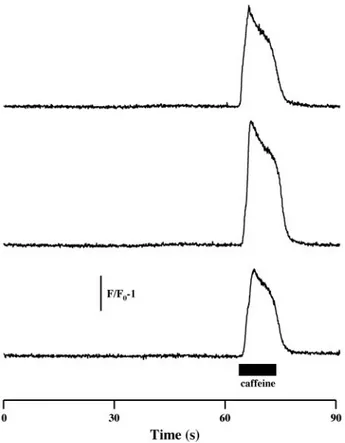
Fig. 5. Dyspedic (mdg) myotubes do not exhibit spontaneous activity. Repre-sentative, normalized time courses of fluo 4 fluorescence of mdg myotubes in response to 40 mM caffeine. Cells were initially perfused with IB, followed by a 10-s perfusion with caffeine (filled bar) (n⫽ 59 cells). The vertical bar represents F/F0fluorescence intensity (au).
Additionally, it has now been suggested that the magnitude of Ca2⫹influx through the skeletal DHPR could be sufficient to
induce CICR (14). Thus it is conceivable that the initiation of Ca2⫹release within each oscillation could be triggered by the
Ca2⫹entry through the␣
1sof DHPRs (5).
In summary, skeletal myotubes exhibit spontaneous oscilla-tions in intracellular Ca2⫹, as well as spontaneous
depolariza-tions of the plasma membrane. Pharmacological data indicate that initiation of Ca2⫹oscillations in developing skeletal
mus-cle cells is dependent on the Ca2⫹ influx through the ␣
1s
-subunit of the DHPR.
ACKNOWLEDGMENTS
The 34C monoclonal antibody developed by J. A. Airey and J. Sutko was obtained from the Developmental Studies Hybridoma Bank developed under the auspices of the National Institute of Child Health and Human Development and maintained by the University of Iowa, Department of Biological Sciences, Iowa City, IA 52242.
GRANTS
This work was supported by National Institute of Arthritis and Musculo-skeletal and Skin Diseases (NIAMS) Grant P01 AR-44650 (to P. D. Allen), Muscular Dystrophy Association Grant MDA 2688 (to P. D. Allen and F. Protasi), and NIAMS Grant AR-49160-2 (to A. Shtifman).
REFERENCES
1. Adams BA and Beam KG. Muscular dysgenesis in mice: a model system for studying excitation-contraction coupling. FASEB J 4: 2809–2816, 1990.
2. Airey JA, Beck CF, Murakami K, Tanksley SJ, Deerink TJ, Ellisman MH, and Sutko JL. Identification and localization of two triad junctional foot protein isoforms in mature fast twitch skeletal muscle. J Biol Chem 265: 14187–14194, 1990.
3. Armstrong CM, Bezanilla FM, and Horowicz P. Twitches in the presence of ethylene glycol bis(aminoethyl ether)-N,N⬘ tetracetic acid. Biochim Biophys Acta 267: 605–608, 1972.
4. Brum G, Stefani E, and Rios E. Simultaneous measurements of Ca2⫹ currents and intracellular Ca2⫹ concentrations in single skeletal muscle fibers of the frog. Can J Physiol Pharmacol 65: 681–685, 1987. 5. Chun LG, Ward CW, and Schneider MF. Ca2⫹spark occurrence in
embryonic mouse skeletal muscle is initiated by Ca2⫹entry and declines during postnatal development. Am J Physiol Cell Physiol 285: C686– C697, 2003.
5a.Endo M, Tanaka M, and Ogawa Y. Calcium induced release of calcium from the sarcoplasmic reticulum of skinned skeletal muscle fibres. Nature 228: 34–36, 1970.
6. Fabiato A and Fabiato F. Calcium-induced release of calcium from the sarcoplasmic reticulum of skinned cells from adult human, dog, cat, rabbit, rat, and frog hearts and from fetal and new-born rat ventricles. Ann NY Acad Sci 307: 491–522, 1978.
7. Feldmeyer D, Melzer W, Pohl B, and Zollner P. Fast gating kinetics of the slow Ca2⫹current in cut skeletal muscle fibres of the frog. J Physiol 425: 347–367, 1990.
8. Ferrari MB, Rohrbough J, and Spitzer NC. Spontaneous calcium transients regulate myofibrillogenesis in embryonic Xenopus myocytes. Dev Biol 178: 484–497, 1996.
9. Flucher BE and Andrews SB. Characterization of spontaneous and action potential-induced calcium transients in developing myotubes in vitro. Cell Motil Cytoskeleton 25: 143–157, 1993.
10. Flucher BE, Andrews SB, Fleischer S, Marks AR, Caswell A, and Powell JA. Triad formation: organization and function of the sarcoplasmic reticulum calcium release channel and triadin in normal and dysgenic muscle in vitro. J Cell Biol 123: 1161–1174, 1993.
11. Garcia J, Avila-Sakar AJ, and Stefani E. Repetitive stimulation in-creases the activation rate of skeletal muscle Ca2⫹currents. Pflu¨gers Arch 416: 210–212, 1990.
12. Garcia J and Beam KG. Measurement of calcium transients and slow calcium current in myotubes. J Gen Physiol 103: 107–123, 1994. 13. Iannaccone ST, Li KX, and Sperelakis N. Transmembrane electrical
characteristics of cultured human skeletal muscle cells. J Cell Physiol 133: 409–413, 1987.
14. Kasielke N, Obermair GJ, Kugler G, Grabner M, and Flucher BE. Cardiac-type EC-coupling in dysgenic myotubes restored with Ca2⫹ channel subunit isoforms␣1Cand ␣1Ddoes not correlate with current density. Biophys J 84: 3816–3828, 2003.
15. Kidokoro Y. Developmental changes of membrane electrical properties in a rat skeletal muscle cell line. J Physiol 244: 129–143, 1975.
16. Knudson CM, Chaudhari N, Sharp AH, Powell JA, Beam KG, and Campbell KP. Specific absence of the␣1subunit of the dihydropyridine receptor in mice with muscular dysgenesis. J Biol Chem 264: 1345–1348, 1989.
17. Lacampagne A, Klein MG, and Schneider MF. Modulation of the frequency of spontaneous sarcoplasmic reticulum Ca2⫹ release events (Ca2⫹ sparks) by myoplasmic [Mg2⫹] in frog skeletal muscle. J Gen Physiol 111: 207–224, 1998.
18. Laver DR, Baynes TM, and Dulhunty AF. Magnesium inhibition of ryanodine-receptor calcium channels: evidence for two independent mech-anisms. J Membr Biol 156: 213–229, 1997.
19. Meissner G and Henderson JS. Rapid calcium release from cardiac sarcoplasmic reticulum vesicles is dependent on Ca2⫹and is modulated by Mg2⫹, adenine nucleotide, and calmodulin. J Biol Chem 262: 3065–3073, 1987.
20. Melzer W, Herrmann-Frank A, and Luttgau HC. The role of Ca2⫹ions in excitation-contraction coupling of skeletal muscle fibres. Biochim Biophys Acta 1241: 59–116, 1995.
21. Neuhaus R, Rosenthal R, and Luttgau HC. The effects of dihydropyr-idine derivatives on force and Ca2⫹current in frog skeletal muscle fibres. J Physiol 427: 187–209, 1990.
22. Perez CF, Voss A, Pessah IN, and Allen PD. RyR1/RyR3 chimeras reveal that multiple domains of RyR1 are involved in skeletal-type E-C coupling. Biophys J 84: 2655–2663, 2003.
23. Pincon-Raymond M, Rieger F, Fosset M, and Lazdunski M. Abnormal transverse tubule system and abnormal amount of receptors for Ca2⫹ channel inhibitors of the dihydropyridine family in skeletal muscle from mice with embryonic muscular dysgenesis. Dev Biol 112: 458–466, 1985. 24. Pizarro G, Fitts R, Uribe I, and Rios E. The voltage sensor of excitation-contraction coupling in skeletal muscle. Ion dependence and selectivity. J Gen Physiol 94: 405–428, 1989.
25. Protasi F. Structural interaction between RYRs and DHPRs in calcium release units of cardiac and skeletal muscle cells. Front Biosci 7: d650– d658, 2002.
26. Protasi F, Paolini C, Nakai J, Beam KG, Franzini-Armstrong C, and Allen PD. Multiple regions of RyR1 mediate functional and structural interactions with␣1S-dihydropyridine receptors in skeletal muscle. Bio-phys J 83: 3230–3244, 2002.
27. Protasi F, Takekura H, Wang Y, Chen SRW, Meissner G, Allen PD, and Franzini-Armstrong C. RyR1 and RyR3 have different roles in the assembly of calcium release units of skeletal muscle. Biophys J 79: 2494–2508, 2000.
28. Rando TA and Blau HM. Primary mouse myoblast purification, charac-terization, and transplantation for cell-mediated gene therapy. J Cell Biol 125: 1275–1287, 1994.
29. Rios E and Brum G. Involvement of dihydropyridine receptors in excitation-contraction coupling in skeletal muscle. Nature 325: 717–720, 1987. 30. Schneider MF and Chandler WK. Voltage dependent charge movement
of skeletal muscle: a possible step in excitation-contraction coupling. Nature 242: 244–246, 1973.
31. Tanabe T, Beam KG, Adams BA, Niidome T, and Numa S. Regions of the skeletal muscle dihydropyridine receptor critical for excitation-con-traction coupling. Nature 346: 567–569, 1990.
32. Tanabe T, Beam KG, Powell JA, and Numa S. Restoration of excitation-contraction coupling and slow calcium current in dysgenic muscle by dihy-dropyridine receptor complementary DNA. Nature 336: 134–139, 1988. 33. Tanaka H, Furuya T, Kameda N, Kobayashi T, and Mizusawa H.
Triad proteins and intracellular Ca2⫹ transients during development of human skeletal muscle cells in aneural and innervated cultures. J Muscle Res Cell Motil 21: 507–526, 2000.
34. Thomson CM and Dryden WF. Different actions of calcium channel blocking agents on resting membrane conductance in developing skeletal muscle. Can J Physiol Pharmacol 59: 335–341, 1981.
35. Zimanyi I, Buck E, Abramson JJ, Mack MM, and Pessah IN. Ryan-odine induces persistent inactivation of the Ca2⫹release channel from skeletal muscle sarcoplasmic reticulum. Mol Pharmacol 42: 1049–1057, 1992.
![Fig. 1. Ryanodine receptor (RyR)-mediated spontaneous oscillations in intra- intra-cellular Ca 2 ⫹ concentration ([Ca 2 ⫹ ] i ) in primary myotubes](https://thumb-eu.123doks.com/thumbv2/123dokorg/4961482.53112/2.931.486.844.714.1009/ryanodine-receptor-mediated-spontaneous-oscillations-cellular-concentration-myotubes.webp)