UNIVERSITY OF MOLISE
Dept. of Medicine and Health Science “V. Tiberio”
PhD course in
TRANSLATIONAL AND CLINICAL
MEDICINE
XXX CYCLE
S.S.D: Area-05-Bio 14 Farmacologia
Doctoral Thesis
GENETIC, PATHOPHYSIOLOGICAL, AND
PHARMACOLOGICAL IMPLICATIONS OF KCNT1 AND
KCNT2 POTASSIUM CHANNELS
IN NEURODEVELOPMENTAL DISORDERS AND
EPILEPTIC ENCEPHALOPATHIES
Tutor: Student:
Prof. Maurizio Taglialatela Laura Manocchio
Coordinator:
Prof. Ciro COSTAGLIOLA
Academic year: 2016/2017
INDEX
INTRODUCTION ... 5
1. THE POTASSIUM CHANNELS FAMILY ... 5
1.1 THE SLO (KCA) FAMILY OF K+ CHANNELS ... 7
1.2 TOPOLOGICAL STRUCTURE OF THE SLO2 a-SUBUNITS ... 9
1.3 SLO2.2 (OR KCNT1 OR SLACK) CHANNELS ... 11
1.4 SLO2.1 (OR KCNT2 OR SLICK) CHANNELS ... 13
1.5 KCNT1 AND KCNT2 CHANNEL SUBUNITS FORMS HETEROMERIC COMPLEXES ... 14
1.6 KCNT1 AND KCNT2 CHANNELS REGULATION ... 14
1.7 DISTRIBUTION OF KCNT1 AND KCNT2 CHANNEL SUBUNITS IN THE CENTRAL NERVOUS SYSTEM (CNS) 18 2. ROLE OF POTASSIUM CHANNELS IN EPILEPSY ... 19
2.1 THE EPILEPTIC ENCEPHALOPATHIES: AN OVERVIEW ... 21
2.2 THE GENETICS OF THE EPILEPTIC ENCEPHALOPATHIES ... 22
2.3 KCNT1 MUTATIONS RESULT IN A WIDE RANGE OF SEIZURE DISORDERS AND INTELLECTUAL DISABILITIES 25 2.3.1 MALIGNANT MIGRATING PARTIAL SEIZURES OF INFANCY ... 25
2.3.2 OHTAHARA SYNDROME ... 27
2.3.3 EARLY MYOCLONIC ENCEPHALOPATHY ... 28
2.3.4 WEST SYNDROME ... 28
2.3.5 AUTOSOMAL DOMINANT NOCTURNAL FRONTAL LOBE EPILEPSY ... 28
3. PHARMACOLOGY OF SODIUM-ACTIVATED POTASSIUM CHANNELS ... 30
4. AIMS OF THE STUDY ... 33
5. MATERIALS AND METHODS ... 37
5.1 SITE-DIRECTED MUTAGENESIS ... 37
5.2 BACTERIAL TRANSFORMATION AND PLASMIDIC DNA PREPARATION ... 38
5.3 CELL CULTURES AND TRANSIENT TRANSFECTION WITH LIPOFECTAMINE 2000 ... 39
5.4 PATCH-CLAMP RECORDINGS ... 40
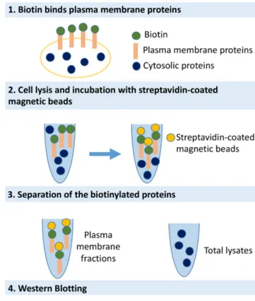
5.5 PLASMA MEMBRANE PROTEIN BIOTINYLATION AND WESTERN BLOTTING ... 41
5.6 MOLECULAR MODELLING ... 43
5.7 STATISTICS ... 43
6. RESULTS ... 44
6.1 CLINICAL FEATURES OF PATIENTS AFFECTED BY EPILEPTIC ENCEPHALOPATHY CARRYING KCNT1 OR KCNT2 MUTATIONS ... 44
6.2 IDENTIFICATION OF G288S AND M516V DE NOVO KCNT1 MUTATIONS IN PATIENTS AFFECTED BY MALIGNANT MIGRATING PARTIAL SEIZURES (MMPSI) ... 46
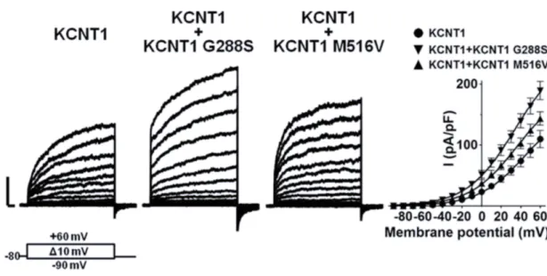
6.3 FUNCTIONAL CHARACTERIZATION OF HOMOMERIC WILD-TYPE AND MUTANT G288S AND M516V CHANNELS ... 46
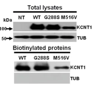
6.4 BIOCHEMICAL ASSAYS OF TOTAL AND PLASMA MEMBRANE EXPRESSION OF WILD-TYPE AND MUTANT KCNT1 SUBUNITS ... 50
6.5 FUNCTIONAL CHARACTERIZATION OF WILD-TYPE AND MUTANT KCNT1 IN HETEROMERIC CONFIGURATION ... 52
6.6 PHARMACOLOGICAL CHARACTERIZATION OF G288S AND M516V MUTANT CHANNELS ... 54
6.7 BIOCHEMICAL AND FUNCTIONAL CHARACTERIZATION OF KCNT1-E893K AND KCNT1-R950Q MUTANT SUBUNITS ... 56
6.9 BIOCHEMICAL EVIDENCE FOR KCNT2 CHANNELS EXPRESSION IN TOTAL LYSATES OF CHO AND HEK 293
CELLS ... 62
6.10 FUNCTIONAL EFFECTS OF THE C484Y VARIANT ON KCNT2 CHANNEL FUNCTION ... 63
6.11 BIOCHEMICAL AND FUNCTIONAL EFFECTS OF THE R190H AND R190P MUTANT SUBUNITS ... 65
6.12 FUNCTIONAL CHARACTERIZATION OF HETEROMERIC CHANNELS FORMED BY WILD-TYPE OR MUTANT KCNT2 SUBUNITS ... 68
6.13 MOLECULAR MODELLING OF THE MECHANISM THROUGH WHICH THE R190 RESIDUE PARTICIPATES IN THE STABILIZATION OF THE CLOSED STATE OF KCNT2 CHANNELS ... 70
6.14 PHARMACOLOGICAL MODULATION OF WILD-TYPE AND MUTANT KCNT2 SUBUNITS ... 71
7. DISCUSSION ... 73
8. REFERENCES ... 81
9. ACKNOWLEDGEMENTS ... 87
ABBREVIATIONS
EE: Epileptic Encephalopathy NEE: Neonatal Epileptic Encephalopathy MMPSI: Malignant Migrating Partial Seizure of Infancy ADNFLE: Autosomal Dominant Nocturnal Frontal Lobe Epilepsy RCK: Regulators of K+ Conductance CHO: Chinese Hamster Ovary HEK: Human Embryonic Kidney RT: Room Temperature GOF: Gain-of-function
INTRODUCTION
1. The Potassium channels family
Potassium channels (K+) are membrane proteins that allow rapid and selective flow of K+ ions across the cell membrane, thus generating electrical signals in many excitable or unexcitable cells. Potassium channels play an important role in several biological functions: their contribution is important in determining the shape and the duration of the action potential, in the control of the membrane potential, in the modulation of hormone secretion and epithelial function.
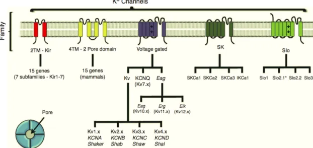
The potassium channel family can be classified based on their structure in channels with two transmembrane segments (2TM), four transmembrane segments (4TM; 2-pore domain), six transmembrane segments (6TM; voltage-gated K+ channels and SK) or seven transmembrane segments (7TM; SLO, Figure 1).
The inward rectifier (Kir) family contains subunits with a topological structure
characterized by two TM segments flanking the pore-forming domain; they assemble as tetramers. They are involved in the control of several biological functions in different cell types, such as cardiac myocytes and endothelial cells. The two-pore 4TM segments K+ channels (K 2P) family assemble as dimers and play an important role in several neuronal functions.
The 6TM segments (called from S1 to S6) K+ channels family includes several subfamilies
called from Kv1.x to Kv12.x. Voltage-dependent K+ channels contain a voltage-sensor
domain (VSD; from S1 to S4 segments) with the S4 segment containing
positively-charged amino acids that constitute the voltage-sensing elements. The small conductance K+ channel family (SK channels, SKCa, KCa2) also have 6TM and are activated by an increase in intracellular Ca2+ ions [Gonzalez et al., 2012].
In particular, the SLO channel subfamily will be the focus of the present Doctoral Thesis and will be extensively discussed in the next sessions.
Figure 1 Potassium channels family classification accordingly to their subunit structure. Potassium channel
families are classified in those having two transmembrane domains (2TM; Kir ), 4TM (2-pore domain), 6TM (voltage-gated and SK) and 7TM (SLO). The large-conductance SLO channel family include the SLO2.x channels, which have a topological structure showing six transmembrane domains. [From Gonzalez et al., 2012].
1.1 The SLO (K
Ca) family of K
+channels
The SLO family encompasses four genes in the mammalian genome that code for high-conductance K+ channels (Table 1). The first member of this family, SLO1, is also known
as “big” K+ channel (BK) or maxi-K channels as these channels have an unusual large
conductance (about 250 pS). Other two highly similar SLO2 paralogues, SLO2.1 (also known as Slick, KCNT2 or calcium-activated K+ channels 4.2) and SLO2.2 (also known as
Slack, KCNT1 or calcium-activated K+ channels 4.1) are members of the SLO2 channels
family. The last member of SLO family is called SLO3. All four genes encode a-subunits that forms heterotetrameric channels that gate a K+-selective current, with marked
differences in their gating properties [Salkoff et al., 2006], as detailed below:
Channel Alternative names Gene
symbol (human)
Chromosomal localization SLO1 BK, KCa, Maxi-K, KCa1.1 KCNMA1 10q22
SLO2.1 Slick, KNa, KCa4.2 KCNT2 1q31.3
SLO2.2 Slack, KNa, KCa4.1 KCNT1 9q34.3
SLO3 Potassium large conductance pH-sensitive channel, KCa5.1
KCNU1 8p11.2
Table 1 SLO channels family genes. [Adapted from Salkoff et al., 2006].
SLO1: SLO1 channels are expressed in many tissues where they elicited an outward K+
current with an unusual dependence on both membrane depolarization and influx of Ca2+ ions [Gorman et al.,1980]. The SLO1 gene was cloned from the Drosophila mutants Slowpoke, which exhibited flight problems and altered response to heat shock. Voltage- clamp recordings of currents recorded in Drosophila mutant revealed that the calcium-dependent regulation of the outward K+ current was absent. The SLO1 gene codes for a protein with homology to voltage-dependent K+ channels, but it has an extra TM segment (S0), allowing the N-terminus to face the extracellular side of the membrane. In
addition, these channels also contain hydrophobic domains at the C-terminus, called RCK (Regulators of K+ conductance): when Ca2+ ions bind these sites, the membrane depolarization needed to open channel decreases, rending SLO1 both Ca2+
- and voltage-dependent channels.
SLO2 is the second member of the SLO channels family that include two paralogues called KCNT1 and KCNT2. Functional studies have revealed that KCNT1 and KCNT2, similarly to BK channels, have high conductance but, differently from BK are potentiated by Na+ (not Ca2+) ions, thereby underlying Na+-activated K+ currents (IK
Na). These
currents have been described in many cells [Hartung et al., 1985], including guinea pig cardiomyocytes [Kameyama et al., 1984] and a wide variety of mammalian neurons, such as those located in the dorsal root ganglia (DRG) [Bischoff et al., 1998].
The identification of the Na+-activated K+ currents has been achieved by means of different experimental tools; including the replacement of the external sodium by
lithium ions. Lithium is a much weaker activator of KNa channels than sodium. Because
lithium promptly enters cells through voltage-dependent sodium channels, lithium replacement in voltage-clamp experiments reduces the net outward currents if KNa currents are present [Hage et al., 2012].
The IKNa currents are believed to play an important role under ischemic conditions in
cardiomyocytes, due to a protective role prompted by the activation of large K+ conductances upon dangerous increase in [Na+]
i [Mitani et al., 1992]. Similarly, in many
neurons IKNa currents contribute to a long-lasting slow afterhyperpolarization (sAHP),
which results from a slowly developing outward current evoked during sustained stimulation. The period of reduced excitability afforded by sAHP is thought to protect the cell from repetitive and tetanic activity. It has been shown that, whereas the early part of sAHP is dependent on Ca2+ influx during stimulation, the late part of sHAP is Na+ sensitive [Schwindt et al., 1998a,b]. Similar Na+-dependent sHAP has also been observed in a number of other neurons, such as hippocampal pyramidal cells [Gustafsson and Wigstrom, 1983] and spinal cord neurons [Wallen et al., 2007].
The fact that KCNT1 and KCNT2 represent the molecular basis of IKNa currents has been
confirmed in mouse models lacking both SLO2.2 and SLO2.1 genes: these mice show a complete absence of the KNa current in DRG neurons, which promotes an increased
excitability in response to depolarizing stimuli. This increased neuronal firing is thought to be responsible for the enhanced itching and pain sensations observed in this mouse
model [Martinez-Espinosa et al., 2015].
SLO3 is the third member of the SLO family channel. The SLO3 gene was identified by bioinformatic approaches characterizing an EST (expressed sequence tag) encoding a sequence with high similarity to the SLO1 channels. Like SLO2, SLO3 channels lack the Ca2+-dependent gating but they are dependent on internal pH values (Figure 2). Figure 2 Phylogenetic tree of SLO channels family in mammals. [From: Gonzalez et al., 2012].
1.2 Topological structure of the SLO2 a-subunits
The topological structure of the a-subunits of SLO2 channels resembles that of voltage-gated K+ channels in having a symmetrical arrangement of membrane-spanning segments clustered around a water-filled, K+ ion-selective pore.The a-subunit of SLO2 channels has six hydrophobic transmembrane segments (S1-S6)
along with a pore-lining loop that is found between the S5-S6 segments. The SLO2
channels also exhibit a large C-terminal region (over 900 amino acids in length) that greatly exceeds the length of the region encompassing the membrane-spanning domains and contains both hydrophobic and hydrophilic domains. Both SLO2.1 and SLO2.2 channels lack the canonical gating charges in the S4 membrane-spanning
segment usually associated with voltage sensing and both exhibit a low intrinsic voltage dependence of activation [Yuan et al., 2003]. The N- and C-terminus of SLO2 channels are located on the intracellular side. The C-terminus of SLO2 subunits contains two predicted regulators of K+ conductance (RCK) domains that stack on the top of each
other and form a gating ring underneath the channel pore (Figure 3). X-ray crystallographic studies of C-terminal domain of chicken KCNT1 channels have confirmed that the structure of RCK domains is likely to resemble that of SLO1 channels.
Figure 3 Topological structure of SLO2 a-subunit. SLO2 channels have six transmembrane domains. The
transmembrane domains are labeled as S1-S6, with the P-loop located between the S5-S6 segments. Both the N- and C-terminal ends are cytosolic. The C-terminal domain contains two Regulators of K+ conductance (RCK). The S4 segment lack the canonical positive charges that confers voltage sensitivity. Four of these a subunits assemble into a functional channel. (Adapted from: Salkoff et al., 2006).
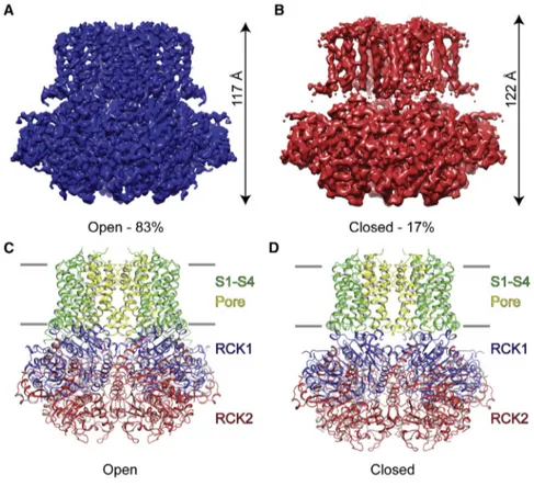
More recently, structural studies have revealed with high accuracy the transition of the KCNT1 channels into the open configuration as a function of Na+ concentration: in fact, by using Cryo-Electron Microscopy, MacKinnon and coll. (2017) solved the crystal structure of KCNT1 channels in the open configuration and revealed structural features that were not present in the closed configuration of KCNT1 channels. In particular, in the open configuration, the gating ring is firmly compressed against the transmembrane domain. In addition, a subdomain structure of the membrane-facing surface of the gating ring (RCK1 N-lobe) showed an expanded conformation induced by Na+ ions, if compared to the closed configuration. This structure is also responsible for pore opening, by causing the displacement of the S6 helices radially away from the pore axis
[Hite et al., 2017]. By contrast, in the closed and Na+-unbound state, the S6 helices were
found to be constricted, thus leading to the observation that the channel was closed (Figure 4). Such observation was also supported by the absence of channels activity in membrane with 0 mM cytoplasmic Na+ [Hite et al., 2015].
Figure 4 Architecture of closed and open KCNT1 channel subunits by cryo-electron microscopy. (A, B) Cryo EM
density map of open (A) and closed (B) KCNT1 channel. (C, D) Domain organization of open (A) and closed (B) chicken KCNT1. The S1-S4 domains are colored in green, the pore domain is yellow, the RCK 1 domain is blue and the RCK2 domain is red. [From: Hite et al., 2017]
1.3 SLO2.2 (or KCNT1 or Slack) channels
The term “Slack” derived from “sequence like A calcium activated K+ channels” because
part of the pore domain and the S6 domain is similar to that of SLO1 (BK) large
conductance calcium-activated K+ channels [Dryer, 1994].
KCNT1 channel was first cloned and expressed in 1998 [Joiner et al., 1998] and was found to be regulated by intracellular Na+ ions in 2003 [Yuan et al., 2003]. KCNT1
channels elicit outwardly rectifying K+ currents that increase upon depolarization. These currents show complex activation kinetics, with an instantaneous time-independent component, followed by a slower, time-dependent one.
When expressed either in mammalian cell lines or Xenopus oocytes KCNT1 channels have a relatively large unitary conductance (~180 pS) [Yuan et al., 2003]; in addition, multiple subconductance states have been described [Brown et al., 2008].
activation of KCNT1 channels is ~ 40 mM [Tamsett et al., 2009], a concentration highly overpassing physiological Na+ levels but possibly occurring during high frequency
neuronal activity or upon hypoxic insults. These observations suggest that the role of KNa channels may be strictly limited to these physiopathological conditions. However, it
has to be highlighted that another activator of KCNT1 current is the nicotinamide adenine dinucleotide (NAD+) [Tamsett et al., 2009], which induces a significant reduction in the [Na+]
i required for current activation and likely allows KCNT1 current
activation also in physiological conditions. These observations are supported by the fact that both Na+ and NAD+ binding sites are localized in the RCK2 domain. In particular, the D818 and H823 residues of rat KCNT1 channels are crucial for the Na+ sensitivity of KCNT1 currents [Zhang et al., 2010], while a complex domain appears to be crucial for NAD+ binding. KCNT1 channel subunits also contain a PDZ-binding consensus motif at the end of the C-terminus, which is functionally relevant since Na+ channels and glutamate receptors can cluster by binding to the same domain [Tomita et al., 2001]. Different KCNT1 channel subunits are generated by alternative splicing of KCNT1 mRNA. In fact, five different transcripts from the rat KCNT1 gene have been described, and these are predicted to produce KCNT1 channels that differ in their cytoplasmic amino termini. In particular, the N-terminal domain of Slack-A is smaller than that of Slack-B and the sequence of Slack-A N-terminal domain very closely resembles the N-terminus of the KCNT2 subunits.
In functional experiments, these isoforms show different activation kinetics: in fact, while Slack-B subunits elicited very slowly-activating currents, Slack-A channels activate very rapidly upon depolarization [Joiner et al., 1998; Yuan et al., 2003].
The kinetic properties of Slack-A and Slack-B isoforms indicate that the predominance of one or the other of these two isoforms is likely to shape the specific pattern of response to repetitive stimulation, as observed in simulations performed in neuronal models: in particular, Slack-B channels are likely to promote bursting and a graded pattern of adaptation, whereas Slack-A channels produce a strong and fixed pattern of rapid adaptation [Brown et al., 2008].
1.4 SLO2.1 (or KCNT2 or Slick) channels
The term “Slick” stands for “sequence like an intermediate conductance K+ channel” as
KCNT2 channels have a conductance that is intermediate between that of SLO1 (BK) calcium-activated K+ channels and other K+ channels.
Overall, the sequence of KCNT2 channel is 74% identical to that of KCNT1; the transmembrane domains and the RCK domains are almost identical. The greatest divergence between KCNT1 and KCNT2 channels occurs at the distal C-terminal regions. The predicted cytoplasmic N-terminus of KCNT2 is similar to that of the Slack-A isoform. Also the electrophysiological properties of KCNT2 currents, recorded in mammalian cells or Xenopus oocytes, resemble those of Slack-A: in fact, KCNT2 channels activate very rapidly upon depolarization and have multiple subconductance states. Also, KCNT2 currents are activated by Na+ ions in a concentration-dependent and reversible manner.
Concentration-response curves for activation of KCNT2 channels revealed that the effective Na+ concentrations for 50% (EC
50) of KCNT2 channels activation is 89 mM, a
value even greater than that described for KCNT1 channels activation (40 mM). By contrast, whereas KCNT1 channels activation has an absolute requirement of Na+ ions, KCNT2 channels have a basal level of activity even in the absence of Na+ ions [Bhattacharjee et al., 2003].
In addition to their Na+-sensitivity, both KCNT1 and KCNT2 channel subunits can be activated by intracellular chloride ions (Cl-) [Yuan et al., 2000], although there is a large difference in their sensitivity: in fact, there is an approximately five-fold increase in KCNT2 channel activation when cytoplasmic Cl- is raised from 3 to 130 mM in the presence of 5 mM Na+, whereas KCNT1 channel activity is increased only twofold under the same conditions [Bhattacharjee et al., 2003]. The domains that confer the Cl- sensitivity are yet unknown. It has been proposed that, in SLO2, Cl- binds to a stretch of positively charged amino acids that has been referred as “Cl- bowl”, localized in the tail region of the channel [Yuan et al., 2000]. The KCNT2 channels are considered to be hybrid between two classes of K+ channels: Na+-activated (K
Na) and ATP-sensitive (KATP) K + channels. Indeed, KCNT2 channel
subunits also contain a regulatory nucleotide-binding site that is responsible for ATP-mediated inhibition of channel activity, thus suggesting a more specialized protective
role under ischemic conditions and seizure activity in neuronal cells. The ATP-binding site is located in the second RCK domain, making the channel sensitive to cytoplasmic ATP levels [Bhattacharjee et al., 2003]. Like KCNT1, also currents elicited by KCNT2 subunits are potentiated by NAD+ exposure. The NAD binding motif in these channels seem to be located within the second RCK domain of KCNT1 and KCNT2 subunits and includes a “finger-print region” containing babab motif [Tamsett et al.,2009].
1.5 KCNT1 and KCNT2 channel subunits forms heteromeric complexes
Slack and Slick subunits can coassemble to form heterotetrameric channels that differ in their properties from those of homomeric channels [Chen et al., 2009]. The unitary conductance of Slack/Slick heteromeric channels results to be intermediate between that of Slack (~ 180 pS) and Slick (~ 140 pS) homomers. Moreover, assembly of heteromeric Slack/Slick channels appears to be specific for the Slack-B isoform: in fact, when Slick and Slack-B are coexpressed at 1:1 ratio, either in Xenopus oocytes or in mammalian HEK293 cells, a 18-25 fold increase in current amplitude is observed when compared to the currents expressed by either subunit alone [Chen et al., 2009]. This functional effect is due to an increased protein expression at the plasma membrane (as demonstrated in biotinylation experiments). These results were obtained by using chimeric channels that replaced the cytoplasmatic N-terminal domain of KCNT2 with that of Slack-B. The coexpression of this modified Slick channel with wild-type Slick channels in oocytes produced a 30-fold increase in whole-oocyte currents, which is similar to the increase obtained when Slack-B subunits were co-expressed with Slick. Interaction between Slack-B and Slick subunits was also demonstrated by co-immunoprecipitation experiments in rat brain tissue and in heterologous expression system [Chen et al., 2009].1.6 KCNT1 and KCNT2 channels regulation
The activity of KCNT1 and KCNT2 channel subunits, as well as the heterotetrameric KCNT1/KCNT2 channel assembly, is regulated by several signaling pathways:
• Modulation of KCNT1 and KCNT2 by Protein Kinase C (PKC)
Exposure of mammalian cells or Xenopus oocytes expressing KCNT1 channels to activators of protein kinase C (PKC) such as diacylglycerol or phorbol esters, lead to a 2-3 fold increase in current amplitude and a slowing of the rate of activation [Santi et al., 2006]. The PKC-mediated effects involved the phosphorylation of many residues, such as serine residue S407 located in the region of the cytoplasmic C-terminal domain between the S6 transmembrane segment and the first RCK domain [Barcia et al., 2012]
and the T517 residue located in the RCK domain [Santi et al., 2006].
In contrast to KCNT1 channels, the PKC activation induces a decrease in KCNT2 currents and this effect seems to be mediated by modification within the C-terminus of the channels [Santi et al., 2006].
Heteromeric Slack-B/Slick channels also respond to PKC activation in a manner that is distinct from that of either subunit expressed alone: in fact, application of PKC activators in Xenopus oocytes expressing both subunits potently reduces currents by ~90%, an effect much greater than the degree of inhibition measured for homomeric KCNT2 channels.
• Modulation of KCNT1 and KCNT2 by G Protein-Coupled Receptors
KCNT1 and KCNT2 channel have been coexpressed in Xenopus oocytes with the M1 muscarinic receptor and the mGluR1 metabotropic receptor [Santi et al., 2006]. These are Gaq protein-coupled receptors that lead to the activation of PKC. The activation of these receptors leads to an increase in KCNT1 currents and a reduction of KCNT2 currents, consistent with the above mentioned effects prompted by a direct PKC activation. The importance of this regulation is suggested by a wide colocalization of these receptors with the SLO2 channel subunits throughout the nervous system.
• Modulation of SLO2 currents by cyclic AMP
Na+-activated K+ channels are also modulated by the biogenic amines such as dopamine
mushroom body of the cricket Gryllus bimaculatus.
In particular, in these cells OA and DA respectively increased and decreased the open probability of SLO2 channels. In addition, Na+-activated K+ channels are also modulated
by cAMP/PKA (protein kinase A) signaling pathways leading to an increased channel open probability, and cGMP/PKG (protein kinase G) pathways responsible for a reduction in the open probability of SLO2 channels [Aoki et al., 2008].
The connexion between OA and cAMP-mediated signaling pathways is demonstrated by the fact the activation of OCTaR is also associated with a small increase in intracellular cAMP levels through the stimulatory G proteins (Gs), which in turn activate the protein Kinase A (PKA). In addition, insect dopamine receptors are G protein-coupled receptor (GPCR) and are classified into four subtypes: the D1-like dopamine receptors (Dop1), invertebrate dopamine receptors (INDRs, also known as Dop2), the D2-like dopamine receptors (Dop3) and the dopamine/ecdysteroid receptors (DopEcR). Once dopamine receptors are activated by dopamine, downstream signaling systems, such as the cyclic adenosine monophosphate (cAMP) or cyclic second messenger pathway and the inositol trisphosphate (IP3) second messenger pathway, are activated [Watanabe et al., 2013]. A possible connection between DA and cGMP has not yet been established.
• Modulation by PIP2
A variety of ion channels has been shown to be regulated by phosphatidylinositol 4,5-biphosphate (PIP2). The application of PIP2 analogues (Dic8 PI(3,4)P2 or Dic8 PI(4,5)P2) on
the cytoplasmic side increases the amplitude of KCNT1 and KCNT2 currents in Xenopus oocytes. Consistently, the exposure to pharmacological compounds, such as neomycin and wortmannin, that are able to reduce endogenous PIP2 levels, induces a significant reduction of current amplitudes mediated by both channels [Tejada et al., 2012]. • Modulation by Estradiol The open probability of KCNT1 and KCNT2 channels has been found to be increased by 17b-estradiol [Zhang et al., 2005].
• Interactions of KCNT1 channels with the Fragile X Mental Retardation Protein (FMRP)
The Fragile X syndrome is the most common inherited form of intellectual disability in humans. This syndrome is caused by a loss of expression of the RNA-binding protein FMRP (Fragile X mental retardation), also required for the normal activity-dependent protein translation in neurons. The cytoplasmic C-terminal domain of KCNT1 subunits interacts with FMRP that acts as a potent activator of KCNT1 channels. This protein-protein interaction is associated with the almost complete elimination of subconductance states. Furthermore, the application of a recombinant FMRP (1-298), which contains only the interaction domains of FMRP with KCNT1, reversibly increases the channel opening probability by two- to three-fold. On the other hand, FMRP was also found to have no effect on KCNT1 truncated channels (Slack-BD804), lacking sites essential for the interaction with FMRP [Brown et al., 2010].
1.7 Distribution of KCNT1 and KCNT2 channel subunits in the Central
Nervous System (CNS)
Cloning of KCNT1 and KCNT2 genes and the development of specific antibodies have provided the unique opportunity to carry out a detailed study of the regional distribution of the encoded subunits. The highest levels of KCNT1 and KCNT2 channels have been found in the brain, with lower expression levels in the heart and kidney [Joiner at., 1998; Yuan et al., 2003; Bhattachaerjee and Kaczmarek, 2005]. KCNT2 is highly expressed in neurons, whereas no staining is found in glial cells. Strong hybridization was found throughout the brain, including the cerebral cortex, hippocampus, deep cerebellar nuclei, cerebellar Purkinje cells, reticular tegmental nucleus of the pons, preoptic nucleus, substantia nigra, and auditory brainstem nuclei [Joiner et al., 1998].
More recently, distribution pattern in the mouse brain of KCNT1 and KCNT2 channel subunits has been also described in more detail. The two channels exhibit distinct distribution, but their expression overlaps in some regions, including the olfactory bulb, subfornical organ, substantia nigra, pars compacta, oculomotor and red nuclei, interpeduncular and rhabdoid nuclei, nucleus of the trapezoid body, reticulotegmental nucleous of pons, and the inferior olivary complex. Several brain structures also exhibit expression pattern in which KCNT1 and KCNT2 do not overlap. In particular, KCNT1 immunoreactivity was found alone in islands of Calleja, nuclei of the extended amygdala, hippocampal formation, and ventromedial hypothalamic and arcuate nuclei. In contrast, a marked KCNT2 channel immunolabeling was found in globus pallidus, substantia nigra, pars reticulate and nigrostriatal bundle, and mesencephalic and parvicellular trigeminal nuclei and in parts of cerebellar cortex [Rizzi et al., 2016].
2. Role of Potassium channels in Epilepsy
Potassium (K+) channels underlie outward K+ currents that contribute to membrane
repolarization and hyperpolarization, thus limiting the neuronal excitability. Such channels are the only ion-selective cation channels that have an equilibrium potential near the typical cellular resting potential.
Potassium channels are expressed in almost every cell, particularly in neurons and excitable tissues, where they regulate the shape and duration of action potentials, the firing rate and the overall excitability of cells. The functional heterogeneity of K+ currents expressed in excitable and non-excitable cells is primarily due to the large number of genes either encoding pore-forming (a) or accessory (b) subunits in the mammalian genome. Additional factors, such as alternative splicing, RNA editing, and ability to form homomeric or heteromeric complexes among pore-forming and accessory subunits also contribute to the K+ channel properties diversification and expression levels. More than 80 genes encoding for K+ channels have been cloned
representing the largest group of ion channels regulating the electrical activity of cells in different tissues. It is therefore not surprising that mutations in these genes leading to ion channels dysfunctions cause several diseases and, in particular, epilepsy in humans and animal models [Villa and Combi, 2016].
Epilepsies are common neurological disorders in infancy, childhood and adolescence characterized predominantly by recurrent and unpredictable interruptions of normal brain function, called epileptic seizures (Fisher et al., 2005).
According to the International League Against Epilepsy (ILAE), an epileptic seizure is “a transient occurrence of signs and/or symptoms due to abnormal excessive or synchronous neuronal activity in the brain”.
The definition of epilepsy also implies the occurrence of at least one epileptic seizure, where the term “seizure” indicates an “abnormal and synchronous excitation of a neuronal population lasting seconds or minutes”.
Epileptic seizures arise when functional alterations of neurons occur, causing an excessive and transient discharge of action potentials. Since the epileptic seizure can
involve a specific neuronal population or the whole brain, the semiology of the epileptic seizures varies according to the neuronal populations and/or cerebral circuits involved. Generally, seizures arise when there is a disruption of the mechanisms controlling the physiological balance between neuronal excitation and inhibition. Cell membrane of neurons have several different ion channels, including Na+ and K+ leak channels as well
as voltage-gated Na+ and K+ channels that allow the passage of ions as a function of their electrochemical gradients. Cl- and Na+ are mainly located in the extracellular fluid,
while K+ ions and negatively charged proteins are located in the intracellular fluid therefore K+ ions leak out of the cell, moving down their concentration gradient. As K+ leaves the cell, the negatively charged proteins are unable to follow because the cell membrane is not permeable to them.
The loss of positive ions from the cell creates an electrical gradient. Because opposite charges attract each other, the negative proteins inside the cell try to pull K+ back into the cell. At some point in this process, the electrical force attracting K+ into the cell becomes equal in magnitude to the chemical concentration gradient driving K+ out of the cell. At that point, called equilibrium potential for K+ (-90 mV), net movement of K+ across the membrane stops. By contrast, because Na+ is more concentrated outside the
cell, some Na+ ions moves into the cell and accumulates there until the equilibrium potential for Na+ (+60 mV) as most cells are about 40 times more permeable to K+ than
to Na+ cell membrane resting potential is closer to the K+, rather than Na+ equilibrium
potential (-90 mV).
Therefore it is plausible that abnormalities in Na+ or K+ channel function can
dramatically alter neuronal firing, through different mechanism depending on the possibility that specific classes of ion channels are expressed in inhibitory and/or excitatory neurons.
The concept that an alteration of the balance between excitation and inhibition processes is responsible for the onset of epileptic seizures has led to the design of anticonvulsant drugs able to restore the physiological neuronal excitability. The traditional antiepileptic drugs, in fact, act mainly through inhibition of Na+ (phenytoin,
inhibitory GABAergic transmission (phenobarbital, benzodiazepines, tiagabine, vigabatrin).
The etiology of epilepsy is very heterogeneous: many different genetic and pathophysiological factors, alone or in combination, can underlie an increased risk of developing a seizure disorder [Berg et al., 2010].
The contribution of genetic factors is observed when the epileptic seizures are the direct result of a known or presumed genetic defect. Also, genetic causes can be heterogeneous: in fact, epilepsy-causing mutations have been identified in different genes; in addition, these mutations can be inherited, can occur de novo in the affected individuals, or in rare case can consist in chromosomal abnormalities (e.g., trisomy 21). The assignment of the disorder as genetic does not exclude the possibility that environmental factors may contribute to the expression of disease.
Epilepsy may be also related also to structural or metabolic causes: lesions, such as those induced by trauma, stroke, infection, or cerebral tumors, may be associated to a substantially increased risk of developing epilepsy. They may also be of genetic origin as
it occurs for tuberous sclerosis.
In some cases, the nature of underlying cause is unknown or is the consequence of a separate unrecognized disorder.
2.1 The Epileptic Encephalopathies: an overview
One third of the epilepsies is refractory to medical treatments, and an important fraction of them have a significant detrimental effect on cognitive and brain functions. These conditions in which the epileptic activity during brain maturation is the main causative factor of severe cognitive and behavioral impairments, are referred to as
epileptic encephalopathies (EEs), a group of devastating epileptic disorders that occur early in life and are often characterized by drug resistance, persistent severe electroencephalographic abnormalities, cognitive dysfunction or decline with poor developmental outcome [Scheffer et al., 2017]. The etiologies of an encephalopathy are heterogeneous and the brain dysfunction can occur either acutely or chronically and can be static or degenerative [Helbig et al., 2017]. Refractory seizures, severe EEG
abnormalities, and developmental delay/regression or intellectual disability are the three main features of EEs.
2.2 The Genetics of the Epileptic Encephalopathies
The current genetic landscape and functional framework in which the epileptic encephalopathies exist were established by stages of gene discovery that occurred over the last 15 years (Figure 5). At the beginning, in the era of family studies, the first genes for familial epilepsies were discovered by systematic analysis of large families with mild dominant epilepsies. These findings, including the discoveries of CHRNA4, SCN1A,
SCN1B, KCNQ2 and GABRG2, laid the foundation for the channelopathy concept of
human epilepsies [Steinlein et al., 1995; Singh et al., 1998; Claes et al., 2001; Sugawara et al., 2002]. Figure 5 Gene discovery in human epilepsies [Helbig et al., 2017] The initial era of gene discovery from large epilepsy families was followed by a period of relative stagnation when very few novel genes were identified. Only in recent years, the advent of large-scale next generation sequencing technologies has strongly increased the speed of gene discovery, leading to the identification of a growing number of ion
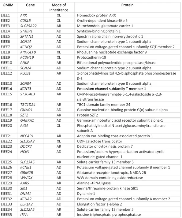
and non-ion-channels genes in sporadic severe and treatment-resistant epileptic encephalopathies (Table 2). An important contribution to the identification of novel genes responsible for Epileptic Encephalopathies has been given by new genetic technologies for mutation detection in human genome, such as Next-Generation Sequencing (NGS) and/or Whole Exome Sequencing (WES) [Choi et al., 2009]. The NGS technology, also known as “massive parallel sequencing”, has largely replaced traditional Sanger sequencing in the research and clinical laboratories allowing the simultaneous sequencing of millions of short fragments of DNA. The NGS technology is rapidly becoming a fundamental tool for genetic and functional genomics, since this technique allows the sequencing of entire gene panels, ranging from a few to several hundred genes. In addition, advances in genome technologies have allowed WES, in which the exon sequence of nearly all ~ 2000 human genes are sequenced in order to identify genetic mutations [Mefford, 2015]. Each of these approaches has been fundamental for the discovery that about 75% of patients with epileptic encephalopathies have de novo mutations, with only a small subset of patients with dominant, recessive or X-linked inherited mutations [Helbig et al., 2016]. Application of NGS technologies has also strongly contributed to demonstrate the significant genetic heterogeneity of EEs, as summarized in Table 2.
OMIM Gene Mode of Inheritance
Protein
EIEE1 ARX XL Homeobox protein ARX EIEE2 CDKL5 XL Cyclin-dependent kinase-like 5 EIEE3 SLC25A22 AR Mitochondrial glutamate carrier 1 EIEE4 STXBP1 AD Syntaxin-binding protein 1
EIEE5 SPTAN1 AD Spectrin alpha chain, non-erythrocytic 1 EIEE6 SCN1A AD Sodium channel protein type 1 subunit alpha
EIEE7 KCNQ2 AD Potassium voltage-gated channel subfamily KQT member 2 EIEE8 ARHGEF9 XL Rho guanine nucleotide exchange factor 9
EIEE9 PCDH19 XL Protocadherin-19
EIEE10 PNKP AR Bifunctional polynucleotide phosphatase/kinase EIEE11 SCN2A AD Sodium channel protein type 2 subunit alpha
EIEE12 PLCB1 AR 1-phosphatidylinositol 4,5-bisphosphate phosphodiesterase β-1
EIEE13 SCN8A AD Sodium channel protein type 8 subunit alpha
EIEE14 KCNT1 AD Potassium channel subfamily T member 1
EIEE15 ST3GAL3 AR CMP-N-acetylneuraminate-β-1,4-galactoside a-2,3-sialyltransferase
EIEE16 TBC1D24 AR TBC1 domain family member 24
EIEE17 GNAO1 AD Guanine nucleotide-binding protein G(o) subunit alpha EIEE18 SZT2 AR Protein SZT2
EIEE19 GABRA1 AD Gamma-aminobutyric acid receptor subunit alpha-1 EIEE20 PIGA XL Phosphatidylinositol N-acetylglucosaminyltransferase
subunit A
EIEE21 NECAP1 AR Adaptin ear-binding coat-associated protein 1 EIEE22 SLC35A2 XL UDP-galactose translocator
EIEE23 DOCK7 AR Dedicator of cytokinesis protein 7
EIEE24 HCN1 AD Potassium/sodium hyperpolarization-activated cyclic nucleotide-gated channel 1
EIEE25 SLC13A5 AR Solute carrier family 13 member 5
EIEE26 KCNB1 AD Potassium voltage-gated channel subfamily B member 1 EIEE27 GRIN2B AD Glutamate receptor ionotropic, NMDA 2B
EIEE28 WWOX AR WW domain-containing oxidoreductase EIEE29 AARS AR Alanine--tRNA ligase
EIEE30 SIK1 AD Serine/threonine-protein kinase SIK1 EIEE31 DNM1 AD Dynamin-1
EIEE32 KCNA2 AD Potassium voltage-gated channel subfamily A member 2 EIEE33 EEF1A2 AD Elongation factor 1-alpha 2
EIEE34 SLC12A5 AR Solute carrier family 12 member 5 EIEE35 ITPA AR Inosine triphosphate pyrophosphatase
Table 2 Early-Infantile-Epileptic-Encephalopaties (EIEEs) associated genes; XL=x-linked, AR=autosomal recessive,
2.3 KCNT1 mutations result in a wide range of seizure disorders and
Intellectual Disabilities
Mutations in KCNT1 gene have been identified in widely-diverging clinical conditions, ranging from very severe form of epilepsy (MMPSI) to milder epileptic diseases (ADNFLE) [Lim et al., 2016]. In vitro functional studies revealed that all KCNT1 mutations are able to induce an increase in current density when compared to wild-type channels, a result referred to as gain-of-function effect. In contrast, only one single study has reported a KCNT1 mutation causing a reduction in channel activity, also referred to as loss-of-function effect [Evely et al., 2017].
Clinical features of most epileptic conditions associated to KCNT1 mutations are described below.
2.3.1 Malignant Migrating Partial Seizures of Infancy
The syndrome of Malignant Migrating Partial Seizures of Infancy (MMPSI) or EIEE14 (Early Infantile Epileptic Encephalopathy 14) is a severe form of epilepsy that begins very early in life. Recurrent seizures begin before the age of 6 months, but commonly start within a few weeks after birth. Although affected patients may develop normally at first, progression stalls and skills decline when seizures begin; as a result, affected individuals have profound developmental delay. The seizures in MMPSI are described as partial (or focal) because seizure activity occurs in specific regions of the brain rather than affecting the entire brain. Seizure activity may appear in multiple locations of the brain or move, hence the definition of migrating seizures, from one brain region to another during an episode (Figure 6).
Migrating focal motor seizures at onset, nearly continuous multifocal seizures migrating between cortical regions and hemispheres, resistance to antiepileptic drugs, lack of demonstrable etiology, and severe psychomotor delay on follow-up are typical in MMPSI patients [Coppola et al., 1995].
The natural history of this syndrome allows recognition of three distinct phases. A first phase, generally starting in the first semester after birth, is often characterized by sporadic seizures, usually recurring in a few weeks or months. Seizure onset may also occur since the first day of life [Hmaimess et al., 2006]. Seizures are mainly focal motor
with rapid secondary generalization; autonomic manifestations such as apnea, flushing, or cyanosis frequently occur. This phase usually lasts a few weeks or months. However, seizures sometimes occur at the onset of the second phase. Interictal electroencephalography (EEG) in this first period shows increasing diffuse slowing of background activity with prevalence of slow waves often shifting from one hemisphere to the other. Shortly, multifocal discharges poorly activated by sleep are present in all cases.
The second phase could also be defined as a ‘‘stormy phase.’’ At an age ranging from about 1–12 months, focal polymorphous seizures shortly become very frequent, occurring in clusters of 5–30 several times a day or being almost continuous for several days. With increasing age, the amplitude of the ictal discharge tends to increase, and frontal areas are more frequently affected [Dulac, 2005]. The age at onset of the third phase may vary highly, ranging from the end of the first year to 5 years of age and over. This phase is a relatively seizure-free period, although spontaneous intercurrent illnesses would easily trigger clusters of seizures or occasional status epilepticus.
Computed tomography (CT) and magnetic resonance imaging (MRI) are generally normal at the beginning of the illness. During follow-up, there may be a mild to moderate enlargement of both subarachnoid and ventricular spaces.
Overall, the long-term outcome of migrating focal seizures in infancy, with reference to seizures and psychomotor development, remains very severe. Even when it becomes possible to shorten the duration of migrating status epilepticus and/or increase the intervals between active phases with different combination therapies, psychomotor abilities are poor, inevitably evolving into mental retardation. Not surprisingly, absence of language and hypotonia are also commonly noted in MMPSI patients [McTague et al., 2013]. Because of the serious health problems caused by MMPSI, many affected individuals do not survive past infancy or early childhood.
Possible causes for seizure development in MMPSI patients has remained elusive until recently. Neurometabolic, blood gas and serum tests are typically normal, and brain lesions are rarely observed in affected patients [Nabbout and Dulac, 2008]. Genetic etiologies for MMPSI were first identified in 2011, with the discovery of SCN1A (Nav 1.1)
mutations [Carranza Rojo et al., 2011], followed by TBC1D24 [Milh et al., 2103] and KCNT1 (Slack) mutations [Barcia et al., 2012]. Figure 6 Ictal EEG of a MMPSI patients. Ictal focus is located in the left temporal area at the onset (narrow arrow), whereas it is shifted to the right occipital area during seizure (broad narrow). [Ishii et al., 2013].
2.3.2 Ohtahara syndrome
Ohtahara syndrome (OS) was originally described as an early infantile epileptic encephalopathy with suppression bursts [Ohtahara et al., 1976]. OS is one of the earliest seizures in its presentation. Infants acutely develop tonic spasms that can be either generalized or lateralized, can occur both singly or in clusters, and are independent of the sleep cycle. A majority of OS patients show severe developmental delay, including intellectual disability. More than 80% of OS patients reported in the literature have a developmental delay, while only 10% are described as showing normal development. It remains a challenge to reverse or overcome this poor prognosis, since these seizures have pronounced pharmacological resistance [Beal et al., 2012]. Genetic etiologies have also been identified in a subset of OS patients. To date, alterations in five genes have been found in OS patients: ion channels KCNQ2 (Kv7.2) [Saitsu et al., 2012a], SCN2A (Nav1.2) [Nakamura et al., 2013], KCNT1 (Slack) [Martin et al., 2014]; the transcription factors ARX and the synaptic binding protein STXBP1. Approximately one third of patients with Ohtahara syndrome will also develop other seizure types, most commonly focal motor seizures, hemiconvulsions, or generalized tonic-clonic seizures. OS may evolve to West syndrome and further to Lennox-Gastaut syndrome during age progression with a poor prognosis.
2.3.3 Early Myoclonic Encephalopathy
Similar to OS, the pathogenesis of early myoclonic encephalopathy (EME) is variable, with structural, metabolic, and genetic abnormalities all playing a role. Many seizure types may occur, but myoclonic seizures (very frequent, brief, single or repetitive, erratic, and nearly continuous body jerks) characterize this epilepsy syndrome and differentiate it from OS. Some may also have focal motor seizures, tonic seizures, or rarely tonic spasms, which appear later. ErbB4 is one gene example associated with EME [Backx et al., 2009], although mutations in SLC25A22 gene have reported in patients with EME [Molinari et al., 2005].
2.3.4 West Syndrome
West syndrome (WS) is the most common form of early onset epileptic encephalopathy and typically begins between 3 and 7 months of age. WS is characterized by epileptic spasms, hypsarrhythmia and developmental cessation or regression. The epileptic spasms typical of WS are brief seizures with flexion or extension of the arms and legs and/or head and torso that occur in clusters usually upon awakening [Nieh et al., 2014].
2.3.5 Autosomal dominant nocturnal frontal lobe epilepsy
Autosomal dominant nocturnal frontal lobe epilepsy (ADNFLE) is characterized by clusters of nocturnal motor seizures, which are often stereotyped and brief (5 seconds to 5 minutes). They vary from simple arousals from sleep to dramatic, often bizarre, hyperkinetic events with tonic or dystonic features. Affected individuals may experience aura. Retained awareness during seizures is common. A minority of individuals experience daytime seizures. Onset ranges from infancy to adulthood. About 80% of individuals develop ADNFLE in the first two decades of life; mean age of onset is ten years. Clinical neurologic examination is normal and intellect is usually preserved, but reduced intellect, psychiatric comorbidity, or cognitive deficits may occur. Within a family, the manifestations of the disorder may vary considerably. ADNFLE is lifelong but not progressive. As an individual reaches middle age, attacks may become milder and
less frequent. Molecular genetic testing reveals pathogenic variants in CHRNA4, CHRNB2, CHRNA2, KCNT1, DEPDC5, or CRH in approximately 20% of individuals with a positive family history and fewer than 5% of individuals with a negative family [Kurahashi et al., 2015].
3. Pharmacology of Sodium-activated potassium channels
Given their role in the regulation of neuronal excitability and their involvement in the pathogenetic mechanisms of several epileptic disorders, these sodium-activated potassium channels are important pharmacological targets.
The pharmacological properties of KCNT1 and KCNT2 channels are similar to each other, but differ from those of many other potassium channels. In fact, KCNT1 and KCNT2 channels are only weakly sensitive to the aspecific potassium channel blocker
tetraethylammonium (TEA): in particular, 1 mM TEA had little effect on both channels while a concentration of 20 mM was required to block 60% of KCNT1 and KCNT2 currents. In addition, Ba+ (1 mM), another aspecific K+ channels blockers, also inhibited
KCNT1 and KCNT2 channels with time- and voltage-dependent effects [Bhattacharjee et al., 2003].
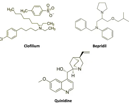
Studies on native IKNa channels in cardiac cells indicate that several antiarrhythmic drugs
inhibit KCNT1 and KCNT2 channels in cardiac cells [Mori et al., 1996; Li et al., 1999]. Some of these compounds, such as clofilium, bepridil, and quinidine (Figure 7), have been also found to be very effective and reversible blockers of KCNT1 and KCNT2 currents expressed in oocytes and mammalian cells. Clofilium inhibits both channels and this compound seems to be more potent on KCNT1 channels: in fact, the EC50 for
KCNT1 is 109 µM, while the EC50 for KCNT2 is 331 µM [Tejada et al., 2012]. Two other
blockers, namely bepridil and quinidine, inhibit KCNT1 currents in a concentration-dependent manner and decrease channel activity in excised membrane patches. Similarly to oocytes, the application of quinidine (1 µM-10 mM) in HEK293 cells expressing KCNT1 produced a rapid decrease in current amplitude, with an EC50 of 89.6
µM [Yang et al., 2006]. The KCNT1 currents are also sensitive to bepridil-induced blockade, with the latter being more potent than quinidine (EC50 of 1.0 µM) [Yang et al.,
2006]. Although these drugs are able to block KNa channels, they are also active on a
variety of other channel types, thus rendering difficult the identification of their precise mechanism of action during their clinical use.
Figure 7 Chemical structure of KCNT1 and KCNT2 blockers.
A variety of compounds that activate SLO2 K+ channels are also known. The first to be described was the bis-phenol anti-parasitic compound bithionol, which in KCNT1-expressing HEK cells produces a robust increase in current amplitude, whit an EC50 of
0.77 µM [Yang et al., 2006]. Similar bithionol-induced current increases are observed in KCNT1-expressing oocytes and for native KNa currents in neurons of the auditory
brainstem. Bithionol reversibly activates KCNT1 channels even when applied to the extracellular face of excised patches; however, bithionol is not selective for KNa channels
because it also activates SLO1 calcium-activated potassium channels [Yang et al., 2006; Yang et al., 2007]. A screen of pharmacologically active compounds using a rubidium flux assay against KCNT1 channels expressed in Chinese Hamster Ovary (CHO) cells has revealed new activators: these include riluzole, loxapine, an antipsycotic agent, and
niclosamide, an anthelmintic agent (Figure 8). Loxapine was found to be more selective than bithionol since no effects on SLO1 calcium-activated potassium channels were observed. Electrophysiological experiments revealed that loxapine is effective on recombinant human and rat KCNT1 channels and that it activates native KNa channels in
isolated DRG neurons [Biton et al., 2012].
Furthermore, KCNT2 channels can be activated by fenamates such as niflumic acid (NFA, Figure 6), even in the absence of intracellular Na+. In Xenopus oocytes, KCNT2
currents were rapidly activated by extracellular application of NFA (EC50 2.1 mM) or flufenamic acid (EC50 1.4 mM) [Dai et al., 2010]. Figure 8 Chemical structure of KCNT1 and KCNT2 activators.
4.
AIMS OF THE STUDY
Epilepsy has a highly heterogeneous etiology with a strong genetic contribution. In particular, mutations in the KCNT1 gene cause a wide spectrum of seizure phenotypes including ADFNLE, multifocal epilepsy, Ohtahara syndrome, leukoencephalopathies and the rare devastating epileptic encephalopathy MMPSI.
However, the identification of one of the many various gene variants identified in each patient with state-of-the-art technologies as pathogenic, requires the convergence of data deriving from various sources. Among these, functional, pharmacological and biochemical in vitro studies are essential to understand the pathogenetic contribution of a specific variant found in patients affected by this severe genetic disease. Therefore, in order to facilitate the identification of specific gene variants as pathogenic in specific individuals affected by rare epileptic conditions, and to provide clues on the potential pathogenetic mechanisms, in the present Doctoral Thesis, I have investigated the biochemical, functional, and pharmacological properties of KCNT1 and KCNT2 channels carrying mutations identified in patients affected by Epileptic Encephalopathy. The genetic variants investigated in the present study are listed in following Table:
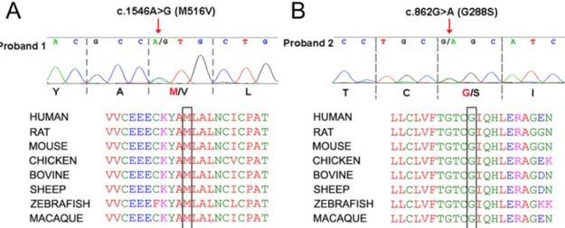
Gene Nucleotide substitution Amino acids substitution Reference KCNT1/SLO2.2 c.862 G>A p. G288S Ishii et al., 2013 [Rizzo et al., 2016] KCNT1/SLO2.2 c.1546 A>G p. M516V [Rizzo et al., 2016]
KCNT1/SLO2.2 c.2677 G>A p. E893K newly-identified
KCNT1/SLO2.2 c.2849 G>A p. R950Q Moller et al., 2016
KCNT2/SLO2.1 c.1451 G>A p. C484Y newly-identified
KCNT2/SLO2.1 c.569 G>C p. R190H newly-identified KCNT2/SLO2.1 c.569 G>C p. R190P [DDDS, Nature 2017] Table 3 Naturally-occurring mutations found in KCNT1 and KCNT2 genes studied in the present doctoral thesis. (“c.” indicates nucleotide substitutions, “p.” indicates amino acid mutations).
In particular, I have studied four mutations (G288S, M516V, E893K and R950Q) in the KCNT1 gene and three mutations (C484Y, R190H and R190P) in the KCNT2 gene. Among them, two KCNT1 mutations (G288S and R950Q) have already been reported in the literature [Ishii et al., 2013; Moller et al., 2015]. The identification of KCNT1 mutations has been made possible thanks to collaboration with a multidisciplinary team formed by child neurologists and human geneticists. Two MMPSI-affected patients have been identified by Prof. G. Coppola, who described this disease for the first time [Coppola et al., 1995], dr. G. Casara (Department of Pediatrics, Regional Hospital of Bolzano), and dr. M. Vecchi (Department of Child and Mother Health, University of Padua); in these patients, the genetic analysis performed by the group of Prof. A. Weisz (University of Salerno) identified the recurrent KCNT1-G288S and the novel KCNT1-M516V mutations. In addition, the novel KCNT1-E893K variant has been identified by the group of dr. J. DiFrancesco at the “Carlo Besta” Institute, while the recurrent KCNT1-R950Q variant has been found in a patient by dr. R. Dilena (Fondazione IRCSS Ca Granda Ospedale Maggiore Policlinico, Milan). As reported in Figure 9, M516V, E893K and R950Q mutations affect residues localized in the KCNT1 C-terminal region: M516V falls within the RCK1 domain, whereas E893K and R950Q are localized in the RCK2 domain; by contrast, G288S falls in the S5 segment of the KCNT1 channel subunit. Figure 9 Schematic topology of a KCNT1 (SLO2.2) channel subunit and location of the mutations studied in the present work. Colored circles indicate the location of the mutations investigated: the G288S mutation (blue circle) is localized in the S5 segment of the channel, while the other mutations are located in the C-terminal region. The M516V mutation (green circle) fall in the RCK1 (Regulator of K+ conductance) domain of the KCNT1 subunits, while the E893K (red circle) and R950Q (yellow circle) mutations are localized in the RCK2 domain. For numbering mutations, numeration complies with that of isoform 2 (NM_020822).
Also KCNT2 mutations herein investigated have been identified through a network of collaboration in Italy and abroad: in particular, the KCNT2-R190H variant has been identified in collaboration with the Prof. Johannes Lemke (Germany), while the KCNT2-R190P variant was recruited through the Deciphering Developmental Disorders Study [DDDS, Nature 2017]. In addition, I have also investigated the functional consequences of another variant in KCNT2 (C484Y) found in a patient with Autosomal Dominant Nocturnal Frontal Lobe Epilepsy (ADFNLE) and identified by Prof. Francesca Bisulli (University of Bologna); this variant has been inherited from father’s patient. As shown in Figure 10, the C484Y mutation falls within the C-terminal region of the KCNT2 subunit, while both mutations affecting the same residue (R190H and R190P) are located in the S4-S5 linker region.
Figure 10 Schematic topology of a SLO2.1 channel subunit and location of the mutations studied in the present work. Colored circles indicate the location of mutations investigated: de novo R190H and R190P mutations are
localized within the S4-S5 linker region, while the inherited C484Y mutation is located in the C-terminal region of the KCNT2 channel subunit. For numbering mutations, the numeration complies with that of isoform NM_001287820.2.
To study the biochemical, functional, and pharmacological consequences prompted by these mutations in SLO2 family genes, the following experiments have been performed: a) for KCNT1 mutations: 1. engineering of each mutation in a plasmid containing the cDNA for a myc-DDK tagged human isoform of KCNT1 (RC214820; Origene, Rockville, MD, USA); 2. patch-clamp recordings of macroscopic currents from Chinese Hamster Ovary (CHO) cells transiently-transfected with wild-type or mutant subunits, in homomeric or heteromeric configurations with wild-type and mutant KCNT1 subunits; 3. biochemical evaluation of total and plasma membrane expression of wild-type and mutant KCNT1 subunits; 4. patch-clamp recordings of macroscopic currents in the presence of two well-known KCNT1 blockers (bepridil and quinidine) in order to evaluate their ability to restore the functional alterations induced by the presence of each mutation. b) for KCNT2 mutations: 1. engineering of mutations in a plasmid containing the cDNA for a turbo-GFP tagged human isoform of KCNT2 (RC216225; Origene, Rockville, MD, USA);
2. biochemical studies by using CHO or HEK-293 total lysates in order to identify the most efficient heterologous expression system for KCNT2 subunits;
3. patch-clamp recordings of macroscopic currents from Human Embryonic Kidney (HEK-293) cells transiently-transfected with wild-type or mutant subunits, in homomeric or heteromeric configurations with wild-type and mutant KCNT2 subunits;
4. patch-clamp recordings of macroscopic currents in the presence of the KCNT2-blocker quinidine in order to evaluate its ability to restore the functional alteration induced by the presence of each mutation;
5. molecular modelling studies to formulate hypothesis on the potential structural consequences of the R190 variants.
5. MATERIALS AND METHODS
5.1 Site-Directed Mutagenesis
Each KCNT1 mutation was engineered by Quick-change Site-Directed Mutagenesis (Agilent Technologies) in a pCMV6-KCNT1 (RC214820; Origene, Rockville, MD, USA) plasmid containing the cDNA for myc-DDK tagged human transcript variant of KCNT1 (accession number: NM_020822; 1256 aminoacids).
Similarly, mutations in KCNT2 gene were obtained by Quick-change Site-Directed Mutagenesis in a pCMV6-KCNT2 plasmid (RG216225; Origene, Rockville, MD, USA) encoding for the tGFP-tagged human transcript variant of KCNT2 (accession number: NM_198503; 1135 aminoacids).
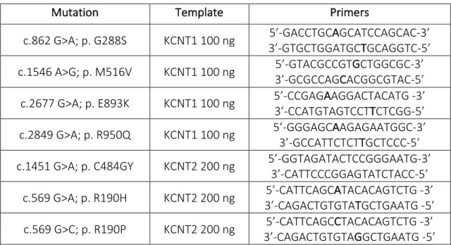
The mutations were engineered in each plasmid by Polymerase Chain Reaction (PCR), using a pair of primers (forward and reverse), incorporating the nucleotide mutation found in each patient (see Table 4).
Mutation Template Primers
c.862 G>A; p. G288S KCNT1 100 ng 3’-GTGCTGGATGC5’-GACCTGCAGCATCCAGCAC-3’ TGCAGGTC-5’ c.1546 A>G; p. M516V KCNT1 100 ng 5’-GTACGCCGT3’-GCGCCAGCACGGCGTAC-5’ GCTGGCGC-3’
c.2677 G>A; p. E893K KCNT1 100 ng 5’-CCGAG3’-CCATGTAGTCCTAAGGACTACATG -3’ TCTCGG-5’ c.2849 G>A; p. R950Q KCNT1 100 ng 5’-GGGAGC3’-GCCATTCTCTAAGAGAATGGC-3’ TGCTCCC-5’ c.1451 G>A; p. C484GY KCNT2 200 ng 5’-GGTAGATACTCCGGGAATG-3’ 3’-CATTCCCGGAGTATCTACC-5’
c.569 G>A; p. R190H KCNT2 200 ng 3’-CAGACTGTGTA5’-CATTCAGCATACACAGTCTG -3’ TGCTGAATG -5’ c.569 G>C; p. R190P KCNT2 200 ng 3’-CAGACTGTGTA5’-CATTCAGCCTACACAGTCTG -3’ GGCTGAATG -5’
Table 4 Experimental conditions used for PCR reaction. Column 1 indicates the mutations found in patients affected by Epileptic Encephalopathy. Column 2 shows the templates and related quantity used for each reaction. Column 3 contains nucleotide sequences of primers used for PCR reaction.
The amplification reaction was performed in a final volume of 50 µL containing the following components: 100-200 ng of plasmids for wild-type KCNT1 or wild-type KCNT2 as template, 0.6 µM primer forward, 0.6 µM primer reverse, 3U of Q5 DNA Polymerase, 0.2 mM dNTP mix, 1X buffer Q5 and 1X buffer GC. The PCR consisted of 30 cycles, with each cycle consisting of three temperature steps, that allow the denaturation of the DNA double helix (95° C for 1’), the annealing of the primers to the single strand of DNA (the temperature was modified according to the nucleotide sequence of each couple of primers) and the extension of the primers (73°C for 5’) (Figure 11). Figure 11 Polymerase Chain Reaction (PCR) protocol. PCR protocol consists of 3 phases. Phase 1: denaturation of the DNA. Phase 2: annealing of the mutated primers to specific complementary region of the DNA. Phase 3: The Q5 polymerase synthesizes a new DNA strand complementary to the DNA template strand.
5.2 Bacterial Transformation and plasmidic DNA preparation
The amplification reaction contains both methylated (parental) and unmethylated (neo-synthesized) DNA. In order to remove the parental DNA, the entire volume reaction was exposed to enzymatic digestion with DpnI enzyme, able to digest only methylated DNA. After the enzymatic digestion with DpnI, competent E.coli DH5a cells were transformed with the PCR product by chemical transformation procedure (30’ at 4°C, shock step at 42° for 45’’ followed by 2’ at 4°C). To help the bacterial cells to recover from the heat shock, cells were incubated with SOC medium (2% tryptone, 0.5% yeast extract, 10 mM
NaCl, 2.5 mM KCl, 10 mM MgCl2, 10 mM MgSO4, 20 mM glucose), for 1h at 37°C. After
this step, the cells were seeded into LB+agar plates (containing 10g/L tryptone, 5 g/L yeast extract, 5 g/L NaCl, agar 15 g/L) containing the specific antibiotic to which plasmids are resistant (in our experiments ampicillin 100 µg/µL) to allow the selective growth of E.coli cells transformed with myc-DDK pCMV6-KCNT1 or tGFP-pCMV6-KCNT2. Plates were then incubated upside down at 37°C for about 17h to allow bacterial growth.
Each colony grown on the LB medium was inoculated in 6 mL of fresh LB medium with ampicillin for selection, at 37°C/220 rpm overnight. Plasmidic DNA was extracted by using a commercially available kit (NucleoSpin Plasmid EasyPure, Promega, Milan, Italy). The successful insertion of each desired mutation was verified by direct sequencing (Eurofins Genomic, Milan, Italy). In order to obtain a large amount of DNA, one of the positive clones, was amplified on a large scale (500 mL) and plasmidic DNA was obtained by using a commercially available kit (PureYield Plasmid Maxiprep System, Promega). The cDNA was sequenced to confirm the presence of the mutation of interest and to exclude the presence of additional mutations in the coding sequence.
5.3 Cell cultures and transient transfection with Lipofectamine 2000
CHO (Chinese Hamstery Ovary) or HEK-293 (Human Embryonic Kidney) cells were grown in plastic Petri’s dishes (100 mm, 60 mm or 40 mm, according to the different experimental procedures) in DMEM (Dulbecco’s Minimum Eagle Medium) supplemented with 10% Fetal Bovine Serum (FBS) decomplemented at 56°C for 30’, 1% L-glutamine (2 mM in 0.85% NaCl), 1% penicillin (50 U/mL) and streptomycin (50 µg/mL) in a humidified atmosphere at 37°C with 5% CO2. The CHO cells were transiently
transfected using Lipofectamine 2000, according to the manufacturer protocol (LifeTechonologies, Milan, Italy). In each transfection reaction, a plasmid encoding for the EGFP (Enhanced Green Fluorescent Protein) was used to confirm the successful transfection procedure. Total cDNA in the transfection mixture was kept constant at 4 µg for electrophysiological experiments and 6 µg for Western blotting experiments.
![Table 1 SLO channels family genes. [Adapted from Salkoff et al., 2006].](https://thumb-eu.123doks.com/thumbv2/123dokorg/4793561.49020/7.892.149.760.492.721/table-slo-channels-family-genes-adapted-from-salkoff.webp)